orji genius presentation
TRANSCRIPT

DIFFERENCES BETWEEN SMOOTH AND SKELETAL
MUSCLE
ORJI JONATHAN ORJIDE GENIUS
PRESENTED BY:

walls of hollow organs•Lack striations
•Contractions are involuntary (not voluntary)
Smooth muscle
Muscle tissue found in hollow internal organs is called SMOOTH MUSCLE
• Spindle Shaped• Central nuclei• Lack Striations, transverse tubules, and lack welldeveloped sacroplasmic reticulum• Actin and myosin thin and randomly distributed
TYPES OF SMOOTH MUSCLE•Multi-unit smooth muscle-Separate units e.gciliary muscle of the eye, the iris muscle of the eye, and the piloerector muscles that cause erection of the hairs when stimulated by the sympathetic nervous system.•Unitary smooth muscle-It is also called visceral smooth muscle because it is found in the walls of most viscera of the body, including the gastrointestinal tract, bile ducts, ureters, uterus, and many blood vessels.
Smooth muscle Contraction

Gap junctions assure contraction as “single unit.”
It has a mass of hundreds to thousands of smooth muscle fibers that contract together as a single unit. The fibers usually are arranged in sheets or bundles, and their cell membranes are adherent to one another at multiple points so that force generated in one muscle fiber can be transmitted to the next.

Fewer or no gap junctions assure independent contraction and more precise control.
This type of smooth muscle is composed of discrete, separate smooth muscle fibers. Each fiber works independently of the others and often is innervated by a single nerve ending, as occurs for skeletal muscle fibers.
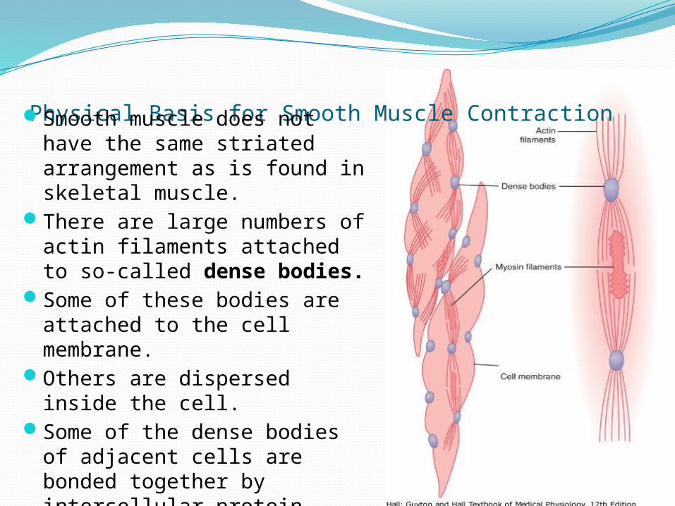
Physical Basis for Smooth Muscle Contraction Smooth muscle does not have
the same striated arrangement as is found in skeletal muscle.
There are large numbers of actin filaments attached to so-called dense bodies.
Some of these bodies are attached to the cell membrane.
Others are dispersed inside the cell.
Some of the dense bodies of adjacent cells are bonded together by intercellular protein bridges. through these bonds that the force of contraction is transmitted from one cell to the next.

• Attach to and move skeleton
• 40% of body weight• Fibers = multinucleate cells (embryonic cells fuse)
• Cells with obvious striations
• Contractions are voluntary
Skeletal muscle
Muscle tissue that attaches to and move bones is called SKELETAL MUSCLE
• Fast, slow and intermediate• Whether or not they predominantly use oxygen
to produce ATP (the energy molecule used in muscle contraction)– Oxidative – aerobic (use oxygen)– Glycolytic – make ATP by glycolysis (break
down of sugars without oxygen=anaerobic) • Fast fibers: “white fibers” – large, predominantly
anaerobic, fatigue rapidly (rely on glycogen reserves); most of the skeletal muscle fibers are fast
• Slow fibers: “red fibers” – half the diameter, 3X slower, but can continue contracting; aerobic, more mitochondria, myoglobin
• Intermediate: in between
Types of skeletal muscle fibers
Low Energy Requirement to Sustain Smooth Muscle Contraction
The smooth muscle is required far less E to sustain the same tension of contraction than in skeletal muscle.
This sparsity of E utilization by smooth muscle is important to the overall E economy of the body because organs such as the intestines, urinary bladder, gallbladder, and other viscera often maintain tonic muscle contraction almost indefinitely.

• A skeletal muscle contracts when its motor units are stimulated
• Amount of tension depends on1. the frequency of stimulation2. the number of motor units involved
• Single, momentary contraction is called a muscle twitch
• All or none principle: each muscle fiber either contracts completely or not at all
• Amount of force: depends on how many motor units are activated
• Muscle tone– Even at rest, some motor units are active:
tense the muscle even though not causing movement: “resting tone”
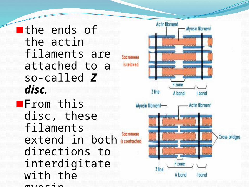
the ends of the actin filaments are attached to a so-called Z disc. From this disc, these filaments extend in both directions to interdigitate with the myosin filaments.
