neuroblastoma - renaissance.stonybrookmedicine.edu · neuroblastoma (nb) is the most common...
TRANSCRIPT

NeuroblastomaParadigm for Precision Medicine
Meredith S. Irwin, MDa, Julie R. Park, MDb,*
KEYWORDS
� Neuroblastoma � Risk stratification � MYCN� Segmental chromosome aberrations (SCA) � ALK (anaplastic lymphoma kinase)� Phox2B � Myeloablative therapy (MAT) � Immunotherapy
KEY POINTS
� Neuroblastoma (NB) is the most common extracranial pediatric tumor, most frequentlydiagnosed cancer in infancy, and has a heterogeneous presentation and prognosis.
� Clinical and biological prognostic factors are used to risk stratify patients into groups withlow, intermediate, and high risk for recurrence; most protocols now use the InternationalNeuroblastoma Risk Group classification system.
� Age, stage, histology, and amplification of the MYCN oncogene are currently the mostrobust prognostic factors.
� Outcomes for low- and intermediate-risk NB are excellent, but survival for high-risk NB isless than 50%.
� High-risk NB tumors contain many segmental chromosome aberrations (eg, loss of het-erozygosity 1p, 11q); but recurrent somatic mutations are rare, with anaplastic lymphomakinase (ALK) being the most commonly altered gene in approximately 10% of NB.
� Survival after relapse of metastatic NB is uncommon; current and upcoming trials will relyon incorporation of novel immunotherapies, inhibitors of aberrant pathways (eg MYC,ALK), and radioisotope-containing regimens, such as high-dose iodine-131-metaiodo-benzylguanidine.
INTRODUCTION
Neuroblastoma (NB), the most common extracranial tumor of childhood, is a cancer ofprimordial neural crest cells that give rise to sympathetic neural ganglia and adrenalmedulla. NB has a diverse pattern of clinical presentation and prognosis that ranges
a Division of Hematology-Oncology, Hospital for Sick Children, University of Toronto, 555 Uni-versity Ave, Toronto, ON M5G1X8, Canada; b Division of Hematology-Oncology, Seattle Chil-dren’s Hospital, University of Washington School of Medicine, Fred Hutchinson CancerResearch Center, 4800 Sandpoint Way NE, Seattle, WA 98105, USA* Corresponding author.E-mail address: [email protected]
Pediatr Clin N Am 62 (2015) 225–256http://dx.doi.org/10.1016/j.pcl.2014.09.015 pediatric.theclinics.com0031-3955/15/$ – see front matter � 2015 Elsevier Inc. All rights reserved.
Downloaded from ClinicalKey.com at Stony Brook State University of New York August 17, 2016.For personal use only. No other uses without permission. Copyright ©2016. Elsevier Inc. All rights reserved.
Irwin & Park226
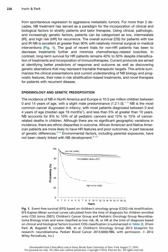
from spontaneous regression to aggressive metastatic tumors. For more than 2 de-cades, NB treatment has served as a paradigm for the incorporation of clinical andbiological factors to stratify patients and tailor therapies. Using clinical, pathologic,and increasingly genetic factors, patients can be categorized as low, intermediate(IR), and high risk (HR) for recurrence. The overall survival (OS) for patients with lowand IR NB is excellent at greater than 90% with relatively minimal surgical or medicalinterventions (Fig. 1). The goal of recent trials for non-HR patients has been todecrease treatments further and minimize chemotherapy-related toxicities. Incontrast, long-term survival for HR patients remains 40% to 50% despite intensifica-tion of treatments and incorporation of immunotherapies. Current protocols are aimedat identifying better predictors of response and outcome as well as discoveringgenetic aberrations that may represent tractable therapeutic targets. This article sum-marizes the clinical presentations and current understanding of NB biology and prog-nostic features, their roles in risk stratification–based treatments, and novel therapiesfor patients with recurrent disease.
EPIDEMIOLOGY AND GENETIC PREDISPOSITION
The incidence of NB in North America and Europe is 10.5 per million children between0 and 14 years of age, with a slight male predominance (1.2:1.0).1–4 NB is the mostcommon cancer diagnosed in infancy, with most patients diagnosed between 0 and4 years of age (median age 19 months5), and less than 5% at greater than 10 years.NB accounts for 8% to 10% of all pediatric cancers and 12% to 15% of cancer-related deaths in children. Although there are no significant geographic variations inincidence, there are ethnic disparities in outcome. African American and Native Amer-ican patients are more likely to have HR features and poor outcomes, in part becauseof genetic differences.6–8 Environmental factors, including parental exposures, havenot been clearly linked with NB development.9,10
Fig. 1. Event-free survival (EFS) based on children’s oncology group (COG) risk stratification.EFS Kaplan-Meier survival curves calculated from the time of diagnosis for children enrolledonto COG (since 2001); Children’s Cancer Group and Pediatric Oncology Group Neuroblas-toma Biology trials and were classified as low risk, IR, or HR at the time of diagnosis basedon clinical and biological factors (current COG classification is summarized in Table 2). (FromPark JR, Bagatell R, London WB, et al. Children’s Oncology Group 2013 blueprint forresearch: neuroblastoma. Pediatr Blood Cancer 2013;60(6):986, with permission. � 2012Wiley Periodicals, Inc.)
Downloaded from ClinicalKey.com at Stony Brook State University of New York August 17, 2016.For personal use only. No other uses without permission. Copyright ©2016. Elsevier Inc. All rights reserved.
Neuroblastoma 227
NB is the only solid tumor of childhood for which there have been large screeninginitiatives, pioneered largely in Japan. Universal screening of 6-month-old asymptom-atic infants by detection of elevated urinary catecholamines resulted in a 2-fold in-crease in NB incidence to 20.1 per million children; however, most of the detectedtumors had favorable clinical and biological characteristics.11–13 Studies in Germanyand Quebec also demonstrated an increased incidence and detection of tumorswith favorable biology and pathology.14,15 In general, universal screening has notdetected poor prognosis disease, which usually presents at an older age and, thus,has not affected mortality rates.16 In contrast, in selected populations with an inheritedgenetic predisposition to NB, screening may be indicated.
Genetic Predisposition
The incidence of familial NB is estimated at 1% to 2%.17 Cases often involve multifocaland/or bilateral adrenal primary tumors with a median age of onset of 9 months. Thepattern of inheritance is autosomal dominant with incomplete penetrance. NB canoccur in patients with other neural crest disorders, such as Hirschsprung disease(HSCR), congenital central hypoventilation syndrome (CCHS), and neurofibromatosistype 1 (NF1). Mutations in the Phox2b homeobox gene have been detected in subsetsof patients with familial NB and usually are associated with other neurocristopathies,such as HSCR and CCHS.18–20 Phox2bmutations have also been detected in approx-imately 2% of sporadic NB. There are many reports of NB in patients with NF;however, there are conflicting data as to whether germline NF1 mutations are associ-ated with an increased risk to develop NB.21,22
Linkage studies in familial NB pedigrees identified candidate chromosomal predis-position regions including 16p, 12p, and 2p23–26 and led to the identification of germ-line mutations in the tyrosine kinase domain of the anaplastic lymphoma kinase (ALK)oncogene.27,28 ALK is involved in nervous system development,29 and central nervoussystem (CNS) anomalies have been reported in some patients with germline ALK mu-tations.29 Sporadic NB tumors also harbor ALK abnormalities, including genomicamplification (2%–3%) and missense mutations (8%–12%)27,28,30–33 (see “SomaticGene Mutations”), that can be targeted by pharmacologic inhibitors.34–36 Studies ofALK inhibitors in NB and other tumors with ALK aberrations (eg, anaplastic largecell lymphoma) have shown promising results.37 NB cases are also detected in otherfamilial cancer syndromes, including Beckwith-Wiedemann syndrome,38 Li-Fraumeni,39,40 Noonan (PTPN11), some subtypes of Fanconi anemia, and some chro-mosomal breakage syndromes.41,42
Recent genome-wide association studies using peripheral blood from thousands ofpatients with NB have also identified germline genetic variants that may predispose tothe development of sporadic NB. These variants include single nucleotide polymor-phisms (SNPs) in LINC00340, BARD1, LMO1, DUSP12, DDX4, LIN28B, HACE1, andTP53.43–48 Unlike the rare germline mutations in ALK and Phox2B described earlier,these SNPs are more frequent but individually have less dramatic impacts on theNB risk.49 The interplay between multiple germline variants and somatic alterations,discussed later, may influence the initiation and progression of NB.
PRESENTATION, DIAGNOSIS, AND STAGINGSymptoms
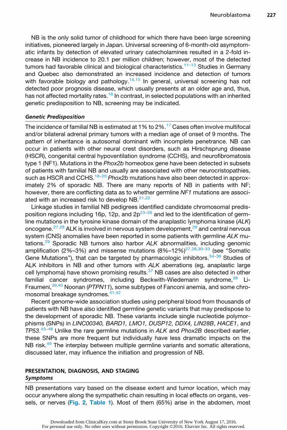
NB presentations vary based on the disease extent and tumor location, which mayoccur anywhere along the sympathetic chain resulting in local effects on organs, ves-sels, or nerves (Fig. 2, Table 1). Most of them (65%) arise in the abdomen, most
Downloaded from ClinicalKey.com at Stony Brook State University of New York August 17, 2016.For personal use only. No other uses without permission. Copyright ©2016. Elsevier Inc. All rights reserved.
Fig. 2. Percent distribution of NBs by primary site and age; Surveillance, Epidemiology andEnd Results Program (1975 to 1995). (Adapted from Ries L, Smith M, Gurney J, et al. Cancerincidence and survival among children and adolescents: United States SEER Program 1975–1995, National Cancer Institute, SEER Program. Bethesda (MD): NIH Pub; 1999. p. 99–4649.NIH Publication No. 99–4649.)
Irwin & Park228
commonly the adrenal gland, and may be asymptomatic or associated with hyperten-sion, abdominal pain, distension, and constipation. Other sites include the neck,chest, and pelvis. The primary site location is associated with age and outcome.50
Cervical and thoracic tumors are more common in infants and may present withHorner syndrome (unilateral ptosis, anhidrosis, and myosis) and respiratory
Table 1Clinical presentation and symptoms of NB
Location Signs and Symptoms
Abdomen/pelvis Pain, constipation, distension, urinary retention,hypertension
Thorax Respiratory distress, Horner syndrome
Presacral and paraspinal (includesabdominal and thoracic masses)
Symptoms of cord compression (urinaryretention, paraplegia/paraparesis, clonus)
Neck Mass/swelling
Metastases Irritability, bone pain, cytopenias (petechiae,ecchymoses, pallor), periorbital ecchymoses, fever,weight loss, lymphadenopathy
4S/4M metastases Hepatomegaly, coagulopathy, hyperbilirubinemia,respiratory distress (from abdominal enlargement),skin nodules
Paraneoplastic syndromes � OMS: myoclonic jerking and random eye movement,with or without cerebellar ataxia
� VIP secreting tumors: intractable secretory diarrheacaused by tumor secretion of VIP
Patients may be asymptomatic or may have one or more of the listed symptoms or findings onexam.
Abbreviations: OMS, opsoclonus myoclonus ataxia syndrome; VIP, vasoactive intestinal peptide.
Downloaded from ClinicalKey.com at Stony Brook State University of New York August 17, 2016.For personal use only. No other uses without permission. Copyright ©2016. Elsevier Inc. All rights reserved.
Neuroblastoma 229
symptoms. Epidural or intradural tumor extension occurs in 5% to 15% of patients andmay result in spinal cord compression and paraplegia.51 Two rare paraneoplasticsyndromes associated with NB include secretory diarrhea caused by tumor produc-tion of vasoactive intestinal peptide52,53 and opsoclonus myoclonus ataxia syndrome(OMS). OMS is reported in 2% to 3% of patients and is commonly associated withfavorable well-differentiated tumors.54,55 OMS is characterized by myoclonic jerksand random eye movements with or without ataxia, is attributed to immune-mediated effects, and often persists after resection, resulting in significant neurodeve-lopmental sequelae.Approximately half of patients present with localized or regional disease, and 35%
have regional lymph node spread at the time of diagnosis. Distant metastases aredetected in 50% of patients at diagnosis and occur through both lymphatic and hema-togenous routes. The most common sites include bone, bone marrow, and liver. NBhas a particular predilection to spread to metaphyseal, skull, and orbital bone sites,resulting in a classic presentation characterized by periorbital ecchymoses (raccooneyes), proptosis, and potentially visual impairment. In contrast to the frequent lackof symptoms for locoregional tumors, patients with widespread disease are often illappearing with fever, pain, irritability, and weight loss. Less common sites of metas-tases at diagnosis include the lung56 and brain; however, CNS disease at relapse isincreasingly common.57,58 In infants there is an unusual pattern of metastases, stage4S or MS (see “Staging” later), characterized by skin nodules and/or diffuse liverinvolvement and hepatomegaly often associated with respiratory compromise.59
Diagnosis is confirmed either by (1) tumor tissue biopsy and histopathology (Fig. 3)or (2) a combination of NB tumor cells detected in bone marrow together with elevatedurine or serum catecholamine or catecholamine metabolites (dopamine, vanillylman-delic acid, and homovanillic acid). Evaluation includes cross-sectional imaging withcomputed tomography or MRI to determine size, regional extent (including intraspinalinvasion), distant spread to neck, thorax, abdomen and pelvis (see Fig. 4).60,61 Bilat-eral iliac crest bone marrow aspirates and biopsies are required to determine tumorinvolvement by histology. Radioiodine-labeled metaiodobenzylguanidine (MIBG), anorepinephrine analogue that selectively concentrates in sympathetic nervous tissue,is used to detect primary tumors and metastatic sites.62 Approximately 90% of pa-tients have MIBG-avid disease, and semiquantitative scoring systems are being inte-grated into NB response criteria.63,64 [(18)F-fluorodeoxyglucose positron emissiontomography (FDG-PET) scans are recommended for detecting metastatic disease inpatients whose tumors are not MIBG avid.65–67 Technetium bone scans can beused to detect cortical bone disease if MIBG and PET scan are not available.
Staging
Until recently, the criteria for diagnosis and staging were based on the surgical-pathologic International Neuroblastoma Staging System (INSS) (Box 1).68,69 INSSstages 1 to 3 are localized tumors that are classified based on the amount of resection,local invasion, and node involvement. Stage 4 is defined as distant metastases; 4S(4Special) is characterized by metastases to the liver, skin, and/or marrow in infants,which is usually associated with favorable biological features and can undergo spon-taneous regression. In 2009, the International Neuroblastoma Risk Group’s (INRG)stratification system was developed by representatives from a major consortium inNorth America (Children’s Oncology Group [COG]) Europe (SIOPEN, InternationalSociety of Pediatric Oncology European Neuroblastoma), and Germany, Japan, andAustralia. The INRG staging system (INRGSS) uses surgical risk factors (SRFs), whichare preoperative radiological features to distinguish locoregional tumors that do not
Downloaded from ClinicalKey.com at Stony Brook State University of New York August 17, 2016.For personal use only. No other uses without permission. Copyright ©2016. Elsevier Inc. All rights reserved.

Fig. 3. Histopathology and fluorescence in situ hybridization (FISH) assays (A–C). Shown arerepresentative images (hematoxylin-eosin, original magnification �200 [A and C] �400)from 3 different histologic appearances of NB: (A) poorly differentiated NB, (B) differenti-ating NB, and (C) ganglioneuroblastoma (stroma-rich NB). The fluffy pink material sepa-rating the cells is neuropil (categorized as stroma-poor). (A) The poorly differentiated NBcells have minimal cytoplasm, discernible only as purple-stained nuclei. (B) The neuroblastsare differentiating as reflected by defined pink cytoplasm and larger nuclei. (C) The neuro-blasts have the features of fully differentiated ganglion cells, and the spindle cell areas inthe 4 corners are composed of Schwann cells (categorized as stroma-rich). (D) FISH showingMYCN amplification (MYCNA). The presence of multiple copies of MYCN is detected intumor cells using a labeled probe (red) for the chromosomal location 2p region that includesthe MYCN gene. MYCNA is defined as greater than 10 copies. (E) FISH showing 1p loss ofheterozygosity (LOH). Cells show 2 signals from the control 1q probe (green) and 1 signalfor the 1p 36 probe (red) indicating that there is loss of one copy of 1p36 loci (LOH) and2 normal copies of 1q. (Courtesy of Dr Paul Thorner, Pathology Department, and Dr MaryShago, Cytogenetics Laboratory, Hospital for Sick Children, Toronto.)
Irwin & Park230
involve local structures (INRGS L1) from locally invasive tumors with imaging-definedrisk factors (IDRFs) (INRGS L2) (Boxes 2 and 3).70,71 INRGS M and MS refer to tumorswith distant metastases and have the INSS 4 or 4S pattern of spread, respectively.
CLINICAL AND BIOLOGICAL RISK FACTORS, PROGNOSIS, AND RISK STRATIFICATION
NB is classified into low risk, IR, and HR based on clinical and biological factors thathave been shown to predict prognosis and risk of recurrence, including age, stage,histopathology, DNA index (ploidy), and MYCN amplification (MYCNA) and are usedto assign treatment (Table 2). In comparison, the recently developed INRG classifica-tion system defines similar cohorts using the INRG database (8800 patients treatedbetween 1990–2002) to facilitate comparisons across international clinical trials(Box 4).70
Downloaded from ClinicalKey.com at Stony Brook State University of New York August 17, 2016.For personal use only. No other uses without permission. Copyright ©2016. Elsevier Inc. All rights reserved.

Fig. 4. Diagnostic imaging of NB. Shown are representative images of NB tumors fromdifferent primary locations from diagnostic evaluations. (A) Computed tomography (CT)scan (axial view) shows a typical retroperitoneal mass arising from the adrenal with calcifica-tions (white speckles in tumor mass, black arrows) and tumor encasement of vessels (aorta,white arrow). The left kidney demonstratesmild pelviectasis, which is commonly seen second-ary to themass effect. (B) CTscan (coronal view)of very large liverwithmultipleNB tumornod-ules (darker than surrounding liver parenchyma), which is typically seen in infants withInternational Neuroblastoma Staging System stage 4S/INRG MS. (C) MRI scan (sagittal view)shows a paraspinal thoracicmass (arrows) with intraspinal extension and spinal cord compres-sion. (D) Brain and orbital CT (axial) with large metastases involving the orbits, with moreextensive involvement on the left (arrow). (E) I-123 metaiodobenzylguanidine scan demon-strates widespread bony metastases in the extremities, vertebrae, and pelvis (darker lesions).Note the normal physiologic uptake in the heart, liver, and bladder.
231
Downloaded from ClinicalKey.com at Stony Brook State University of New York August 17, 2016.For personal use only. No other uses without permission. Copyright ©2016. Elsevier Inc. All rights reserved.
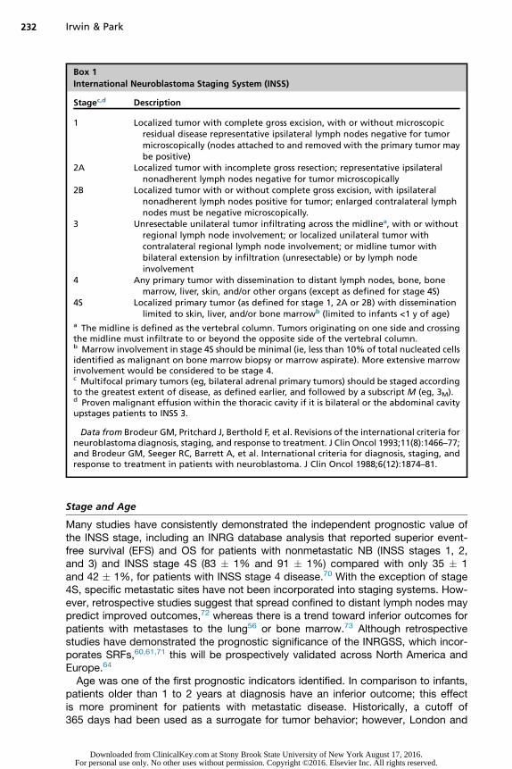
Box 1
International Neuroblastoma Staging System (INSS)
Stagec,d Description
1 Localized tumor with complete gross excision, with or without microscopicresidual disease representative ipsilateral lymph nodes negative for tumormicroscopically (nodes attached to and removed with the primary tumor maybe positive)
2A Localized tumor with incomplete gross resection; representative ipsilateralnonadherent lymph nodes negative for tumor microscopically
2B Localized tumor with or without complete gross excision, with ipsilateralnonadherent lymph nodes positive for tumor; enlarged contralateral lymphnodes must be negative microscopically.
3 Unresectable unilateral tumor infiltrating across the midlinea, with or withoutregional lymph node involvement; or localized unilateral tumor withcontralateral regional lymph node involvement; or midline tumor withbilateral extension by infiltration (unresectable) or by lymph nodeinvolvement
4 Any primary tumor with dissemination to distant lymph nodes, bone, bonemarrow, liver, skin, and/or other organs (except as defined for stage 4S)
4S Localized primary tumor (as defined for stage 1, 2A or 2B) with disseminationlimited to skin, liver, and/or bone marrowb (limited to infants <1 y of age)
a The midline is defined as the vertebral column. Tumors originating on one side and crossingthe midline must infiltrate to or beyond the opposite side of the vertebral column.b Marrow involvement in stage 4S should be minimal (ie, less than 10% of total nucleated cellsidentified as malignant on bone marrow biopsy or marrow aspirate). More extensive marrowinvolvement would be considered to be stage 4.c Multifocal primary tumors (eg, bilateral adrenal primary tumors) should be staged accordingto the greatest extent of disease, as defined earlier, and followed by a subscript M (eg, 3M).d Proven malignant effusion within the thoracic cavity if it is bilateral or the abdominal cavityupstages patients to INSS 3.
Data from Brodeur GM, Pritchard J, Berthold F, et al. Revisions of the international criteria forneuroblastoma diagnosis, staging, and response to treatment. J Clin Oncol 1993;11(8):1466–77;and Brodeur GM, Seeger RC, Barrett A, et al. International criteria for diagnosis, staging, andresponse to treatment in patients with neuroblastoma. J Clin Oncol 1988;6(12):1874–81.
Irwin & Park232
Stage and Age
Many studies have consistently demonstrated the independent prognostic value ofthe INSS stage, including an INRG database analysis that reported superior event-free survival (EFS) and OS for patients with nonmetastatic NB (INSS stages 1, 2,and 3) and INSS stage 4S (83 � 1% and 91 � 1%) compared with only 35 � 1and 42 � 1%, for patients with INSS stage 4 disease.70 With the exception of stage4S, specific metastatic sites have not been incorporated into staging systems. How-ever, retrospective studies suggest that spread confined to distant lymph nodes maypredict improved outcomes,72 whereas there is a trend toward inferior outcomes forpatients with metastases to the lung56 or bone marrow.73 Although retrospectivestudies have demonstrated the prognostic significance of the INRGSS, which incor-porates SRFs,60,61,71 this will be prospectively validated across North America andEurope.64
Age was one of the first prognostic indicators identified. In comparison to infants,patients older than 1 to 2 years at diagnosis have an inferior outcome; this effectis more prominent for patients with metastatic disease. Historically, a cutoff of365 days had been used as a surrogate for tumor behavior; however, London and
Downloaded from ClinicalKey.com at Stony Brook State University of New York August 17, 2016.For personal use only. No other uses without permission. Copyright ©2016. Elsevier Inc. All rights reserved.

Box 2
International Risk Group Staging System (INRGSS)
Stagea Description
L1 Localized tumor not involving vital structures as defined by the list of image-definedrisk factors and confined to one body compartment
L2 Locoregional tumor with presence of one or more IDRFs (see Box 1)M Distant metastatic disease (except stage MS)MS Metastatic disease in children younger than 18 mo with metastases confined to skin,
liver, and/or bone marrowa Patients with multifocal primary tumors should be staged according to the greatest extent ofdisease as defined in the table.
Data from Monclair T, Brodeur GM, Ambros PF, et al. The International Neuroblastoma RiskGroup (INRG) staging system: an INRG task force report. J Clin Oncol 2009;27(2):298–303.
Neuroblastoma 233
colleagues5 studied the continuous nature of age for 3666 patients and concluded thatthe most prognostic cutoff was 460 days (15.1 months). Several retrospective studiesspecifically examined whether 18 months might represent a more clinically relevantcutoff and demonstrated that EFS and OS for patients with INSS stage 4 diseaseaged 12 to 18 months (with favorable tumor biology) was similar to that of patientsaged less than 12 months.74,75 Similarly, patients with INSS stage 3 disease aged12 to 18 months had a superior outcome to those older than 18 months.76 ProspectiveCOG trials will determine whether reduction of therapy for toddlers aged 12 to18 months with biologically favorable tumors, traditionally treated with more intensiveregimens, will still provide superior outcomes.77 Older children, adolescents, andyoung adults with NB have a more indolent course and worse overall outcome despiteinfrequent MYCN oncogene amplification (MYCNA); however, no specific prognosticage cutoffs greater than 18 months have been identified.78
Histopathology
Pathologic characteristics have been used to further classify tumors into favorableand unfavorable categories, initially using a system developed by Shimada andcolleagues79 that provided the basis for the more recently revised InternationalNeuroblastoma Pathology Committee (INPC) criteria. The prognostic value of INPCclassification, based on age, presence of Schwannian stroma, grade of neuroblasticdifferentiation, andMitosis-karyorrhexis index, has been validated in large cooperativegroup studies80,81 to identify specific patient risk groups that may benefit from modi-fied therapy. In the COG P9641, patients with INSS stage 1 and 2 disease with favor-able histology had a significantly better outcome than those with unfavorable histology(UH) (EFS 90 � 3% and 72 � 7%, OS 99 � 1% and 86 � 5%).82
Tumor Genetics
NB genetic features have been used for risk stratification for more than 20 years. Twobroad categories of genetic aberration patterns include (1) tumors with whole chromo-some gains, lack of structural changes, and hyperdiploid karyotype and (2) tumorswith segmental chromosomal aberrations (SCAs) and diploid DNA content, whichare often associated with poor outcomes. SCAs often include partial gains and lossesof chromosomal regions predicted to encode oncogenes and tumor suppressors,respectively.
Downloaded from ClinicalKey.com at Stony Brook State University of New York August 17, 2016.For personal use only. No other uses without permission. Copyright ©2016. Elsevier Inc. All rights reserved.
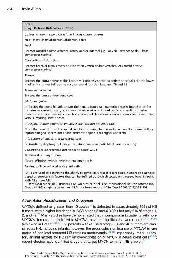
Box 3
Image Defined Risk Factors (IDRFs)
Ipsilateral tumor extension within 2 body compartments
Neck-chest, chest-abdomen, abdomen-pelvis
Neck
Encases carotid and/or vertebral artery and/or internal jugular vein; extends to skull base;compresses trachea
Cervicothoracic junction
Encases brachial plexus roots or subclavian vessels and/or vertebral or carotid artery;compresses trachea
Thorax
Encases the aorta and/or major branches; compresses trachea and/or principal bronchi; lowermediastinal tumor infiltrating costovertebral junction between T9 and 12
Thoracoabdominal
Encases the aorta and/or vena cava
Abdomen/pelvis
Infiltrates the porta hepatis and/or the hepatoduodenal ligament; encases branches of thesuperior mesenteric artery at the mesenteric root or origin of celiac axis and/or superiormesenteric artery; invades one or both renal pedicles; encases aorta and/or vena cava or iliacvessels, crossing sciatic notch
Intraspinal tumor extension whatever the location provided that
More than one-third of the spinal canal in the axial plane invaded and/or the perimedullaryleptomeningeal spaces not visible and/or the spinal cord signal abnormal
Infiltration of adjacent organs/structures
Pericardium, diaphragm, kidney, liver, duodeno-pancreatic block, and mesentery
Conditions to be recorded but not considered IDRFs
Multifocal primary tumors
Pleural effusion, with or without malignant cells
Ascites, with or without malignant cells
IDRFs are used to determine the ability to completely resect locoregional tumors at diagnosisbased on surgical risk factors that can be defined by IDRFs detected on cross-sectional imagingwith CT and/or MRI.
Data from Monclair T, Brodeur GM, Ambros PF, et al. The International Neuroblastoma RiskGroup (INRG) staging system: an INRG task force report. J Clin Oncol 2009;27(2):298–303.
234 Irwin & Park
Allelic Gains, Amplifications, and Oncogenes
MYCNA defined as greater than 10 copies83 is detected in approximately 20% of NBtumors, with a higher incidence in INSS stages 3 and 4 (40%) but only 5% of stages 1,2, and 4s.70 Many studies have demonstrated that in comparison to patients with non-MYCNA tumors, patients with MYCNA have a significantly worse outcome84,85
(reviewed in Refs.49,86,87). All patients withMYCNA stage 3, 4 and 4S tumors are clas-sified as HR, including infants; however, the prognostic significance ofMYCNA in rarecases of localized resected NB remains controversial.88,89 Importantly, most labora-tory animal models for NB rely on overexpression of MYCN in neural crest cells36,90;recent studies have identified drugs that target MYCN to inhibit NB growth.91
Downloaded from ClinicalKey.com at Stony Brook State University of New York August 17, 2016.For personal use only. No other uses without permission. Copyright ©2016. Elsevier Inc. All rights reserved.
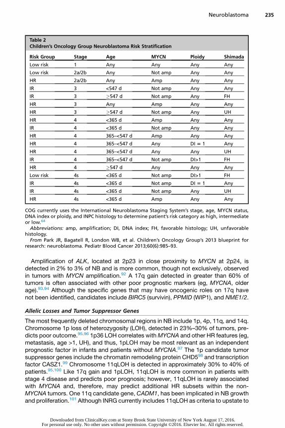
Table 2Children’s Oncology Group Neuroblastoma Risk Stratification
Risk Group Stage Age MYCN Ploidy Shimada
Low risk 1 Any Any Any Any
Low risk 2a/2b Any Not amp Any Any
HR 2a/2b Any Amp Any Any
IR 3 <547 d Not amp Any Any
IR 3 �547 d Not amp Any FH
HR 3 Any Amp Any Any
HR 3 �547 d Not amp Any UH
HR 4 <365 d Amp Any Any
IR 4 <365 d Not amp Any Any
HR 4 365–<547 d Amp Any Any
HR 4 365–<547 d Any DI 5 1 Any
HR 4 365–<547 d Any Any UH
IR 4 365–<547 d Not amp DI>1 FH
HR 4 �547 d Any Any Any
Low risk 4s <365 d Not amp DI>1 FH
IR 4s <365 d Not amp DI 5 1 Any
IR 4s <365 d Not amp Any UH
HR 4s <365 d Amp Any Any
COG currently uses the International Neuroblastoma Staging System’s stage, age, MYCN status,DNA index or ploidy, and INPC histology to determine patient’s risk category as high, intermediateor low.64
Abbreviations: amp, amplification; DI, DNA index; FH, favorable histology; UH, unfavorablehistology.
From Park JR, Bagatell R, London WB, et al. Children’s Oncology Group’s 2013 blueprint forresearch: neuroblastoma. Pediatr Blood Cancer 2013;60(6):985–93.
Neuroblastoma 235
Amplification of ALK, located at 2p23 in close proximity to MYCN at 2p24, isdetected in 2% to 3% of NB and is more common, though not exclusively, observedin tumors with MYCN amplification.92 A 17q gain detected in greater than 60% oftumors is often associated with other poor prognostic markers (eg, MYCNA, olderage).93,94 Although the specific genes that may have oncogenic roles on 17q havenot been identified, candidates include BIRC5 (survivin), PPMID (WIP1), and NME1/2.
Allelic Losses and Tumor Suppressor Genes
The most frequently deleted chromosomal regions in NB include 1p, 4p, 11q, and 14q.Chromosome 1p loss of heterozygosity (LOH), detected in 23%–30% of tumors, pre-dicts poor outcome.95,96 1p36 LOH correlates withMYCNA and other HR features (eg,metastasis, age >1, UH), and thus, 1pLOH may be most relevant as an independentprognostic factor in infants and patients without MYCNA.97 The 1p candidate tumorsuppressor genes include the chromatin remodeling protein CHD598 and transcriptionfactor CASZ1.99 Chromosome 11qLOH is detected in approximately 30% to 40% ofpatients.95,100 Like 17q gain and 1pLOH, 11qLOH is more common in patients withstage 4 disease and predicts poor prognosis; however, 11qLOH is rarely associatedwith MYCNA and, therefore, may predict additional HR subsets within the non-MYCNA tumors. One 11q candidate gene, CADM1, has been implicated in NB growthand proliferation.101 Although INRG currently includes 11qLOH as criteria to upstate to
Downloaded from ClinicalKey.com at Stony Brook State University of New York August 17, 2016.For personal use only. No other uses without permission. Copyright ©2016. Elsevier Inc. All rights reserved.

Box 4
INRG consensus pretreatment classification schema
INRGStage Age (mo) Histologic Category Grade of Tumor Differentiation MYCN 11q Aberration Ploidy
Pretreatment RiskGroup
L1/L2 — GN maturing; GNB intermixed — — — — A. Very lowL1 — Any, except GN maturing or GNB
intermixed— NA — — B. Very low
Amp — — K. HighL2 <18 Any, except GN maturing or GNB
intermixed— NA No — D. Low
Yes — G. Intermediate�18 GNB nodular; neuroblastoma Differentiating NA No — E. Low
Yes — —Poorly differentiated or undifferentiated NA — — H. Intermediate
Amp — — N. HighM <18 — — NA — Hyperdiploid F. Low
<12 — — NA — Diploid I. Intermediate12–<18 — — NA — Diploid J. Intermediate<18 — — Amp — — O. High�18 — — — — — P. High
MS <18 — — NA No — C. Very lowYes — Q. High
Amp — — R. High
Classification schema is based on analysis of 8800 patients in the INRG database (1990–2002). Risk groups are very low risk (5-year event-free survival[EFS] >85%); low risk (5-year EFS >75% to �85%); IR (5-year EFS �50% to �75%); HR (5-year EFS <50%).
Staging of L1, L2, M, and MS described in Fig. 2B.Abbreviations: Amp, amplified; EFS, event-free survival; GN, ganglioneuroma; GNB, ganglioneuroblastoma; NA, not amplified.
Adapted from Cohn SL, Pearson AD, LondonWB, et al. The International Neuroblastoma Risk Group [INRG] classification system: an INRG task force report. JClin Oncol 2009;27(2):295.
Irwin
&Park
236
Dow
nloaded from C
linicalKey.com
at Stony Brook State U
niversity of New
York A
ugust 17, 2016.For personal use only. N
o other uses without perm
ission. Copyright ©
2016. Elsevier Inc. A
ll rights reserved.

Neuroblastoma 237
HR classification, prospective trials are ongoing to determine whether 11qLOHpredicts poor outcomes for non-HR patients.
Segmental Chromosome Aberrations
Historically, individual chromosomal loci were analyzed using polymerase chainreaction or fluorescent in situ hybridization–based assays. Recent studies using tech-niques that assess the whole genome, such as comparative genome hybridization andSNP arrays, demonstrate that it is the genomic pattern and not individual losses/gainsthat is most prognostic. Tumors with numerical chromosomal abnormalities (NCAs)characterized by whole chromosome gains and losses have an excellent outcome,even in patients greater than 18 mo. In contrast, patients with segmental chromosomeaberrations (SCA), characterized by gains and losses of smaller fragments, have aninferior outcome.102,103 SCAs may be a particularly strong predictor of poor outcomein infants with locally unresectable or metastatic non-MYCN amplified tumors.104 Pro-spective trials in North America and Europe will determine whether the presence ofSCAs (�1 of the following: segmental loss at 1p, 3p, 4p, 11q or gain at 1q, 2p, or17q) can distinguish less favorable subsets of patients within the non-HR groups ofpatients and potentially replace tests that detect single gene losses/gains.
DNA Content
Ploidy or tumor DNA content (chromosome number) is a powerful predictor of survival.Hyperdiploid tumors (DNA index >1) with an increased amount of DNA in comparisonwith diploid tumors (DNA index 5 1) are associated with a more favorable prog-nosis.105,106 Ploidy is most prognostic in infants and patients with localized dis-ease74,107 and has been used prospectively to inform risk assignment and tailortherapy for patients with non-HR NB.108
Somatic Gene Mutations
Recently, next-generation sequencing approaches have revealed that, in contrast toadult carcinomas, there is a striking lack of recurrent NB tumor (somatic) muta-tions.109–112 The most commonly mutated gene is ALK (8%–10%), with an additional3% harboring ALK amplification.10 ALK genomic aberrations are detected in allrisk groups and are associated with an adverse outcome,33 and high levels of ALKprotein or amplification may correlate with poor outcomes independent of mutationstatus.32,113,114 Mutations in ATRX (alpha thalassemia/mental retardation syndromeX linked), which is involved in telomere maintenance, are detected more frequentlyin older patients with NB.112 Deletions and point mutations of the chromatin remodel-ing proteins AT-rich interactive domain 1A and B (ARID1a/1b) were detected in 11% oftumors.111 Other mutations detected in less than 5% of tumors include MYCN, TP53,PTPN11, and genes involved in Ras/MAPK signaling. Current studies are exploringwhether mutations may be more common at recurrence115 and whether epigeneticregulation of transcription and genomic organization, which has recently been re-ported to be involved in the medulloblastoma,116 may be playing similar roles in NB.
Molecular Factors and Expression Signatures
Because recurrent mutations are not frequent in NB, the identification of genes andsignaling pathways with altered expression have also been used to discover additionalprognostic factors and therapeutic targets involved in NB differentiation, apoptosis,drug resistance, angiogenesis, metastasis, and inflammation. Extensive reviews ofthese molecular factors have been the subject of several recent reviews,49,117,118
and a subset of the most well studied are included later.
Downloaded from ClinicalKey.com at Stony Brook State University of New York August 17, 2016.For personal use only. No other uses without permission. Copyright ©2016. Elsevier Inc. All rights reserved.

Irwin & Park238
Neurotrophin signaling has central roles in normal neuronal cell development, andthe clinical and biological roles of TRK receptors (NTRK1, 2, 3 encoding TrkA, B, C)and their ligands (NGF, BDNF, and NT-3) have been extensively studied in NB(reviewed in Ref.119). TrkA expression is highest in tumors with favorable biologicalcharacteristics and outcomes, and TrkA induces apoptosis and/or differentiationin vitro. TrkA signaling has been implicated in mediating spontaneous regressionthat is often observed in infants with localized or stage 4S disease.120 In contrast,TrkB has pro-proliferative and migratory properties, enhances chemoresistance,and is highly expressed in biologically unfavorable MYCNA NB. Although the TrkB in-hibitor lestaurtinib did not show efficacy in a phase 1 trial,121 trks and proteins involvedin neural crest development and differentiation pathways may still represent potentialtherapeutic targets.Disruption of proteins involved in apoptotic pathways, including multidrug-
resistance proteins, such as MDR-1, bcl-2 family proteins, caspase-8, mTOR/PI3kinase, and TP53/HDM2, have also been shown to play important roles in NB initiationand progression. There are many ongoing pre-clinical studies to determine the abilityto pharmacologically target these pathways.122–126 Many genes involved in NB,including caspase 8 and the RASSF1A tumor suppressor, are inactivated by the pro-moter hypermethylation,126–128 which contributes to resistance to apoptosis inducedby many therapies. Demethylating agents, such as decitabine, have been tested inphase I studies.129 Enhanced angiogenesis and high expression of proangiogenic fac-tors, such as vascular endothelial growth factor and basic fibroblast growth factor areassociated with more aggressive NB tumors; early phase clinical trials of drugs thatblock these pathways have been completed.130–132
Rather than focusing on specific candidate genes, several investigators have iden-tified multigene expression profiles that predict outcome and may lead to furtherrefinement of risk categories. One large study demonstrated that the expression of59 genes was an independent predictor of outcome, even after controlling for currentlyused risk factors, with an odds ratio of 19.3 for OS and 3.96 for progression-freesurvival.133 Additional retrospective studies have identified other multigene classifiers(ranging from 3 to >50 genes).134–138 Although most of these signatures have not beenstudied in specific NB risk groups, Asgharzadah and collegues139,140 recently demon-strated that a 14-gene classifier can be used to specifically identify subsets of HR pa-tients with the worst prognosis. Many of these signatures include genes implicated inNB pathogenesis, neural development, and inflammation/immune response. Recentreports also demonstrate prognostic profiles of microRNAs, small 22 to 25 nucleotideRNAs that inhibit protein translation or target mRNA degradation141–143 (reviewed inRef.144).
MANAGEMENT GOALS
Diagnosis and therapy requires a multidisciplinary approach. Surgical biopsy is usuallyrequired to assess tumor genetic and histologic features and is most critical forpatients less than 18 months of age with metastatic disease and those withlocalized unresectable tumors. The improved understanding of NB biology and itsimpact on prognosis has resulted in successful tailoring based on risk stratification(low risk, IR, and HR) using many of the pretreatment clinical and biological riskfactors discussed earlier (see Box 4, Table 2). The requirements for further surgicalresection, chemotherapy, radiotherapy and/or immunotherapy is based on thepatients’ specific risk category (Table 3) and, in part, response as outlined in the Inter-national Neuroblastoma Response Criteria, which is currently under revision. When
Downloaded from ClinicalKey.com at Stony Brook State University of New York August 17, 2016.For personal use only. No other uses without permission. Copyright ©2016. Elsevier Inc. All rights reserved.

Neuroblastoma 239
possible, exposure to chemotherapy is limited for patients with regional disease,whereas radiotherapy is limited to those with advanced disease with unfavorablecharacteristics.
Low Risk
Survival rates for patients with INSS stage 1 disease, regardless of biological factors,are excellent with surgery alone and rare recurrences can often be cured with salvagechemotherapy.145,146 Similarly, chemotherapy can be omitted for most patients withbiologically favorable but incompletely resected localized tumors (INSS 2A, 2B),with survival rates greater than 95%.82,146–148 In general, for patients with INSS stage1, 2A, and 2B (mostly INRG stage L1), chemotherapy is reserved for patients withlocalized NB who have life- or organ-threatening symptoms or the minority of patientswho experience recurrence or progressive disease.Because previous infant screening studies15,16,149–151 and European trials152,153
have suggested that subsets of biologically favorable NB can spontaneously differen-tiate and regress, a recent COG trial (ANBL00P2) studied whether infants less than6 months of age with small localized adrenal masses (including those detected by pre-natal ultrasound) could be observed without biopsy, surgery, or chemotherapy.154
Eighty-one percent of patients demonstrated spontaneous regression without surgicalintervention; the 3-year EFS and OS were 97% and 100%, respectively.Like many localized tumors in infants, most of stage 4S NBwithoutMYCNA undergo
spontaneous regression.59,155 Chemotherapy or low-dose radiotherapy is reserved forsymptoms of large tumors or massive hepatomegaly causing mechanical obstruction,respiratory distress, and/or liver dysfunction and should be initiated as soon aspossible to prevent the morbidity and mortality frequently associated with this formof the disease, especially in very young infants.105,156,157
Overall, these data support continued reduction of chemotherapy exposure andsurgery for most low risk asymptomatic patients, while strategies to improve survivalfor the rare subsets of non-HR patients with unfavorable pathology or biology (eg,
Table 3Treatment strategies based on risk group (COG)
Low (40%) IR (20%) HR (40%)
Survival (EFS) >95 80–95 40–50
Patient/tumorcharacteristics
� Localized, resectabletumors
� Localizedunresectable
� Infants withmetastases(no MYCNA)
� Metastases >18 mo� Unresectable with
unfavorable biology(eg, MYCNA)
Treatment Observation OR surgery(chemotherapy only forsymptoms (eg, stage 4Sor cord compression))
Chemotherapy(2–8 cycles basedon biology),surgery
Chemotherapy, surgery,radiation, myeloablativetherapy with autologousstem cell rescue,immunotherapy andbiological agents(isotretinoin)
Summarized are general treatment strategies and characteristics for each risk group based onrecent COG trials. This chart includes the most common characteristics for each group and overalltreatment strategies. These treatments may vary across different cooperative groups internation-ally and change based on ongoing and future clinical trials. The approximate relative proportion ofpatients in each risk group is based on data from the COG ANBL00B1 Biology Study (since 2001).64
Downloaded from ClinicalKey.com at Stony Brook State University of New York August 17, 2016.For personal use only. No other uses without permission. Copyright ©2016. Elsevier Inc. All rights reserved.
Irwin & Park240
diploid tumors with SCAs)82,158 are being examined in prospective SIOPEN and COGtrials.64
Intermediate Risk
IR classification encompasses a wide spectrum of disease for which surgical resectionand moderate-dose multiagent chemotherapy are the backbone of most regimens. IRincludes subsets of patients with INSS stage 3 (mostly INRG L2) disease and infantswith stage 4/M disease with favorable biological features. Survival following surgicalresection and moderate-dose chemotherapy, including carboplatin or cisplatin, doxo-rubicin, etoposide, and cyclophosphamide, is greater than 90% for children whosetumors exhibit favorable characteristics, including infants with stage 4/M who lackMYCNA.159–161 These high survival rates were maintained in 2 prospective COG IRtrials in which therapy was reduced further based on histology, ploidy, and 1p and11qLOH status.108,157 Small series have suggested that IR patients with localizedNB with favorable biology can be observed without chemotherapy.153,162 Ongoingprospective international trials will determine whether SCA status can be used torefine treatment assignment to further reduce, and in some cases eliminate, therapyfor most IR patients with favorable histology and genomics.
High Risk
Outcome of HR patients (mainly stage 4 > 18 months of age and stage 3 MYCNA orstage 3 > 18 months with unfavorable histology tumors) remains poor despite im-provements in survival (Fig. 5).163–168 Standard HR therapy involves 3 components:(1) induction chemotherapy and local control, (2) consolidation, and (3) postconsolida-tion/maintenance. These regimens have evolved significantly over the past 20 yearsbased on work by several international cooperative groups and smaller cohort studiessummarized later.
Induction therapyThere is a correlation between survival and end-of-induction response63,169; despitechemotherapy dose intensification, approximately 20% of patient will progress orhave inadequate response to induction therapy. Standard North American (COG) in-duction regimens include combinations of anthracyclines, alkylators, platinum com-pounds, and topoisomerase II inhibitors delivered every 21 days for 5 to 7 cycles.SIOPEN uses a rapid regimen whereby cycles are delivered every 10 days basedon results that demonstrated superior 5-year EFS of 30%, compared with 18% forstandard interval chemotherapy.165 The topoisomerase I inhibitor topotecan, whichhas demonstrated efficacy in recurrent NB,170 has recently been incorporated intoCOG induction regimens.64,171
Local controlOptimal local control is achieved with a combination of aggressive surgical resectionand external beam radiotherapy to the primary tumor. Surgery of the primary andbulky metastatic disease is usually delayed until after 4 to 6 cycles of chemotherapyto improve resectability and minimize complications172; however, there are conflictingreports as to whether complete primary tumor resection impacts patient outcomes inHR NB.173–176
NB is one of the most radiosensitive pediatric solid tumors, and doses of 2160 cGyin daily 180 cGY fractions to the primary sites decrease local recurrence rates for HRpatients.177,178 A recently completed prospective COG trial will determine whetherhigher radiation doses delivered to incompletely resected tumors improves local con-trol rates. Radiation is also often delivered to residual MIBG-avid metastatic sites, and
Downloaded from ClinicalKey.com at Stony Brook State University of New York August 17, 2016.For personal use only. No other uses without permission. Copyright ©2016. Elsevier Inc. All rights reserved.

Fig. 5. Survival for HR patients with NB based on treatment era. The EFS (A) and OS (B)Kaplan-Meier survival curves calculated from the time of diagnosis for children enrolledonto COG (since 2001) and Children’s Cancer Group and Pediatric Oncology Group Neuro-blastoma Biology trials between 1990 and 2010 (N 5 3389) shown in 5-year intervals, begin-ning in 1990. (With permission from Children’s Oncology Group Statistical Data Center.)
241
Downloaded from ClinicalKey.com at Stony Brook State University of New York August 17, 2016.For personal use only. No other uses without permission. Copyright ©2016. Elsevier Inc. All rights reserved.

Irwin & Park242
a recent report suggests that nonirradiated lesions have a higher likelihood of involve-ment at the time of first relapse.179
Myeloablative consolidation therapyOver the past 2 decades, several clinical trials performed in Germany, Europe, andNorth America demonstrated improved outcomes following myeloablative therapy(MAT) with autologous bone marrow or, more recently, autologous peripheral bloodstem cell rescue as compared with maintenance chemotherapy or observa-tion.167,169,180,181 These data together with a recent Cochrane systems meta-analysis suggest that MAT has resulted in improvements in EFS.182 Recent andongoing trials are aimed at identifying the optimal intensity and chemotherapy combi-nations for MAT regimens. Preliminary SIOPEN results suggest that patients random-ized to a Busulfan-Melphalan (Bu-Mel) regimen had outcomes superior to those whoreceived carboplatin-etoposide-melphalan.175 Before adoption of Bu-Mel, the COGand other groups are examining the efficacy and toxicities of Bu-Mel MAT in combi-nation with different induction regimens and postconsolidation immunotherapy.64 Inaddition, data will soon be available from COG study ANBL0532, which randomizedpatients to single and tandem MAT and was based on a limited institution tandemMAT study with 3- and 5-year EFS rates of 55% and 47%.183 Future trials will alsoaim to identify those at highest risk for recurrence and assess whether additional ther-apies during induction or consolidation improve their outcome.64
Postconsolidation biologic and immunotherapiesInitial results from CCG-3891 demonstrated efficacy for the synthetic retinoid isotret-inoin [cis-retinoic acid (cis-RA)] in treating minimal residual NB after MAT and estab-lished a standard for the use of noncytotoxic differentiation therapy for minimalresidual disease.164 A recent randomized-controlled trial led by Yu and colleagues166
demonstrated that the addition of the anti-GD2 chimeric monoclonal antibody (mAb) inconjunction with cytokines (granulocyte-macrophage colony-stimulation factor andinterleukin 2) improved survival, establishing a role for immunotherapy in the standardtreatment of HR patients. Additional studies have shown efficacy for different anti-GD2regimens at diagnosis and recurrence.184,185 Future immunotherapy regimens areaimed at determining the importance of cytokines andmAb and examining biomarkersthat may predict which patients are most likely to respond favorably to this regimen,which has many side effects, including allergic reactions, fever, hypotension, capillaryleak syndrome, and pain (caused by cross-reactivity with GD2 expressed on periph-eral nerve cells). Early phase trials are also examining different antibodies and additionof immunomodulators (see “Recurrence” section).
LATE EFFECTS
There are few comprehensive reports of the prevalence of long-term effects in NB sur-vivors, in part because of the poor prognosis for HR NB. Late effects are generallyrelated to chemotherapy/radiation dose intensities, with the highest toxicities inpatients who underwent MAT.186–190 Recent pharmacogenomic studies have begunto identify germline variants or SNPs that may predict which patients are most suscep-tible to specific chemotherapy toxicities.191 Ototoxicity, renal dysfunction, and endo-crine late effects, including hypothyroidism, ovarian dysfunction and infertility, havebeen detected in most HR patients with NB.186 Secondary cancers, most commonlymyelodysplastic syndrome and acute myelogenous leukemia, have been reported in1% to 8% of patients enrolled on trials and small series of NB survivors187,192,193
and have been attributed to etoposide exposure, radiation, high-dose MIBG, and
Downloaded from ClinicalKey.com at Stony Brook State University of New York August 17, 2016.For personal use only. No other uses without permission. Copyright ©2016. Elsevier Inc. All rights reserved.
Neuroblastoma 243
other agents. In addition to hematopoietic malignancies, solid tumors of the thyroid,bone, and kidney have been reported. Patients may also have effects related to tumorlocation, such as visual impairment caused by orbital metastases and neurologic com-plications or scoliosis following spinal cord compression.194,195
RECURRENCE
Despite recent advances, greater than 50% of patients with HR NB experience tumorrecurrence. Although there are no proven curative therapies, some patients achieveprolonged survival even after multiple relapses. In the INRG database, low/IR patientswith NB who relapsed had an OS of 65% 5 years after recurrence, whereas for thosewith metastatic disease, 5-year OS was 8%.96 Thus, research into novel therapies is ahigh priority and has been the subject of several recent reviews.117,196,197
Relapse strategies can be divided into chemotherapies, MIBG/radioisotopes, im-munotherapies, and targeted therapies. Current phase I and II trials often involve com-binations of these approaches. Cytotoxic chemotherapies commonly used for relapseinclude topotecan or irinotecan-based regimens170,198–200 as well as ifosfamide, car-boplatin, etoposide201 and often result in transient responses or stable disease butpoor long-term survival. Iodide-131- MIBG, which targets high doses of radiation toNB cells, is the most effective single agent for relapsed NB, with response ratesgreater than 30%.202–205 Current MIBG trials will determine the efficacy of concurrentradiosensitizing chemotherapies and feasibility of delivering MIBG followed by MAT topotentially incorporate MIBG into upfront therapy for HR NB.64
Building on the success of anti-GD2 mAbs, novel approaches to enhance mAbefficacy, such as the addition of lenolidomide,206 which activates natural killer cells,and active immunization with anti-idiotype antibodies, are being studied in relapsedpatients.207 Among the most promising phase I trials are those that use a patient’sown cytotoxic T cells (CTLs) that can be redirected against tumor-associated antigens(eg, GD2, L1CAM). Autologous CTLs engineered to overexpress chimeric antigen re-ceptors are infused and have been shown to persist and demonstrate antitumor activ-ity in patients with NB.208–211
There are several potential targets, and respective inhibitors, for recurrent NBbased on preclinical and, in certain cases, phase I trials. A subset of ALK aberrant tu-mors can be targeted with crizotinib, and trials with second-generation ALK inhibitorsand combinations with chemotherapy are underway.37,92,212 For patients withMYCNA, preclinical studies suggest that bromodomain and extraterminal domain(BET) inhibitors can induce cell death by interfering with MYCN transcription.91 Otherdrugs that have effects on MYCN stability (aurora kinase A and mTOR inhibitors)213 aswell as those that target MYC-dependent metabolic changes214 are being studied.There is significant interest in drugs, such as histone deacetylase inhibitors, that areless targeted and instead modulate the expression of many genes to induce death,differentiation, and enhance the response to chemotherapies in NB cells.215 Otherdrugs targeting cell cycle (eg, Chk1, Wee-1, CDK4/6), angiogenesis, and differentia-tion are also under investigation.196
Current trial designs for patients with relapsed NB have incorporated novelapproaches, such as pick the winner whereby patients are randomized to receivedifferent novel agents in combination with a common chemotherapy backbone regi-mens. In addition, many early phase NB trials will incorporate precision medicine bytailoring treatment based on individual patient tumor aberrations. These studies willincreasingly depend on genomic and molecular studies of tumors, particularly at thetime of relapse, when mutations may be more common.216
Downloaded from ClinicalKey.com at Stony Brook State University of New York August 17, 2016.For personal use only. No other uses without permission. Copyright ©2016. Elsevier Inc. All rights reserved.

Irwin & Park244
FUTURE DIRECTIONS
NB is a heterogeneous tumor for which molecular and genetic determinants affectclinical behavior. Further advances in the understanding of aberrantly expressedgenes and pathways will continue to inform and refine risk stratification and treatmentand identify novel therapeutic targets. For patients with low risk and IR NB, thesegenetic factors will help to identify rare patients who still require treatment as wecontinue to reduce exposures to chemotherapy and surgery for most non-HRpatients. In contrast, for HR patients, we need to better predict those at greatestrisk of treatment failure or recurrence, either at diagnosis (eg, genetic signatures) orbased on their response to treatment (eg, persistent MIBG positive metastases).Furthermore, molecular and genetic studies of tumors at the time of recurrence willbe required to specifically identify targets in this chemoresistant population. Interna-tional collaborations, including INRG databases, are critical for the development ofrisk stratification and response classifications as well as advances in basic and trans-lational studies, especially for rare populations (eg 4S, OMS). Future studies will movetoward more refined risk classifications and treatments based on individual tumor ab-errations as well as more attention to survivors to better understand the extent andindividual susceptibility to long-term side effects of our treatments.
REFERENCES
1. Ries L, Smith M, Gurney J, et al. Cancer incidence and survival among childrenand adolescents: United States SEER Program 1975–1995, National CancerInstitute, SEER Program. Bethesda (MD): NIH Pub; 1999. p. 99–4649.
2. Stiller CA, Parkin DM. International variations in the incidence of neuroblastoma.Int J Cancer 1992;52(4):538–43.
3. Heck JE, Ritz B, Hung RJ, et al. The epidemiology of neuroblastoma: a review.Paediatr Perinat Epidemiol 2009;23(2):125–43.
4. Spix C, Pastore G, Sankila R, et al. Neuroblastoma incidence and survival inEuropean children (1978–1997): report from the automated childhood cancerinformation system project. Eur J Cancer 2006;42(13):2081–91.
5. London WB, Castleberry RP, Matthay KK, et al. Evidence for an age cutoffgreater than 365 days for neuroblastoma risk group stratification in the Chil-dren’s Oncology Group. J Clin Oncol 2005;23(27):6459–65.
6. Johnson KA, Aplenc R, Bagatell R. Survival by race among children with extra-cranial solid tumors in the United States between 1985 and 2005. Pediatr BloodCancer 2011;56(3):425–31.
7. Henderson TO, Bhatia S, Pinto N, et al. Racial and ethnic disparities in risk andsurvival in children with neuroblastoma: a Children’s Oncology Group study.J Clin Oncol 2011;29(1):76–82.
8. Pinto N, Cipkala DA, Ladd PE, et al. Treatment of two cases with refractory, met-astatic intermediate-risk neuroblastoma with isotretinoin alone or observation.Pediatr Blood Cancer 2014;61(6):1104–6.
9. Zahm SH, Devesa SS. Childhood cancer: overview of incidence trendsand environmental carcinogens. Environ Health Perspect 1995;103(Suppl 6):177–84.
10. Connelly JM, Malkin MG. Environmental risk factors for brain tumors. CurrNeurol Neurosci Rep 2007;7(3):208–14.
11. Yamamoto K, Hayashi Y, Hanada R, et al. Mass screening and age-specific inci-dence of neuroblastoma in Saitama Prefecture, Japan. J Clin Oncol 1995;13(8):2033–8.
Downloaded from ClinicalKey.com at Stony Brook State University of New York August 17, 2016.For personal use only. No other uses without permission. Copyright ©2016. Elsevier Inc. All rights reserved.
Neuroblastoma 245
12. Yamamoto K, Ohta S, Ito E, et al. Marginal decrease in mortality and marked in-crease in incidence as a result of neuroblastoma screening at 6 months of age:cohort study in seven prefectures in Japan. J Clin Oncol 2002;20(5):1209–14.
13. Hiyama E, Iehara T, Sugimoto T, et al. Effectiveness of screening for neuroblas-toma at 6 months of age: a retrospective population-based cohort study. Lancet2008;371(9619):1173–80.
14. Schilling FH, Spix C, Berthold F, et al. Neuroblastoma screening at one year ofage. N Engl J Med 2002;346(14):1047–53.
15. Woods WG, Gao RN, Shuster JJ, et al. Screening of infants and mortality due toneuroblastoma. N Engl J Med 2002;346(14):1041–6.
16. Maris JM, Woods WG. Screening for neuroblastoma: a resurrected idea? Lancet2008;371(9619):1142–3.
17. Shojaei-Brosseau T, Chompret A, Abel A, et al. Genetic epidemiology of neuro-blastoma: a study of 426 cases at the Institut Gustave-Roussy in France. PediatrBlood Cancer 2004;42(1):99–105.
18. Trochet D, Bourdeaut F, Janoueix-Lerosey I, et al. Germline mutations of thepaired-like homeobox 2B (PHOX2B) gene in neuroblastoma. Am J Hum Genet2004;74(4):761–4.
19. Rohrer T, Trachsel D, Engelcke G, et al. Congenital central hypoventilation syn-drome associated with Hirschsprung’s disease and neuroblastoma: case ofmultiple neurocristopathies. Pediatr Pulmonol 2002;33(1):71–6.
20. Mosse YP, Laudenslager M, Khazi D, et al. Germline PHOX2B mutation in hered-itary neuroblastoma. Am J Hum Genet 2004;75(4):727–30.
21. Brems H, Beert E, de Ravel T, et al. Mechanisms in the pathogenesis of malig-nant tumours in neurofibromatosis type 1. Lancet Oncol 2009;10(5):508–15.
22. Clausen N, Andersson P, Tommerup N. Familial occurrence of neuroblastoma,von Recklinghausen’s neurofibromatosis, Hirschsprung’s agangliosis and jaw-winking syndrome. Acta Paediatr Scand 1989;78(5):736–41.
23. Longo L, Panza E, Schena F, et al. Genetic predisposition to familial neuroblas-toma: identification of two novel genomic regions at 2p and 12p. Hum Hered2007;63(3–4):205–11.
24. Tonini GP, McConville C, Cusano R, et al. Exclusion of candidate genes andchromosomal regions in familial neuroblastoma. Int J Mol Med 2001;7(1):85–9.
25. Maris JM, Kyemba SM, Rebbeck TR, et al. Familial predisposition to neuroblas-toma does not map to chromosome band 1p36. Cancer Res 1996;56(15):3421–5.
26. Maris JM, Weiss MJ, Mosse Y, et al. Evidence for a hereditary neuroblastomapredisposition locus at chromosome 16p12-13. Cancer Res 2002;62(22):6651–8.
27. Mosse YP, Laudenslager M, Longo L, et al. Identification of ALK as a majorfamilial neuroblastoma predisposition gene. Nature 2008;455(7215):930–5.
28. Janoueix-Lerosey I, Lequin D, Brugieres L, et al. Somatic and germline acti-vating mutations of the ALK kinase receptor in neuroblastoma. Nature 2008;455(7215):967–70.
29. de Pontual L, Kettaneh D, Gordon CT, et al. Germline gain-of-function mutationsof ALK disrupt central nervous system development. Hum Mutat 2011;32(3):272–6.
30. George RE, Sanda T, Hanna M, et al. Activating mutations in ALK provide a ther-apeutic target in neuroblastoma. Nature 2008;455(7215):975–8.
31. Chen Y, Takita J, Choi YL, et al. Oncogenic mutations of ALK kinase in neuro-blastoma. Nature 2008;455(7215):971–4.
Downloaded from ClinicalKey.com at Stony Brook State University of New York August 17, 2016.For personal use only. No other uses without permission. Copyright ©2016. Elsevier Inc. All rights reserved.

Irwin & Park246
32. Schulte JH,BachmannHS,BrockmeyerB, et al.HighALK receptor tyrosinekinaseexpression supersedes ALK mutation as a determining factor of an unfavorablephenotype in primary neuroblastoma. Clin Cancer Res 2011;17(15):5082–92.
33. Weiser DA, Bresler SC, Laudenslager M, et al. Stratification of patients with neu-roblastoma for targeted ALK inhibitor therapy. J Clin Oncol 2011;29 [abstract:9514].
34. Berry T, Luther W, Bhatnagar N, et al. The ALK (F1174L) mutation potentiates theoncogenic activity of MYCN in neuroblastoma. Cancer Cell 2012;22(1):117–30.
35. Heukamp LC, Thor T, Schramm A, et al. Targeted expression of mutated ALK in-duces neuroblastoma in transgenic mice. Sci Transl Med 2012;4(141):141ra191.
36. Zhu S, Lee JS, Guo F, et al. Activated ALK collaborates with MYCN in neuroblas-toma pathogenesis. Cancer Cell 2012;21(3):362–73.
37. Mosse YP, Lim MS, Voss SD, et al. Safety and activity of crizotinib for paediatricpatients with refractory solid tumours or anaplastic large-cell lymphoma: a chil-dren’s oncology group phase 1 consortium study. Lancet Oncol 2013;14(6):472–80.
38. DeBaun MR, Tucker MA. Risk of cancer during the first four years of life inchildren from The Beckwith-Wiedemann Syndrome Registry. J Pediatr 1998;132(3 Pt 1):398–400.
39. Birch JM, Alston RD, McNally RJ, et al. Relative frequency and morphology ofcancers in carriers of germline TP53 mutations. Oncogene 2001;20(34):4621–8.
40. Rossbach HC, Baschinsky D, Wynn T, et al. Composite adrenal anaplastic neu-roblastoma and virilizing adrenocortical tumor with germline TP53 R248W muta-tion. Pediatr Blood Cancer 2008;50(3):681–3.
41. Bissig H, Staehelin F, Tolnay M, et al. Co-occurrence of neuroblastoma andnephroblastoma in an infant with Fanconi’s anemia. Hum Pathol 2002;33(10):1047–51.
42. Reid S, Schindler D, Hanenberg H, et al. Biallelic mutations in PALB2 causeFanconi anemia subtype FA-N and predispose to childhood cancer. Nat Genet2007;39(2):162–4.
43. Maris JM, Mosse YP, Bradfield JP, et al. Chromosome 6p22 locus associatedwith clinically aggressive neuroblastoma. N Engl J Med 2008;358(24):2585–93.
44. Diskin SJ, Capasso M, Schnepp RW, et al. Common variation at 6q16 withinHACE1 and LIN28B influences susceptibility to neuroblastoma. Nat Genet2012;44(10):1126–30.
45. Capasso M, Devoto M, Hou C, et al. Common variations in BARD1 influencesusceptibility to high-risk neuroblastoma. Nat Genet 2009;41(6):718–23.
46. Diskin SJ, Hou C, Glessner JT, et al. Copy number variation at 1q21.1 associ-ated with neuroblastoma. Nature 2009;459(7249):987–91.
47. Wang K, Diskin SJ, Zhang H, et al. Integrative genomics identifies LMO1 as aneuroblastoma oncogene. Nature 2011;469(7329):216–20.
48. Diskin SJ, Capasso M, Diamond M, et al. Rare variants in TP53 and susceptibil-ity to neuroblastoma. J Natl Cancer Inst 2014;106(4):dju047.
49. Maris JM. Recent advances in neuroblastoma. N Engl J Med 2010;362(23):2202–11.
50. Vo KT, Matthay KK, Neuhaus J, et al. Clinical, biological and prognostic differencesbased on primary tumor site in neuroblastoma: a report from the International Neu-roblastoma Risk Group (INRG) project. J Clin Oncol 2014;32(28):3169–76.
51. De Bernardi B, Pianca C, Pistamiglio P, et al. Neuroblastoma with symptomaticspinal cord compression at diagnosis: treatment and results with 76 cases.J Clin Oncol 2001;19(1):183–90.
Downloaded from ClinicalKey.com at Stony Brook State University of New York August 17, 2016.For personal use only. No other uses without permission. Copyright ©2016. Elsevier Inc. All rights reserved.
Neuroblastoma 247
52. El Shafie M, Samuel D, Klippel CH, et al. Intractable diarrhea in children withVIP-secreting ganglioneuroblastomas. J Pediatr Surg 1983;18(1):34–6.
53. Scheibel E, Rechnitzer C, Fahrenkrug J, et al. Vasoactive intestinal polypeptide(VIP) in children with neural crest tumours. Acta Paediatr Scand 1982;71(5):721–5.
54. Matthay KK, Blaes F, Hero B, et al. Opsoclonus myoclonus syndrome in neuro-blastoma a report from a workshop on the dancing eyes syndrome at theadvances in neuroblastoma meeting in Genoa, Italy, 2004. Cancer Lett 2005;228(1–2):275–82.
55. Gorman MP. Update on diagnosis, treatment, and prognosis in opsoclonus-myoclonus-ataxia syndrome. Curr Opin Pediatr 2010;22(6):745–50.
56. Dubois SG, London WB, Zhang Y, et al. Lung metastases in neuroblastoma atinitial diagnosis: a report from the International Neuroblastoma Risk Group(INRG) project. Pediatr Blood Cancer 2008;51(5):589–92.
57. Kramer K, Kushner B, Heller G, et al. Neuroblastoma metastatic to the centralnervous system. The Memorial Sloan-Kettering Cancer Center experience anda literature review. Cancer 2001;91(8):1510–9.
58. Matthay KK, Brisse H, Couanet D, et al. Central nervous system metastases inneuroblastoma: radiologic, clinical, and biologic features in 23 patients. Cancer2003;98(1):155–65.
59. Evans AE, Chatten J, D’Angio GJ, et al. A review of 17 IV-S neuroblastomapatients at the Children’s Hospital of Philadelphia. Cancer 1980;45(5):833–9.
60. Simon T, Hero B, Benz-Bohm G, et al. Review of image defined risk factors inlocalized neuroblastoma patients: results of the GPOH NB97 trial. Pediatr BloodCancer 2008;50(5):965–9.
61. Cecchetto G, Mosseri V, De Bernardi B, et al. Surgical risk factors in primary sur-gery for localized neuroblastoma: the LNESG1 study of the European Interna-tional Society of Pediatric Oncology Neuroblastoma Group. J Clin Oncol 2005;23(33):8483–9.
62. Messina JA, Cheng SC, Franc BL, et al. Evaluation of semi-quantitative scoringsystem for metaiodobenzylguanidine (mIBG) scans in patients with relapsedneuroblastoma. Pediatr Blood Cancer 2006;47(7):865–74.
63. Yanik GA, Parisi MT, Shulkin BL, et al. Semiquantitative mIBG scoring as a prog-nostic indicator in patients with stage 4 neuroblastoma: a report from the chil-dren’s oncology group. J Nucl Med 2013;54(4):541–8.
64. Park JR, Bagatell R, London WB, et al. Children’s Oncology Group’s 2013blueprint for research: neuroblastoma. Pediatr Blood Cancer 2013;60(6):985–93.
65. Sharp SE, Shulkin BL, Gelfand MJ, et al. 123I-MIBG scintigraphy and 18F-FDGPET in neuroblastoma. J Nucl Med 2009;50(8):1237–43.
66. Zhang H, Huang R, Cheung NK, et al. Imaging the norepinephrine transporter inneuroblastoma: a comparison of [18F]-MFBG and 123I-MIBG. Clin Cancer Res2014;20(8):2182–91.
67. Taggart DR, Han MM, Quach A, et al. Comparison of iodine-123 metaiodoben-zylguanidine (MIBG) scan and [18F]fluorodeoxyglucose positron emissiontomography to evaluate response after iodine-131 MIBG therapy for relapsedneuroblastoma. J Clin Oncol 2009;27(32):5343–9.
68. Brodeur GM, Pritchard J, Berthold F, et al. Revisions of the international criteriafor neuroblastoma diagnosis, staging, and response to treatment. J Clin Oncol1993;11(8):1466–77.
Downloaded from ClinicalKey.com at Stony Brook State University of New York August 17, 2016.For personal use only. No other uses without permission. Copyright ©2016. Elsevier Inc. All rights reserved.

Irwin & Park248
69. Brodeur GM, Seeger RC, Barrett A, et al. International criteria for diagnosis,staging, and response to treatment in patients with neuroblastoma. J Clin Oncol1988;6(12):1874–81.
70. Cohn SL, Pearson AD, London WB, et al. The International Neuroblastoma RiskGroup (INRG) classification system: an INRG task force report. J Clin Oncol2009;27(2):289–97.
71. Monclair T, Brodeur GM, Ambros PF, et al. The International Neuroblastoma RiskGroup (INRG) staging system: an INRG task force report. J Clin Oncol 2009;27(2):298–303.
72. Morgenstern DA, London WB, Stephens D, et al. Metastatic neuroblastomaconfined to distant lymph nodes (stage 4N) predicts outcome in patients withstage 4 disease: a study from the International Neuroblastoma Risk Group Data-base. J Clin Oncol 2014;32(12):1228–35.
73. Hartmann O, Valteau-Couanet D, Vassal G, et al. Prognostic factors in meta-static neuroblastoma in patients over 1 year of age treated with high-dosechemotherapy and stem cell transplantation: a multivariate analysis in 218patients treated in a single institution. Bone Marrow Transplant 1999;23(8):789–95.
74. George RE, London WB, Cohn SL, et al. Hyperdiploidy plus nonamplified MYCNconfers a favorable prognosis in children 12 to 18 months old with disseminatedneuroblastoma: a pediatric oncology group study. J Clin Oncol 2005;23(27):6466–73.
75. Schmidt ML, Lal A, Seeger RC, et al. Favorable prognosis for patients 12 to 18months of age with stage 4 nonamplified MYCN neuroblastoma: a Children’sCancer Group study. J Clin Oncol 2005;23(27):6474–80.
76. Park JR, Villablanca JG, London WB, et al. Outcome of high-risk stage 3 neuro-blastoma with myeloablative therapy and 13-cis-retinoic acid: a report from thechildren’s oncology group. Pediatr Blood Cancer 2009;52(1):44–50.
77. Park JR, Eggert A, Caron H. Neuroblastoma: biology, prognosis, and treatment.Hematol Oncol Clin North Am 2010;24(1):65–86.
78. Mosse YP, Deyell RJ, Berthold F, et al. Neuroblastoma in older children, adoles-cents and young adults: a report from the International Neuroblastoma RiskGroup project. Pediatr Blood Cancer 2014;61(4):627–35.
79. Shimada H, Chatten J, Newton WA Jr, et al. Histopathologic prognostic factors inneuroblastic tumors: definition of subtypes of ganglioneuroblastoma and an age-linked classification of neuroblastomas. J Natl Cancer Inst 1984;73(2):405–16.
80. Shimada H, Stram DO, Chatten J, et al. Identification of subsets of neuroblas-tomas by combined histopathologic and N-myc analysis. J Natl Cancer Inst1995;87(19):1470–6.
81. Shimada H, Ambros IM, Dehner LP, et al. The International NeuroblastomaPathology Classification (the Shimada system). Cancer 1999;86(2):364–72.
82. Strother DR, London WB, Schmidt ML, et al. Outcome after surgery alone orwith restricted use of chemotherapy for patients with low-risk neuroblastoma: re-sults of children’s oncology group study P9641. J Clin Oncol 2012;30(15):1842–8.
83. Ambros PF, Ambros IM, Brodeur GM, et al. International consensus for neuro-blastoma molecular diagnostics: report from the International NeuroblastomaRisk Group (INRG) Biology Committee. Br J Cancer 2009;100(9):1471–82.
84. Seeger RC, Brodeur GM, Sather H, et al. Association of multiple copies of theN-myc oncogene with rapid progression of neuroblastomas. N Engl J Med1985;313(18):1111–6.
Downloaded from ClinicalKey.com at Stony Brook State University of New York August 17, 2016.For personal use only. No other uses without permission. Copyright ©2016. Elsevier Inc. All rights reserved.

Neuroblastoma 249
85. Brodeur GM, Seeger RC, Schwab M, et al. Amplification of N-myc in untreatedhuman neuroblastomas correlates with advanced disease stage. Science 1984;224(4653):1121–4.
86. Huang M, Weiss WA. Neuroblastoma and MYCN. Cold Spring Harb PerspectMed 2013;3(10):a014415.
87. Maris JM, Hogarty MD, Bagatell R, et al. Neuroblastoma. Lancet 2007;369(9579):2106–20.
88. Cohn SL, Look AT, Joshi VV, et al. Lack of correlation of N-myc gene amplifica-tion with prognosis in localized neuroblastoma: a Pediatric Oncology Groupstudy. Cancer Res 1995;55(4):721–6.
89. Bagatell R, Beck-Popovic M, London WB, et al. Significance of MYCN amplifica-tion in international neuroblastoma staging system stage 1 and 2 neuroblas-toma: a report from the International Neuroblastoma Risk Group database.J Clin Oncol 2009;27(3):365–70.
90. Weiss WA, Aldape K, Mohapatra G, et al. Targeted expression of MYCN causesneuroblastoma in transgenic mice. EMBO J 1997;16(11):2985–95.
91. Puissant A, Frumm SM, Alexe G, et al. Targeting MYCN in neuroblastoma byBET bromodomain inhibition. Cancer Discov 2013;3(3):308–23.
92. Carpenter EL, Mosse YP. Targeting ALK in neuroblastoma–preclinical and clin-ical advancements. Nat Rev Clin Oncol 2012;9(7):391–9.
93. Bown N, Cotterill S, Lastowska M, et al. Gain of chromosome arm 17q andadverse outcome in patients with neuroblastoma. N Engl J Med 1999;340(25):1954–61.
94. Meddeb M, Danglot G, Chudoba I, et al. Additional copies of a 25 Mb chromo-somal region originating from 17q23.1-17qter are present in 90% of high-gradeneuroblastomas. Genes Chromosomes Cancer 1996;17(3):156–65.
95. Attiyeh EF, London WB, Mosse YP, et al. Chromosome 1p and 11q deletions andoutcome in neuroblastoma. N Engl J Med 2005;353(21):2243–53.
96. Caron H, van Sluis P, de Kraker J, et al. Allelic loss of chromosome 1p as a pre-dictor of unfavorable outcome in patients with neuroblastoma. N Engl J Med1996;334(4):225–30.
97. Riley RD, Heney D, Jones DR, et al. A systematic review of molecular and bio-logical tumor markers in neuroblastoma. Clin Cancer Res 2004;10(1 Pt 1):4–12.
98. Fujita T, Igarashi J, Okawa ER, et al. CHD5, a tumor suppressor gene deletedfrom 1p36.31 in neuroblastomas. J Natl Cancer Inst 2008;100(13):940–9.
99. Liu Z, Yang X, Li Z, et al. CASZ1, a candidate tumor-suppressor gene, sup-presses neuroblastoma tumor growth through reprogramming gene expression.Cell Death Differ 2011;18(7):1174–83.
100. Guo C, White PS, Weiss MJ, et al. Allelic deletion at 11q23 is common in MYCNsingle copy neuroblastomas. Oncogene 1999;18(35):4948–57.
101. Nowacki S, Skowron M, Oberthuer A, et al. Expression of the tumour suppressorgene CADM1 is associated with favourable outcome and inhibits cell survival inneuroblastoma. Oncogene 2008;27(23):3329–38.
102. Janoueix-Lerosey I, SchleiermacherG,Michels E, et al.Overall genomicpattern isa predictor of outcome in neuroblastoma. J Clin Oncol 2009;27(7):1026–33.
103. Schleiermacher G, Mosseri V, London WB, et al. Segmental chromosomal alter-ations have prognostic impact in neuroblastoma: a report from the INRG project.Br J Cancer 2012;107(8):1418–22.
104. Schleiermacher G, Michon J, Huon I, et al. Chromosomal CGH identifies pa-tients with a higher risk of relapse in neuroblastoma without MYCN amplification.Br J Cancer 2007;97(2):238–46.
Downloaded from ClinicalKey.com at Stony Brook State University of New York August 17, 2016.For personal use only. No other uses without permission. Copyright ©2016. Elsevier Inc. All rights reserved.

Irwin & Park250
105. Katzenstein HM, Bowman LC, Brodeur GM, et al. Prognostic significance of age,MYCN oncogene amplification, tumor cell ploidy, and histology in 110 infants withstageD(S) neuroblastoma: the Pediatric OncologyGroup experience–a pediatriconcology group study. J Clin Oncol 1998;16(6):2007–17.
106. Look AT, Hayes FA, Shuster JJ, et al. Clinical relevance of tumor cell ploidy andN-myc gene amplification in childhood neuroblastoma: a Pediatric OncologyGroup study. J Clin Oncol 1991;9(4):581–91.
107. Bowman LC, Castleberry RP, Cantor A, et al. Genetic staging of unresectable ormetastatic neuroblastoma in infants: a Pediatric Oncology Group study. J NatlCancer Inst 1997;89(5):373–80.
108. Baker DL, Schmidt ML, Cohn SL, et al. Outcome after reduced chemotherapyfor intermediate-risk neuroblastoma. N Engl J Med 2010;363(14):1313–23.
109. Molenaar JJ, Koster J, Zwijnenburg DA, et al. Sequencing of neuroblastomaidentifies chromothripsis and defects in neuritogenesis genes. Nature 2012;483(7391):589–93.
110. Pugh TJ, Morozova O, Attiyeh EF, et al. The genetic landscape of high-risk neu-roblastoma. Nat Genet 2013;45:279–84.
111. Sausen M, Leary RJ, Jones S, et al. Integrated genomic analyses identifyARID1A and ARID1B alterations in the childhood cancer neuroblastoma. NatGenet 2013;45(1):12–7.
112. Cheung NK, Zhang J, Lu C, et al. Association of age at diagnosis and geneticmutations in patients with neuroblastoma. JAMA 2012;307(10):1062–71.
113. De Brouwer S, De Preter K, Kumps C, et al. Meta-analysis of neuroblastomasreveals a skewed ALK mutation spectrum in tumors with MYCN amplification.Clin Cancer Res 2010;16(17):4353–62.
114. Passoni L, Longo L, Collini P, et al. Mutation-independent anaplastic lymphomakinase overexpression in poor prognosis neuroblastoma patients. Cancer Res2009;69(18):7338–46.
115. Schleiermacher G, Javanmardi N, Bernard V, et al. Emergence of new ALKmutations at relapse of neuroblastoma. J Clin Oncol 2014;32:2727–34.
116. Hovestadt V, Jones DT, Picelli S, et al. Decoding the regulatory landscape ofmedulloblastoma using DNA methylation sequencing. Nature 2014;510(7506):537–41.
117. Cole KA, Maris JM. New strategies in refractory and recurrent neuroblastoma:translational opportunities to impact patient outcome. Clin Cancer Res 2012;18(9):2423–8.
118. Cheung NK, Dyer MA. Neuroblastoma: developmental biology, cancer geno-mics and immunotherapy. Nat Rev Cancer 2013;13(6):397–411.
119. Brodeur GM, Minturn JE, Ho R, et al. Trk receptor expression and inhibition inneuroblastomas. Clin Cancer Res 2009;15(10):3244–50.
120. Diede SJ. Spontaneous regression of metastatic cancer: learning from neuro-blastoma. Nat Rev Cancer 2014;14(2):71–2.
121. Minturn JE, Evans AE, Villablanca JG, et al. Phase I trial of lestaurtinib for chil-dren with refractory neuroblastoma: a new approaches to neuroblastoma ther-apy consortium study. Cancer Chemother Pharmacol 2011;68(4):1057–65.
122. Pastorino F, Di Paolo D, Loi M, et al. Recent advances in targeted anti-vasculature therapy: the neuroblastoma model. Curr Drug Targets 2009;10(10):1021–7.
123. Barone G, Tweddle DA, Shohet JM, et al. MDM2-p53 interaction in paediatricsolid tumours: preclinical rationale, biomarkers and resistance. Curr Drug Tar-gets 2014;15(1):114–23.
Downloaded from ClinicalKey.com at Stony Brook State University of New York August 17, 2016.For personal use only. No other uses without permission. Copyright ©2016. Elsevier Inc. All rights reserved.
Neuroblastoma 251
124. Goldsmith KC, Lestini BJ, Gross M, et al. BH3 response profiles from neuroblas-toma mitochondria predict activity of small molecule Bcl-2 family antagonists.Cell Death Differ 2010;17(5):872–82.
125. Henderson MJ, Haber M, Porro A, et al. ABCC multidrug transporters in child-hood neuroblastoma: clinical and biological effects independent of cytotoxicdrug efflux. J Natl Cancer Inst 2011;103(16):1236–51.
126. Teitz T, Wei T, Valentine MB, et al. Caspase 8 is deleted or silenced preferentiallyin childhood neuroblastomas with amplification of MYCN. Nat Med 2000;6(5):529–35.
127. Yang Q, Zage P, Kagan D, et al. Association of epigenetic inactivation ofRASSF1A with poor outcome in human neuroblastoma. Clin Cancer Res 2004;10(24):8493–500.
128. Astuti D, Agathanggelou A, Honorio S, et al. RASSF1A promoter region CpGisland hypermethylation in phaeochromocytomas and neuroblastoma tumours.Oncogene 2001;20(51):7573–7.
129. George RE, Lahti JM, Adamson PC, et al. Phase I study of decitabine with doxo-rubicin and cyclophosphamide in children with neuroblastoma and other solidtumors: a children’s oncology group study. Pediatr Blood Cancer 2010;55(4):629–38.
130. Rossler J, Taylor M, Geoerger B, et al. Angiogenesis as a target in neuroblas-toma. Eur J Cancer 2008;44(12):1645–56.
131. Glade Bender J, Yamashiro DJ, Fox E. Clinical development of VEGF signalingpathway inhibitors in childhood solid tumors. Oncologist 2011;16(11):1614–25.
132. Glade Bender J, Blaney SM, Borinstein S, et al. A phase I trial and pharmaco-kinetic study of aflibercept (VEGF Trap) in children with refractory solid tumors:a children’s oncology group phase I consortium report. Clin Cancer Res 2012;18(18):5081–9.
133. Vermeulen J, De Preter K, Naranjo A, et al. Predicting outcomes for childrenwith neuroblastoma using a multigene-expression signature: a retrospectiveSIOPEN/COG/GPOH study. Lancet Oncol 2009;10(7):663–71.
134. De Preter K, Vermeulen J, Brors B, et al. Accurate outcome prediction inneuroblastoma across independent data sets using a multigene signature.Clin Cancer Res 2010;16(5):1532–41.
135. Oberthuer A, Berthold F, Warnat P, et al. Customized oligonucleotide microarraygene expression-based classification of neuroblastoma patients outperformscurrent clinical risk stratification. J Clin Oncol 2006;24(31):5070–8.
136. Oberthuer A, Hero B, Berthold F, et al. Prognostic impact of gene expression-based classification for neuroblastoma. J Clin Oncol 2010;28(21):3506–15.
137. Garcia I, Mayol G, Rios J, et al. A three-gene expression signature model for riskstratification of patients with neuroblastoma. Clin Cancer Res 2012;18(7):2012–23.
138. Stricker TP, Morales La Madrid A, Chlenski A, et al. Validation of a prognosticmulti-gene signature in high-risk neuroblastoma using the high throughput dig-ital NanoString nCounter system. Mol Oncol 2014;8(3):669–78.
139. Asgharzadeh S, Pique-Regi R, Sposto R, et al. Prognostic significance of geneexpression profiles of metastatic neuroblastomas lacking MYCN gene amplifica-tion. J Natl Cancer Inst 2006;98(17):1193–203.
140. Asgharzadeh S, Salo JA, Ji L, et al. Clinical significance of tumor-associatedinflammatory cells inmetastatic neuroblastoma. JClinOncol 2012;30(28):3525–32.
141. Buckley PG, Alcock L, Bryan K, et al. Chromosomal and microRNA expressionpatterns reveal biologically distinct subgroups of 11q- neuroblastoma. Clin Can-cer Res 2010;16(11):2971–8.
Downloaded from ClinicalKey.com at Stony Brook State University of New York August 17, 2016.For personal use only. No other uses without permission. Copyright ©2016. Elsevier Inc. All rights reserved.

Irwin & Park252
142. Chen Y, Stallings RL. Differential patterns of microRNA expression in neuroblas-toma are correlated with prognosis, differentiation, and apoptosis. Cancer Res2007;67(3):976–83.
143. De Preter K, Mestdagh P, Vermeulen J, et al. miRNA expression profiling en-ables risk stratification in archived and fresh neuroblastoma tumor samples.Clin Cancer Res 2011;17(24):7684–92.
144. Buechner J, Einvik C. N-myc and noncoding RNAs in neuroblastoma. MolCancer Res 2012;10(10):1243–53.
145. Alvarado CS, London WB, Look AT, et al. Natural history and biology of stage Aneuroblastoma: a Pediatric Oncology Group study. J Pediatr Hematol Oncol2000;22(3):197–205.
146. Perez CA, Matthay KK, Atkinson JB, et al. Biologic variables in the outcome ofstages I and II neuroblastoma treated with surgery as primary therapy: aChildren’s Cancer Group study. J Clin Oncol 2000;18(1):18–26.
147. Simon T, Spitz R, Faldum A, et al. New definition of low-risk neuroblastoma usingstage, age, and 1p and MYCN status. J Pediatr Hematol Oncol 2004;26(12):791–6.
148. Simon T, Spitz R, Hero B, et al. Risk estimation in localized unresectable singlecopy MYCN neuroblastoma by the status of chromosomes 1p and 11q. CancerLett 2006;237(2):215–22.
149. Yamamoto K, Hanada R, Kikuchi A, et al. Spontaneous regression oflocalized neuroblastoma detected by mass screening. J Clin Oncol 1998;16(4):1265–9.
150. Woods WG, Tuchman M, Robison LL, et al. A population-based study ofthe usefulness of screening for neuroblastoma. Lancet 1996;348(9043):1682–7.
151. Oue T, Inoue M, Yoneda A, et al. Profile of neuroblastoma detected by massscreening, resected after observation without treatment: results of the waitand see pilot study. J Pediatr Surg 2005;40(2):359–63.
152. Rubie H, De Bernardi B, Gerrard M, et al. Excellent outcome with reduced treat-ment in infants with nonmetastatic and unresectable neuroblastoma withoutMYCN amplification: results of the prospective INES 99.1. J Clin Oncol 2011;29(4):449–55.
153. Hero B, Simon T, Spitz R, et al. Localized infant neuroblastomas often showspontaneous regression: results of the prospective trials NB95-S and NB97.J Clin Oncol 2008;26(9):1504–10.
154. Nuchtern JG, London WB, Barnewolt CE, et al. A prospective study of expectantobservation as primary therapy for neuroblastoma in young infants: a children’soncology group study. Ann Surg 2012;256(4):573–80.
155. D’Angio GJ, Evans AE, Koop CE. Special pattern of widespread neuroblastomawith a favourable prognosis. Lancet 1971;1(7708):1046–9.
156. Nickerson HJ, Matthay KK, Seeger RC, et al. Favorable biology and outcome ofstage IV-S neuroblastoma with supportive care or minimal therapy: a Children’sCancer Group study. J Clin Oncol 2000;18(3):477–86.
157. Twist C, London WB, Naranjo AN, et al. Maintaining outstanding outcomes usingresponse- and biology-based therapy for intermediate-risk neuroblastoma: areport from the Children’s Oncology Group study ANBL0531. J Clin Oncol2014;32:5s [suppl; abstract: 10006].
158. De Bernardi B, Mosseri V, Rubie H, et al. Treatment of localised resectable neu-roblastoma. Results of the LNESG1 study by the SIOP Europe NeuroblastomaGroup. Br J Cancer 2008;99(7):1027–33.
Downloaded from ClinicalKey.com at Stony Brook State University of New York August 17, 2016.For personal use only. No other uses without permission. Copyright ©2016. Elsevier Inc. All rights reserved.

Neuroblastoma 253
159. Matthay KK, Perez C, Seeger RC, et al. Successful treatment of stage III neuro-blastoma based on prospective biologic staging: a Children’s Cancer Groupstudy. J Clin Oncol 1998;16(4):1256–64.
160. Schmidt ML, Lukens JN, Seeger RC, et al. Biologic factors determine prognosisin infants with stage IV neuroblastoma: a prospective Children’s Cancer Groupstudy. J Clin Oncol 2000;18(6):1260–8.
161. Meany HJ, London WB, Ambros PF, et al. Significance of clinical and biologicfeatures in Stage 3 neuroblastoma: a report from the International Neuroblas-toma Risk Group Project. Pediatr Blood Cancer 2014;61:1932–9.
162. Kushner BH, Cheung NK, LaQuaglia MP, et al. Survival from locally invasive orwidespread neuroblastoma without cytotoxic therapy. J Clin Oncol 1996;14(2):373–81.
163. Ladernstein RL, Poetschger U, Luksch R, et al. Busulfan-melphalan as a mye-loablative therapy (MAT) for high risk neuroblastoma: results from the HR-NBL1/SIOPEN trial. ASCO Annual Meeting Proceedings. J Clin Oncol 2011;29[abstract: 2].
164. Matthay KK, Reynolds CP, Seeger RC, et al. Long-term results for children withhigh-risk neuroblastoma treated on a randomized trial of myeloablative therapyfollowed by 13-cis-retinoic acid: a Children’s Oncology Group study. J ClinOncol 2009;27(7):1007–13 [Erratum appears in J Clin Oncol 2014;32:1862–3].
165. Pearson AD, Pinkerton CR, Lewis IJ, et al. High-dose rapid and standard induc-tion chemotherapy for patients aged over 1 year with stage 4 neuroblastoma: arandomised trial. Lancet Oncol 2008;9(3):247–56.
166. Yu AL, Gilman AL, Ozkaynak MF, et al. Anti-GD2 antibody with GM-CSF, inter-leukin-2, and isotretinoin for neuroblastoma. N Engl J Med 2010;363(14):1324–34.
167. Berthold F, Boos J, Burdach S, et al. Myeloablative megatherapy with autolo-gous stem-cell rescue versus oral maintenance chemotherapy as consolidationtreatment in patients with high-risk neuroblastoma: a randomised controlled trial.Lancet Oncol 2005;6(9):649–58.
168. Kreissman SG, Seeger RC, Matthay KK, et al. Purged versus non-purgedperipheral blood stem-cell transplantation for high-risk neuroblastoma (COGA3973): a randomised phase 3 trial. Lancet Oncol 2013;14(10):999–1008.
169. Matthay KK, Villablanca JG, Seeger RC, et al. Treatment of high-risk neuroblas-toma with intensive chemotherapy, radiotherapy, autologous bone marrow trans-plantation, and 13-cis-retinoic acid. Children’s Cancer Group. N Engl J Med1999;341(16):1165–73.
170. London WB, Frantz CN, Campbell LA, et al. Phase II randomized comparison oftopotecan plus cyclophosphamide versus topotecan alone in children withrecurrent or refractory neuroblastoma: a Children’s Oncology Group study.J Clin Oncol 2010;28(24):3808–15.
171. Park JR, Scott JR, Stewart CF, et al. Pilot induction regimen incorporating phar-macokinetically guided topotecan for treatment of newly diagnosed high-riskneuroblastoma: a Children’s Oncology Group study. J Clin Oncol 2011;29(33):4351–7.
172. Adkins ES, Sawin R, Gerbing RB, et al. Efficacy of complete resection for high-risk neuroblastoma: a Children’s Cancer Group study. J Pediatr Surg 2004;39(6):931–6.
173. Zwaveling S, Tytgat GA, van der Zee DC, et al. Is complete surgical resection ofstage 4 neuroblastoma a prerequisite for optimal survival or may >95 % tumourresection suffice? Pediatr Surg Int 2012;28(10):953–9.
Downloaded from ClinicalKey.com at Stony Brook State University of New York August 17, 2016.For personal use only. No other uses without permission. Copyright ©2016. Elsevier Inc. All rights reserved.

Irwin & Park254
174. Simon T, Haberle B, Hero B, et al. Role of surgery in the treatment of patientswith stage 4 neuroblastoma age 18 months or older at diagnosis. J Clin Oncol2013;31(6):752–8.
175. Holmes K, ASarnacki S, Poetschger U, et al. Influence of surgical excision on sur-vival of patients with high risk neuroblastoma: report from Study 1 of SIOPEN. Ad-vances inNeuroblastomaResearchMeeting,May15,2014.Cologne,2014;PL-012.
176. VonAllmenD,Davidoff A, LondonWB, et al. Influence of extent of resection on sur-vival in high risk neuroblasotma: report fromCOGA3793 Study. Advances in Neu-roblastoma Research Meeting, May 15, 2014. Cologne, 2014. 2014; OR-067.
177. Haas-Kogan DA, Swift PS, Selch M, et al. Impact of radiotherapy for high-riskneuroblastoma: a Children’s Cancer Group study. Int J Radiat Oncol Biol Phys2003;56(1):28–39.
178. Kushner BH, Wolden S, LaQuaglia MP, et al. Hyperfractionated low-dose radio-therapy for high-risk neuroblastoma after intensive chemotherapy and surgery.J Clin Oncol 2001;19(11):2821–8.
179. Polishchuk AL, Li R, Hill-Kayser C, et al. Likelihood of bone recurrence in priorsites of metastasis in patients with high-risk neuroblastoma. Int J Radiat OncolBiol Phys 2014;89(4):839–45.
180. Ladenstein R, Potschger U, Hartman O, et al. 28 years of high-dose therapy andSCT for neuroblastoma in Europe: lessons from more than 4000 procedures.Bone Marrow Transplant 2008;41(Suppl 2):S118–27.
181. Pritchard J, Cotterill SJ, Germond SM, et al. High dosemelphalan in the treatmentof advanced neuroblastoma: results of a randomised trial (ENSG-1) by the Euro-pean Neuroblastoma Study Group. Pediatr Blood Cancer 2005;44(4):348–57.
182. Yalcin B, Kremer LC, Caron HN, et al. High-dose chemotherapy and autologoushaematopoietic stem cell rescue for children with high-risk neuroblastoma.Cochrane Database Syst Rev 2013;(8):CD006301.
183. George RE, Li S, Medeiros-Nancarrow C, et al. High-risk neuroblastoma treatedwith tandem autologous peripheral-blood stem cell-supported transplantation:long-term survival update. J Clin Oncol 2006;24(18):2891–6.
184. Simon T, Hero B, Faldum A, et al. Long term outcome of high-risk neuroblastomapatients after immunotherapy with antibody ch14.18 or oral metronomic chemo-therapy. BMC Cancer 2011;11:21.
185. Cheung NK, Cheung IY, Kushner BH, et al. Murine anti-GD2 monoclonal anti-body 3F8 combined with granulocyte-macrophage colony-stimulating factorand 13-cis-retinoic acid in high-risk patients with stage 4 neuroblastoma in firstremission. J Clin Oncol 2012;30(26):3264–70.
186. Cohen LE, Gordon JH, Popovsky EY, et al. Late effects in children treated withintensive multimodal therapy for high-risk neuroblastoma: high incidence ofendocrine and growth problems. Bone Marrow Transplant 2014;49(4):502–8.
187. Laverdiere C, Liu Q, Yasui Y, et al. Long-term outcomes in survivors of neuro-blastoma: a report from the Childhood Cancer Survivor Study. J Natl CancerInst 2009;101(16):1131–40.
188. Moreno L, Vaidya SJ, Pinkerton CR, et al. Long-term follow-up of children withhigh-risk neuroblastoma: the ENSG5 trial experience. Pediatr Blood Cancer2013;60(7):1135–40.
189. Perwein T, Lackner H, Sovinz P, et al. Survival and late effects in children withstage 4 neuroblastoma. Pediatr Blood Cancer 2011;57(4):629–35.
190. Trahair TN, Vowels MR, Johnston K, et al. Long-term outcomes in children withhigh-risk neuroblastoma treated with autologous stem cell transplantation. BoneMarrow Transplant 2007;40(8):741–6.
Downloaded from ClinicalKey.com at Stony Brook State University of New York August 17, 2016.For personal use only. No other uses without permission. Copyright ©2016. Elsevier Inc. All rights reserved.
Neuroblastoma 255
191. Ross CJ, Katzov-Eckert H, Dube MP, et al. Genetic variants in TPMT and COMTare associated with hearing loss in children receiving cisplatin chemotherapy.Nat Genet 2009;41(12):1345–9.
192. Martin A, Schneiderman J, Helenowski IB, et al. Secondary malignant neo-plasms after high-dose chemotherapy and autologous stem cell rescue forhigh-risk neuroblastoma. Pediatr Blood Cancer 2014;61(8):1350–6.
193. Kushner BH, Kramer K, Modak S, et al. Reduced risk of secondary leukemiawith fewer cycles of dose-intensive induction chemotherapy in patients with neu-roblastoma. Pediatr Blood Cancer 2009;53(1):17–22.
194. De Bernardi B, Quaglietta L, Haupt R, et al. Neuroblastoma with symptomaticepidural compression in the infant: the AIEOP experience. Pediatr Blood Cancer2014;61(8):1369–75.
195. London WB, Castel V, Monclair T, et al. Clinical and biologic features predictiveof survival after relapse of neuroblastoma: a report from the InternationalNeuroblastoma Risk Group project. J Clin Oncol 2011;29(24):3286–92.
196. Morgenstern DA, Baruchel S, Irwin MS. Current and future strategies forrelapsed neuroblastoma: challenges on the road to precision therapy.J Pediatr Hematol Oncol 2013;35(5):337–47.
197. Modak S, Cheung NK. Neuroblastoma: therapeutic strategies for a clinicalenigma. Cancer Treat Rev 2010;36(4):307–17.
198. Di Giannatale A, Dias-Gastellier N, Devos A, et al. Phase II study of temozolomidein combination with topotecan (TOTEM) in relapsed or refractory neuroblastoma:a European Innovative Therapies for Children with Cancer-SIOP-EuropeanNeuroblastoma study. Eur J Cancer 2014;50(1):170–7.
199. Kushner BH, Kramer K, Modak S, et al. Differential impact of high-dose cyclo-phosphamide, topotecan, and vincristine in clinical subsets of patients with che-moresistant neuroblastoma. Cancer 2010;116(12):3054–60.
200. Garaventa A, Luksch R, Biasotti S, et al. A phase II study of topotecan withvincristine and doxorubicin in children with recurrent/refractory neuroblastoma.Cancer 2003;98(11):2488–94.
201. Kushner BH, Modak S, Kramer K, et al. Ifosfamide, carboplatin, and etoposidefor neuroblastoma: a high-dose salvage regimen and review of the literature.Cancer 2013;119(3):665–71.
202. Lashford LS, Lewis IJ, Fielding SL, et al. Phase I/II study of iodine 131 metaio-dobenzylguanidine in chemoresistant neuroblastoma: a United Kingdom Chil-dren’s Cancer Study Group investigation. J Clin Oncol 1992;10(12):1889–96.
203. Matthay KK, Tan JC, Villablanca JG, et al. Phase I dose escalation of iodine-131-metaiodobenzylguanidine with myeloablative chemotherapy and autologousstem-cell transplantation in refractory neuroblastoma: a new approaches toNeuroblastoma Therapy Consortium Study. J Clin Oncol 2006;24(3):500–6.
204. Matthay KK, DeSantes K, Hasegawa B, et al. Phase I dose escalation of 131I-metaiodobenzylguanidine with autologous bone marrow support in refractoryneuroblastoma. J Clin Oncol 1998;16(1):229–36.
205. Matthay KK, Yanik G,Messina J, et al. Phase II study on the effect of disease sites,age, and prior therapy on response to iodine-131-metaiodobenzylguanidinetherapy in refractory neuroblastoma. J Clin Oncol 2007;25(9):1054–60.
206. Xu Y, Sun J, SheardMA, et al. Lenalidomide overcomes suppression of human nat-ural killer cell anti-tumor functions by neuroblastomamicroenvironment-associatedIL-6 and TGFbeta1. Cancer Immunol Immunother 2013;62(10):1637–48.
207. Koehn TA, Trimble LL, Alderson KL, et al. Increasing the clinical efficacy of NKand antibody-mediated cancer immunotherapy: potential predictors of
Downloaded from ClinicalKey.com at Stony Brook State University of New York August 17, 2016.For personal use only. No other uses without permission. Copyright ©2016. Elsevier Inc. All rights reserved.

Irwin & Park256
successful clinical outcome based on observations in high-risk neuroblastoma.Front Pharmacol 2012;3:91.
208. Heczey A, Louis CU. Advances in chimeric antigen receptor immunotherapy forneuroblastoma. Discov Med 2013;16(90):287–94.
209. Louis CU, Savoldo B, Dotti G, et al. Antitumor activity and long-term fate ofchimeric antigen receptor-positive T cells in patients with neuroblastoma. Blood2011;118(23):6050–6.
210. Pule MA, Savoldo B, Myers GD, et al. Virus-specific T cells engineered to coex-press tumor-specific receptors: persistence and antitumor activity in individualswith neuroblastoma. Nat Med 2008;14(11):1264–70.
211. Park JR, Digiusto, DL, Slovak M, et al. Adoptive transfer of chimeric antigen re-ceptor re-directed cytolytic T lymphocyte clones in patients with neuroblastoma.Mol Ther 2007;15(4):825–33.
212. Bresler SC, Wood AC, Haglund EA, et al. Differential inhibitor sensitivity ofanaplastic lymphoma kinase variants found in neuroblastoma. Sci Transl Med2011;3(108):108ra114.
213. Barone G, Anderson J, Pearson AD, et al. New strategies in neuroblastoma:therapeutic targeting of MYCN and ALK. Clin Cancer Res 2013;19(21):5814–21.
214. Hogarty MD, Norris MD, Davis K, et al. ODC1 is a critical determinant of MYCNoncogenesis and a therapeutic target in neuroblastoma. Cancer Res 2008;68(23):9735–45.
215. Witt O, Deubzer HE, Lodrini M, et al. Targeting histone deacetylases in neuro-blastoma. Curr Pharm Des 2009;15(4):436–47.
216. Schleirmacher G, Javanmardi N, Bernard V, et al. Emergence of new ALKmutations at relapse in neuroblastoma. J Clin Oncol 2014;32(25):2727–34.
Downloaded from ClinicalKey.com at Stony Brook State University of New York August 17, 2016.For personal use only. No other uses without permission. Copyright ©2016. Elsevier Inc. All rights reserved.