microscopic and uv/vis spectrophotometric characterization ... · and uv/vis spectrophotometric...
TRANSCRIPT

O
Mo
Na
b
a
ARAA
KELMMMP
I
hriASiin
dum“1“I1
0
Revista Brasileira de Farmacognosia 26 (2016) 135–146
ww w.elsev ier .com/ locate /b jp
riginal Article
icroscopic and UV/Vis spectrophotometric characterizationf Cissampelos pareira of Brazil and Africa
iara Moura Portoa, Yuri Lima de Barrosb, Ionaldo J.L. Diniz Basíliob, Maria de Fátima Agraa,∗
Laboratório de Taxonomia e Farmacobotânica, Programa de Pós-graduac ão em Produtos Naturais e Sintéticos Bioativos, Universidade Federal da Paraíba, João Pessoa, PB, BrazilDepartamento de Ciências Farmacêuticas, Centro de Ciências da Saúde, Universidade Federal da Paraíba, João Pessoa, PB, Brazil
r t i c l e i n f o
rticle history:eceived 18 May 2015ccepted 20 October 2015vailable online 31 December 2015
eywords:thnomedicineeaf anatomy
a b s t r a c t
Cissampelos pareira L., belonging to Menispermaceae family, has worldwide distribution, occurring intropical and subtropical regions of the Americas, Africa and Asia. It is the most popular species of Cissam-pelos, known for its medicinal uses of leaves and roots. The study aims to find distinctive leaf anatomicalcharacters, and also demonstrate the importance of spectral data to identify C. pareira samples, in order tocontribute to its taxonomy and quality control of its drugs. Anatomical leaf analyses were performed byoptical and scanning electron microscopy. The spectral profile was obtained from methanolic extracts ofC. pareira samples from Brazil and Africa, with application of UV–vis spectrophotometry data, which wereanalyzed by principal component analysis (PCA). Some anatomical characters such as leaf epidermal cells
edicinal plantenispermaceaeorphoanatomy
harmacobotany
walls, stomata, trichomes, mesophyll, features of midrib and petiole, and the spectral profile within thewavelength ranging between 770 and 240 nm (eight bands) differs between Brazilian and African sam-ples. The results represent an additional support to the taxonomy of C. pareira, and the quality control oftheir leaf drugs, mainly in relation to misidentified samples.
© 2015 Sociedade Brasileira de Farmacognosia. Published by Elsevier Editora Ltda. All rights reserved.
ntroduction
Cissampelos pareira L., belonging to Menispermaceae family,as worldwide distribution, occurring in tropical and subtropicalegions of the Americas, Africa and Asia (Ortiz, 2001). In Brazil,t is encountered in different types of vegetation, from Caatinga,tlantic Forest and Amazon forest (Braga, 2015). According tochmelzer and Gurib-Fakim (2008), in Africa this species occursn subtropical forest, savannah, deciduous shrubs, often persistingn cleared land and plantations, also in secondary vegetation andear rock outcrops.
It is the most popular species of Cissampelos not only for its wideistribution, but mainly because its leaves and roots are widelysed as medicinal. According to Napralert (2013), C. pareira hasore than eighty folk names. In Brazil, it is known as “parreira”,
abuta”, and “parreira-brava” (Lewis and Elvin-Lewis, 1977; Rury,983); in Africa, it is called in folk medicine as “chegonde” and
karigi-munana” (Hedberg et al., 1983; Rukunga et al., 2009); and inndia, it is known as “ambastha”, “patha” and “laghupatha” (Vaidya,988).∗ Corresponding author.E-mail: [email protected] (M. de Fátima Agra).
http://dx.doi.org/10.1016/j.bjp.2015.10.006102-695X/© 2015 Sociedade Brasileira de Farmacognosia. Published by Elsevier Editora
In many ethnobotanical reports, the leaves of C. pareira are rec-ognized as a natural medicine for various purposes. The leaf juiceis used as antiseptic, anthelmintic, insecticidal and parasiticidal,and against dermatitis (Singh and Ali, 1992), asthmas (Singh andMaheshwari, 1994), genitourinary disorders (Sanchez Medina et al.,2001), diarrhea, dysenteries and gastrointestinal disorders (Kumaret al., 2006; Kamble et al., 2008), antifertility (Ganguly et al., 2007;Priya et al., 2012), and antidiabetic (Yadav et al., 2013). The topicaluse of leaves is indicated to treat hemorrhages from cuts, burns andwounds (Ramasubramaniaraja and Babu, 2010; Shukla et al., 2012),and also to treat abscesses (Abbasi et al., 2010; Haque et al., 2011). Inaddition, in India, the leaves are also used as cattle feed to increasemilk production, and also in some food systems as thickeners,gelling agents, texture modifiers and stabilizers (Vardhanabhutiand Ikeda, 2006; Priya et al., 2012), inter alia.
The leaves of C. pareira have been reported to be a richsource of isoquinoline and bisbenzylisoquinoline alkaloids (Shuklaet al., 2012), such as berberine (Kupchan et al., 1960a), curine(Chowdhury, 1972), hayatine (Sharma, 1987) and magnoflorine(Ahmad et al., 1992). In addition, have also been isolated essen-
tial oil (Kupchan et al., 1960b), flavonoids (Ramirez et al., 2003;Amresh et al., 2007a), polysaccharides (Vardhanabhuti and Ikeda,2006), and pectin (Singthong et al., 2004; Arkarapanthu et al., 2005)have also been isolated.Ltda. All rights reserved.

1 a de Fa
pis(ieicedea
hmvhaAf(
tait(
tc
TS
36 N.M. Porto et al. / Revista Brasileir
Biological and pharmacological activities of leaves and aerialarts (leaves and branches) of C. pareira were demonstrated
n several studies. The cissampeloflavone, isolated from leaves,howed activity against Trypanosoma cruzi and T. brucei rhodesienseRamirez et al., 2003). The plant extract exhibited antifungal activ-ty against Aspergillus niger and Saccharomyces cerevisiae (Kumart al., 2006). The ethanol extract of the aerial parts showed anti-nflammatory and analgesic activities (Amresh et al., 2007b). Theontraceptive and cytotoxic effects were demonstrated by Priyat al. (2012) and Ganguly et al. (2007), respectively. The anti-iabetic activity was confirmed by Jannu et al. (2011) and Yadavt al. (2013). In addition, a preliminary study carried out by Thakurnd Rana (2013) confirmed the anxiolytic effect of C. pareira leaves.
According to Rhodes (1975) and Hoot et al. (2009), C. pareiraas problems in its interspecific delimitation with imprecise limits,ainly caused by its wide distribution and great plasticity of their
egetative forms. On the other hand, the leaf anatomical studiesave shown to be an additionall support to the plant taxonomy, aslready done in Solanum (Nurit-Silva et al., 2007; Nurit-Silva andgra, 2011; Sampaio et al., 2014), and also to the Menispermaceae
amily, including Cissampelos by De Wet et al. (2002), Porto et al.2008, 2011, 2012), for example.
The spectroscopic chemical techniques have emerged and con-ributed as an additional tool to contribute to plant taxonomy, andlso as a support to the quality control of herbal drugs, allowingnformation to be obtained without the need for previous isola-ion of chemical constituents, as demonstrated before for Baccharis
Lonni et al., 2005) and Solanum (Basílio et al., 2012).Although the leaves of C. pareira are commonly used in tradi-ional medicine, and there is evidence of many activities of theirompounds, a literature survey showed a lack of studies of the
able 1elected voucher specimens of Cissampelos pareira and species of outgroup.
Species Specimen code Country, State
Anomospermum chloranthum AC Brazil, Pará, San
Anomospermum steyermarkii AS Brazil, Roraima
Cissampelos andromorpha
CA1 Brazil, Paraíba,CA2 Brazil, Pará, BeCA3 Brazil, PernamCA4 Brazil, Espírito
Cissampelos pareira
CP1 Brazil, RondônCP2 Brazil, Pará, MoCP3 Brazil, Distrito
CP4 Brazil, Santa CaCP5 Brazil, Goiás, PCP6 Brazil, Mato GrCP7 Africa, EthiopiaCP8 Africa, TanzaniCP9 Africa, UgandaCPa Brazil, Bahia, FiCPb Brazil, Mato GrCPc Brazil, Bahia, CCPd Brazil, Paraíba,CPe Brazil, Santa Ca
Cissampelos sympodialisCS1 Brazil, Paraíba,CS2 Brazil, Ceará, FCS3 Brazil, Bahia, Ju
Cissampelos tropaeolifolia
CT1 Brazil, Sergipe,CT2 Brazil, AlagoasCT3 Brazil, MaranhCT4 Brazil, Pará, Co
Hyperbaena domingensis HD Brazil, Pernam
Orthomene hirsuta OH Brazil, Amazon
Orthomene schomburgkii OS Brazil, Pernam
Sciadotenia brachypoda SB Brazil, Amazon
rmacognosia 26 (2016) 135–146
leaf comparative anatomy, as well as spectroscopic analysis ofUV–visible of the leaf extracts. In this way, this study aimed to findleaf anatomical characters, distinctive to C. pareira, on samples ofplants from Brazil and Africa, revealing the importance of anatom-ical studies combined with spectral data, would be useful to thequality control of its drugs, as well as to the taxonomy of C. pareira.
Materials and methods
Plant material
Botanical expeditions and field observations were carried outby N.M. Porto, in areas of Atlantic Forest and Rain Forest, for sam-ple collection of Menispermaceae, including leaves of Cissampelospareira L. in the following Brazilian States: Alagoas, Pará Maranhão,Paraíba, Pernambuco and Sergipe (Table 1). For each individual, anaverage of three leaf samples were taken from the second to thefifth nodes of the leaf blades and the proximal, median and distalportions, and petiole were fixed in FAA (50%) for 24 h (Johansen,1940), and preserved in ethanol 70 GL. The other part of fertilematerial was pressed and dried for herbaria, according to Bridsonand Forman (1999). The voucher specimens were deposited at theHerbarium Prof. Lauro Pires Xavier (JPB), of the Universidade Fed-eral da Paraíba.
In addition, leaf samples from herbarium specimens identi-fied as C. pareira were also analyzed from the following herbaria,acronyms by Thiers (2015): Herbarium of Centro de Pesquisas
do Cacau (CEPEC), Herbarium Prof. Jayme Coelho de Morais(EAN), Herbarium of Embrapa Amazônia Oriental (IAN), HerbárioProf. Lauro Pires Xavier (JPB), Herbário Museu Paraense EmílioGoeldi (MG), Herbarium Jardim Botânico do Rio de Janeiro (RB),and Municipality Voucher specimen Herbarium
tarém M Silva 2619 MG
, Uaicá GT Prance s/n MG
Conde NM Porto 30 RBlém NM Porto 45 JPBbuco, Catende NM Porto 07 JPB
Santo, Santa Teresa W Pizziolo 329 RB
ia, Porto Velho JA Silva 39 IANnte Alegre RL Fróes 30443 IANFederal, Brasília HS Irwin s/n IANtarina, Ipumirim AL Gasper 2020 RB
irenópolis HS Irwin s/n RBosso do Sul, Corumbá A C. Cervi 3276 RB, Ghion JW Ash 655 MOa, Tanga district H Faulkner 5631 MO, Kyadondo PK Rwaburindore 205 MOladélfia AM Giulietti 1886 CEPECosso do Sul, Corumbá A Pott 3158 RBoribe MM Lopes 1374 CEPEC
Maturéia MF Agra 5061 JPBtarina, Capão Alto M Verdi 1156 RB
João Pessoa MF Agra 7133 JPBortaleza Celismar s/n JPBazeiro Zehntren 211 RB
Capela NM Porto 19 JPB, Coruripe NM Porto 47 JPBão, Ribeirãozinho NM Porto 48 JPBnceic ão do Araguaia T Plowman 8755 IAN
buco, Sirinhaém M Oliveira 1553 UFP
as, São Gabriel GA Black 48-2473 IAN
buco, Igarassu BS Amorim 1668 UFP
as, São Paulo Olivenc a NT Silva 4146 IAN

de Fa
HddT
A
ymoquwas(i
Dsvst
S
fes(lNq
Ft
N.M. Porto et al. / Revista Brasileira
erbarium Prof. Geraldo Mariz (UFP) of the Universidade Federale Pernambuco (UFPE), and Herbarium of Missouri Botanical Gar-en (MO). A list of voucher specimens used in this study is given inable 1.
natomical and histochemical analysis
The plant material was divided in two portions, one for anal-sis by optical microscopy and the other by scanning electronicroscopy (SEM). Transverse sections were performed on leaves
f C. pareira by free hand using commercial razor blades. Subse-uently, the sections were cleared by sodium hypochlorite (20%)ntil complete clarification, neutralized with acetic acid (0.2%),ashed in distilled water, and stained with a solution of Astra blue
nd Safranin, modified by Bukatsch (1972). Leaf epidermis waseparated from the mesophyll by dissociation in Jeffrey solutionJohansen, 1940), and then stained with Safranin with 1% solutionn 50% alcohol, according to Franklin (1945).
For alkaloid detection, transverse sections were treated withittman’s and Wagner reagents (Furr and Mahlberg, 1981). All leaf
ections were mounted in glycerinated gelatin (50%). The obser-ations and microphotographs were performed by a photomicro-cope (Leica DM750), with image processing software (Qwin Sys-em) coupled to a video camera (Leica ICC50 HD) for image capture.
canning electron microscopy (SEM)
Scanning electron microscopy (SEM) of leaf epidermis was per-ormed in dry material to optimize the observation of waxes andpidermal appendages. Leaf fragments of 1 cm2 were fixed in aolution of 4% glutaraldehyde in 0.1 M potassium phosphate buffer
pH 7.0) for 24 h, at 48 ◦C, then washed in 0.1 M sodium cacody-ate buffer (pH 7.0), followed by post fixation in 1% OsO4, in 0.1 Ma-cacodylate buffer (pH 7.0) for 1 h, at room temperature. Subse-uently, the fragments were dehydrated in a crescent ethylic series,ig. 1. Cissampelos pareira (MF Agra 5061), front view: (A) adaxial surface laxe-pilose; (B)he adaxial surface by light microscopy; (D) Leaf epidermis with stomata (st1, anomocyti
rmacognosia 26 (2016) 135–146 137
and dried at the critical point, placed on aluminum stubs withdouble-sided tape, air-dried and, finally, coated with gold. Finally,photomicrographs and microscopic analysis were performed byscanning electron microscopy (JEOL JSM-5600), on leaf epidermis,at an accelerating voltage of 15 KV. Micromorphological character-ization was complemented by the analysis of epicuticular waxes ofthe leaf epidermis that were classified here according to Barthlottet al. (1998).
Chemical analysis
Samples of dried leaves of C. pareira (from Brazil and Africa)and of nine species used as an outgroup of Menispermaceae fam-ily were investigated: Cissampelos andromorpha DC., Cissampelossympodialis Eichl., Cissampelos tropaeolifolia DC., Anomospermumchloranthum Diels, Anomospermum steyermarkii Krukoff & Barneby,Hyperbaena domingensis (DC.) Benth., Orthomene hirsuta (Krukoff& Moldenke) Barneby & Krukoff, Orthomene schomburgkii (Miers)Barneby & Krukoff, Sciadotenia brachypoda Diels. All UV spectro-photometric analysis were performed using a modified version of apreviously published method (Basílio et al., 2012). Briefly, methanolextracts of dried leaves were prepared by ultrasound extraction for20 min at room temperature, and then filtered through membraneswith a pore size of 0.45 mm, modified from Basílio et al. (2012).
The UV/Vis spectra were recorded with a spectrophotometer(UV-1650PC, Shimadzu, Kyoto, Japan). For all absorbance measure-ments Quartz cells (1 cm) were used. The spectra were recordedin triplicate from 770 to 200 nm. The standardized procedure wasrepeated for all species and the data was automatically reduced toan ASCII file.
The spectra were normalized by setting the absorbance at770 nm equal to zero and subsequently mean centered. The datamatrix was processed by FITOPAC v.2.1.2 software. Finally, a prin-cipal component analysis (PCA) was performed using the methods
abaxial surface pubescent; (C) leaf epidermis with waved anticlinal walls cells onc and st2, anisocytic) on the abaxial surface by light microscopy.
1 a de Farmacognosia 26 (2016) 135–146
vs
R
A
wsfCcSpeo1At(di
dsoDahciM
Fetuotaieoe
fipamwr
s(wpcttW
oef
anat
omic
al
char
acte
rs
of
Ciss
ampe
los
pare
ira
L.
and
rela
ted
, an
d
thei
r
stat
es.
Cou
ntr
y
Spec
imen
cod
eC
har
acte
rs
An
ticl
inal
wal
lsof
epid
erm
alce
lls
Epid
erm
alcu
ticl
eEp
ider
mal
pap
illa
eSt
omat
ale
vel
Laye
rs
ofp
alis
ade
par
ench
yma
Cry
stal
s
Mid
rib
Vas
cula
rbu
nd
les
Alk
aloi
d
Secr
etor
yca
viti
es
AD
AB
AD
AB
AB
AB
AD
ME
BL
PT
ME
AP
MI
PT
s
pare
ira
and
rela
ted
Afr
ica
CP1
wv
wv
−
−
−
el
1
st
bc
6–8
+
−
−
−C
P2
wv
wv
−
−
−
el
1
st
bc
6–8
+
−
−
−C
P3
wv
wv
−
−
+
el
1
st
bc
9–12
−
−
+
+C
P4
wv
wv
−
−
− el
1
−
bc
6–8
+
−
−
−C
P5
wv
wv
−
−
+ el
1
st
bc
9–12
−
−
+
+C
P6
st
st
+
+
− ae
2
st
pc
9–12
−
−
+
+
Bra
zil
CPa
wv
wv
−
− +
el
1
st
bc
9–12
−
−
+
+C
Pb
st
st
+
+ −
ae
2
st
pc
9–12
−
−
+
+C
Pc
wv
wv
−
− +
el
1
st
bc
9–12
−
−
+
+C
Pd
wv
wv
− −
−
el
1
st
bc
6–8
+
−
−
−C
Pe
wv
wv
− −
−
el
1
st
bc
6–8
+
−
−
−C
P7
wv
wv
− −
+
el
2
st
pc
6–8
+
−
+
−C
P8
wv
wv
+
−
+
ae
2
dr,
st
pc
6–8
−
+
+
+C
P9
wv
wv
+
−
+
ae
2
dr,
st,
pc
6–8
−
+
+
+
bove
the
epid
erm
al
leve
l;
AB
, aba
xial
surf
ace;
AD
, ad
axia
l su
rfac
e;
AP,
apex
leaf
; BL,
blad
e
leaf
; bc,
bico
nve
x;
dr,
dru
ses;
el, a
t
epid
erm
al
leve
l ME,
Mes
oph
yll;
MI,
Mid
rib;
PT, p
etio
le; p
c,
pla
ne-
con
vex;
st, s
tilo
yds;
st,
rve;
wv,
wav
ed;
−,
abse
nt;
+,
pre
sen
t;
1,
un
iser
iate
2,
biss
eria
te.
38 N.M. Porto et al. / Revista Brasileir
ariance-covariance between the groups with Past software, ver-ion 2.15 (Hammer et al., 2001).
esults and discussion
natomic study
The leaf epidermis of C. pareira, in front view, presented cellsith curve to waved anticlinal walls on the glabrescent adaxial
urface (Fig. 1A and C), and waved and hairy on the abaxial sur-ace (Table 2, Fig. 1B and D). In transverse section, the epidermis of. pareira was uniseriate with tabular cells, and a thin and smoothuticle (Fig. 3A). Metcalfe and Chalk (1950), Porto et al. (2011) andudhakaran (2012) have reported this pattern of cell walls to C.areira. It was also recorded in other species of Cissampelos (Hongt al., 2001; De Wet et al., 2002; Porto et al., 2011), and other generaf Menispermaceae as Cocculus and Stephania (Metcalfe and Chalk,950). Only two samples from Brazil (CP6 and CPb), and two fromfrica (CP8 and CP9) showed thickened cuticle (Figs. 4E and 5C, E)
hat differed from the C. pareira pattern (Table 2). To Wilkinson1979), in general the presence and thickness of the cuticle isetermined by environmental factor and does not have taxonomic
mportance.Eight specimens identified as C. pareira displayed the leaf epi-
ermis pattern different from those known for this species. Twoamples from Brazil (CP6 and CPb) showed straight anticlinal wallsn the adaxial, and curve on the abaxial (Table 2, Fig. 4C and). According to Stace (1965), the features of anticlinal walls are
mesomorphic character, and environmental conditions such asumidity play a significant role in determining the pattern of anti-linal cell walls. These features also have been reported to varyn response to changes in light regimes by Rôc as et al. (2001) and
antuano et al. (2006).Four samples from Brazil, CP3, CP5, CPa and CPc (Table. 2,
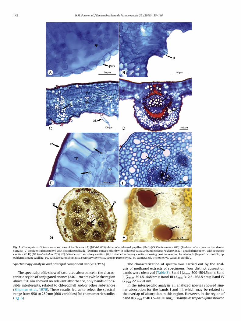
ig. 4G), and three from Africa (CP7, CP8 and CP9) showed anpidermal papillae (Table. 2, Fig. 5A). The presence, number and dis-ribution of papillae constitute an important feature that has beensed to define boundaries at specific and generic level in many tax-nomic groups (Hong et al., 2001). According to Bone et al. (1985),he cell walls of the leaf epidermis with convex curvature would bedvantageous to increase the energy capture efficiency, which ismportant for plants that must survive at extremely low light lev-ls. Furthermore, papillae in the leaf epidermis minimize the areaf contact causing a very low adhesion of water on leaf (Ensikatt al., 2011).
Epicuticular waxes as tubules and clusters of tubules were con-rmed in SEM, in both surfaces of C. pareira (Fig. 2C and D), asreviously referred by Porto et al. (2011). The epicuticular waxeslso have taxonomic value in the characterization of the leaf epider-is, according to Barthlott et al. (1998). Moreover, the presence ofax tubules in leaf epidermis could be related to the higher water
epellency (Ensikat et al., 2011).The leaves were hypostomatic with anomocytic and anisocytic
tomata occurring simultaneously, both at the epidermal levelFig. 3A), however, the anomocytic type was predominant (Fig. 1D),hich corroborate with the pattern previously described to C.
areira by De Wet et al. (2002) and Porto et al. (2011). Simple bi-elled to many-celled trichomes were present on both surfaces ofhe leaf epidermis (Figs. 1A, B and 2A, B), which are common onhe leaf epidermis of some Menispermaceae species, according to
ilkinson (1978).
Dorsiventral leaves with adaxial mesophyll and a single layerf palisade was the pattern for C. pareira (Table 2, Fig. 3A). How-ver, bisseriate palisade was observed in five samples, being tworom Brazil (CP6 and CPb), and three from Africa (CP7, CP8, CP9) Ta
ble
2Se
lect
ed
leaf
Spec
ies
Ciss
ampe
lo
Lege
nds:
ae, a
stra
igh
t
to
cu

N.M. Porto et al. / Revista Brasileira de Farmacognosia 26 (2016) 135–146 139
F adaxio s tubue
(Rktlb
wdCep
n3pactCmFCti
cctbrC(
ig. 2. Cissampelos pareira (RL Fróes 30443), leaf epidermis in front view by SEM: (A)n the abaxial surface; (C) detail of the abaxial epidermis with epicuticular waxes, apicuticular wax; st, stomata; tri, trichome).
Table 2, Fig. 5C) According to Esau (1972), Levitt (1980) andozema et al. (1997), palisade and spongy parenchyma are tissuesnown to reveal responses related to light and soil water varia-ions. All samples showed 3–5-layered spongy parenchyma witharge intercellular spaces (Fig. 3A), and several collateral vascularundles distributed throughout the mesophyll.
The leaf margin was slightly curved toward the abaxial surfaceith a many-layered collenchyma and a single vascular bundle. Theorsiventral organization of the leaf mesophyll is characteristic toissampelos, according to Metcalfe and Chalk (1979) and De Wett al. (2002), and does not constitute an exclusive character of C.areira.
The midrib was biconvex, in transverse section, more promi-ent and rounded to the abaxial surface with a sub-epidermal–5-layered lacunar collenchyma, followed by the fundamentalarenchyma. The vascular system has a single vascular bundle with
sclerenchymatous ring at the middle portion, and 1–2 smaller vas-ular bundles at the base and apex (Table 2, Fig. 3B). Five samples,hree from Africa (CP7, CP8 and CP9) and two from Brazil (CP6 andPb) showed midrib with plane-convex shape, differing from theore common biconvex pattern characteristic of C. pareira (Table 2,
igs. 4D and 5D). In our studies, the morphology of the midrib inissampelos and other genera of Menispermaceae (in prep.), consti-utes an important feature to separate taxa at specific level, whichs also corroborated by studies of De Wet et al. (2002).
In transverse section, the petiole of C. pareira showed circularontour with uniseriate epidermis and simple long trichomes; theortex under the epidermis has continuous collenchyma similar tohe midrib; the vascular system is formed by 6–8 collateral vascular
undles (Fig. 3C–E), which are surrounded by a sclerenchymaticing at the middle portion (Table 2, Fig. 3D). Six samples (CP3, CP5,P6, CPa, CPb and CPc) showed 9–12 collateral vascular bundlesTable 2, Fig. 4E), thus differing from the pattern observed for C.al surface with long finger hairs; (B) detail of the wrinkled epidermis and trichomesle; (D) stoma in detail and epicuticular waxes on the abaxial surface (Legends: epw,
pareira and therefore suggesting these samples belong to differenttaxa.
According to Sinnot (1914), Metcalfe and Chalk (1950) andHoward (1979), the characteristics of the angiosperms have greattaxonomic significance because the environment does not influ-ence it. Moreover, the petiole vascularization is diagnostic todistinguish some genera and species and also has taxonomic impor-tance (Wilkinson, 1978, 1986, 1989). On the other hand, thesclerenchymatic sheath was also referred to C. sympodialis by Portoet al. (2008), and to other South African Cissampelos species by DeWet et al. (2002). It is common in vegetative organs of Menisper-maceae, especially in lianas (Carlquist, 1996).
Idioblasts were observed in the vascular tissue of the corti-cal parenchyma of the midrib and petiole and showed a positivereaction to alkaloids in all samples of C. pareira (Table 2, Fig. 3F).Differently, the samples CP3, CP5, CP6, CP8, CP9, CPa, CPb andCP5 showed no positive reaction to alkaloids (Fig. 5G and H).These results corroborate with existing chemical studies of Cis-sampelos (Semwal et al., 2014), as well as confirm the localizationof the production and/or accumulation of these compounds inthe leaf (Chowdhury, 1972; Cavalcanti et al., 2014). According toMenachery (1996) and Barbosa-Filho et al. (1997), the occurrenceof alkaloids can be a chemotaxonomic character of Menisperma-ceae.
Secretory cavities were observed at the leaf apex of two samplesfrom Africa (CP8 and CP9), and also in the midrib and petiole of eightsamples (Table 2, Figs. 4H and 5E, F), three from Africa (CP7, CP8 andCP9), and six from Brazil (CP3, CP5, CP6, CPa, CPb and CPc). Althoughthis character has been reported for some species of Cissampelos
and other genera of Menispermaceae (Metcalfe and Chalk, 1950,1979; Wilkinson, 1989; De Wet et al., 2002), it was never men-tioned before to C. pareira, and provides further evidence that thesesamples with secretory cavities belong to different taxa. According
140 N.M. Porto et al. / Revista Brasileira de Farmacognosia 26 (2016) 135–146
Fig. 3. Cissampelos pareira (AL Gasper 2020) in transverse sections: (A) dorsiventral mesophyll; (B) midrib with collateral vascular bundle; (C) vascular bundles of petiole inthe basal portion; (D) vascular bundles of petiole in the median portion with a sclerenchymatous ring; (E) detail of collateral vascular bundle; (F) stained idioblasts showingp ep, ep ing; sb
ta
iCtalocesaep
wa
ositive reaction for alkaloids (Legends: col, collenchyma; ef, exchange fascicular;ericycle; ph, phloem; pp, palisade parenchyma; pt, pith; rsc, sclerenchymatous rundle; xyl, xylem).
o Fahn (1988), secretory cavities are distinctive for angiospermsnd have important taxonomic value.
The results of the leaf anatomy of thirteen samples analyzeddentified as C. pareira, revealed that only five of these samples (CP1,P2, CP4, CPd and CPe), all from Brazil, showed a set of charactershat match the pattern described for C. pareira by Porto et al. (2011)nd Sudhakaran (2012), which are summarized here: hypostomaticeaf blades; anomocytic and anisocytic stomata with predominancef the first type at the epidermis level; epidermal anticlinal wallsurves on the adaxial surface, and wavy on the abaxial surface;pidermis with thin cuticle and simple uniseriate trichomes; dor-iventral mesophyll with uniseriate palisade parenchyma, with fewnd rare styloid cristals (Table 2, Fig. 4H); biconvex midrib; collat-ral vascular system; petiole with 6–7 vascular bundles; idioblasts
ositive for alkaloids; and no secretory cavities.Nine samples (CP3, CP5, CP6, CP7, CP8, CP9, CPa, CPb and CPc)ere mistakenly identified as C. pareira, as they have a set of char-
cters different of the pattern of C. pareira. These set of different
pidermis; fc, fascicular cambium; fp, fundamental parenchyma; id, idioblast; per,cl, sclerenchyma; sp, spongy parenchyma; st, stomata; tri, trichome; vb, vascular
characters displayed by these samples allow to separate them intothree groups, which could belong to distinct and unidentified taxa(Table 2): Cissampelos sp1, CP3, CP5, CPa and CPc (Fig. 4A and B);Cissampelos sp2 (Fig. 4C–H), CP6 and CPb; and Cissampelos sp3, CP7,CP8 and CP9 (Fig. 5A–H).
Cissampelos sp1 and Cissampelos sp3 have secretory cavities,biconvex midrib, and epidermal papillae on the abaxial surface.However, they present distinctive characters: Cissampelos sp1 hasuniseriate palisade parenchyma, and the petiole with 9–12 vas-cular bundles (Table 2); while in Cissampelos sp3 the palisadeparenchyma is biseriate and the petiole has 6–8 vascular bundles.
On the other hand, samples of Cissampelos sp2 differs fromCissampelos sp1 and Cissampelos sp3 mainly by the anticlinal wallsof epidermal cells that are straight and smooth with thin cuticle
(Fig. 4C and D), without papillae. However, it shows a biseriatemesophyll and plane-convex midrib, similar to Cissampelos sp3,but different from Cissampelos sp1 that has an uniseriate palisade,and biconvex midrib. The number of petiole vascular bundles of
N.M. Porto et al. / Revista Brasileira de Farmacognosia 26 (2016) 135–146 141
Fig. 4. (A, B) Cissampelos sp1 (HS Irwin s/n – IAN), leaf epidermis in front view: (A) Leaf epidermis with straight anticlinal cells walls on the adaxial surface; (B) Leaf epidermiswith curve anticlinal cells walls on the abaxial surface; (C–H) Cissampelos sp2 (AC Cervi 3276), transverse sections: (C) leaf margin somewat rounded; (D) Plane convexmidrib with collateral vascular bundle; (E) Petiole of 12 collateral vascular bundles in the basal portion; (F) detail of parenchyma with styloids cristals, (G) detail of epidermalp egends t, stom
Cb
m
apillae on the abaxial surface; (H) detail of mesophyll with secretory cavities; (Lecretory cavity; scl, sclerenchyma; sct, scar of trichome; sp, spongy parenchyma; s
issampelos sp2 is similar to that showed by Cissampelos sp1 (9–12),ut different from Cissampelos sp3, with 6–8 vascular bundles.
The evidences found in this work suggest that nine samples wereistakenly identified as C. pareira, and probably belong to three
s: cr, crystals; ep, epidermis; pap, papillae; pp, palisade parenchyma; pt, pith; sc,ata; tri, trichome; vb, vascular bundle).
different taxa, as they have a distinctive set of characters from thoseobserved C. pareira. With regard to identification of these inde-terminate taxa, further investigations are required with a highersampling allowing for the identification of the taxa involved.
142 N.M. Porto et al. / Revista Brasileira de Farmacognosia 26 (2016) 135–146
F of epids b withc tainede paren
S
tas(r(
ig. 5. Cissampelos sp3, transverse sections of leaf blades. (A) (JW Ash 655): detail
urface; (C) dorsiventral mesophyll with bisseriate palisade; (D) plane-convex midriavities; (F, H) (PK Rwaburindore 205): (F) Palisade with secretory cavities; (G, H) spidermis; pap: papillae; pp, palisade parenchyma; sc, secretory cavity; sp, spongy
pectroscopy analysis and principal component analysis (PCA)
The spectral profile showed saturated absorbance in the charac-eristic region of conjugated enones (240–190 nm) while the regionbove 550 nm showed no relevant absorbance, only bands of pos-
ible interferents, related to chlorophyll and/or other substancesShipman et al., 1976). These results led us to select the spectralange from 550 to 250 nm (600 variables) for chemometric studiesFig. 6).ermal papillae; (B–D) (PK Rwaburindore 205): (B) detail of a stoma on the abaxial collateral vascular bundle; (E) (H Faulkner 5631): detail of mesophyll with secretory
secretory cavities showing positive reaction for alkaloids (Legends: ct, cuticle; ep,chyma; st, stomata; tri, trichome; vb, vascular bundle).
The characterization of spectra was carried out by the anal-ysis of methanol extracts of specimens. Four distinct absorptionbands were observed (Table 3): Band I (�max 500–504.5 nm); BandII (�max 391.5–468 nm); Band III (�max 312.5–368.5 nm); Band IV(�max 223–291 nm).
In the interspecific analysis all analyzed species showed sim-ilar absorption for the bands I and III, which may be related tothe overlap of absorption in this region. However, in the region ofband II (�max at 403.5–410.0 nm), Cissampelos tropaeolifolia showed

N.M. Porto et al. / Revista Brasileira de Farmacognosia 26 (2016) 135–146 143
1.0
0.9
0.8
0.7
0.6
0.5
0.4
0.3
0.2
0.1
0.0250 300 350 400
Wavelength (nm)
Arb
itrar
y un
its
450 500 550
CA4
CP1 CP3 CP5 CP7 CP9 CA2
CA3HD
AE
CA1AC
SB
CP8
OH
OSCP6CT3
CT2
CP4CT1
CS3
CP2CS2
CS1
F mom
sc
sbasBc
TM
ig. 6. Cissampelos species: UV–vis spectra of in the spectral region selected for che
ignificantly different absorption values at �max 466.5 nm, whenompared with other species.
The intraspecific analysis of samples of C. pareira revealedhifts in the wavelength of maximum absorption in the band IVetween Brazilian (�max at 261.5–272.0 nm) and African (�max
t 282.0–284.0 nm) specimens, except for the sample CP9 thathowed no band. The spectral analysis of C. pareira samples fromrazil revealed absorption maxima at 261.5 and 272 nm, which areharacteristic of the C N chromophore (Nagarajan et al., 2011).
able 3aximum absorption values (�max) for Cissampelos species and out-group.
Species (Country) Collector (Herbarium) Code
Anomospermum chloranthum M Silva 2619 (MG) AC
Anomospermum steyermarkii GT Prance s/n (MG 42173) AS
Cissampelos andromorpha
NM Porto 30 (JPB) CA1
NM Porto 45 (JPB) CA2
NM Porto 07 (JPB) CA3
W Pizziolo 329 (RB) CA4
Cissampelos pareira (Brazil)
JA Silva 39 (IAN) CP1
RL Fróes 30443 (IAN) CP2
HS Irwin s/n (IAN 129416) CP3
AL Gasper 2020 (RB) CP4
HS Irwin s/n (RB 144809) CP5
A Pott 3158 (RB) CP6
Cissampelos pareira (Africa)JW Ash 655 (MO) CP7
H Faulkner 5631 (MO) CP8
PK Rwaburindore 205 (MO) CP9
Cissampelos sympodialisAgra 7133 (JPB) CS1
Celismar s/n (JPB) CS2
Zehntren 211 (RB) CS3
Cissampelos tropaeolifoliaNM Porto 19 (JPB) CT1
NM Porto 47 (JPB) CT2
T Plowman 8755 (IAN) CT4
Hyperbaena domingensis M Oliveira 1553 (UFP) HD
Orthomene hirsuta GA Black 48-2473 (IAN) OH
Orthomene schomburgkii BS Amorim 1668 (UFP) OS
Sciadotenia brachypoda NT Silva 4146 (IAN) SB
a Wavelengths of maximum UV/Vis absorbance (≥0.01 nm) between 260 and 505 nm.
etric analysis (550–250 nm). For the numerical code of the species see Table 1.
Based on the data available, such absorbance can be related to thepresence of tropoisoquinoline alkaloids, which were already iso-lated from C. pareira (Morita et al., 1993), and is also common inother species of Cissampelos (Menachery, 1996).
The spectral data of the outgroup samples, A. chloranthum, A.
steyermarkii, H. domingensis, O. hirsuta, O. schomburgkii, S. brachy-poda were different, as expected, from C. pareira and other speciesof the genus, especially C. andromorpha, C. sympodialis and C. tropae-olifolia in the PC1 factor (Fig. 7A).�max (nm)a
Band IV Band III Band II Band I
271.5 319.5 – 501.5
– – 405.5 501.5
270.5 and 279.5 323 and 366.5 409.5 –269 – 409 503270 320.5 406.5 –– – 408 504.5
267.5 – 407 504.5270 334 407.5 504.5269.5 320.5 405.5 504.5272 312.5 408 504.5261.5 – 407.5 504.5271.5 319.5 404.5 503.5
283 336.5 – –284 – 407 –– 335.5 – –
270 318.5 and 361.5 407 503.5270 326 403.5 503.5269.5 317.5 406 504
257 326 410 and 468 –– 320.5 and 368.5 409.5 and 466.5 –278 and 282.5 – – –
273 318 – 504.5
– – 406.5 –
276 318.5 – 503
269.5 328 403 503

144 N.M. Porto et al. / Revista Brasileira de Farmacognosia 26 (2016) 135–146
40
32
24
–24
–24–36–48
16
16
–16
8
8
4
–4
–8
–12
–16
–20
Component 1
Com
pone
nt 3
–8
–12 12
12
12–12–24–36
B
–48 24 36 48 60
24
Component 1
A
Com
pone
nt 2
36 48 60
C. pareira (Brazil)
Anomospermum chloranthum
Anomospermum steyermarki
Orthomene hirsuta eOrthomene schomburgkii
C. pareira (Africa)
C. andromorpha
C. sympodialis
C. tropaeolifolia
Sciadotenia brachypoda
Hyperbaena domingensis
B) PC1
doBopwgCdw
tcgB
Fig. 7. Cissampelos species (A) PC1 × PC2; (
The principal component analysis of spectral data allowed theifferentiation of samples of C. pareira of African and Brazilianrigin mainly based on their score values at PC3. The spectra ofrazilian samples CP4 and CP6 showed proximity with the samplesf the African group, CP7, CP8 and CP9 (Fig. 8). The samples of C.areira specimens were close to C. tropaeolifolia and C. sympodialisith positive score values in the PC3 (Fig. 7B). It is worth noting a
roup of African (CP7, CP8 and CP9) and Brazilian samples (CP4 andP6) that, although similar, has spread throughout the dispersioniagram (Fig. 8). Based on the results, in general, the PCA analysisas able to delimit the species of Cissampelos analyzed.
The results showed that application of UV/Vis spectropho-
ometry is a valid technique for identifying Cissampelos samples,onfirming the observations already made for other angiospermsroups such as Asteraceae by Lonni et al. (2005) and Solanum byasílio et al. (2012).× PC3: PCA spectral profile (770–250 nm).
The chemical data corroborates with the anatomical featuresof C. pareira samples, which differentiates them from samples ofother species with the following characters: biseriate parenchyma,stomata above the level of the epidermis, presence of epidermalpapillae as well as secretory cavities in the mesophyll.
The principal components analysis (PCA) provided importantspectral information, which allowed the separation of African-tropical and Neotropical samples. These differences may be relatedto geographical distribution, considered by Griffin and Lin (2000)as an aspect that influences the chemical composition of plants.However, when analyzing chemical data together with anatom-ical characters, usually neglected in taxonomic studies, these
differences suggest that the studied samples belong to differenttaxa, probably in speciation, after being subjected to differentselection processes, with the development of adaptive charac-ters.
N.M. Porto et al. / Revista Brasileira de Fa
2.4
–2.4
–3.0
Component 1
Com
pone
nt 2
1.8
–1.8
1.2
–1.2
0.6
+
+
–0.6–12 12–24 24–36 36 48 60
C. pareira (Brazil)
C. pareira (Africa)
Fs
C
ltdacnsi
A
ahppdota
C
A
fDttsMm
R
A
ig. 8. Cissampelos pareira and relatives from Brazil and Africa (PC1 × PC2): PCApectral profile (770–250 nm).
onclusions
Characters of epidermal cells, mesophyll (palisade parenchymaayers), midrib shape, number of vascular bundles in petiole, secre-ory cavities, and the chemical results proved to be useful andistinctive to separate C. pareira from related species, providing andditional tool for its taxonomy, and quality control of their drugsomposed by leaves. Further chemical and taxonomic studies areeeded to enable the identification of all taxa, as well as to explainome variations in spectral and anatomical data to C. pareira andts related species.
uthors’ contributions
NMP (PhD student) contributed in collecting plant samplesnd identification, and running the laboratory work (preparingerbaria samples, chemical extraction, obtaining of spectra andlant anatomy studies), analysis of the data and preparation of theaper. YLB contributed to UV analysis, supervised by IJLDB. MFAesigned the study with Cisampelos pareira, and supervised all lab-ratory and field work, as well as contributed to critical reading ofhe manuscript. All the authors have read the final manuscript andpproved the submission.
onflicts of interest
The authors declare no conflicts of interest.
cknowledgments
The authors are grateful to CAPES for financial support, and CNPqor the scholarships to NM Porto and MF Agra. The authors thank tor. Eduardo de Jesus Oliveira for the English revision and sugges-
ions, and Dr. Rosa Ortiz for her valuable comments and help withhe bibliography; to Missouri Botanical Garden for the institutionalupport; the curators of herbaria CEPEC, IAN, JPB, MG, RB, UFP andO for their pleasant cooperation during our visits and loan of plantaterial; and Dulce Gonc alves for her technical support.
eferences
bbasi, A.M., Khan, M.A., Ahmad, M., Zafar, M., Jahan, S., Sultana, S., 2010. Ethnophar-macological application of medicinal plants to cure skin diseases and in folk
rmacognosia 26 (2016) 135–146 145
cosmetics among the tribal communities of North-West Frontier Province, Pak-istan. J. Ethnopharmacol. 128, 322–335.
Ahmad, R., Malik, M.A., Zia-ul-Haq, M., 1992. Alkaloids of Cissampelos pareira. Fitoter-apia 63, 282–285.
Amresh, G., Zeashan, H., Rao, C.V., Singh, P.N., 2007a. Prostaglandin mediated anti-inflammatory and analgesic activity of Cissampelos pareira. Acta Pharm. Sci. 49,153–160.
Amresh, G., Zeashan, H., Gupta, R.J., Kant, R., Rao, C.V., Singh, P.N., 2007b. Gastro-protective effects of ethanolic extract from Cissampelos pareira in experimentalanimals. J. Nat. Med. 61, 323–328.
Arkarapanthu, A., Chavasit, V., Sungpuag, P., Phuphathanaphong, L., 2005. Gelextracted from Khruea-ma-noi (Cyclea barbata Miers) leaves: chemical com-position and gelation properties. J. Sci. Food Agric. 85, 1741–1749.
Barbosa-Filho, J.M., Agra, M.F., Thomas, G., 1997. Botanical, chemical and pharma-cological investigation on Cissampelos species from Paraíba (Brazil). Sci. Cult. 49,386–394.
Barthlott, W., Neinhuis, C., Cutler, D., Ditsch, F., Meusel, I., Theisen, I., Wilhelmi, H.,1998. Classification and terminology of plant epicuticular waxes. J. Linn. Soc.126, 237–260.
Basílio, I.J.L.D., Bhattacharyya, J., Moura, R.K.P., Agra, M.F., 2012. Application ofUV/VIS spectrophotometry and multivariate analysis to characterization of thespecies of Solanum sect. Erythrotrichum CHILD. Chem. Biodivers. 9, 1114–1124.
Bone, R.E., Lee, D.W., Norman, J.M., 1985. Epidermal cells functioning as lenses inleaves of tropical rainforest shade plants. Appl. Opt. 24, 1408–1412.
Braga, J.M.A. Menispermaceae in Lista de Espécies da Flora do Brasil. Jardim Botânicodo Rio de Janeiro. http://floradobrasil.jbrj.gov.br/jabot/floradobrasil/FB10031(accessed July 2015).
Bridson, D., Forman, L., 1999. The Herbarium Handbook, 3rd ed. Royal Botanic Gar-dens, Kew.
Bukatsch, F., 1972. Azul de Astra e Safranina. In: Kraus, J., Arduin, M. (Eds.), Manualbásico de métodos em morfologia vegetal. Edur, Seropédia, Rio de Janeiro, p. 26.
Carlquist, S., 1996. Wood and stem anatomy of Menispermaceae. Aliso 14, 155–170.Cavalcanti, A.C., Gomes, A.N.P., Porto, N.M., Agra, M.F., Moura, T.F.A.L., Oliveira, E.J.,
2014. Phamacognostic evaluation of Cissampelos sympodialis Eichl leaves. S. Afr.J. Bot. 93, 70–78.
Chowdhury, A.R., 1972. Chemical investigations on Cissampelos pareira. Sci. Cult. 38,358.
De Wet, H., Tilney, P.M., Van Wyk, B.E., 2002. Vegetative morphology and anatomyof Cissampelos in South Africa. S. Afr. J. Bot. 68, 181–190.
Ensikat, H.J., Ditsche-Kuru, P., Neinhuis, C., Barthlott, W., 2011. Superhydrophobicityin perfection: the outstanding properties of the lotus leaf. Beilstein J. Nanotech-nol. 2, 152–161.
Esau, K., 1972. Anatomia Vegetal. Omega, Barcelona.Franklin, G.L., 1945. Preparation of thin sections of synthetic resins and wood-resin
composites, and a new macerating method for wood. Nature 155, 51.Fahn, A., 1988. Secretory tissues in vascular plants. New Phytol. 108, 229–257.Furr, M., Mahlberg, P.G., 1981. Histochemical analyses of laticifers and glandular
trichomes in Cannabis sativa. J. Nat. Prod. 44, 153–159.Ganguly, M., KrBorthakur, M., Devi, N., Mahanta, R., 2007. Antifertility activity of
the methanolic leaf extract of Cissampelos pareira in female albino mice. J.Ethnopharmacol. 111, 688–691.
Griffin, W.J., Lin, G.D., 2000. Chemotaxonomy and geographical distribution oftropane alkaloids. Phytochemistry 53, 623–637.
Hammer, Ø., Harper, D.A.T., Ryan, P.D., 2001. PAST: Paleontological statistics soft-ware package for education and data analysis. Palaeontol. Electron. 4, 1–9.
Haque, M.A., Shaha, M.K., Ahmed, S.U., Akter, R., Rahman, H., Chakravotry, S.,Imran, A.H.M.N., Islam, M.T., Das, R.C., Rahmatullah, M., 2011. Use of inorganicsubstances in folk medicinal formulations: a case study of a folk medicinalpractitioner in Tangail district, Bangladesh. Am. Eurasian J. Sustain. Agric. 5,415–423.
Hedberg, I., Hedbrerg, O., Madati, P.J., Mshigeni, K.E., Mshiu, E.N., Samuelsson, G.,1983. Inventory of plants used in traditional medicine in Tanzania. II. Plants ofthe families Dilleniaceae-Opiliaceae. J. Ethnopharmacol. 9, 105–127.
Hong, Y.P., Pan, K.Y., Chen, Z.D., Lu, A.M., 2001. Characters of leaf epidermis and theirsystematic significance in Menispermaceae. Acta Bot. Sin. 43, 615–623.
Hoot, S.B., Zautke, H., Harris, D.J., Crane, P.R., Neves, S.S., 2009. Phylogenetic patternsin Menispermaceae based on multiple chloroplast sequence data. Syst. Bot. 34,44–56.
Howard, R., 1979. The Petiole. In: Metcalfe, C.R., Chalk, L. (Eds.), Anatomy of theDicotyledons, 1. Clarendon Press, Oxford, pp. 88–96.
Jannu, V., Sai Vishal, D., Ranjith Babu, V., Harisha, B., Ravi Chandra Sekhara Reddy,D., 2011. Antidiabetic activity of hydro-alcoholic extract of Cissampelos pareiraLinn. Leaves in streptozotocin induced diabetic rats. Int. J. Pharm. Technol. 3,3601–3611.
Johansen, D.A., 1940. Plant Microtechnique. McGraw Hill, New York.Kamble, S.Y., More, T.N., Patil, S.R., Pawar, S.G., Ram Bindurani Bodhankar, S.L., 2008.
Plants used by tribes of Northwest Maharashtra for the treatment of gastroin-testinal disorders. Indian J. Tradit. Knowl. 7, 321–325.
Kumar, V.P., Chauhan, N.S., Padh, H., Rajani, M., 2006. Search for antibacterial andantifungal agents from selected Indian medicinal plants. J. Ethnopharmacol. 107,182–188.
Kupchan, S.M., Yokoyama, N., Beal, J.L., 1960a. Menispermaceae alkaloids. I. The alka-loids of Cissampelos pareira Linn. and the origin of radix pareira brave. J. Am.Pharm. Assoc. 49, 727.
Kupchan, S.M., Slade, P., Young, R.J., 1960b. Intramolecular catalysis. Facilitation ofalkaline hydrolysis of alicyclic 1,2-diol monoesters. Tetrahedron Lett. 1, 22–25.

1 a de Fa
L
L
L
M
M
M
M
M
N
N
N
N
O
P
P
P
P
R
R
RR
R
46 N.M. Porto et al. / Revista Brasileir
evitt, J., 1980. Responses of Plants to Environmental Stresses, vol. 2. New York,Academic Press.
ewis, W.H., Elvin-Lewis, M.P.F., 1977. Medical Botany. Wiley-Interscience, NewYork.
onni, A.A.S.G., Scarminio, I.S., Silva, L.M.C., Ferreira, D.T., 2005. Numerical taxonomiccharacterization of Baccharis genus species by ultravioleta-visible spectropho-tometry. Anal. Sci. 21, 235–239.
antuano, D.G., Barros, C.F., Scarano, F.R., 2006. Leaf anatomy variation within andbetween three restinga populations of Erythroxylum ovalifolium Peyr. (Erythrox-ylaceae) in Southeast Brazil. Rev. Bras. Bot. 29, 209–215.
enachery, M.D. 1996. The alkaloids of South American Menispermaceae. In S.W.Pelletier (org), Alkaloids: Chemical and Biological Perspectives. New York: Ed.Pergamon, p 269–302.
etcalfe, C.R., Chalk, L., 1950. Anatomy of Dicotyledons: Leaves, Stem, and Woodsin Relation to Taxonomy with Notes on Economic Uses, vol. 1. Clarendon Press,Oxford, United Kingdom, pp. 52–62.
etcalfe, C.R., Chalk, L., 1979. Anatomy of the Dicotyledons: Systematic Anatomy ofLeaf and Stem, with a Brief History of the Subject, vol. 1., 2nd ed. Claredon Press,Oxford/United Kingdom, pp. 267.
orita, H., Matsumoto, K., Takeya, K., Itokawa, H., Iitaka, Y., 1993. A novelantileukemic tropoloisoquinoline alkaloid, pareirubrine, from Cissampelospareira. Chem. Lett., 339–342.
agarajan, K., Chauhan, N., Mittal, A., Singh, V., Bodla, R.B., Tiwari, R.K., 2011. Phyto-chemical extraction, optimization and physico-chemical characterization of twobioactive isolates from the leaves and stem of Cissampelos pareira. Der PharmaChem. 3, 327–337.
APRALERT, Natural Products Alert. http://www.napralert.org/ (accessed March2013).
urit-Silva, K., Basilio, I.J.L.D., Agra, M.F., 2007. Estudo farmacobotânico comparativoentre Solanum paniculatum L. e Solanum rhytidoandrum Sendtn. Rev. Bras. Biol.5, 243–245.
urit-Silva, K., Agra, M.F., 2011. Leaf epidermal characters of Solanum sect. poly-trichum (Solanaceae) as taxonomic evidence. Microsc. Res. Tech. 74, 1186–1191.
rtiz, R., 2001. Menispermaceae. In Stevens, W.D., Ulloa Ulloa, C., Pool, A., Montiel,O. M. (orgs.), Flora de Nicaragua. Missouri: Monographs in Systematic Botanyfrom the Missouri Botanical Garden, p. 1432–1442.
orto, N.M., Basílio, I.J.L.D., Agra, M.F., 2008. Pharmacobotanical study of the leavesof Cissampelos sympodialis Eichl., (Menispermaceae). Braz. J. Pharmacogn. 18,102–107.
orto, N.M., Figueiredo, R.C.B.Q., Oliveira, A.F.M., Agra, M.F., 2011. Leaf epidermalcharacteristics of Cissampelos L. (Menispermaceae) species from NortheasternBrazil. Microsc. Res. Tech. 74, 370–376.
orto, N.M., Araújo, N.D., Basílio, I.J.L.D., Agra, M.F., 2012. Analysis of leaf epidermalcharacters of medicinal and poisonous Brazilian Menispermaceae. Planta Med.78, 1111.
riya, G., Saravanan, K., Renuka, C., 2012. Medicinal plants with potential antifertilityactivity – a review of sixteen years of herbal medicine research (1994–2010).Int. J. Pharm. Tech. Res. 4, 481–494.
amasubramaniaraja, R., Babu, N.M., 2010. Antihelminthic studies and medicinalherbs – an overview. Int. J. Pharm. Sci. Rev. Res. 5, 39–47.
amirez, I., Carabot, A., Melendez, P., Carmona, J., Jimenez, M., Patel, A.V., Crab,T.A., Blunden, G., Cary, P.D., Croft, S.L., Costa, M., 2003. Cissampeloflavone,a chalcone–flavone dimer from Cissampelos pareira. Phytochemistry 64,645–647.
hodes, D.G., 1975. A revision of the genus Cissampelos. Phytologia 30, 415–485.ôc as, G., Scarano, F.R., Barros, C.F., 2001. Leaf anatomical variation in Alchornea
triplinervia (Spreng.) Müll. Arg. (Euphorbiaceae) under distinct light and soil
water regimes. Bot. J. Linn. Soc. 136, 1–8.ozema, J., Chardonnens, A., Tosserams, M., Hafkenscheid, R., Bruijnzeel, S.,1997. Leaf thickness and UV-B absorbing pigments of plants in relation toan elevational gradient along the Blue Mountains, Jamaica. Plant Ecol. 128,150–159.
rmacognosia 26 (2016) 135–146
Rukunga, G.M., Gathirwa, J.W., Omar, S.A., Muregi, F.W., Muthaura, C.N., Kirira, P.G.,Mungai, G.M., Kofi-Tsekpo, W.M., 2009. Anti-plasmodial activity of the extractsof some Kenyan medicinal plants. J. Ethnopharmacol. 121, 282–285.
Rury, P.M., 1983. Pareira Brava: 19th Century notes and commercial samples fromE. R. Squibb M. D. Bot. Mus. Leaf. 29, 27–48.
Sampaio, V.S., Araújo, N.D., Agra, M.F., 2014. Characters of leaf epidermis of Solanumof the Brevantherum Clade from Atlantic Forest of Northeastern Brazil. S. Afr. J.Bot. 74, 108–113.
Sanchez Medina, A., Garcia Sosa, K., May Pat, F., Pena Rodriguez, L.M., 2001. Antioxi-dant, antimicrobial and beta-glucosidase inhibition activities. Phytomedicine 8,144–151.
Schmelzer, G.H., Gurib-Fakim, A. (Eds.), 2008. Plant Resources of TropicalAfrica 11(1). Medicinal Plants 1. PROTA Foundation/Backhuys Publishers/CTA,Wageningen, Netherlands/Leiden, Netherlands/Wageningen, Netherlands, 791pp.
Semwal, D.K., Semwal, R.B., Vermaak, I., Viljoen, A., 2014. From arrow poison toherbal medicine: the ethnobotanical, phytochemical and pharmacological sig-nificance of Cissampelos (Menispermaceae). J. Ethnopharmacol. 155, 1011–1028.
Sinnot, E.W., 1914. The anatomy of the node as an aid in the classification ofangiosperms. Am. J. Bot. 7, 303–322.
Sharma, V., 1987. Biosynthesis of hayatin. Indian J. Chem. 26, 589–591.Shipman, L.L., Cotton, T.M., Norris, J.R., Katz, J.J., 1976. An analysis of the visible
absorption spectrum of chlorophyll a monomer, dimer, and oligomers in solu-tion. J. Am. Chem. Soc. 98, 8222–8230.
Singh, V.K., Ali, Z.A., 1992. A contribution to the ethnopharmacological study of theUdaipur forests of Rajasthan, India. Fitoterapia 63, 136–144.
Singh, K.K., Maheshwari, J.K., 1994. Traditional phytotherapy of some plants used bythe tharus of the Nainital District, Uttar Pradesh, India. Int. J. Pharmacogn. 32,51–58.
Stace, C.A., 1965. Cuticular studies as an aid to plant taxonomy. Bull. Br. Mus. Nat.Hist. Bot. 4, 1–78.
Singthong, J., Cui, S.W., Ningsanond, S., Douglas Goff, H., 2004. Structural character-ization, degree of esterification and some gelling properties of Krueo Ma Noy(Cissampelos pareira) pectin. Carbohydr. Polym. 58, 391–400.
Sudhakaran, M.V., 2012. Histo-morphological, fluorescent and powder microscopiccharacterization of Cissampelos pareira Linn. Phcog. J. 4, 57–68.
Shukla, P., Shukla, P., Gopalakrishna, B., 2012. Investigation of in-vitro anthelminticactivity of Cissampelos pareira Linn against Pheretima posthuma. Int. J. Pharm. Sci.Res. 3, 265–267.
Thakur, P., Rana, A.C., 2013. Effect of Cissampelos pareira leaves on anxiety-likeBehavior in experimental animals. J. Tradit. Complement. Med. 3, 188–193.
Thiers, B. (continuously updated) Index Herbariorum: A Global Directory of PublicHerbaria and Associated Staff. New York Botanical Garden’s Virtual Herbar-ium. New York Botanical Garden. Available from: http://sweetgum.nybg.org/ih/(accessed 29.10.15).
Vaidya, B.G., 1988. Nighantu Adarsh, vol. 1. Chaukhambha Bharti Academy Publica-tions, Varanasi, India, pp. 44–45.
Vardhanabhuti, B., Ikeda, S., 2006. Isolation and characterization of hydrocolloidsfrom monoi (Cissampelos pareira) leaves. Food Hydrocolloids 20, 885–891.
Yadav, K.S., Yadav, N.P., Shanker, K., Thomas, S.C., Srivastav, S., Srivastava, S., KumarRai, V., Mishra, N., Sinha, P., 2013. Assessment of antidiabetic potential of Cis-sampelos pareira leaf extract in streptozotocinenicotinamide induced diabeticmice. J. Pharm. Res. 6, 874–878.
Wilkinson, P., 1978. Leaf anatomy of the Tribe Coscinieae Hook. f. & Thoms. (Menis-permaceae). Kew Bull. 32, 347–360.
Wilkinson, H.P., 1979. Plant surface. In: Metcalfe, C.R., Chalk, L. (Eds.), Anatomy ofthe Dicotyledon. Claredon Press, Oxford, pp. 97–162.
Wilkinson, H.P., 1986. Leaf anatomy of Tinomiscium and Fibraurea (Menisperma-ceae tribe Fibraureeae) with special reference to laticifers and astrosclereids.Kew Bull. 41, 153–169.
Wilkinson, H.P., 1989. Leaf anatomy of the Menispermaceae tribe Tiliacoreae Miers.Bot. J. Linn. Soc. 99, 125–174.