malaria journal resources... · detecting placental infection (mcnemar’s x2< 3.84). presence...
TRANSCRIPT

This Provisional PDF corresponds to the article as it appeared upon acceptance. Fully formattedPDF and full text (HTML) versions will be made available soon.
Placental Plasmodium falciparum malaria infection: operational accuracy ofHRP2 rapid diagnostic tests in a malaria endemic setting
Malaria Journal 2011, 10:306 doi:10.1186/1475-2875-10-306
Daniel J Kyabayinze ([email protected])James K Tibenderana ([email protected])
Mercy Nassali ([email protected])Lynette K Tumwine ([email protected])
Clare Riches ([email protected])Mark Montague ([email protected])Helen Counihan ([email protected])
Prudence Hamade ([email protected])Jean-Pierre Van Geertruyden ([email protected])
Sylvia Meek ([email protected])
ISSN 1475-2875
Article type Research
Submission date 1 August 2011
Acceptance date 18 October 2011
Publication date 18 October 2011
Article URL http://www.malariajournal.com/content/10/1/306
This peer-reviewed article was published immediately upon acceptance. It can be downloaded,printed and distributed freely for any purposes (see copyright notice below).
Articles in Malaria Journal are listed in PubMed and archived at PubMed Central.
For information about publishing your research in Malaria Journal or any BioMed Central journal, goto
http://www.malariajournal.com/authors/instructions/
For information about other BioMed Central publications go to
Malaria Journal
© 2011 Kyabayinze et al. ; licensee BioMed Central Ltd.This is an open access article distributed under the terms of the Creative Commons Attribution License (http://creativecommons.org/licenses/by/2.0),
which permits unrestricted use, distribution, and reproduction in any medium, provided the original work is properly cited.

http://www.biomedcentral.com/
Malaria Journal
© 2011 Kyabayinze et al. ; licensee BioMed Central Ltd.This is an open access article distributed under the terms of the Creative Commons Attribution License (http://creativecommons.org/licenses/by/2.0),
which permits unrestricted use, distribution, and reproduction in any medium, provided the original work is properly cited.

Placental Plasmodium falciparum malaria infection: operational accuracy of
HRP2 rapid diagnostic tests in a malaria endemic setting
Daniel J Kyabayinze 1,2*
, James K Tibenderana1,3
, Mercy Nassali 4, Lynette K
Tumwine 5, Clare Riches
1, Mark Montague
6, Helen Counihan
6, Prudence Hamade
6
Jean-Pierre Van Geertruyden2, Sylvia Meek
6
1 Malaria Consortium, Upper Naguru East Road, P.O. Box 8045, Kampala, Uganda
2 Unit International Health, ESOC Department, Faculty of Medicine, Antwerp
University, Universiteiplein 1, BE-2610 Antwerpen, Belgium
3 London School of Hygiene and Tropical Medicine, Keppel Street, London, WC1E
7HT, UK
4 Mbale Regional Referral Hospital, Ministry of Health, PO Box 192, Mbale, Uganda
5 Makerere University, College of Health Sciences, PO Box 7072, Kampala, Uganda
6 Malaria Consortium, Development House, 56-64 Leonard Street, London, EC2A
4LT, UK
*Corresponding author
e-mail addresses:
DJK: [email protected]
JKT: [email protected]
LKT: [email protected]


Abstract
Background
Malaria has a negative effect on the outcome of pregnancy. Pregnant women are at
high risk of severe malaria and severe haemolytic anaemia, which contribute 60–70%
of foetal and perinatal losses. Peripheral blood smear microscopy under-estimates
sequestered placental infections, therefore malaria rapid diagnostic tests (RDTs)
detecting histidine rich protein-2 antigen (HRP-2) in peripheral blood are a potential
alternative.
Methods
HRP-2 RDTs accuracy in detecting malaria in pregnancy (MIP >28 weeks gestation)
and placental Plasmodium falciparum malaria (after childbirth) were conducted using
Giemsa microscopy and placental histopathology respectively as the reference
standard. The study was conducted in Mbale Hospital, using the midwives to perform
and interpret the RDT results. Discordant results samples were spot checked using
PCR techniques.
Results
Among 433 febrile women tested, RDTs had a sensitivity of 96.8% (95% CI 92-98.8),
specificity of 73.5% (95% CI 67.8-78.6), a positive predictive value (PPV) of 68.0%
(95% CI 61.4-73.9), and negative predictive value (NPV) of 97.5% (95% CI 94.0-
99.0) in detecting peripheral P. falciparum malaria during pregnancy. At delivery, in
non-symptomatic women, RDTs had a 80.9% sensitivity (95% CI 57.4-93.7) and a
87.5% specificity (95%CI 80.9-92.1), PPV of 47.2% (95% CI 30.7-64.2) and NPV of
97.1% (95% CI 92.2-99.1) in detecting placental P. falciparum infections among 173
samples. At delivery, 41% of peripheral infections were detected by microscopy
without concurrent placental infection. The combination of RDTs and microscopy

improved the sensitivity to 90.5% and the specificity to 98.4% for detecting placental
malaria infection (McNemar’s X2> 3.84). RDTs were not superior to microscopy in
detecting placental infection (McNemar’s X2< 3.84). Presence of malaria in pregnancy
and active placental malaria infection were 38% and 12% respectively. Placental
infections were associated with poor pregnancy outcome [pre-term, still birth and low
birth weight] (aOR=37.9) and late pregnancy malaria infection (aOR=20.9). Mosquito
net use (aOR 2.1) and increasing parity (aOR 2.7) were associated with lower risk for
malaria in pregnancy.
Conclusion
Use of HRP-2 RDTs to detect malaria in pregnancy in symptomatic women was
accurate when performed by midwives. A combination of RDTs and microscopy
provided the best means of detecting placental malaria. RDTs were not superior to
microscopy in detecting placental infection. With a high sensitivity and specificity,
RDTs could be a useful tool for assessing malaria in pregnancy, with further (cost-)
effectiveness studies.

Background
Plasmodium falciparum malaria remains a major contributor of the disease burden in
sub-Saharan Africa (SSA). Children under five years of age and pregnant women
represent the most vulnerable population. During pregnancy, malaria infection leads
to parasite sequestration in the maternal placental vascular space, with the
consequence of increased risks of abortion, stillbirth, prematurity, intra-uterine growth
retardation (IUGR) and maternal anaemia [1]. Malaria in pregnancy (MIP) is
associated with increased risk of low birth weight (LBW) and prenatal, neonatal and
infant mortality [2-5]. Infection with P. falciparum towards the end of gestation
increases the likelihood of placental infection [6]. A previous review by Desai et al
estimated that approximately one in every four pregnant women in malaria endemic
areas has evidence of placental infection at the time of delivery [7].
The diagnostic challenge is that conventional peripheral blood microscopy is unable
to detect all infections as parasites can be sequestered in the placenta [8-10]. Such
sub-microscopic infections increase the risk of anaemia and poor foetal outcomes in
seemingly aparasitaemic pregnant women [9, 11] thus increasing the risk of maternal
and perinatal mortality. Consequently, some clinicians are reluctant to withhold anti-
malarials for febrile pregnant women, arguing that microscopy misses placental
infections.
However, malaria rapid diagnostic tests (RDTs) may provide a solution as emerging
evidence suggests that RDTs are capable of detecting placental malaria better than
microscopy and may detect sub-microscopic infections [12-13]. RDTs are lateral flow
immuno-chromatographic dipstick assays that detect either histidine rich protein-2

(HRP-2) or Plasmodium lactate dehydrogenase (pLDH) produced by infected red
blood cells. Although the large majority of RDTs comprise mAbs raised against
HRP2 or pLDH, it should be noted here that there are an increasing number of tests
that also include a Pan anti-aldolase test line. Whereas good quality microscopy is
lacking in many resource-limited settings, as it requires well-trained, competent
personnel, infrastructure as well as effective quality control and quality assurance
[13], RDTs are becoming increasingly available and affordable[14].
With many countries in SSA following World Health Organization (WHO) policy
[15] and scaling up parasite-based diagnosis for malaria through the use of
microscopy and RDTs, establishing the effectiveness of RDTs in comparison to
microscopy in detecting malaria during pregnancy has become a priority.
Although there is limited evidence on the use of RDTs among pregnant women [16-
17], RDTs have been found to be comparable in sensitivity to microscopy for malaria
[18] and to improve malaria diagnosis and quality of care at lower level health
facilities [19-21]. The few studies that have been done have based the accuracy of
RDTs on either placental smear or placental blood polymerase chain reaction (PCR)
and not histological examination of the placenta at delivery [10-11, 22-24].
This study compares HRP-2 RDTs to microscopy for detecting peripheral malaria in
feverish pregnant women at time of recruitment in a hospital setting. In addition, at
delivery both RDTs and microscopy are compared to placental histopathology
performed to detect placental malaria.

Methods
Study design and site
Study site: Mbale is a mountainous region with moderately low temperatures and
humidity, approximately 300 km north east of Kampala, Uganda. It is a hyper-
endemic region for malaria with an estimated parasite prevalence rate of 56% among
children under five years [25]. The hospital conducts over 300 deliveries with less
than 30% having attended antenatal care (ANC) in the same hospital. Insecticide-
treated net (ITN) ownership in the region is 52% and net use by pregnant women is
42% [25]. Use of ANC services has been reported to be as high as 90%, and coverage
with the second dose of intermittent preventive treatment (IPT) of malaria in
pregnancy (IPTp-2) with sulphadoxine-pyrimethamine (SP) is below 50% [26]. The
operational research was to determine the effectiveness of RDTs when used in a
hospital setting. Cross-sectional evaluation at time of recruitment compared accuracy
of RDTs to microscopy as a reference standard in detecting peripheral parasitaemia
during pregnancy. At delivery, a second cross-sectional evaluation compared accuracy
of RDTs and microscopy to the reference standard of placental histology performed to
detect placental malaria.
Study participants. Patients with fevers (temperature >37.2) or history of fever in the
last 24 hours, at a gestational age >28 weeks by dates, were screened and consented to
participate in the study while at the antenatal clinic. Enrolled women were clinically
examined and information was collected on social demographics and health-seeking

behaviour using a pre-tested questionnaire. A code was placed on the ANC card of the
participating mothers to enable easy identification and follow-up at time of labour.
Pregnant women were encouraged to come and deliver in the hospital. All study
participants were given transport refund as well as a free delivery set (maama kit) to
ensure safe delivery. A finger prick was performed to test for malaria using both
RDTs and microscopy smears. In addition, a sample of blood was collected on filter
paper. Haemoglobin (Hb) testing was done using a portable spectrophotometer
(HemoCue, Angelholm, Sweden). A routine (non-study) field stain smear, was
performed to guide. Women with malaria, according to routine microscopy, were
treated with either quinine or artemisinin combination therapy (ACT) according to the
national treatment guideline.
During labour, participating mothers were identified based on a code placed on their
ANC cards. A finger prick was performed to test for malaria using both RDTs and
microscopy and to estimate Hb. A sample of blood was again collected on filter paper
for PCR. After delivery, the outcomes and weight of the child were documented. A
placental biopsy for histopathology was also performed. Mothers were requested to
bring children back to the hospital for immunization and review after six weeks.
Laboratory procedures
Rapid diagnostic testing for malaria. The commercially available RDT kit,
“diagnosticks” Malaria P. falciparum cassette (SSA Diagnostics and Biotech Systems,
Goa, India) was used. The lot number was: S21004, manufactured in September 2009
and expiry date of August 2011. Each box of 25 individually sealed strips with a
desiccant was supplied with loops, alcohol swabs and lancets. Temperature and

humidity conditions were monitored during transportation and storage. After
performing a finger prick, two midwives independently read each of the RDTs, 15
minutes after adding five drops of the wash buffer according to the manufacturer’s
instructions. A result was interpreted as positive when both the test line and control
line showed pink and the test line was also graded as either a strong or faint band. The
result was recorded as negative when only the control line showed pink or invalid
when the control line did not show up at all. Only one test was performed for each
patient. No repeat test was done for invalid tests and used RDTs were discarded the
same day. First reading was considered for analysis in the case of discordant result
between readers.
Microscopy. Smears for the study were stained with 10% Giemsa for 30 minutes and
examined under X100 objective lens of the microscope by two independent laboratory
technologists blinded to each other’s results. Microscopy examination of smears was
done under oil immersion. The number of asexual parasites was counted against 200
leucocytes assuming an average leucocytes count of 8,000/µL. Before a smear was
declared negative, 200 high power fields were examined.
Histopathology assessment of placental tissue. For every delivery conducted,
placental biopsy was collected within 30 minutes to determine malaria infection. The
specimen of the placental tissue (1.5 cm3) was excised away from the centre of the
edge of the placenta through the full thickness of the placenta including the
membranes [27]. Placental tissues were fixed in 10% neutral buffered formalin and
transferred to the pathology laboratory at Makerere University, College of Health
Sciences. At the pathology laboratory, the fixed placental biopsy specimens were

grossed into 2-3 mm sections, processed using standard procedures and embedded in
paraffin wax. They were sectioned using a microtometo 4µm [27]. The final tissue
sections were stained with haematoxylin and eosin for detection of active malaria.
Placental malaria infection was classified using the following definitions: 1) no
infection (no evidence of parasites or pigment), 2) active acute infection (parasites in
the maternal erythrocytes in the intervillous space but no/minimal pigment in
fibrin/cells within fibrin), 3) active chronic infection (parasites in maternal
erythrocytes in intervillous space, pigment in erythrocytes and circulating monocytes
within intervillous space and pigment in fibrin or cells within fibrin and /or chronic
villous syncytiotrophoblast /stroma and, 4) past infection (parasites not present,
pigment confined to fibrin or cells within fibrin [28]. The enrolled sample of 434
pregnant febrile women at ANC was sufficient to detect a minimum sensitivity of
90% and specificity of 75% at a precision of 0.05 and power of 80%. The prevalence
of malaria was estimated at 40% [28].
Molecular assessments
A convenient sample of 50% of all discordant results (RDTs and microscopy) was
selected for molecular assessment due to limited resources. Using the filter papers
samples collected, PCR was performed to identify presence of infection in samples
positive by microscopy, that had negative RDTs and samples negative by microscopy
but positive by RDTs. Parasite DNA was isolated from filter paper using the chelex
extraction method [29]. To detect P. falciparum, the block-3 region of merozoite
surface protein-2 (MSP-2) was amplified by nested PCR with primers corresponding
to conserved sequences flanking this region [30], followed by primers to amplify the

IC3D7 and FC27 allelic family, using conditions described previously [31]. PCR
products were analysed by electrophoresis using 2% agarose gel.
Quality control measures
All study research assistants (midwives) were centrally trained and had continuous
support supervision by the laboratory technician and site supervisor. The study RDTs
were centrally purchased and delivered to the hospital by the study coordinator, with a
reserve kept at the Malaria Consortium offices. Manufacturer’s storage temperature
specifications (4-30°C) were monitored and maintained both at storage and during
transportation. The slides and RDTs were read by two persons blinded to each other’s
outcome. One data tracker was responsible for aggregating the data onto the case
record forms. The practice of recording this information was checked during monthly
support supervision visits. Placental histology smears were first examined by a senior
pathologist and later, blindly, quality controlled by a second reader at Mbarara
University pathology department. No tie-breaker reading was performed.
Statistical methods
Clinical and social demographic data were collected at the time of enrolment and
linked through unique identifier numbers. At delivery, outcomes of smear and
placental histology results were also recorded in addition to repeat RDTs and
peripheral microscopy. Data were entered using EpiData ("The EpiData Association"
Odense, Denmark) and analysed with SPSS 12 (SPSS USA 2005). For malaria in
pregnancy, results were categorized as positive and negative for both RDTs and
microscopy. Sensitivity, specificity PPV and NPV were calculated by comparing the
proportion of positive and negative results for each RDT with expert field microscopy

(these are not certified WHO experts). Categorical variables were compared using chi
2test. Weighted positive likelihood ratio (LR) was calculated by dividing probability
that a positive test result is a true positive by probability that a positive test result is a
false positive as LR= (prevalence)(sensitivity)/ (1-prevalence)(1-specificity) and the
negative LR =(prevalence)(1-sensitivity)/(1-prevalence)(specificity). Logistic
regression analysis was conducted to determine factors that predicted malaria test
results and level of associations presented as odds ratios.
Placental results were categorized as positive (acute and chronic infections) or
negative (past and not infected) for histopathology. Associations between placental
infection and independent variables were assessed using odds ratios with 95%
confidence intervals calculated and interpreted as significant if two-sided p-value was
less than 0.05. For placental malaria, the sensitivity, specificity, PPV and NPV were
calculated for both RDTs and microscopy in comparison to the reference standard
placental histology. The McNemar test (X2) was used to examine whether the two
proportions derived from the marginal sums of the matched sample contingency table
significantly differed. McNemar’s test result was computed: X2 = ∑ (O-E)
2/E where O
is observed , E is expected value at 5% significant level [32]. Odds ratios and 95%
confidence intervals of the discordant cells were computed to show the level of this
association.
Ethical considerations
The study was conducted according to the principles of the declaration of Helsinki
and the international guidelines of biomedical research involving human subjects. The
study protocol was approved by Mbale Hospital Research and Ethical Committee and

Uganda National Council of Science and Technology. All participants provided
written informed consent to participate in the study.
Results
Patient profile
Between February and July 2010, 534 febrile women attending ANC clinic were
screened for eligibility to participate in the study, out of which 434 were enrolled. The
screen failures were due to low gestational age (< 28 weeks of amenorrhoea). After
testing with RDTs at time of enrolment, four women had invalid results and were
excluded from analysis while one had no microscopy results. At delivery, 208/434
(48%) returned to the hospital. From the delivery data, 173/208 (83%) was analysed
after excluding 28 without placental biopsies histology results and seven women who
lacked unique linking data for both microscopy and RDTs results (Figure 1).
Malaria in pregnancy (gestational malaria)
One third of the febrile women were primigravidae with a mean age of 19 years. Of
these, 406/434 (94%) had attended ANC two or more times with just 58/434 (13%)
having received a second dose of sulphadoxine-pyrimethamine for IPTp at time of
enrolment. Among 421 respondents, 343 (82%) reported having slept under a
mosquito net the night before. Prevalence of clinical malaria in pregnancy was
164/433 (38%) based on peripheral blood Giemsa smears microscopy, while RDTs
were positive among 225/430 (52%)(Table 1). RDTs had a sensitivity of 96.8% (95%
CI 92.0-98.8), specificity of 73.5 % (95% CI 67.8-78.6), Positive Predictive Value

(PPV) of 68.0 % (95% CI 61.4-73.9), and Negative Predictive Value (NPV) of 97.5 %
(95% CI 94.0-99.0). The weighted likelihood ratios (LR) of positive test (LR+) and
negative tests (LR-) were calculated. The LR+ was 2.1 (95% CI 1.7-2.6) which means
that a pregnant woman with malaria is about two times more likely to have a positive
RDT test than a woman who has not got malaria in pregnancy. A LR- of 0.025 (95%
CI 0.01-0.06) means that the probability of having a negative test for pregnant women
with malaria is 0.025 times that of those without malaria. Women without malaria are
about 40 times more likely to have negative test than women with malaria. Given the
LR, RDTs were better at ‘ruling out malaria infection’ than in confirming active
malaria infection (Table 2). PCR was selectively conducted as a spot check to
evaluate discordant results on a total of 45 ‘false RDTs’ samples. PCR was positive
for 1/37 RDT false negative and negative for 2/8 RDT false positive samples. These
spot check PCR results did not affect the computed accuracy of the RDTs.
Factors associated with malaria in pregnancy
Primigravidae were more than twice as likely to have clinical malaria when compared
with multigravidae women (aOR 2.5, p<0.001). Women with low Hb were more at
risk of having malaria than those with Hb greater than 11g/dl (aOR 1.6, p=0.024).
Women who reported having slept under a mosquito net the night before were two
times less likely to have malaria than those who did not use a mosquito net (aOR 0.44
p=0.001) (Table 3).
Placental malaria and its associated factors
At the end of gestation, 208/434 (48%) enrolled mothers returned for hospital
delivery. The delivery outcomes were 186/208 (89%) live births, 11 pre-term, eight

still births (five fresh still births), and three unrecorded outcomes. Six (3%) were
surgical deliveries. One maternal death occurred due to post-partum haemorrhage and
three neonatal deaths on the first-day of life due to prematurity. Further analysis was
then restricted to 173 patients with complete histopathology results.
Of the 178 placental tissue examined, 38/178 (21.3% [95% CI 16.0-27.9]) had malaria
infection, of which 21/173(12.1% [95% CI 7.8-17.4]) were active infections (16 acute
and five chronic) and the rest past infections. RDTs had a sensitivity of 80.9 % (95%
CI 57.4-93.7), specificity of 87.5 % (95% CI 80.9-92.1), PPV of 47.2% (95% CI 30.7-
64.2) and NPV of 97.1% (95% CI 92.2-99.1) against the reference standard of
placental histology in detecting placental P. falciparum infections. Combination
results of RDTs and microscopy improved the total sensitivity to 90.5 (95% CI 68.2-
98.3) for detecting placental malaria infection and NPV to 98.4 (95% CI 93.9-99.7)
but lowered the specificity and PPV (Table 4). The McNemar’s test (X2) was
performed to test whether RDTs were better than microscopy in detecting placental
malaria. The X2 values among the placental-infected and non-infected mothers were
0.25 and 1.565 respectively, both less than the 3.84 cut-off for a significant difference
at 5% (Additional file 1). The extended McNemar X2 confirmed that combined tests
of RDTs and microscopy were significantly better than a single test (X2 of 6.125 for
infected and 9.00 none infected both >than 5.00 cut off). At delivery, the mean age
was 25 (SD 6.0) years and 47/166 (28%) were teenagers. The mean haemoglobin
concentration was 11 (SD 3.0) g/dL. Overall, 64/161(40%) of the mothers were
anaemic at delivery (Hb <11.g/dL). Using a logistic regression model, the clinical
factors associated with placental infection were late pregnancy malaria infection
(aOR=20.9, p<0.001) and poor foetal outcome (aOR=37.9, p=0.007). There was no

statistically significant association between placental infection and ITN use, IPTp use
and birth weight (Table 5).
Discussion
This study found a malaria prevalence of 38% using microscopy and 54% with RDTs
among febrile pregnant women at ANC. When compared with microscopy, RDTs
demonstrated acceptable sensitivity (97%) and specificity (74%) for the diagnosis of
P. falciparum malaria among febrile pregnant women attending ANC at a hyper-
endemic region in eastern Uganda. The procedures in this study were performed by
nurses and midwives at the hospital ANC clinic. The operational nature of this
evaluation allowed the investigators to demonstrate that RDTs are reliable for
diagnosis of P. falciparum malaria in pregnancy when performed by midwives. These
findings are comparable to previous reports of diagnosed malaria in pregnancy
ranging from 9% to 60% in sub-Saharan Africa [33].
The high sensitivity of RDTs among symptomatic pregnant mothers is comparable to
results from a previous operational study among outpatients in a similar setting [34].
The high sensitivity gives confidence to clinicians that RDTs are unlikely to miss
malaria infections in pregnancy. The PPV was low and this could be due to false
positive results attributable to the persistent nature of HRP-2 antigenemia already
documented by previous studies [34-36]. The false positive may also have been due to
better detection of malaria by HRP2 test in respect to the microscopy used as
reference [37]. Using PCR to validate the false positives did not alter this result [37].
This was unexpected as the sensitivity of PCR has been documented as significantly
higher than RDTs [17].
This study found a relatively low specificity but a high negative predictive value of
97.5%, almost certainly excluding malaria infection. This NPV result gives
confidence that if RDTs are used to diagnose malaria in pregnancy very few infected
women will be missed. Moreover, false negatives were linked to very low parasite
densities that are unlikely to be the cause of illness in such an endemic setting. The
consequences of a false negative in an endemic area, particularly when related to low
parasitaemia, mean that the patient is unlikely to die. The infection may either clear
without illness, as people living in endemic areas have partial immunity, or clinical
symptoms will recur and they would seek care again.
For clinical application, sensitivity and specificity of the RDTs can be combined into
a single measure called likelihood ratio (LR). LR provides a summary of how many
times more (or less) likely patients with malaria are to have a positive (or negative)
result than women without malaria. This study found a LR+ of two which means that
a pregnant woman with malaria is about two times more likely to have a positive RDT
test than a woman who has not got malaria in pregnancy. It also found a LR- of 0.025
which means that the probability of having a negative test for pregnant women with
malaria is 0.025 times of that of those without malaria. Therefore, women without
malaria are about 40 times more likely to have negative test than women with malaria.
These results imply that RDTs were better at ‘ruling out malaria infection’ than in
confirming active malaria infection. The benefit of likelihood ratios is that they can be
used to help the clinician adapt the sensitivity and specificity to tests of individual

patients [38-39]. In summary, the high sensitivity, specificity, NPV and the LR of
RDTs in symptomatic pregnant women, demonstrated in this study, gives confidence
to the midwife (or physician) to treat all RDT-positive and not treat women with a
negative RDT. However, to make sure that all pregnant women receive the doses of
SP for IPT that will hopefully clear any infection and placental infection if present,
the midwife should investigate for non-malarial causes of fever when the RDT is
negative.
This study found that there was an association between RDT result and clinical or
demographic factors. During routine care, the clinician takes a history and examines
the patient to estimate the chance (probability) of malaria prior to performing a test. A
patient’s probability of having malaria after the test result is known (post-test
probability) is what the clinicians are most interested in as it can help in deciding
where to confirm a diagnosis or rule it out. The actions taken after receiving the test
results are weighed in light of the history and examination obtained prior to testing.
The factors that influenced post-test probability in this study are consistent with
known risk factors associated with malaria infection, and include parity, low maternal
haemoglobin, sleeping under a mosquito net and advanced gestational age. Previous
studies have shown that primigravidae are at increased risk for malaria in pregnancy
and most cases of malaria in pregnancy happen towards the end of gestation [1, 6, 40-
41]. The findings affirm the notion that HRP2 RDTs are most useful when the clinical
and social demographic context is known because of varying risk levels for malaria
during pregnancy [42].
In this study, direct examination of the placental tissue showed that 21% of women
had placental infection of which 12% were active infections on the day of delivery.
The study demonstrated a modest level of accuracy (80.9% sensitivity, 87.5%
specificity, 98% NPV) of RDTs in detecting placental malaria using peripheral blood
at time of delivery. The study allowed the investigators to characterize the burden of
placental infection among pregnant women with history of febrile illness during the
third trimester. The prevalence of placental malaria is similar to that reported from
Nigeria [43]. However active placental malaria infection at the time of delivery in this
study was lower than the reports from Cameroon at 60% [9] and 34% in a study in
Ghana [11]. The possible reason for the difference with previous studies is that those
studies used placental microscopy smears rather than histology to detect placental
infection. In addition, it is possible that this study presents lower rates of placental
malaria because of the high use of IPT-1 at 70% and ITN use at 84%. Use of IPT and
ITNs reduces the risk of malaria and placental malaria [44]. The current study
highlighted that as parity and use of ITNs increase, there is a significant reduction in
the risk of placental malaria infection. There was no significant difference between
RDTs and microscopy in detecting placental malaria. The combined sensitivity and
specificity of both peripheral microscopy and RDTs detecting placental malaria was
higher than using individual tests and this difference was statistically significant [32].
This finding is similar to that reported in a study in Cameroon where a combination of
microscopy and RDTs detected more placental infection [10]. This implies that RDTs
could complement peripheral microscopy in excluding placental malaria. The clinical
relevance of this is that women with negative tests for both microscopy and RDTs
should not be treated with anti-malarial drugs as chances of having placental malaria
are minimal. However, intermittent presumptive treatment needs to be advocated for

and promoted to minimize the risks of placental infection and the associated
complications to the mother and foetus [3, 11, 45]. Malaria in pregnancy and acute
placental infection increase the risk of clinical malaria in the mother, severe anaemia,
intra uterine growth retardation and death in infancy [1]. In RDT negative pregnant
women, IPT with SP should be given. Although some studies have reported good
efficacy of SP, its future usefulness is questionable [46] given the current prevalence
of mutant parasites [47-48], and yet alternative effective and safe replacement remains
unclear. The sensitivity of the RDT in this study was acceptably high and any
clinician who does not treat the patient with a positive RDT would be negligent, but
should also explore other reasons for fever, which could in any case coexist with
malaria. Some mothers had false positive RDTs without placental or peripheral
infection detected. The consequence of false positive result is that someone may be
treated for malaria when they are not infected. Based on these study results, health
workers will over-treat fewer mothers than if they treated all symptomatic women. It
is also likely that some of these malaria infections were identified early before
migration of infection to the placenta as shown by positive peripheral smear readings
[43].
This study was integrated into the existing health system and done by the human
resources who would be available for eventual scale-up and in close collaboration
with National Malaria Control Programme. The study built on previous work
published by others by using an implementation/prospective approach and providing
an appropriate context in which malaria is diagnosed. This approach in study conduct
was important to validate effectiveness of RDTs given this can be context specific

[42]. It has been argued that the effectiveness of RDTs is dependent on malaria
transmission intensity and the operational context.
The main limitations of this study included the low hospital delivery rate of 47%,
which made it impossible to analyse the deliveries out of the hospital. However the
hospital delivery rate was higher than the national rate of 30% and the authors
attribute this to the study mothers being incentivized to deliver in the hospital. Some
samples were not analysed due to missing data, an expected challenge when using
data from a routine ANC care setting. These limitations are to be expected in most
operational studies but this approach allows the team to learn through implementation.
Another limitation is that the RDTs (Diagnosticks) selected for the study displayed a
very low sensitivity (59%) against low parasitaemia blood samples in an independent
evaluation conducted by FIND/WHO [49]. Additionally, it has been noted that this
RDT also has quite a high false positivity rate of 8% (8 out of 100 negative blood
samples tested positive for P. falciparum). The inherently higher rate of false
positives recorded for this test may explain the poor PPV observed in the study and
also account for lower specificity percentages. It is recommended by the WHO that an
RDT should have at least 95% sensitivity at 100p/µl [50]. It is possible that another
HRP-2 RDT would perform better in terms of sensitivity in this context. In order to
provide a more comprehensive and thorough assessment of performance of an RDT
when compared to microscopy or any other experimental method, future studies
should make use of the WHO interactive online guide for selection of RDTs. Studies
should include well characterized RDTs with much higher sensitivities and
specificities and more than one test evaluated to provide a more robust assessment of
their performance in this context.

Conclusions
Prevalence of active placental malaria infection was 12% in a malaria endemic setting
in Uganda. RDTs were accurate and comparable to microscopy for diagnosing
malaria in pregnancy when performed by midwives during ANC. RDTs were not
statistically better than microscopy in detecting placental infection and both methods
were less than optimal. Combined use of RDTs and microscopy on peripheral blood
improved detection of placental malaria infection. RDTs high sensitivity and
specificity of RDTs means they can complement microscopy for diagnosing P.
falciparum malaria in pregnancy with further (cost-) effectiveness studies.
Competing interests
The authors declare that they have no competing interests.
Authors' contributions
DJK participated in the conception, study design, protocol development, training, and
was responsible for project management, data analysis and interpretation, and
developed the draft manuscript. NM, CR, DN, and MM participated in the
development of tools, interpretation of results and writing of the manuscript. LKT and
MN participated in the training of health workers and acquisition of data. HC, JP, SM
and JKT participated in the conception study, interpretation of results, provided
technical over sight and leadership in writing of the manuscript. All authors read and
approved the final manuscript.
Acknowledgements
The authors thank all the patients who participated in this study. They recognize the
contributions of S Takali, I Otim, M Adeke, B Namwase, Dr A Diaz, Moses
Kiggundu and C Apio. Special thanks to Drs P Mufubega, D Chandamohan, A
Talisuna and D Nakanjako for reviewing the proposal and manuscript. The research
was funded by United Kingdom Department for International Development (DFID)
through COMDIS Research Programme Consortium

References
1. Bardaji A, Sigauque B, Sanz S, Maixenchs M, Ordi J, Aponte JJ, Mabunda S,
Alonso PL, Menendez C: Impact of malaria at the end of pregnancy on
infant mortality and morbidity. J Infect Dis 2011, 203:691-699.
2. Shulman CE, Dorman EK: Importance and prevention of malaria in
pregnancy. Trans R Soc Trop Med Hyg 2003, 97:30-35.
3. van Geertruyden JP, Thomas F, Erhart A, D'Alessandro U: The contribution
of malaria in pregnancy to perinatal mortality. Am J Trop Med Hyg 2004,
71:35-40.
4. Adam I: Impact of maternal Plasmodium falciparum malaria and
haematological parameters on pregnancy and its outcome in southeastern
Nigeria. J Vector Borne Dis 2008, 45:78-79; author reply 79-80.
5. Adebami OJ, Owa JA, Oyedeji GA, Oyelami OA, Omoniyi-Esan GO:
Associations between placental and cord blood malaria infection and fetal
malnutrition in an area of malaria holoendemicity. Am J Trop Med Hyg
2007, 77:209-213.
6. Cottrell G, Deloron P, Fievet N, Sow S, Gaye O, Le Hesran JY: Prediction of
Plasmodium falciparum placental infection according to the time of
infection during pregnancy. Acta Trop 2006, 98:255-260.
7. Desai M, ter Kuile FO, Nosten F, McGready R, Asamoa K, Brabin B,
Newman RD: Epidemiology and burden of malaria in pregnancy. Lancet
Infect Dis 2007, 7:93-104.
8. Adam I, IE AE, Salih I, Elbashir MI: Submicroscopic Plasmodium
falciparum infections during pregnancy, in an area of Sudan with a low
intensity of malaria transmission. Ann Trop Med Parasitol 2005, 99:339-
344.
9. Anchang-Kimbi JK, Achidi EA, Nkegoum B, Sverremark-Ekstrom E, Troye-
Blomberg M: Diagnostic comparison of malaria infection in peripheral
blood, placental blood and placental biopsies in Cameroonian parturient
women. Malar J 2009, 8:126.
10. Leke RG, Djokam R, Mbu R, Leke J, Fogako J, Megnekou R, Metenou S,
Sama G, Zhou A, Cadigan TJ, Parra M, Taylor DW: Detection of Plasmodium
falciparum antigen Histidine-Rich Protein-2 in blood of pregnat women:
Implecations ofr diagnosing placental malaria. J Clin Microbiol 1999,
37:2992-2996.
11. Mockenhaupt FP, Bedu-Addo G, von Gaertner C, Boye R, Fricke K, Hannibal
I, Karakaya F, Schaller M, Ulmen U, Acquah PA, Dietz E, Eggelte TA, Bienzle
U: Detection and clinical manifestation of placental malaria in southern
Ghana. Malar J 2006, 5:119.
12. Uneke CJ, Iyare FE, Oke P, Duhlinska DD: Assessment of malaria in
pregnancy using rapid diagnostic tests and its association with HIV
infection and hematologic parameters in South-Eastern Nigeria. Haematologica 2008, 93:143-144.
13. Wongsrichanalai C, Barcus MJ, Muth S, Sutamihardja A, Wernsdorfer WH: A
review of malaria diagnostic tools: microscopy and rapid diagnostic test
(RDT). Am J Trop Med Hyg 2007, 77:119-127.
14. Abba K, Deeks JJ, Olliaro P, Naing CM, Jackson SM, Takwoingi Y, Donegan
S, Garner P: Rapid diagnostic tests for diagnosing uncomplicated P.

falciparum malaria in endemic countries. Cochrane Database Syst Rev
2011, 7:CD008122.
15. World Health Organisation: Guidelines for the treatment of malaria. Second
edn. Geneve: WHO Press; 2010.
16. Nosten F, McGready R, Mutabingwa T: Case management of malaria in
pregnancy. Lancet Infect Dis 2007, 7:118-125.
17. Campos IM, Uribe ML, Cuesta C, Franco-Gallego A, Carmona-Fonseca J,
Maestre A: Diagnosis of gestational, congenital, and placental malaria in
Colombia: comparison of the efficacy of microscopy, nested polymerase
chain reaction, and histopathology. Am J Trop Med Hyg 2011, 84:929-935.
18. Reyburn H, Mbakilwa H, Mwangi R, Mwerinde O, Olomi R, Drakeley C,
Whitty CJ: Rapid diagnostic tests compared with malaria microscopy for
guiding outpatient treatment of febrile illness in Tanzania: randomised
trial. BMJ 2007, 334:403.
19. Kyabayinze DJ, Asiimwe C, Nakanjako D, Nabakooza J, Counihan H,
Tibenderana JK: Use of RDTs to improve malaria diagnosis and fever case
management at primary health care facilities in Uganda. Malar J 2010,
9:200.
20. Msellem MI, Martensson A, Rotllant G, Bhattarai A, Stromberg J, Kahigwa E,
Garcia M, Petzold M, Olumese P, Ali A, Bjorkman A: Influence of rapid
malaria diagnostic tests on treatment and health outcome in fever
patients, Zanzibar: a crossover validation study. PLoS Med 2009,
6:e1000070.
21. Reyburn H, Ruanda J, Mwerinde O, Drakeley C: The contribution of
microscopy to targeting antimalarial treatment in a low transmission area
of Tanzania. Malar J 2006, 5:4.
22. Malhotra I, Dent A, Mungai P, Muchiri E, King CL: Real-time quantitative
PCR for determining the burden of Plasmodium falciparum parasites
during pregnancy and infancy. J Clin Microbiol 2005, 43:3630-3635.
23. Mankhambo L, Kanjala M, Rudman S, Lema VM, SJ R: Evaluation of the
OptiMAL rapid antigen test and species-specific PCR to detect placental
Plasmodium falciparum infection at delivery. J Clin Microbiol 2002,
40:155-158.
24. Mockenhaupt FP, Rong B, Till H, Eggelte TA, Beck S, Gyasi-Sarpong C,
Thompson WN, Bienzle U: Submicroscopic Plasmodium falciparum
infections in pregnancy in Ghana. Trop Med Int Health 2000, 5:167-173.
25. Uganda Bureau of Statistics, ICF Macro: Uganda malaria indicator survey
2009. Calverton ,Maryland, USA: UBOS and ICF Macro.; 2010.
26. Ndyomugyenyi R, Katamanywa J: Intermittent preventive treatment of
malaria in pregnancy (IPTp): do frequent antenatal care visits ensure
access and compliance to IPTp in Ugandan rural communities? Trans R
Soc Trop Med Hyg 2010, 104:536-540.
27. Ismail MR, Ordi J, Menendez C, Ventura PJ, Aponte JJ, Kahigwa E, Hirt R,
Cardesa A, Alonso PL: Placental pathology in malaria: a histological,
immunohistochemical, and quantitative study. Hum Pathol 2000, 31:85-93.
28. Carley S, Dosman S, Jones SR, Harrison M: Simple nomograms to calculate
sample size in diagnostic studies. Emerg Med J 2005, 22:180-181.
29. Plowe CV, Djimde A, Bouare M, Doumbo O, Wellems TE: Pyrimethamine
and proguanil resistance-conferring mutations in Plasmodium falciparum

dihydrofolate reductase: polymerase chain reaction methods for
surveillance in Africa. Am J Trop Med Hyg 1995, 52:565-568.
30. Zwetyenga J, Rogier C, Tall A, Fontenille D, Snounou G, Trape JF,
Mercereau-Puijalon O: No influence of age on infection complexity and
allelic distribution in Plasmodium falciparum infections in Ndiop, a
Senegalese village with seasonal, mesoendemic malaria. Am J Trop Med
Hyg 1998, 59:726-735.
31. Cattamanchi A, Kyabayinze D, Hubbard A, Rosenthal PJ, Dorsey G:
Distinguishing recrudescence from reinfection in a longitudinal
antimalarial drug efficacy study: comparison of results based on
genotyping of msp-1, msp-2, and glurp. Am J Trop Med Hyg 2003, 68:133-
139.
32. Hawass NE: Comparing the sensitivities and specificities of two diagnostic
procedures performed on the same group of patients. Br J Radiol 1997,
70:360-366.
33. Uneke CJ: Diagnosis of Plasmodium falciparum malaria in pregnancy in
sub-Saharan Africa: the challenges and public health implications. Parasitol Res 2008, 102:333-342.
34. Kyabayinze DJ, Tibenderana JK, Odong GW, Rwakimari JB, Counihan H:
Operational accuracy and comparative persistent antigenicity of HRP2
rapid diagnostic tests for Plasmodium falciparum malaria in a
hyperendemic region of Uganda. Malar J 2008, 7:221.
35. Murray CK, Gasser RA, Jr., Magill AJ, Miller RS: Update on rapid
diagnostic testing for malaria. Clin Microbiol Rev 2008, 21:97-110.
36. Swarthout TD, Counihan H, Senga RK, van den Broek I: Paracheck-Pf
accuracy and recently treated Plasmodium falciparum infections: is there
a risk of over-diagnosis? Malar J 2007, 6:58.
37. Bell DR, Wilson DW, Martin LB: False-positive results of a Plasmodium
falciparum histidine-rich protein 2-detecting malaria rapid diagnostic test
due to high sensitivity in a community with fluctuating low parasite
density. Am J Trop Med Hyg 2005, 73:199-203.
38. Akobeng AK: Understanding diagnostic tests 2: likelihood ratios, pre- and
post-test probabilities and their use in clinical practice. Acta Paediatr
2007, 96:487-491.
39. Halkin A, Reichman J, Schwaber M, Paltiel O, Brezis M: Likelihood ratios:
getting diagnostic testing into perspective. QJM 1998, 91:247-258.
40. Cottrell G, Mary JY, Barro D, Cot M: Is malarial placental infection related
to peripheral infection at any time of pregnancy? Am J Trop Med Hyg
2005, 73:1112-1118.
41. Valente B, Campos PA, do Rosario VE, Varandas L, Silveira H: Prevalence
and risk factors of Plasmodium falciparum infections in pregnant women
of Luanda, Angola. Trop Med Int Health 2011.
42. Pang T, Peeling RW: Diagnostic tests for infectious diseases in the
developing world: two sides of the coin. Trans R Soc Trop Med Hyg 2007,
101:856-857.
43. Mokuolu OA, Falade CO, Orogade AA, Okafor HU, Adedoyin OT, Oguonu
TA, Dada-Adegbola HO, Oguntayo OA, Ernest SK, Hamer DH, Callahan
MV: Malaria at parturition in Nigeria: current status and delivery
outcome. Infect Dis Obstet Gynecol 2009, 2009:473971.
44. Feng G, Simpson JA, Chaluluka E, Molyneux ME, Rogerson SJ: Decreasing
burden of malaria in pregnancy in Malawian women and its relationship
to use of intermittent preventive therapy or bed nets. PLoS One 2010,
5:e12012.
45. Uneke CJ: Impact of placental Plasmodium falciparum malaria on
pregnancy and perinatal outcome in sub-Saharan Africa: part III:
placental malaria, maternal health, and public health. Yale J Biol Med
2008, 81:1-7.
46. Chico RM, Chandramohan D: Intermittent preventive treatment of malaria
in pregnancy: at the crossroads of public health policy. Trop Med Int
Health 2011, 16: 774–785
47. Bertin G, Briand V, Bonaventure D, Carrieu A, Massougbodji A, Cot M,
Deloron P: Molecular markers of resistance to sulphadoxine-
pyrimethamine during intermittent preventive treatment of pregnant
women in Benin. Malar J 2011, 10:196.
48. Kyabayinze D, Cattamanchi A, Kamya MR, Rosenthal PJ, Dorsey G:
Validation of a simplified method for using molecular markers to predict
sulfadoxine-pyrimethamine treatment failure in African children with
falciparum malaria. Am J Trop Med Hyg 2003, 69:247-252.
49. UNICEF/UNDP/World Bank/WHO Special Programme for Research and
Training in Tropical Diseases, Centers for Disease Control (U.S), Foundation
for Innovative New Diagnostics: Malaria rapid diagnostic test
performance: results of WHO product testing malaria RDTs: round 2
(2009). 2009.
50. Bell D, Peeling RW: Evaluation of rapid diagnostic tests: malaria. Nat Rev
Microbiol 2006, 4:S34-38.

Figure legends
Figure 1 - Study profile of participants in the study to detect placental malaria
infection using RDTs in Mbale Regional Hospital, Uganda
The figure shows the study conduct when women were screened, enrolled and
followed up to delivery and the samples included in the final analysis

Tables
Table 1 - Baseline characteristics of 434 febrile pregnant women enrolled in the
malaria rapid diagnostic test in the antenatal clinic, January- Sept 2010 at Mbale
Hospital, Uganda
Demographic
characteristic
All women
(N=434)
Primiparae
131(30%)
Para II& III
153(36%)
Multiparae
150 (34%)
Educational ≤7 y(%,n) 52 (216/434) 41(51/123) 50 (71/151) 70(96/147)
Age yrs, (median, IQR
range)
24(20-28) 19(15-37) 23(16-39) 30(17-42)
Teenager (%,n) 22 (146/434) 75(98/131) 26(40/153) 3(4/150)
WOA(mean, range) 32(28-36) 33(28-41) 32(35-40) 32(23-40)
Married %(n) 91(387/434) 77(101/131) 93(142/149) 96(144/150)
HIV Positive %(n) 5.5(24/434) 5.7(7/122) 5.3(8/151) 6.2(9/144)
Unknown HIV status%(n) 8.5(36/434) 9.0(11/122) 6.5(10/153) 10.4(15/144)
Anaemia % (n) Hb
<11gldl
43(185/150) 57 (74/131) 45(69/153) 39(59/150)
Hb <8g/dl %(n) 4(17/434) 4.6 (5/131)) 2(3/153) 5.3(8/150)
Back pain%(n) 19(82/434) 21(28/131) 22(33/153) 14(21/150)
Abdominal pain %(n) 24(103/434) 22(29/131) 21(32/153) 28(42/150)
Headache % (n) 19 (82/434) 38(50/131) 28(43/153) 36(54/150)
Temperature (oC, mean
±SD)
37.0±0.9 37.2± 0.9 36.9±0.8 36.9±0.8
ANC visits ≥2 visits %(n) 94 (406/434) 94(124/130) 72(94/131) 93(137/147)
IPTp-1%(n) 62 (269) 69(75/108) 72(94/131) 78(100/127)
IPTp-2 %(n) 13.4(58/434) 13(17/131) 15(23/153) 17(26/150)
Slept under net %(n) 82(343/434) 74(96/129) 83(123/148) 88(126/144)
DIAGNOSTIC TOOLS
Routines smear
positive(%,n)
49(205/434) 64(80/125) 53(77/147) 37.2(57/148)
HRP-2 positive(%,n) 52(225/430) 66.4(87/131) 53(81/152) 39.3(59/150)
Microscopy positive (%,n) 38(164/433) 51(67/131) 41(63/152) 23(34/150)
.

Table 2 - Accuracy of peripheral blood RDTs in detecting malaria in pregnancy
among 432 febrile women attending antenatal care at Mbale regional referral
hospital between March and November 2010
Accuracy Parameter¶ Peripheral RDTs
for symptomatic
pregnant
women(95% CI)
Sensitivity 96.8(92.3-98.8)
Specificity: 73.5(67.8-78.6)
Positive predictive value 68.0(61.4-73.9)
Negative predictive value 97.5(94.0-99.0)
Likelihood ratio
Positive LHR[W] 2.13(1.72-2.62)
Negative LHR [w] 0.03(0.01-0.06)
Test sensitivity =conditional probability that the test will be positive if the condition is
present
Test specificity =conditional probability that the test will be negative if the condition
is absent
W= weighted for prevalence
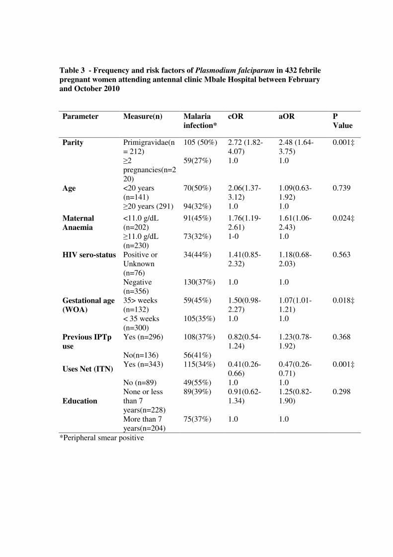
Table 3 - Frequency and risk factors of Plasmodium falciparum in 432 febrile
pregnant women attending antennal clinic Mbale Hospital between February
and October 2010
*Peripheral smear positive
Parameter Measure(n) Malaria
infection* cOR aOR P
Value
Parity Primigravidae(n
= 212) 105 (50%) 2.72 (1.82-
4.07) 2.48 (1.64-
3.75) 0.001‡
≥2
pregnancies(n=2
20)
59(27%) 1.0 1.0
<20 years
(n=141)
70(50%) 2.06(1.37-
3.12)
1.09(0.63-
1.92)
0.739 Age
≥20 years (291) 94(32%) 1.0 1.0
Maternal
Anaemia
<11.0 g/dL
(n=202)
91(45%) 1.76(1.19-
2.61)
1.61(1.06-
2.43)
0.024‡
≥11.0 g/dL
(n=230)
73(32%) 1-0 1.0
Positive or
Unknown
(n=76)
34(44%) 1.41(0.85-
2.32)
1.18(0.68-
2.03)
0.563 HIV sero-status
Negative
(n=356)
130(37%) 1.0 1.0
35> weeks
(n=132)
59(45%) 1.50(0.98-
2.27)
1.07(1.01-
1.21)
0.018‡ Gestational age
(WOA)
< 35 weeks
(n=300)
105(35%) 1.0 1.0
Yes (n=296) 108(37%) 0.82(0.54-
1.24)
1.23(0.78-
1.92)
0.368 Previous IPTp
use
No(n=136) 56(41%)
Uses Net (ITN) Yes (n=343) 115(34%) 0.41(0.26-
0.66)
0.47(0.26-
0.71)
0.001‡
No (n=89) 49(55%) 1.0 1.0
Education
None or less
than 7
years(n=228)
89(39%) 0.91(0.62-
1.34)
1.25(0.82-
1.90)
0.298
More than 7
years(n=204)
75(37%) 1.0 1.0

Table 4 - Accuracy of microscopy and RDT in detecting placental malaria
infection in peripheral blood among 173 women at Mbale regional referral
hospital between March and November 2010
Placental histology
+(n=21) (n=152) Total Accuracy measure(95%CI)
Microscopy + 17 12 29 Sensitivity 76.2(52.4-90.8)
4 140 144 Specificity 92.1(86.3-95.7)
PPV* 57.1(37.4-74.9)
NPV‡ 96.6(91.7-98.7)
RDTs + 17 19 36 Sensitivity 80.9(57.4-93.7)
4 133 137 Specificity 87.5(80.9-92.1)
PPV 47.2(30.7-64.2)
NPV 97.1(92.2-99.1)
Microscopy + 19 27 46 Sensitivity 90.5(68.2-98.3)
+RDTs 2 125 127 Specificity 82.2(75.0-87.7)
PPV 41.3(27.3-56.7)
NPV 98.4(93.9-99.7)

Table 5 - Frequency and risk factors of placental Plasmodium falciparum
infection among 166 delivering mothers in Mbale Hospital between February
and October 2010
*Malaria in pregnancy
aOR = Adjusted odds ratios
‡ Statistically significant
.
Parameter Categories(n) Placental
Infection
cOR aOR P
Value
Parity Primigravidae(n= 83) 14 (17%) 2.60 (0.95-
7.15)
1.01 (0.21-
4.72)
0.994
≥2 pregnancies(n=83) 6(7%) 1.0 1.0
< 20 years (n=47) 9(19%) 2.33 (0.90-
6.05)
1.68 (0.44-
6.46)
0.451 Age
≥20 years (119) 11(9%) 1.0 1.0
Maternal
Anaemia
<11.0 g/dL (64) 11(17%) 2.31 (0.87-
6.10)
2.09 (0.63-
6.96)
0.222
≥11.0 g/dL (n=97) 8(8%) 1.0 1.0
Still birth (n=14) 6(36%) 1.34(0.16-
11.12)
37.9 (2.70-
528.80)
0.007‡ Pregnancy
outcome
Live full term(n=149) 14(9%)
< 2500gm (n=11) 1(9%) 1.34 (0.16-
11.12)
17.04 (0.63-
460.7)
0.092 Birth weight
≥2500 gm(n=97) 8(8%)
Yes (n=118) 17(14%) 0.40 (0.11-
1.42)
0.31 (0.07-
1.40)
0.127 Previous
IPTp use
No(n=30) 7(23%) 1
Uses Net
(ITN)
Yes (n=136) 13(10%) 0.34 (0.13-
0.96)
1.02 (0.22-
4.58)
0.977
No (n=30) 7(23%) 1.0 1.0
MIP* Smear Positive(n=61) 16(26%) 8.98 (2.84-
28.37)
20.95 (3.99-
110.05)
0.001‡
Smear
Negative(n=105)
4(4%) 1.0 1.0
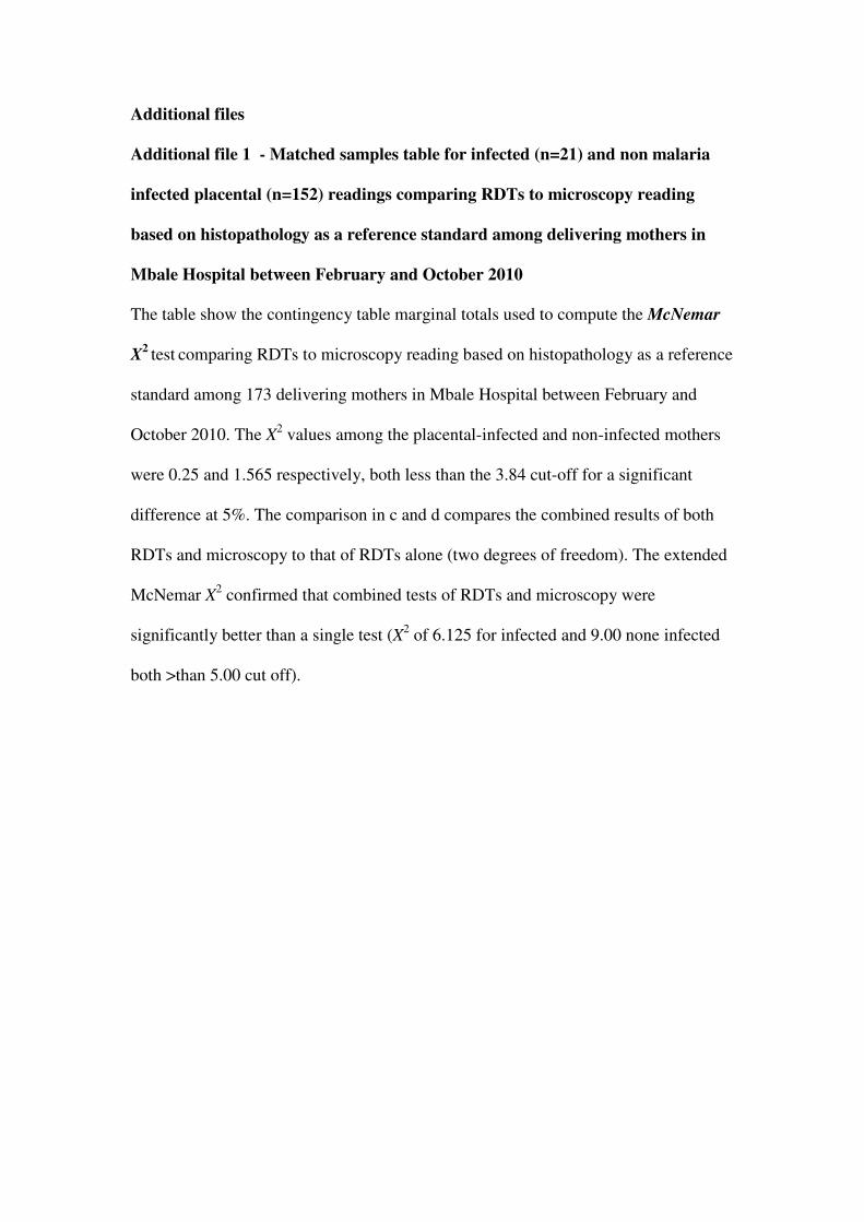
Additional files
Additional file 1 - Matched samples table for infected (n=21) and non malaria
infected placental (n=152) readings comparing RDTs to microscopy reading
based on histopathology as a reference standard among delivering mothers in
Mbale Hospital between February and October 2010
The table show the contingency table marginal totals used to compute the McNemar
X2
test comparing RDTs to microscopy reading based on histopathology as a reference
standard among 173 delivering mothers in Mbale Hospital between February and
October 2010. The X2 values among the placental-infected and non-infected mothers
were 0.25 and 1.565 respectively, both less than the 3.84 cut-off for a significant
difference at 5%. The comparison in c and d compares the combined results of both
RDTs and microscopy to that of RDTs alone (two degrees of freedom). The extended
McNemar X2 confirmed that combined tests of RDTs and microscopy were
significantly better than a single test (X2 of 6.125 for infected and 9.00 none infected
both >than 5.00 cut off).

534 screen
434 febrile women enrolled
164(38%) smear positive
173 Placental histology results
• 16 Active acute infections
• 4 Chronic infections
• 16 Past infection
208(47%) Hospital deliveries
• 186 Full term
• 11 pre-term*
• 8 still births
35 Excluded from analysis
• 13 Poor /missing smear/records
3- Birth before admission
8 missing tissue
9 poor tissue taken/preserved
226 (53%) Delivery outside
the hospital
Post Natal child follow-up
Figure 1

Additional files provided with this submission:
Additional file 1: Additional Table 1.doc, 47Khttp://www.malariajournal.com/imedia/1810923672619126/supp1.doc