paleopathological considerations on malaria infection in
TRANSCRIPT
Review ArticlePaleopathological Considerations on Malaria Infection inKorea before the 20th Century
Dong Hoon Shin,1 Min Seo,2 Jong Ha Hong,1 and Eunju Lee 3
1Lab of Bioanthropology, Paleopathology and History of Diseases, Department of Anatomy/Institute of Forensic Science,Seoul National University College of Medicine, Seoul, Republic of Korea2Department of Parasitology, Dankook University, Cheonan, Republic of Korea3Department of Internal Medicine, Asan Medical Center, University of Ulsan College of Medicine, Seoul, Republic of Korea
Correspondence should be addressed to Eunju Lee; [email protected]
Received 28 December 2017; Accepted 1 April 2018; Published 9 May 2018
Academic Editor: Stefano D’Amelio
Copyright © 2018 DongHoon Shin et al.This is an open access article distributed under theCreativeCommonsAttribution License,which permits unrestricted use, distribution, and reproduction in any medium, provided the original work is properly cited.
Malaria, one of the deadliest diseases in human history, still infects many people worldwide. Among the species of the genusPlasmodium, P. vivax is commonly found in temperate-zone countries including South Korea. In this article, we first review thehistory of malarial infection in Korea by means of studies on Joseon documents and the related scientific data on the evolutionaryhistory of P. vivax in Asia. According to the historical records, malarial infection was not unusual in pre-20th-century Koreansociety.We also found that certain behaviors of the Joseon people might have affected the host-vector-pathogen relationship, whichcould explain why malarial infection prevalence was so high in Korea at that time. In our review of genetic studies on P. vivax, weidentified substantial geographic differentiation among continents and even between neighboring countries. Based on these, wewere able to formulate a strategy for future analysis of ancient Plasmodium strains in Korea.
1. Introduction
Globally, malaria is the fifth deadliest disease, infectingapproximately 200 million people worldwide [1–3]. Malarialinfection is mediated by the arthropod vector Anophelesmosquito. The Plasmodium parasite has a complex life-cycleof sexual reproduction inside the mosquito vector and asex-ual stage in the vertebrate host. In brief, malarial sporozoitesare inoculated into human hosts when a mosquito bites them[4]. After a dormant phase, they differentiate into merozoitesfor release into the bloodstream, upon which they invadeerythrocytes (the beginning of asexual multiplication). Thebursting of infected red blood cells (RBCs) by merozoitemultiplication is responsible for the typical malarial fever [4].Some of themerozoites then develop into gametocytes, whichare taken up by a femalemosquito [4]. Sexual reproduction inthe anopheline mosquito is followed by sporozoite migrationinto the salivary gland, from which they are inoculated into avertebral host, thus beginning a new cycle of malarial infec-tion [4].
In general, five species of genus Plasmodium are knownto cause malaria: P. falciparum, P. vivax, P. malariae, P. ovale,and P. knowlesi [5]. Recent malaria outbreak in Brazil hasalso been traced to new zoonotic transmission of P. simiumfrommonkey [6]. Among them, P. vivax and P. falciparum arethe most commonly detected causative pathogens of humanmalaria. Clinical manifestations of uncomplicated malariaare nonspecific: headache, fever, malaise, myalgia, nausea,vomiting, and abdominal pain. Rare cases of malaria showsevere manifestations including anemia, thrombocytopenia,pulmonary edema, renal failure, hepatic dysfunction, andsplenic rupture [7].
Although malarial species share typical signs and symp-toms such as undulating intermittent fever, they also have dif-ferent traits depending on each subtype. P. falciparum’s symp-toms are serious enough to show the highest mortality ratewhereas P. vivax, P. malariae, and P. ovale exhibit generallynonfatal clinical courses [5].The geographical distribution ofeach Plasmodia subtype differs as well. P. falciparum is more
HindawiBioMed Research InternationalVolume 2018, Article ID 8516785, 14 pageshttps://doi.org/10.1155/2018/8516785
2 BioMed Research International
prevalent in tropical or subtropical zones including Sub-Saharan areas but relatively absent in temperate countries [8].Meanwhile, P. vivax generally infects human populations intemperate and tropical zones but is not so prevalent in Sub-Saharan Africa [1, 3, 9]. P. vivax malaria was endemic evenin some high latitude countries (Finland and Russia, etc.) atcertain points in history [4, 10, 11].
As malaria historically has been, and continues to be, oneof the most serious diseases, it has attracted the attention ofmany paleopathologists. Studies on ancientmalarial infectionhave been conducted by various methods such as osteoar-chaeological and biomedical approaches [12]. As chronic-stage vivax malaria was generally known to induce anemia,further causing porotic hyperostosis (PO) or cribra orbitalia(CO) in the cranium [10], anthropologists have searched forthe presence of PO or CO in skeletal remains as indirectevidences of malarial infection [5, 12–15]. Nevertheless, POor CO has clear limitations with respect to its application tothe study of ancient malaria because Plasmodium infection isnot the only cause of them [12, 16]. Other pathologies suchas inherited hemolytic anemia, scurvy, or malignancies arealso known to induce the same skeletal changes of PO or CO[5, 12].
In recent years, the paleopathological study ofmalaria hasbeen revolutionized by successful applications of immuno-logical and ancient DNA (aDNA) analyses to archaeologicalspecimens. To detect malaria-related proteins, researchersperformed the dipstick assay or new-generation immunoas-says on ancient mummies[17–19] or skeletons [20, 21]. Theimmunological assay became an effective screening methodto secure the evidence of ancient malarial infection [5]. Also,Plasmodium aDNAs reportedly have been obtained fromEgyptian mummies [2, 22–24], an infant skeleton dating toancient Rome [25], 15th-to-19th-century infant of Bavaria[26], and 1st-to-2nd-century adult skeletons of Italy [27].As a paleopathological tool, aDNA analysis is useful forconfirming the presence of malarial genomes remnant inarchaeological specimens as well as for revealing the originand dispersal of the protozoan parasite in evolutionaryhistory [28].
Although immunological and molecular analyses havebecome more reliable tools for the study of ancient malaria,the data obtained to date are not sufficient in terms ofquantity and quality [16]. Moreover, since previous studieshave focused mainly on ancient Egyptian, Roman, andRenaissance European remains thus far [5], such informationas has been obtained from malaria aDNA reflects a seriousgeographical bias. Extensive geographic sampling is thusnecessary in order to understand the demographic history ofmalaria much more comprehensively and clearly [4, 29–31].Like the other continents, Asia is also a region where malariahas historically been epidemic and endemic. In several Asiancountries, many people continue to have suffered from andeven died of Plasmodium infection. Nonetheless, most of therequisite paleopathology still remains to be revealed in Asiaas few medical studies on the ancient malaria have beenreported in the area. Herein, then, we offer this historicalreview as a fundamental basis for future research of ancientmalaria infection in Korea and other Asian countries.
2. Origin and Dispersal of VivaxMalaria Parasite
Parasitologists have speculated that human malaria mighthave been transmitted from nonhuman primates by a host-switch event [11]. Initially, they presumed that P. falciparumwas transmitted from chimpanzees and gorillas in Africa [18]while P. vivax originated from another nonhuman primate,possibly macaques, in Southeast Asia [4, 32, 33]. However,this hypothesis is seriously challenged nowadays by thegenetic analysis of malaria worldwide. Alternatively, a recentstudy revealed that both P. falciparum and P. vivax originatedin Africa and that P. vivax transmission to human beingsmight have occurred much earlier than P. falciparum did by ahost switch [34].
According to the estimated time to most recent commonancestor (TMRCA), the ancestor of the extant P. vivax popu-lations existed between 50 and 550 ka before the present [3].In a demographic history inferred from the P. vivax genomeanalysis, the global population size of vivax malaria mighthave expanded slowly until about 60 ka BP, which is closelyconsistent with the demographic history of mankind [3, 35].Once the divergence of African and Eurasian P. vivax popu-lations occurred at about 51 ka BP, the latter appears to haveundergone a rapid exponential increase in population size[3, 35]. AmongEurasian vivaxmalarias, the EastAsian varietymight have experienced a distinct pattern of populationgrowth [3]. The population of East Asian variety might havebeen relatively stable in its expansion until approximately10,000 years BP [36, 37]. It then began to increase rapidlyonce rice and millet started to be domesticated in the areaand sustained such increase, without tapering off, until thepresent [3]. The inferred hypothesis is suggestive of thedetailed evolutionary history of vivaxmalaria in East Asia [3].
In the phylogenetic tree of P. vivax worldwide, twodivergent groups were identified: a large star-like clusterand a divergent cluster [3]. The latter was composed of twosubclades with different geographical distributions: “Asia a”ofCentral China and “Asia b” ofChina, Korea, and Indonesia.The divergent East Asian P. vivax lineage was connected tothe large star-like cluster by a group of haplotypes found inSoutheast Asia [3]. Based on the phylogenetic analysis, EastAsian P. vivax might have been split from all other vivaxmalaria and developed a distinct demographic history for atleast 121 ka [3]. Meanwhile, mutations of P. vivax-resistantRBCs (Duffy-negative phenotype) occurred in Sub-Saharanpeoples [38].Due to themutations, vivaxmalaria disappearedcompletely from the area until the reintroduction of P. vivaxto East Africa by sea-going traders from Asia [39].
3. History of Malaria Infection in Korea
The historical record is important for understanding thepattern of malarial infection in ancient civilizations[27].In the classical period of Greece, Hippocrates famouslydescribed the typical undulating fever, a very suggestive signof malarial infection [5, 39]. Historians believed that malariabecame hyperendemic in Europe by its spread around theMediterranean area, next along the riverbanks of the Rhine,

BioMed Research International 3
Danube, and Rhone and then further to Northern Europe,while accommodating to colder climatic conditions in thoseareas [40]. Historical studies have shown thatmalaria becameremarkably prevalent in themarshy areas ofNorthern Europein the Early Middle Ages [5, 40, 41]. By the Later Middleto Early Modern Ages, except for Iceland, plenty of reportson malaria were available from every corner of Europe(including the North Sea, Germany, Anglo-Saxon England,and even Scandinavian countries) [40, 42, 43]. In a sense,malaria appears to have been a much more serious diseasethan even the Plague [40, 44].
Malaria must have been endemic in East Asia fromancient times as well, as descriptions about malaria-likesymptoms can be seen in Chinese historical records [45].Although Korea had been in close interaction with Chinafrom earliest times, in Korean history, the first recorded caseof malaria occurred only in the Goryeo Dynasty (918-1392CE) [46]. In a 14th-century record, a Joseon King’s mother(Joseon Dynasty: 1392-1910 CE) was seriously infected withmalaria and eventually died of it [47]. Over the following cen-turies, a wealth of records on the typical signs and symptomsof malarial infection (intermittent fever, repeated every thirdday) can be found in the Korean historical literature [47]. Asmost malarial infection in modern Korea has been revealedto have been caused by P. vivax [48], the Joseon people mighthave suffered from the same Plasmodium subtype. Notwith-standing the benign traits of P. vivax, relapsed infection typ-ically might have exhausted people, often eventually killingthem, as seen in similar clinical reports today [3, 49–53].
Before the first modern medical record on malaria inKorea (1886), prevalences of malarial infection could not bereliably calculated. In the First Annual Report of the KoreanGovernment Hospital, Seoul, Dr. Horace Newton Allendescribed “endemic intermittent fever” (possibly malaria) asthe most commonly observed sign among Korean patientswho visited his hospital [54, 55]. According to him, in the late19th century, hyperendemic malaria posed a serious threatto Koreans throughout the entire Joseon Kingdom. How,exactly, did malaria show such a high infection prevalencein Joseon society? In general, wetlands such as scatteredswamps, bogs, and river valleys have been important habitatsfor anopheline mosquito breeding. As wetlands were dis-tributed widely in Korea at that time, they must have beenintegral to the high malarial transmission rates [34, 40, 56–63].
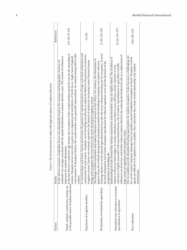
In malariology, however, the waxing and waning ofmalarial infection in a specific area cannot be explained sosimply. In addition to wetlands, environmental alteration ordegradation due to human activity also has a great influenceon the density and activity of mosquito populations and,further, on the prevalence of malaria itself[64–66]. Table 1summarizes the anthropogenic causes of malaria currentlyrecognized by scholars. As is apparent, people’s efforts toexploit environments often induce outbreaks of malaria [40,60, 64, 67]. Indeed, agricultural development and malariaare highly correlated in human history [60, 68–72]. Theexpansion of irrigation facilities, the reclamation of wetlands,economic specialization in agriculture, the simplification ofcrop types, enlargements of rice paddies, high population
densities, deforestation, and still other malaria-inducingfactors have been commonly cited (Table 1).
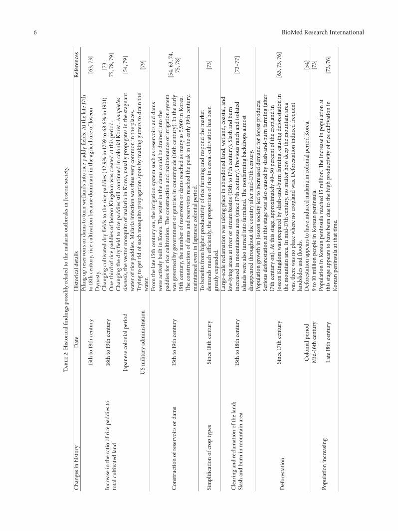
We do not yet know whether the close relationshipbetween environmental change by agriculture and malarialinfection is a universal phenomenonbeyond certain temporaland spatial limits. In a recent cross-national analysis, how-ever, correlations among anthropogenic activity, mosquitopopulation sizes, and malaria rates were seen to have beencommon in many parts of the world [64]. The findings ofTable 1 can thus be applied to our conjecture about Joseonsociety’s vulnerability to malarial infection. In our carefulexamination of the Joseon records, we found many similarmalarial-infection-facilitating situations to those noted inTable 1. The situations in Joseon society are summarized inTable 2.
In brief, the 15th to 19th centuries in Korean history werea turbulent and dynamic period during which the Joseonpeople were highly motivated to be involved in agriculturalinnovation, thereby eventually effecting major changes intheir sociocultural environment (Table 2). By infusion oflabor and capital investments into land development, thestate of the agricultural techniques was advanced. By clearingevery corner of wasteland and reclaiming wetlands, hugeareas of farmland in the Kingdom were newly opened up[73–77]. Farmers cleared slash-and-burn fields even up tothe tops of mountains[63, 73, 76]. By the end of the JoseonDynasty, there was virtually no land remaining that had notbeen utilized for farming purposes (Table 2).
On such lands, Joseon farmers planted crops. Rice wasvery popular, becoming the most preferred crop by the lateJoseon Dynasty [73]. To meet growing market demand forrice, farmers hastily turned their existing dry fields intorice paddies [54, 63, 73–75, 78, 79]. To supply enoughwater for rice cultivation, irrigation systems comprised ofreservoirs and dammed pools were newly constructed inthe Kingdom [54, 63, 74, 75, 78, 80]. Due to such increasedagricultural productivity during the 15th to 19th centuries,the population of the Joseon Kingdom soared [73, 76]. Allof these changes meant that the Joseon people came tolive more and more in highly populated villages, towns,and cities around which rice paddies, reservoirs, and damswere scattered (Table 2). Certainly, as long as this newsituation continued, malarial prevalence was by no meanslowered. In a sense, intensive farming appears to have beena necessary evil for the Joseon people, as, notwithstand-ing the malaria-inductive environments thus created, theincreased food production potentiated and achieved therebywas a great economic as well as social boon to the King-dom.
From the late 19th century, the diagnosis and treatmentof malaria began to be performed by specialists in Westernmedicine. In 1913, for example, an intermittent fever observedamong Korean patients was finally confirmed by a modernmicrobiology technique to be Plasmodium infection [54, 81].During the Japanese colonial period, however, significantreduction of malarial incidence proved difficult, as the envi-ronmental conditions associated with agriculture remainedthe same. Since the end of World War II and subsequentUS army administration, malarial infection as well as its
4 BioMed Research International
Table1:Th
efactorsproven
torelatew
ithhigh
prevalence
ofmalariainfection.
Factors
Details
References
Marsh,w
etland
,coastalarea,swam
p,etc.
asthep
ossib
lesource
ofmalariainfectionIn
theA
frican
coun
tries,anop
helin
elarvaew
erea
bund
antly
foun
din
thes
wam
ps;and
topo
graphicw
etness(of
wetland
s)was
stron
glyassociated
with
thes
patia
ldistrib
utionof
malariainfectioncases.Th
econ
trolofw
etland
sis
impo
rtantfor
malariaelimination.
[34,40
,56–
62]
InthelateB
ronzetoearly
Iron
Age
Europe,the
occupatio
nof
thec
oastalmarshes
pavedthew
ayforthe
spreadingof
malaria.M
osqu
itolarvae
werea
bletogrow
upin
stagnant
poolsa
ndditcheso
fNorth
Seao
rAng
lo-Saxon
England
coastm
arshes.InEu
ropean
histo
ry,the
coastalm
arshes
wereg
enerallyhyperend
emicform
alariadu
ring16th
to18th
century.
Expansionof
irrigationfacilities
InAncient
Egyptand
Rome,Fayum
area
becamethe
granaryby
repeated
projectsof
large-scalelandrecla
mationand
constructio
nof
canalsystem.A
nophele
svectorb
reedingwas
directlyor
indirectlylin
kedto
thep
resenceo
fextensiv
eirr
igationsyste
m.Fayum
was
serio
uslyexpo
sedto
theh
azards
ofmalariaby
increasin
gcontactb
etweenhu
man
beings
andmosqu
itosthatw
ereb
redin
then
ewlydevelopedirr
igationsyste
m.
[2,60]
Recla
mationof
wetland
fora
griculture
Thelinkage
ofland
recla
mationandmalariainfectionwas
proven
inEa
stAfrica.Fo
rinstance,thee
liminationof
papyrusfrom
thew
etland
sduringland
recla
mationprom
otes
theb
reedingof
mosqu
itosa
ndmalariainfection.
Drainaged
itchesinnewlycla
imed
agric
ulturallandwerea
lsothem
ostcom
mon
breeding
sitefor
mosqu
itos.In
brief,
thelandrecla
mationwas
tofoste
rmosqu
itoreprod
uctio
nby
redu
cedvegetatio
ncovera
ndthee
levatio
nof
the
temperature
atbreeding
site.
[2,60,124,125]
Intensified
crop
cultivatio
nor
econ
omic
specializationin
agric
ulture
Epidem
iologicalstudy
show
edthatmalariatransm
issionandintensified
crop
sare
high
lyrelated.Th
eincidence
ofmalariaisabou
tten
times
high
erin
cereals-cultivatio
narea
than
inareasw
ithlesscereals.Intensity
ofcrop
cultivatio
nishigh
lyassociated
with
exacerbatedhu
man
riskof
malaria.Specializationin
agric
ulture
initially
influ
enceso
nforestloss,and
furtherind
uces
malariainfection(byredu
cing
theb
iodiversity
duetoar
eplacemento
fhu
gevarie
tyof
vegetatio
nwith
nonn
ativec
rops).
[2,64,124–
127]
Rice
cultivatio
nTh
ericep
addies
providea
bund
antb
reedingop
portun
ities
form
alariamosqu
itos.Italso
becomes
achalleng
ingsite
forv
ectorc
ontro
l.Im
prop
erdrainage
from
ricep
addies
caused
Ano
phelinem
osqu
itostobreed.Mosqu
itocontrol
agentsared
ifficultto
beappliedto
ricep
addies.R
icec
ultiv
ationhasa
deep-roo
tedrelationshipwith
malaria
transm
ission.
[124,128–133]

BioMed Research International 5
Table1:Con
tinued.
Factors
Details
References
Deforestatio
n
Thep
upationrateof
Anopheles
mosqu
itowas
theh
ighestin
thes
amples
collected
from
deforeste
dareas.Land
cover
patte
rnisak
eyfactor
thatinflu
encesthe
habitatfor
malariamosqu
itos.Re
lationshipbetweendeforestationcaused
byas
mall-scalefarm
ingandAn
opheles
mosqu
itobreeding
was
evidently
proven
inAmazon
region
.
[60,64
,65,134–
144]
Inas
tructuralequ
ationmod
elacross67
(develo
ping
)nations,positive
associationwas
observed
between
deforestationratesa
ndmalariaprevalence.InSub-Saharancoun
tries,livingin
thelandwith
outtrees
ledto
the
increasedris
kof
malariainfection.
Ther
elationshipbetweenland
covera
ndther
eprodu
ctionof
malariavector
mosqu
itosw
asalso
show
nin
Western
Kenyan
Highlands.
Epidem
iologicalaspectsof
ecosystem
change
(deforestatio
n)andmosqu
itohabitatp
roliferation(in
creasedlevelsof
larvae,m
osqu
itopo
pulations,and
actualmalariarates)have
been
studied
extensively
.Significantrelationshipwas
observed
betweenthep
ercentageo
fforestcover
lossandhigh
erinfectionprevalence
ofmalaria.
Deforestatio
nim
pactsm
alariaprevalence
bymultip
lemechanism
s:increase
inthes
unlight
amou
nt,w
arming
temperature
idealfor
thep
upationof
malariavector
larvae,stand
ingwater
after
clearingterrain,
thelandbecoming
flatte
rand
morelikely
tosto
rewater,w
hich
istypically
lessacidicandmorec
ondu
ctivetoAn
opheles
larvae
developm
ent.
Whentheforestisreplacedby
newcrop
land
s,thep
lantsstillprovide
theb
ushy
coverfor
mosqu
itoproliferatio
n,makingthem
alariainfectionprevalence
high
er.
Highpo
pulatio
ndensity
Indeveloping
coun
tries,ruralp
opulationgrow
thandneedstoincrease
food
prod
uctio
nindu
cetheforestloss,
furtherinfl
uencingmalariainfection.
Usin
gthetim
bersforb
uildingandfuelwoo
disalso
oneo
fthe
keycauses
ofdeforestation.
Growingruralp
opulationalso
tend
tolivec
losertothen
aturalhabitatsof
mosqu
itos,further
experie
ncingris
kof
malariainfection.
[40,64
,145–147]
Inmedievalcity
ofGroningen,10percento
fthe
urbanpo
pulationdied
whilethes
urroun
ding
coun
trysides
howed
adeathrateof
5percent.
6 BioMed Research International
Table2:Historicalfin
ding
spossib
lyrelated
tothem
alariaou
tbreaksinJoseon
society.
Changesinhisto
ryDate
Historicaldetails
References
Increase
inther
atio
ofric
epaddies
tototalcultiv
ated
land
15th
to18th
century
Pilin
gup
reservoirsor
damstoturn
wetland
sintoric
epaddy
fields.At
thelate17th
to18th
century,ric
ecultiv
ationbecamed
ominantinthea
griculture
ofJoseon
Dyn
asty.
[63,73]
18th
to19th
century
Changing
cultivateddryfieldstother
icep
addies
(42.9%
in1759
to68.6%in
1901).
One-th
irdof
ricep
addies
ofJoseon
Kingdo
mwas
createdatthisperio
d.[73–
75,78,79]
Japanese
colonialperio
dCh
anging
thed
ryfield
toric
epaddies
stillcontinuedin
colonialKo
rea.An
opheles
sinensis,the
vector
mosqu
itoof
malariain
Korea,usually
prop
agates
inthes
tagn
ant
water
ofric
epaddies.M
alariainfectionwas
thus
very
common
inthep
laces.
[54,79]
USmilitary
administratio
nTrying
togetrid
ofsuch
mosqu
itoprop
agationspotsb
ymakinggutte
rsto
drainthe
water.
[79]
Con
structio
nof
reservoirsor
dams
15th
to19th
century
From
thelate15thcenturyon
,the
irrigationsyste
msuch
asreservoirsanddams
werea
ctively
built
inKo
rea.Th
ewater
inthed
amsc
ould
bedrainedinto
the
padd
iesfor
ricec
ultiv
ation.
Thec
onstr
uctio
nandmaintenance
ofirr
igationsyste
mwas
governed
bygovernmento
rgentriesincoun
tryside(16th
century).Inthee
arly
19th
century,then
umbero
freservoirs
ordamsreached
asmanyas
5,960in
Korea.
Thec
onstr
uctio
nof
damsa
ndreservoirsreachedthep
eakin
thee
arly19th
century,
maintainedeven
inJapanese
colonialperio
d.
[54,63,74,
75,78]
Simplificatio
nof
crop
types
Since18thcentury
Tobenefitsfrom
high
erprod
uctiv
ityof
ricefarmingandrespon
dthem
arket
demands
mucheffi
ciently,the
prop
ortio
nof
riceincerealcultivatio
nhasb
een
greatly
expand
ed.
[73]
Clearin
gandrecla
mationof
theland;
Slashandbu
rnin
mou
ntainarea
15th
to18th
century
Large-scaler
eclamationwas
taking
placeinabando
nedland
,wetland
,coasta
l,and
low-ly
ingareasa
triver
orstr
eam
basin
s(15th
to17th
century).Slash
andbu
rnincreasedin
mou
ntaino
usarea
(since
17th
century).P
reviou
sranch
andiso
lated
islands
werea
lsoturned
into
farm
land
.Then
onfarm
ingbackdrop
salm
ost
disapp
earedthroug
hout
thec
ountry
after
mid-17thcentury.
[73–77]
Deforestatio
nSince17thcentury
Popu
lationgrow
thin
Joseon
societyledto
increaseddemandforforestp
rodu
cts.
Serio
usdeforestationatthisstagew
asalso
caused
bysla
sh-and
-burnfarm
ing(afte
r17th
centuryon
).At
thisstage,approxim
ately
40–50percento
fthe
crop
land
inJoseon
Kingdo
mwas
prepared
bysla
sh-and
-burnfarm
ing,causingdeforestationin
them
ountainarea.Inmid-17thcentury,no
matterh
owdeep
them
ountainarea
was,there
was
noplacew
here
nocrop
land
was.D
eforestatio
nindu
cedfre
quent
land
slidesa
ndflo
ods.
[63,73,76]
Colon
ialp
eriod
Deforestatio
nappearstohave
indu
cedmalariain
colonialperio
dKo
rea.
[54]
Popu
latio
nincreasin
g
Mid-16thcentury
9to
10millionpeop
lein
Korean
peninsula.
[73]
Late18th
century
Popu
lationin
Korean
peninsular
eached
15million.
Theincreaseinpo
pulationat
thisstagea
ppearsto
have
been
duetotheh
ighprod
uctiv
ityof
ricec
ultiv
ationin
Korean
peninsulaa
tthattim
e.[73,76]

BioMed Research International 7
management underwent a revolutionary change in Korea.The US army, which had experienced many deaths frommalaria in the course of the war with Japan, was able to estab-lish an effective means of controlling the Plasmodium infec-tion in Asia [79]. Whereas the Japanese during the period oftheir colonial rule of Korea preferred a strategy entailing cur-ing ofmalaria patients by quinine treatment at the onset of thedisease, the US military administration adopted a far moreaggressive and highly effective policy of controlling malariathrough the use of insecticides [79].
However, we must also consider the possibility thatmalarial prevalence in the country was not reduced byantimalarial medications or insecticides alone. This idea issupported by instances in European history. In the 18th to19th centuries, malaria was still prevalent in Europe, thoughit rapidly declined thereafter, finally disappearing from mostregions by the 1930s [82]. The retreat of malaria from Europewas not the result of medical or chemical innovations, asno such deliberate countermeasures (entailing use of quinineor insecticide, etc.) had been pursued at that time [83].Rather, another factor has been proposed to explain thedecline of malaria in Europe: socioeconomic progress [84].Many studies in fact have shown a positive correlationbetween malaria and poverty [85, 86]. Furthermore, it hasbeen established that malaria was active for much longerperiods of time in regions where modernization was delayed[40]. In Finland, long-term social changes such as landconsolidation, decreasing household size, fewer interactionsbetween families, and the transition from extended familyto nuclear family have been posited as causes of malaria’sdecline during the past 200 years [83]. In fact, the combinedeffects of social innovations and improved standards ofliving were decisive in controlling and eventually eradicatingmalaria in Europe [40, 60]. Arguably, this explanation isapplicable to the decline of malaria in South Korea as well.In that country, malaria was eradicated in the 1970s bythe cooperative efforts of the World Health Organization(WHO) and Korea’s National Malaria Eradication Program[48, 87]. Significantly, this corresponded to the period ofrapid industrialization by which the living standards ofthe Korean people were remarkably improved. In short,socioeconomic development in South Korea might havemade a great contribution to the eradication ofmalaria in thatcountry.
Unfortunately, the reemergence of malaria after long-term eradication is not a rare phenomenon in the world[3,88]. In 1993, after more than two decades of malaria-freestatus, a new Korean malaria patient was reported amongsoldiers who had served near the Demilitarized Zone (DMZ)in South Korea [48, 89]. At the time, as North Korea was suf-feringmalarial infection, the new patient was thought to havebeen infected by an Anopheles mosquito migrating from thenorth [87, 90]. In fact, this cannot explain everything aboutthe reemergence of malaria in South Korea, as, nowadays atleast, foreign travelers and workers from malaria-endemicregions are commonplace [87]. Today, malaria is once againan endemic disease and a source of public concern in SouthKorea, as cases of malarial infection have continued to bereported [87, 90, 91].
4. Genetic Diversity of Modern KoreanP. vivax Isolates
Malarial infection in history often cannot be fully evidencedby the examination of historical documents. This is due todifficulty in accurate diagnosis of ancient malaria cases inhistory. Actually, the signs and symptoms of ancient malariapatients were often vaguely described in historical literaturesby modern clinical medicine standards. In addition, diagno-sis of ancient malaria (solely) by the examination of archaeo-logically obtained skeletal remains is also highly problematicas malaria leaves little traces on bones. In this regard, we notethat DNA-based study could be useful for acquiring scientificevidences of specific diseases prevalent in history[27].
DNA analysis of P. vivax is generally targeted on theprotozoan parasites’ surface proteins by which the erythro-cyte invasion of the vivax-malarial parasite can be triggered[92, 93]. One such surface protein is the P. vivax merozoitesurface protein (PvMSP), which is abundantly expressed onthe merozoites of vivax malaria [94]. Duffy-binding protein(PvDBP) is another membrane protein that is also presenton the P. vivax merozoites and that plays a crucial role inRBC invasion of parasites [92, 93, 95, 96]. As antibodiesagainst these proteins effectively block the invasion of P. vivaxinto human RBC[91], PvMSP and PvDBP are regarded asleading candidates for use in the development of malariavaccines[91, 97–99] though great genetic diversity amongthose surface proteins still represent a major obstacle to thevaccine research [4, 91, 97, 100–103].
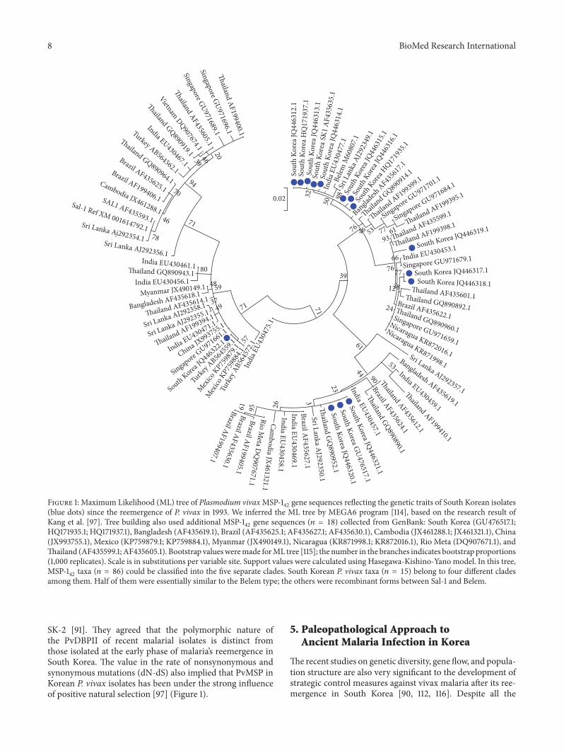
Since the reemergence of P. vivax in South Korea [48, 87],Korean researchers also have aimed to study the genetic traitsof vivax malaria’s PvMSP, PvDBP, circumsprozoite protein(PvCSP), apical membrane antigen-1 (AMA-1), microsatel-lites sequences, and 18S ribosomal RNAgenes [87, 90, 91, 103–108]. Those scientists are indeed eager to analyze the geneticdiversity, population structure, and operation of naturalselection among Korean P. vivax isolates, as the outcomeswould doubtlessly be useful for understanding the natureof the P. vivax population in South Korea [90, 91, 97]. Ingeneral, the genetic diversity of P. vivax is higher than that ofP. falciparum, suggesting that the former has a long, complex,but successful evolutionary history of adaptation [4, 109–112]. However, when P. vivax reemerged in South Korea, theisolates of the years 1993 to 2000 were genetically closelyrelated, meaning that its genetic diversity was very low atthe initial stage of its reintroduction [113]. Since 2001, thereemergent malaria population in South Korea has becomemore heterogeneous, showing increased genetic diversity anda more complex population structure [87, 89, 97, 108]. Theresults clearly indicate that some genotypes that were notfound before 2000 eventually migrated into South Korea ata much later date, as accompanied by outbreeding betweendifferent genotypes [90].
Ju et al. [91] also reported that a phylogenetic analysisbased on PvDBPII sequences showed 3 different clusters (SK-1, SK-2, and SK-3) in Korean P. vivax isolates. Among them,SK-3 was a new clade that had not been identified at theearly phase of reemergence in the same Korean isolates [106]but later became a more prevalent group than either SK-1 or
8 BioMed Research International
Sout
h Ko
rea J
Q44
6312
.1So
uth
Kore
a HQ
1719
37.1
Sout
h Ko
rea J
Q44
6313
.1So
uth
Kore
a SK1
AF4
3563
5.1
Sout
h Ko
rea J
Q44
6314
.1
Indi
a EU
4304
77.1
Belem
M60
807.1
Sri L
anka
AJ2
9234
9.1
Sout
h Kor
ea JQ
4463
15.1
Sout
h Kor
ea JQ
4463
16.1
South
Korea H
Q1719
35.1
Bangla
desh AF43
5617
.1
�aila
nd GQ890914.1
�ailand AF199399.1
Singapore GU971701.1
Singapore GU971684.1
�ailand AF199395.1
�ailand AF435599.1
�ailand AF199398.1
South Korea JQ446319.1
India EU430453.1
Singapore GU971679.1
South Korea JQ446317.1South Korea JQ446318.1
�ailand AF435601.1�ailand GQ890892.1Brazil AF435622.1�ailand GQ890960.1
Singapore GU971659.1
Nicaragua KR872016.1
Nicaragua KR871998.1Sri Lanka AJ292357.1
Bangladesh AF435619.1
India EU430459.1
�ailand AF199410.1
�ailand AF435612.1
Brazil AF435624.1
�ailand GQ890890.1
India EU430457.1
South Korea JQ446321.1
South Korea GU476517.1
South Korea JQ446320.1
�ailand G
Q890952.1
Sri Lanka AJ292350.1
Brazil AF435627.1
India EU430469.1
India EU430458.1
Cambodia JX461321.1
Rio Meta D
Q907671.1
Brazil AF199405.1Brazil AF435630.1
Brazil AF199407.1�
ailand AF199400.1
Singapore GU971696.1
Singapore GU971689.1
�ailand AF435605.1
Vietnam DQ907674.1
�ailand GQ890919.1
India EU430467.1
Turkey AB564562.1
�ailand GQ890964.1
Brazil AF435625.1
Brazil AF199406.1
Cambodia JX461288.1SAL1 AF435593.1
Sal-1 Ref XM 001614792.1Sri Lanka Aj292354.1Sri Lanka AJ292356.1India EU430461.1�ailand GQ890943.1
India EU430456.1Myanmar JX490149.1
Bangladesh AF435618.1
�ailand AF435614.1
Sri Lanka AJ292358.1
Sri Lanka AJ292355.1
�ailand AF199394.1
India EU430471.1
China JX993755.1
Singapore GU971661.1
South Korea JQ
446322.1
Turkey AB56
4559
.1
Mexico
KP75
9879
.1
Mex
ico K
P759
884.1
Turk
ey A
B564
572.1
Indi
a EU43
0475
.1
98204639
9470
7146
78
80
4859
3749 71
5771
39
0.02 32
50
76 46 53 7793
66
61
7677
3812
24
61
53
90
44
23
3
269561
Figure 1: Maximum Likelihood (ML) tree of Plasmodium vivaxMSP-142gene sequences reflecting the genetic traits of South Korean isolates
(blue dots) since the reemergence of P. vivax in 1993. We inferred the ML tree by MEGA6 program [114], based on the research result ofKang et al. [97]. Tree building also used additional MSP-1
42gene sequences (𝑛 = 18) collected from GenBank: South Korea (GU476517.1;
HQ171935.1; HQ171937.1), Bangladesh (AF435619.1), Brazil (AF435625.1; AF435627.1; AF435630.1), Cambodia (JX461288.1; JX461321.1), China(JX993755.1), Mexico (KP759879.1; KP759884.1), Myanmar (JX490149.1), Nicaragua (KR871998.1; KR872016.1), Rio Meta (DQ907671.1), andThailand (AF435599.1; AF435605.1). Bootstrap valuesweremade forML tree [115]; the number in the branches indicates bootstrap proportions(1,000 replicates). Scale is in substitutions per variable site. Support values were calculated using Hasegawa-Kishino-Yano model. In this tree,MSP-1
42taxa (𝑛 = 86) could be classified into the five separate clades. South Korean P. vivax taxa (𝑛 = 15) belong to four different clades
among them. Half of them were essentially similar to the Belem type; the others were recombinant forms between Sal-1 and Belem.
SK-2 [91]. They agreed that the polymorphic nature ofthe PvDBPII of recent malarial isolates is distinct fromthose isolated at the early phase of malaria’s reemergence inSouth Korea. The value in the rate of nonsynonymous andsynonymous mutations (dN-dS) also implied that PvMSP inKorean P. vivax isolates has been under the strong influenceof positive natural selection [97] (Figure 1).
5. Paleopathological Approach toAncient Malaria Infection in Korea
The recent studies on genetic diversity, gene flow, and popula-tion structure are also very significant to the development ofstrategic control measures against vivax malaria after its ree-mergence in South Korea [90, 112, 116]. Despite all the

BioMed Research International 9
Table 3: Archaeological information on mummy samples with liver obtained during autopsy.
Number Mummy Estimated Date Sex Date of excavation(YYYY.MM)
1 Cheongdo 1642a Male 2014.10.
2 Andong 18Cb Male 2013.01.3 Dalsung 16C-17Cc Female 2014.05.4 Hwasung 18Cc Male 2012.12.5 Gangneung 1622
a Male 2007.11.6 Hadong2 Late 16-early 17Ca Female 2009.067 Kunkook Joseon periodc Female Unknown8 Mungyeong 1647
d Female 2010.04.aHistorical documentation. bCarbon dating. cArchaeological evidence. dTree ring.
benefits, however, the overall genetic trends of vivax malaria,especially concerning its evolutionary history, have not yetbeen revealed by the simple genomic assay of modernisolates. In fact, the investigation of P. vivax using present-day DNA extracts from modern Korean isolates often leadsto confusion as to vivaxmalaria’s origin and dispersal [28]. Toovercome this drawback, wemust conduct aDNA analyses onvarious human samples obtained from archaeological sites inKorea to analyze the genetic origin and phylogenetic historyof malaria more accurately and comprehensively.
The significance of aDNA analysis to any derived under-standing of the evolutionary history of malaria recently hasbeen demonstrated by Gelabert et al. [28]. By way of aDNAanalysis on 70-year-old microscopic slides of blood frommalaria-infected people in Spain, they were able to success-fully reconstruct themtDNA sequence of the now-eradicatedEuropean P. vivax malaria. Moreover, as it was proven tobe related to the most common present-day American P.vivax haplotype, the authors were able to confirm that vivaxmalaria entered the Americas by post-Columbian contactwith Europeans [28]. In this way, aDNA assay of ancienthuman remains can be used for finding the missing links inthe origin and spread of ancient malaria.
In aDNA analysis, the types of specimens to choose arevery crucial to the research’s success. To select the speci-mens ideal for aDNA assay purposes, the life-cycle of thevivax-malarial parasite must be considered. In brief, whenmalarial sporozoites are inoculated into human hosts, someof them migrate to the liver wherein they invade the hepaticparenchymal cells [4]. While some sporozoites can maintainthe dormant state there, they can be further differentiated intomerozoites and released into the bloodstream [4]. As seenin vivax malaria’s life-cycle, the liver is the place where thefinal preerythrocytic phase takes place [45]. In this regard,Joseon Dynasty (1392-1910 CE) mummy’s livers might besignificant to our project. For the past 10 years, scientists andarchaeologists in South Korea have been involved in inter-disciplinary work on well-preserved mummies discovered inJoseonDynasty tombs [117–123].The livers that could be usedfor aDNA analysis were obtained from mummies by en blocresection during autopsy (Table 3) [117].
Nevertheless, as the number of malaria sporozoites atliver stage might be actually very small, we should alsoconsider alternate specimens for our aDNA analysis. In thisregard, we note that a small amount (less than 1 g) of spongybones inside vertebrae (possibly containing hemopoietic cellremains) was chosen commonly as specimens for aDNAanalysis ofmalaria; and in another case, first or secondmolarshave been selected forPlasmodium aDNAanalysis [27].Manyfuture studies on ancient malarial genomes will proceed withthese specimens of Korean mummies or skeletons.
6. Conclusion
With respect to human samples obtained from archaeologicalsites, scientific techniques can be done to reveal whether theindividual had suffered from malaria in his lifetime or toobtain phylogenetic information of its ancient genome. Asthe previous studies on ancient malaria have focused mainlyon specimens from Egypt and Europe, however, the currentinformation so far obtained carries a serious geographicalbias. More extensive geographic samplings and assays arethus needed in order to obtain a more comprehensivedemographic evolutionary history of malaria.
Like the other continents, Asia is a region wherein malar-ial infection has been epidemic in history. Nonetheless, verylittle has been done in the way of relevant paleopathologicalstudies on ancient malaria. We thus reviewed the history ofmalaria in Korea and attempted to derive scientific clues tothe evolution of P. vivax there and elsewhere in Asia. Tothose ends, we first examined the historical-documentaryevidence of ancient malarial outbreaks in Joseon society andfound that malarial epidemics were in fact not unusual inpre-20th-century Korea. We detected changes in the host-vector-pathogen relationship, which probably affected theproliferation of the mosquito vector and indeed the preva-lence of ancient malaria in Joseon society. We also noted,in our review of genomic studies on P. vivax, substantialgeographic differentiation of vivax-malarial DNA betweendifferent continents and even neighboring countries. Manyscientific studies on the history of malaria will be done withancient specimens inKorea andAsia, pending the permissionof the relevant medical-ethics review boards.

10 BioMed Research International
Conflicts of Interest
The authors declare that there are no conflicts of interestregarding the publication of this paper.
Authors’ Contributions
Dong Hoon Shin and Min Seo contributed equally to thisstudy as first authors.
Acknowledgments
This work was supported by the National Research Founda-tion of Korea (NRF) grant funded by the Korea government(MSIP) (NRF-2016R1A2B4015669). The authors speciallythank Dr. Min Jae Kim (Department of Infectious Diseases,Asan Medical Center, South Korea), for his great advice withregard to our manuscript.
References
[1] World Health Organization, World malaria report 2011, WorldHealth Organization, Geneva, Switzerland, 2011.
[2] A. Lalremruata, M. Ball, R. Bianucci et al., “Molecular iden-tification of Falciparum malaria and human tuberculosis co-infections in mummies from the Fayum Depression (LowerEgypt),” PLoS ONE, vol. 8, no. 4, Article ID e60307, 2013.
[3] J. E. Taylor, M. A. Pacheco, D. J. Bacon et al., “The evolutionaryhistory of plasmodium vivax as inferred from mitochondrialgenomes: Parasite genetic diversity in the Americas,”MolecularBiology and Evolution, vol. 30, no. 9, pp. 2050–2064, 2013.
[4] O. E. Cornejo and A. A. Escalante, “The origin and age of Plas-modium vivax,” Trends in Parasitology, vol. 22, no. 12, pp. 558–563, 2006.
[5] A.Nerlich, “Paleopathology and paleomicrobiology ofmalaria,”Microbiology Spectrum, vol. 4, no. 6, Article ID PoH-0006-2015,2016.
[6] P. Brasil, M. G. Zalis, A. de Pina-Costa et al., “Outbreakof human malaria caused by Plasmodium simium in theAtlantic Forest in Rio de Janeiro: A molecular epidemiologicalinvestigation,”The Lancet Global Health, 2017.
[7] World Health Organization, Guidelines for the Treatment ofMalaria,WorldHealth Organization, Geneva, Switzweland, 3rdedition, 2015.
[8] R. Carter and K. N. Mendis, “Evolutionary and historicalaspects of the burden ofmalaria,”ClinicalMicrobiology Reviews,vol. 15, no. 4, pp. 564–594, 2002.
[9] M. Miao, Z. Yang, H. Patch, Y. Huang, A. A. Escalante, and L.Cui, “Plasmodium vivax populations revisited: Mitochondrialgenomes of temperate strains inAsia suggest ancient populationexpansion,” BMC Evolutionary Biology, vol. 12, no. 1, article no.22, 2012.
[10] L. Hulden, L. Hulden, andK.Heliovaara, “Endemicmalaria: An’indoor’ disease in northern Europe. Historical data analysed,”Malaria Journal, vol. 4, no. 19, 2005.
[11] R. Culleton, C. Coban, F. Y. Zeyrek et al., “The origins ofafrican plasmodium vivax; insights from mitochondrial ge-nome sequencing,” PLoS ONE, vol. 6, no. 12, Article ID e29137,2011.
[12] T. J. Setzer, “Malaria detection in the field of paleopathology: Ameta-analysis of the state of the art,” Acta Tropica, vol. 140, pp.97–104, 2014.
[13] M. N. Cohen,Health and the Rise of Civilization, Yale UniversityPress, New Haven, CT, 1989.
[14] T. D. White and P. A. Folkens, Human Osteology, AcademicPress, San Diego, 2000.
[15] T. D. White and P. A. Folkens,TheHuman Bone Manual, Elsev-ier Academic, Amsterdam, Boston, 2005.
[16] R. Bianucci, A. Araujo, C. M. Pusch, and A. G. Nerlich, “Theidentification of malaria in paleopathology-An in-depth assess-ment of the strategies to detectmalaria in ancient remains,”ActaTropica, vol. 152, pp. 176–180, 2015.
[17] R. L.Miller, S. Ikram, G. J. Armelagoss et al., “Diagnosis of Plas-modium falciparum infections in mummies using the rapidmanual ParaSightTM-F test,” Transactions of the Royal Societyof Tropical Medicine and Hygiene, vol. 88, no. 1, pp. 31-32, 1994.
[18] E. RabinoMassa, N. Cerutti, A. Marin et al., “Malaria in ancientEgypt: paleoimmunological investigations in predynasticmum-mified remains,” Chungara, vol. 32, no. 1, pp. 7–9, 2000.
[19] R. Bianucci, G. Mattutino, R. Lallo et al., “Immunological evi-dence of Plasmodium falciparum infection in an Egyptian childmummy from the early dynastic period,” Journal of Archae-ological Science, vol. 35, no. 7, pp. 1880–1885, 2008.
[20] G. Fornaciari, V. Giuffra, E. Ferroglio, and R. Bianucci, “MalariaWas “the Killer” of Francesco I de’ Medici (1531-1587),” Ameri-can Journal of Medicine, vol. 123, no. 6, pp. 568-569, 2010.
[21] G. Fornaciari, V. Giuffra, E. Ferroglio, S. Gino, and R. Bianucci,“Plasmodium falciparum immunodetection in bone remainsof members of the Renaissance Medici family (Florence, Italy,sixteenth century),” Transactions of the Royal Society of TropicalMedicine and Hygiene, vol. 104, no. 9, pp. 583–587, 2010.
[22] A. G. Nerlich, B. Schraut, S. Dittrich, T. Jelinek, and A. R. Zink,“Plasmodium falciparum in ancient Egypt,” Emerging InfectiousDiseases, vol. 14, no. 8, pp. 1317–1319, 2008.
[23] Z. Hawass, Y. Z. Gad, S. Ismail et al., “Ancestry and pathologyin King Tutankhamun’s family,” The Journal of the AmericanMedical Association, vol. 303, no. 7, pp. 638–647, 2010.
[24] R. Khairat, M. Ball, C.-C. H. Chang et al., “First insights intothe metagenome of Egyptian mummies using next-generationsequencing,” Journal of Applied Genetics, vol. 54, no. 3, pp. 309–325, 2013.
[25] R. Sallares and S. Gomzi, “Biomolecular archaeology of ma-laria,” Ancient Biomolecules, vol. 3, pp. 195–213, 2001.
[26] A. Zink, C. J. Haas, K. Herberth, andA. G.Nerlich, “PCR ampli-fication of Plasmodium DNA in ancient human remains,” An-cient Biomolecules, vol. 3, p. 293, 2001.
[27] S. Marciniak, T. L. Prowse, D. A. Herring et al., “Plasmodiumfalciparum malaria in 1st–2nd century CE southern Italy,”Current Biology, vol. 26, no. 23, pp. R1220–R1222, 2016.
[28] P. Gelabert, M. Sandoval-Velasco, I. Olalde et al., “Mitochon-drial DNA from the eradicated European Plasmodium vivaxand P. falciparum from 70-year-old slides from the Ebro Deltain Spain,” Proceedings of the National Acadamy of Sciences of theUnited States of America, vol. 113, no. 41, pp. 11495–11500, 2016.
[29] M. F. Hammer, F. Blackmer, D. Garrigan, M. W. Nachman, andJ. A. Wilder, “Human population structure and its effects onsampling Y chromosome sequence variation,”Genetics, vol. 164,no. 4, pp. 1495–1509, 2003.
[30] L. Excoffier, “Patterns of DNA sequence diversity and geneticstructure after a range expansion: lessons from the infinite-island model,” Molecular Ecology, vol. 13, no. 4, pp. 853–864,2004.

BioMed Research International 11
[31] M. Imwong, D. Sudimack, S. Pukrittayakamee et al., “Micro-satellite variation, repeat array length, and population historyof Plasmodium vivax,”Molecular Biology and Evolution, vol. 23,no. 5, pp. 1016–1018, 2006.
[32] A. A. Escalante, O. E. Cornejo, D. E. Freeland et al., “Amonkey’stale: The origin of Plasmodium vivax as a human malariaparasite,” Proceedings of the National Acadamy of Sciences of theUnited States of America, vol. 102, no. 6, pp. 1980–1985, 2005.
[33] J.Mu,D.A. Joy, J. Duan et al., “Host switch leads to emergence ofPlasmodium vivax malaria in humans,” Molecular Biology andEvolution, vol. 22, no. 8, pp. 1686–1693, 2005.
[34] D. E. Loy, W. Liu, Y. Li et al., “Out of Africa: origins and evo-lution of the human malaria parasites Plasmodium falciparumand Plasmodium vivax,” International Journal for Parasitology,vol. 47, no. 2-3, pp. 87–97, 2017.
[35] S. Gravel, B. M. Henn, R. N. Gutenkunst et al., “Demographichistory and rare allele sharing among human populations,”Proceedings of the National Acadamy of Sciences of the UnitedStates of America, vol. 108, no. 29, pp. 11983–11988, 2011.
[36] H. Lu, J. Zhang, and K.-B. Liu, “Earliest domestication of com-mon millet (Panicum miliaceum) in East Asia extended to10,000 years ago,” Proceedings of the National Acadamy of Sci-ences of the United States of America, vol. 106, no. 18, pp. 7367–7372, 2009.
[37] J. Molina, M. Sikora, N. Garud et al., “Molecular evidence for asingle evolutionary origin of domesticated rice,” Proceedings ofthe National Acadamy of Sciences of the United States of America,vol. 108, no. 20, pp. 8351–8356, 2011.
[38] L. L. Cavalli-Sforza, P. Menozzi, and A. Piazza, The Historyand Geography of Human Genes, Princeton University Press,Princeton, New Jersey, USA, 1994.
[39] M. D. Bogdonoff, J. K. Crellin, R. A. Good et al., The GenuineWorks of Hippocrates, Classics of Medicine Library, Birming-ham, UK, 1985.
[40] O. S. Knottnerus, “Malaria Around the North Sea: A Survey,”in in proceedings of Climatic Development and History of theNorth Atlantic Realm: Hanse Conference Report, G.Wefer,W. H.Berger, K. E. Behre, and E. Jansen, Eds., pp. 339–353, Springer-Verlag, Berlin Heidelberg, 2002.
[41] O. S. Knottnerus, “Malaria in denNordseemarschen: Gedankenuber Mensch und Umwelt,” in Dunger und Dynamit: Beitragezur Umweltgeschichte Schleswig-Holsteins und Danemarks, M.Jakubowski-Tiessen and K. J. Lorenzen-Schmidt, Eds., pp. 25–39, Neumunster, 1999.
[42] E. Meineke and K. Schier, “Fieber,” in Reallexikon der germanis-chen Altertumskunde, J. Hoops and H. Jankuhn, Eds., vol. 9, pp.4–11, De Gruyter, Berlin, 2nd edition, 1995.
[43] V. Møller-Christensen, “Feber,” in Kulturhistorisk leksikon fornordisk middelalder fra vikingetid til reformationstid, vol. 4, pp.208–210, København, Rosenkilde og Bagger, Kopenhagen OsloMalmo, 1959.
[44] G. Keil, “Malaria,” in Lexikon des Mittelalters, R. Auty et al., Ed.,vol. 6, pp. 162-163, Munich, Artemis, 1993.
[45] F. E. Cox, “History of the discovery of the malaria parasites andtheir vectors,” Parasites & Vectors, vol. 3, no. 1, article 5, 2010.
[46] H. S. Lee, “Goryeo Period,” in Hangukjeonyeombyueongsa, K.W. Choi et al., Ed., pp. 65–108,The Korean Society of InfectiousDiseases, Gunjachulpansa, Seoul, South Korea, 2009.
[47] S. S. Kim, “The first half of Joseon Period,” inHangukjeonyeom-byueongsa, K. W. Choi et al., Ed., pp. 113–200, The KoreanSociety of Infectious Diseases, Gunjachulpansa, Seoul, SouthKorea, 2009.
[48] I. H. Chai, G. I. Lim, S. N. Yoon, W. I. Oh, S. J. Kim, and J. Y.Chai, “Occurrence of tertian malaria in a male patient who hasnever been abroad,”The Korean Journal of Parasitology, vol. 32,no. 3, pp. 195–200, 1994.
[49] D. K. Kochar, V. Saxena, N. Singh, S. K. Kochar, S. V. Kumar,and A. Das, “Plasmodium vivax malaria,” Emerging InfectiousDiseases, vol. 11, no. 1, pp. 132–134, 2005.
[50] N. M. Anstey, T. Handojo, and M. C. F. Pain, “Lung injuryin vivax malaria: pathophysiological evidence for pulmonaryvascular sequestration and posttreatment alveolar-capillaryinflammation,” The Journal of Infectious Diseases, vol. 195, no.4, pp. 589–596, 2007.
[51] M. A. Alexandre, C. O. Ferreira, A. M. Siqueira et al., “SeverePlasmodium vivaxmalaria, Brazilian Amazon,” Emerging Infec-tious Diseases, vol. 16, no. 10, pp. 1611–1614, 2010.
[52] N.M. Douglas, N.M. Anstey, P. A. Buffet et al., “The anaemia ofPlasmodium vivaxmalaria,”Malaria Journal, vol. 11, article 135,2012.
[53] V. B. Kute, H. L. Trivedi, A. V. Vanikar et al., “Plasmodiumvivax malaria-associated acute kidney injury, India, 2010-2011,”Emerging Infectious Diseases, vol. 18, no. 5, pp. 842–845, 2012.
[54] I. Yeo, “A history ofmalaria inmodernKorea 1876-1945,”KoreanJournal of Medical History, vol. 20, no. 1, pp. 53–82, 2011.
[55] I. Yeo, Allen eui euiryo bogoseo, Yeoksagonggan, Seoul, 2016.[56] J. M. Chase and T. M. Knight, “Drought-induced mosquito
outbreaks in wetlands,” Ecology Letters, vol. 6, no. 11, pp. 1017–1024, 2003.
[57] J. M. Cohen, K. C. Ernst, K. A. Lindblade, J. M. Vulule, C. C.John, and M. L. Wilson, “Local topographic wetness indicespredict household malaria risk better than land-use and land-cover in the western Kenya highlands,” Malaria Journal, vol. 9,no. 1, article no. 328, 2010.
[58] O. Kenea, M. Balkew, and T. Gebre-Michael, “Environmentalfactors associated with larval habitats of anophelinemosquitoes(diptera: Culicidae) in irrigation and major drainage areas inthe middle course of the rift valley, central ethiopia,” Journal ofVector Borne Diseases, vol. 48, no. 2, pp. 85–92, 2011.
[59] Y. Do, H. A. Kim, S. B. Kim et al., “Awareness and exploitationof wetland during the Joseon Dynasty,” Journal of WetlandsResearch, vol. 14, no. 3, pp. 329–340, 2012.
[60] B. Wielgosz, M. Mangheni, D. W. Tsegai, and C. Ringler,“Malaria and Agriculture,” in A global view of the literature witha focus on the application of integrated pest and vector man-agement in East Africa and Uganda, International Food PolicyResearch Institute, Washington DC, USA, 2012, IFPRI Discus-sion Paper 01232.
[61] T. Bousema, J. T. Griffin, R. W. Sauerwein et al., “Hittinghotspots: Spatial targeting of malaria for control and elimina-tion,” PLoS Medicine, vol. 9, no. 1, Article ID e1001165, 2012.
[62] C. J. Shiff, C. Stoyanov, C. Choobwe, A. Kamanga, and V. M.Mukonka, “Measuringmalaria by passive case detection: A newperspective based on Zambian experience,” Malaria Journal,vol. 12, no. 1, article no. 120, 2013.
[63] D. J. Kim, Joseon eui saengtaehwangyeongsa, Purunyeoksa,Seoul, South Korea, 2017.
[64] K. F. Austin,M.O. Bellinger, and P. Rana, “Anthropogenic forestloss and malaria prevalence: a comparative examination of thecauses and disease consequences of deforestation in developingnations,” AIMS Environmental Science, vol. 4, no. 2, pp. 217–231,2017.

12 BioMed Research International
[65] D. E. Norris, “Mosquito-borne Diseases as a Consequence ofLand Use Change,” EcoHealth, vol. 1, no. 1, pp. 19–24, 2004.
[66] S. L. Lewis and M. A. Maslin, “Defining the Anthropocene,”Nature, vol. 519, no. 7542, pp. 171–180, 2015.
[67] P. Reiter, “Climate change and mosquito-borne disease: Know-ing the horse before hitching the cart,” Revue Scientifique etTechnique de l’OIE, vol. 27, no. 2, pp. 383–398, 2008.
[68] W. H. Wernsdorfer and I. McGregor, Malaria: Principles andPractice of Malariology, Churchill Livingstone, Edinburgh,Scotland, 1988.
[69] S. C.Oaks, V. S.Mitchell, G.W. Pearson et al.,Malaria: Obstaclesand Opportunities, National Academy Press, Washington, DC,USA, 1991.
[70] J. A. Najera, “Malaria Control: Achievements, Problems andStrategies,” Parassitologia, vol. 43, no. 1-2, pp. 1–89, 2001.
[71] J. A. Patz, P. Daszak, G. M. Tabor et al., “Unhealthy landscapes:Policy recommendations on land use change and infectious dis-ease emergence,” Environmental Health Perspectives, vol. 112, no.10, pp. 1092–1098, 2004.
[72] R.M. Packard,TheMaking of a Tropical Disease: A Short Historyof Malaria, Johns Hopkins University Press, Baltimore, US,2007.
[73] National Institute of Korean History, Hanguk Munhwasa,National Institute of Korean History, Gwacheon, South Korea,2009.
[74] J. S. Yeom, “Construction of irrigation facilities in Hwaseong-bu and management of Dunjeon in the late 18th century,”Nongeobsayeongu, vol. 9, no. 1, 2010.
[75] J. S. Yeom, “The agricultural techniques and irrigation facilitiesin the medieval age-early modern age,” Journal of CentralInstitute of Cultural Heritage, vol. 10, pp. 99–151, 2012.
[76] C. S. Song, “17, 18 Segi shinjeongaegan eui hwakdae wa gyeon-gyeongheyongtae,” Hanguksaron, vol. 12, pp. 231–304, 1985,Dept. of Korean History, Seoul National University.
[77] J. S. Yeom, “Reclamation of river islands at lower Daedong riverand transition of Gungbangjeon in the Late Joseon Dynasty,”Gyujanggak, vol. 37, pp. 101–130, 2010.
[78] Y. J. Kim, A review for diffusion of rice transplantation and irri-gation system during latter period of the Chosun Era [Master,thesis], Inha University, Incheon, South Korea, 2001.
[79] I.-S. Yeo, “U.S. military administration’s malaria control activi-ties (1945-1948),” Korean Journal of Medical History, vol. 24, no.1, pp. 35–65, 2015.
[80] C.-Y. Jung, “Regional characteristics of Cheon during the LateChoson Era as seen through the Yojitoso,” Journal of the KoreanGeographical Society, vol. 43, no. 4, pp. 620–637, 2008 (Korean).
[81] C. Y. Chow, “Arthropods of public health importance in Korea,”Korean Journal of Entomology, vol. 3, no. 1, pp. 43-44, 1973.
[82] L. Hulden, L. Hulden, andK.Heliovaara, “Endemicmalaria: An’indoor’ disease in northern Europe. Historical data analysed,”Malaria Journal, vol. 25, no. 4, 2005.
[83] L. Hulden and L. Hulden, “The decline of malaria in Finland -The impact of the vector and social variables,”Malaria Journal,vol. 8, no. 1, article no. 94, 2009.
[84] J. de Zulueta, “Malaria and ecosystems: from prehistory to pos-teradication.,” Parassitologia, vol. 36, no. 1-2, pp. 7–15, 1994.
[85] J. L. Gallup and J. D. Sachs, “The economic burden of malaria,”TheAmerican Journal of Tropical Medicine and Hygiene, vol. 64,no. 1-2, pp. 85–96, 2001.
[86] R. I. Chima, C. A. Goodman, and A. Mills, “The economicimpact of malaria in Africa: A critical review of the evidence,”Health Policy, vol. 63, no. 1, pp. 17–36, 2003.
[87] Y.-K. Choi, K.-M. Choi, M.-H. Park et al., “Rapid disseminationof newly introduced Plasmodium vivax genotypes in SouthKorea,”TheAmerican Journal of Tropical Medicine and Hygiene,vol. 82, no. 3, pp. 426–432, 2010.
[88] C. A. Guerra, S. I. Hay, L. S. Lucioparedes et al., “Assembling aglobal database of malaria parasite prevalence for the MalariaAtlas Project,”Malaria Journal, vol. 6, article no. 17, 2007.
[89] J. W. Park, T. A. Klein, H. C. Lee et al., “Vivax malaria: A con-tinuing health threat to the Republic of Korea,” The AmericanJournal of Tropical Medicine and Hygiene, vol. 69, pp. 159–167,2003.
[90] H. Honma, J.-Y. Kim, N. M. Q. Palacpac et al., “Recent increaseof genetic diversity in Plasmodium vivax population in theRepublic of Korea,”Malaria Journal, vol. 10, article no. 257, 2011.
[91] H.-L. Ju, J.-M. Kang, S.-U. Moon et al., “Genetic diversity andnatural selection of Duffy binding protein of Plasmodium vivaxKorean isolates,” Acta Tropica, vol. 125, no. 1, pp. 67–74, 2013.
[92] J. W. Barnwell, M. E. Nichols, and P. Rubinstein, “In vitroevaluation of the role of the Duffy blood group in erythrocyteinvasion by Plasmodium vivax,” The Journal of ExperimentalMedicine, vol. 169, no. 5, pp. 1795–1802, 1989.
[93] J. H. Adams, B. K. L. Sim, S. A.Dolan, X. Fang,D. C. Kaslow, andL.H.Miller, “A family of erythrocyte binding proteins ofmalariaparasites,” Proceedings of the National Acadamy of Sciences of theUnited States of America, vol. 89, no. 15, pp. 7085–7089, 1992.
[94] A. A. Holder, J. A. Guevara Patino, and C. Uthaipibull, “Mero-zoite surface protein 1, immune evasion, and vaccines againstasexual blood stage malaria,” Parasitologia, vol. 41, pp. 409–414,1999.
[95] S. P. Wertheimer and J. W. Barnwell, “Plasmodium vivax inter-action with the human Duffy blood group glycoprotein: Identi-fication of a parasite receptor-like protein,” Experimental Para-sitology emphasizes, vol. 69, no. 3, pp. 340–350, 1989.
[96] R. Horuk, C. E. Chitnis, W. C. Darbonne et al., “A receptorfor the malarial parasite Plasmodium vivax: The erythrocytechemokine receptor,” Science, vol. 261, no. 5125, pp. 1182–1184,1993.
[97] J.-M. Kang, H.-L. Ju, Y.-M. Kang et al., “Genetic polymorphismand natural selection in the C-terminal 42kDa region ofmerozoite surface protein-1 among Plasmodium vivax Koreanisolates,”Malaria Journal, vol. 11, article no. 206, 2012.
[98] M.Arevalo-Herrera and S.Herrera, “Plasmodiumvivaxmalariavaccine development,”Molecular Immunology, vol. 38, no. 6, pp.443–455, 2001.
[99] A. A. Holder, “Malaria vaccines: Where next?” PLoS Pathogens,vol. 5, no. 10, Article ID e1000638, 2009.
[100] M. C. Bruce, M. R. Galinski, J. W. Barnwell, G. Snounou, andK. P. Day, “Polymorphism at the merozoite surface protein-3𝛼 locus of Plasmodium vivax: global and local diversity,” TheAmerican Journal of Tropical Medicine and Hygiene, vol. 61, no.4, pp. 518–525, 1999.
[101] J. C. Rayner, V. Corredor, D. Feldman et al., “Extensive polymor-phism in the Plasmodium vivax merozoite surface coat proteinMSP-3𝛼 is limited to specific domains,” Parasitology, vol. 125,no. 5, pp. 393–405, 2002.
[102] M. C. Leclerc, C. Gauthier, L. Villegas, and L. Urdaneta, “Genet-ic diversity of merozoite surface protein-1 gene of Plasmodiumvivax isolates in mining villages of Venezuela (Bolivar State),”Acta Tropica, vol. 95, no. 1, pp. 26–32, 2005.

BioMed Research International 13
[103] J.-Y. Chai, Y.-K. Park, S.-M. Guk et al., “A trial for a DNAdiagnosis of Plasmodium vivax malaria recently reemerging inthe republic of korea usingmicrotiter plate hybridization assay,”TheAmerican Journal of Tropical Medicine and Hygiene, vol. 63,no. 1-2, pp. 80–84, 2000.
[104] C. S. Lim, S. H. Kim, S. I. Kwon, J.-W. Song, K.-J. Song, andK.N.Lee, “Analysis of Plasmodium vivaxmerozoite surface protein-1gene sequences from resurgent Korean isolates,” The AmericanJournal of Tropical Medicine and Hygiene, vol. 62, no. 2, pp. 261–265, 2000.
[105] C. S. Lim, Y. K. Kim, K. N. Lee et al., “ The analysis of circum-sporozoite-protein gene sequences from South Korean isolatesof ,” Annals of Tropical Medicine & Parasitology, vol. 95, no. 3,pp. 229–235, 2016.
[106] W. G. Kho, J. Y. Chung, E. J. Sim, D. W. Kim, and W. C.Chung, “Analysis of polymorphic regions of Plasmodium vivaxDuffy binding protein of Korean isolates.,” The Korean Journalof Parasitology, vol. 39, no. 2, pp. 143–150, 2001.
[107] J.-Y. Chung, E.-H. Chun, J.-H. Chun, and W.-G. Kho, “Analysisof the Plasmodium vivax apical membrane antigen-1 gene fromre-emerging Korean isolates,” Parasitology Research, vol. 90, no.4, pp. 325–329, 2003.
[108] D. H. Nam, J. S. Oh, M. H. Nam et al., “Emergence of newalleles of the MSP-3alpha gene in Plasmodium vivax isolatesfrom Korea,” The American Journal of Tropical Medicine andHygiene, vol. 82, no. 4, pp. 522–524, 2010.
[109] D. E. Neafsey, K. Galinsky, R. H. Y. Jiang et al., “The malariaparasite Plasmodium vivax exhibits greater genetic diversitythan Plasmodium falciparum,” Nature Genetics, vol. 44, no. 9,pp. 1046–1050, 2012.
[110] P. Orjuela-Sanchez, J. M. Sa, M. C. C. Brandi et al., “Highermicrosatellite diversity in Plasmodium vivax than in sympatricPlasmodium falciparum populations in Pursat, Western Cam-bodia,” Experimental Parasitology emphasizes, vol. 134, no. 3, pp.318–326, 2013.
[111] W. Liu, Y. Li, K. S. Shaw et al., “African origin of the malariaparasite Plasmodium vivax,” Nature Communications, vol. 5, p.3346, 2014.
[112] A. E. Barry, A. Waltmann, C. Koepfli, C. Barnadas, and I.Mueller, “Uncovering the transmission dynamics of Plasmod-ium vivax using population genetics,” Pathogens and GlobalHealth, vol. 109, no. 3, pp. 142–152, 2015.
[113] M. Iwagami, S.-Y. Hwang, S.-H. Kim et al., “Microsatellite DNAAnalysis Revealed a Drastic Genetic Change of Plasmodiumvivax Population in the Republic of Korea During 2002 and2003,” PLOS Neglected Tropical Diseases, vol. 7, no. 10, ArticleID e2522, 2013.
[114] K. Tamura, G. Stecher, D. Peterson, A. Filipski, and S. Kumar,“MEGA6: Molecular Evolutionary Genetics Analysis version6.0,”Molecular Biology and Evolution, vol. 30, no. 12, pp. 2725–2729, 2013.
[115] B. G. Hall, “Building phylogenetic trees from molecular datawith MEGA,” Molecular Biology and Evolution, vol. 30, no. 5,pp. 1229–1235, 2013.
[116] L. Cui, A. A. Escalante, M. Imwong, and G. Snounou, “Thegenetic diversity of Plasmodium vivax populations,” Trends inParasitology, vol. 19, no. 5, pp. 220–226, 2003.
[117] Y. S. Kim, M. J. Kim, and J. H. Hong, “The Scientific and EthicalBackground of the Invasive Studies on the KoreanMummies ofthe Joseon Dynasty,” Asian Journal of Paleopathology, vol. 1, pp.5–11, 2017.
[118] I. S. Lee, E.-J. Lee, J. B. Park et al., “Acute traumatic death ofa 17th century general based on examination of mummifiedremains found in Korea,” Annals of Anatomy, vol. 191, no. 3, pp.309–320, 2009.
[119] D.H. Shin, C. S.Oh, S. J. Lee et al., “Ectopic paragonimiasis from400-year-old female mummy of Korea,” Journal of Archaeologi-cal Science, vol. 39, no. 4, pp. 1103–1110, 2012.
[120] D. H. Shin, C. S. Oh, H. J. Lee et al., “Ancient DNA analysis onClonorchis sinensis eggs remained in samples from medievalKorean mummy,” Journal of Archaeological Science, vol. 40, no.1, pp. 211–216, 2013.
[121] E.-J. Lee, C. S. Oh, S. G. Yim et al., “Collaboration of Archae-ologists, Historians and Bioarchaeologists During Removal ofClothing from Korean Mummy of Joseon Dynasty,” Interna-tional Journal of Historical Archaeology, vol. 17, no. 1, pp. 94–118,2013.
[122] M. Seo, A. Araujo, K. Reinhard, J. Y. Chai, andD.H. Shin, “Pale-oparasitological studies on mummies of the Joseon Dynasty,Korea,” The Korean Journal of Parasitology, vol. 52, no. 3, pp.235–242, 2014.
[123] Y.-S. Kim, I. S. Lee, C. S.Oh,M. J. Kim, S. C. Cha, andD.H. Shin,“Calcified pulmonary nodules identified in a 350-year-old-joseon mummy: The first report on ancient pulmonary tuber-culosis from archaeologically obtained pre-modern Koreansamples,” Journal of Korean Medical Science, vol. 31, no. 1, pp.147–151, 2016.
[124] J. Keiser, M. C. De Castro, M. F. Maltese et al., “Effect of irri-gation and large dams on the burden of malaria on a global andregional scale,” The American Journal of Tropical Medicine andHygiene, vol. 72, no. 4, pp. 392–406, 2005.
[125] K. Asenso-Okyere, F. A. Asante, J. Tarekegn et al., “The Linkagesbetween Agriculture and Malaria. Issues for Policy, ResearchandCapacity Strengthening,” inProceedings of the IFPRIDiscus-sion Paper 00861, International Food Policy Research Institute,Washington DC, USA, 2009.
[126] S. S. Myers, L. Gaffikin, C. D. Golden et al., “Human healthimpacts of ecosystem alteration,” Proceedings of the NationalAcadamy of Sciences of the United States of America, vol. 110, no.47, pp. 18753–18760, 2013.
[127] G. Z. Laporta, P. I. K. L. D. Prado, R. A. Kraenkel, R. M.Coutinho, andM.A.M. Sallum, “Biodiversity CanHelp PreventMalariaOutbreaks in Tropical Forests,”PLOSNeglected TropicalDiseases, vol. 7, no. 3, Article ID e2139, 2013.
[128] J. A. Carney, Black Rice: The African Origins of Rice Cultivationin the Americas, Harvard University Press, Cambridge, MA,USA, 2001.
[129] J. N. Ijumba and S. W. Lindsay, “Impact of irrigation on malariain Africa: Paddies paradox,” Medical and Veterinary Entomol-ogy, vol. 15, no. 1, pp. 1–11, 2001.
[130] E. Klinkenberg, W. Takken, F. Huibers, and Y. T. Toure, “Thephenology of malaria mosquitoes in irrigated rice fields inMali,” Acta Tropica, vol. 85, no. 1, pp. 71–82, 2003.
[131] G. Dolo, O. J. T. Briet, A. Dao et al., “Malaria transmission inrelation to rice cultivation in the irrigated Sahel of Mali,” ActaTropica, vol. 89, no. 2, pp. 147–159, 2004.
[132] P. N. Ng’ang’a, J. Shililu, G. Jayasinghe et al., “Malaria vectorcontrol practices in an irrigated rice agro-ecosystem in centralKenya and implications for malaria control,” Malaria Journal,vol. 7, article no. 146, 2008.
[133] J. M. Mwangangi, J. Shililu, E. J. Muturi et al., “Anopheles larvalabundance and diversity in three rice agro-village complexes
14 BioMed Research International
Mwea irrigation scheme, central Kenya,” Malaria Journal, vol.9, no. 1, article no. 228, 2010.
[134] J. A. Patz, T. K. Graczyk, N. Geller, and A. Y. Vittor, “Effects ofenvironmental change on emerging parasitic diseases,” Interna-tional Journal for Parasitology, vol. 30, no. 12-13, pp. 1395–1405,2000.
[135] S. Munga, N. Minakawa, G. Zhou et al., “Association betweenland cover and habitat productivity of malaria vectors in west-ern Kenyan highlands,” The American Journal of Tropical Med-icine and Hygiene, vol. 74, no. 1, pp. 69–75, 2006.
[136] A. Y. Vittor, R. H. Gilman, J. Tielsch et al., “The effect of defor-estation on the human-biting rate of Anopheles darlingi, theprimary vector of Falciparummalaria in the PeruvianAmazon,”The American Journal of Tropical Medicine and Hygiene, vol. 74,no. 1, pp. 3–11, 2006.
[137] J. Yasuoka and R. Levins, “Impact of deforestation and agricul-tural development on anopheline ecology andmalaria epidemi-ology,”The American Journal of Tropical Medicine and Hygiene,vol. 76, no. 3, pp. 450–460, 2007.
[138] K. C. Ernst, K. A. Lindblade, D. Koech et al., “Environmental,socio-demographic and behavioural determinants of malariarisk in the western Kenyan highlands: A case-control study,”Tropical Medicine & International Health, vol. 14, no. 10, pp.1258–1265, 2009.
[139] A. Y. Vittor, W. Pan, R. H. Gilman et al., “Linking deforestationto malaria in the Amazon: characterization of the breedinghabitat of the principal malaria vector, Anopheles darlingi,”TheAmerican Journal of Tropical Medicine and Hygiene, vol. 81, no.1, pp. 5–12, 2009.
[140] E. Teferi, S. Uhlenbrook, W. Bewket, J. Wenninger, and B.Simane, “The use of remote sensing to quantify wetland loss inthe Choke Mountain range, Upper Blue Nile basin, Ethiopia,”Hydrology and Earth System Sciences, vol. 14, no. 12, pp. 2415–2428, 2010.
[141] B. H. Manh, A. C. A. Clements, N. Q. Thieu et al., “Social andenvironmental determinants of malaria in space and time inViet Nam,” International Journal for Parasitology, vol. 41, no. 1,pp. 109–116, 2011.
[142] A. Midekisa, G. B. Senay, and M. C. Wimberly, “Multisensorearth observations to characterize wetlands and malaria epi-demiology in Ethiopia,” Water Resources Research, vol. 50, no.11, pp. 8791–8806, 2014.
[143] W. Hanandita and G. Tampubolon, “Geography and socialdistribution of malaria in Indonesian Papua: A cross-sectionalstudy,” International Journal of Health Geographics, vol. 15, no. 1,article no. 13, 2016.
[144] X. Wang, G. Zhou, D. Zhong et al., “Life-table studies revealedsignificant effects of deforestation on the development andsurvivorship of Anophelesminimus larvae,” Parasites &Vectors,vol. 9, no. 1, article no. 323, 2016.
[145] A. K. Jorgenson and T. J. Burns, “Effects of rural and urban pop-ulation dynamics and national development on deforestation inless-developed countries, 1990-2000,” Sociological Inquiry, vol.77, no. 3, pp. 460–482, 2007.
[146] T. Bensel, “Fuelwood, deforestation, and land degradation: 10years of evidence from Cebu province, the Philippines,” LandDegradation & Development, vol. 19, no. 6, pp. 587–605, 2008.
[147] K. F. Austin, M. D. Noble, and M. T. Mejia, “Gendered vulner-abilities to a neglected disease: A comparative investigation ofthe effect of women’s legal economic rights and social status onmalaria rates,” International Journal of Comparative Sociology,vol. 55, no. 3, pp. 204–228, 2014.

Hindawiwww.hindawi.com
International Journal of
Volume 2018
Zoology
Hindawiwww.hindawi.com Volume 2018
Anatomy Research International
PeptidesInternational Journal of
Hindawiwww.hindawi.com Volume 2018
Hindawiwww.hindawi.com Volume 2018
Journal of Parasitology Research
GenomicsInternational Journal of
Hindawiwww.hindawi.com Volume 2018
Hindawi Publishing Corporation http://www.hindawi.com Volume 2013Hindawiwww.hindawi.com
The Scientific World Journal
Volume 2018
Hindawiwww.hindawi.com Volume 2018
BioinformaticsAdvances in
Marine BiologyJournal of
Hindawiwww.hindawi.com Volume 2018
Hindawiwww.hindawi.com Volume 2018
Neuroscience Journal
Hindawiwww.hindawi.com Volume 2018
BioMed Research International
Cell BiologyInternational Journal of
Hindawiwww.hindawi.com Volume 2018
Hindawiwww.hindawi.com Volume 2018
Biochemistry Research International
ArchaeaHindawiwww.hindawi.com Volume 2018
Hindawiwww.hindawi.com Volume 2018
Genetics Research International
Hindawiwww.hindawi.com Volume 2018
Advances in
Virolog y Stem Cells International
Hindawiwww.hindawi.com Volume 2018
Hindawiwww.hindawi.com Volume 2018
Enzyme Research
Hindawiwww.hindawi.com Volume 2018
International Journal of
MicrobiologyHindawiwww.hindawi.com
Nucleic AcidsJournal of
Volume 2018
Submit your manuscripts atwww.hindawi.com