inhibition of pikfyve kinase prevents infection by zaire
TRANSCRIPT
Inhibition of PIKfyve kinase prevents infection by Zaireebolavirus and SARS-CoV-2Yuan-Lin Kanga,b,1, Yi-ying Choua,b,2, Paul W. Rothlaufc,d, Zhuoming Liuc
, Timothy K. Sohd, David Curetond,e,
James Brett Casef, Rita E. Chenf,g, Michael S. Diamondc,f,g, Sean P. J. Whelanc,3
, and Tom Kirchhausena,b,h,3
aDepartment of Cell Biology, Harvard Medical School, Boston, MA 02115; bProgram in Cellular and Molecular Medicine, Boston Children’s Hospital, Boston,MA 02115; cDepartment of Molecular Microbiology, Washington University in St. Louis, St. Louis, MO 63110; dProgram in Virology, Harvard Medical School,Boston, MA 02115; eBoehringer Ingelheim Animal Health, Inc. Duluth, GA 30096; fDepartment of Medicine, Washington University in St. Louis, St. Louis, MO63110; gDepartment of Pathology & Immunology, Washington University in St. Louis, St. Louis, MO 63110; and hDepartment of Pediatrics, Harvard MedicalSchool, Boston, MA 02115
Edited by Peter Palese, Icahn School of Medicine at Mount Sinai, New York, NY, and approved July 22, 2020 (received for review April 22, 2020)
Virus entry is a multistep process. It initiates when the virusattaches to the host cell and ends when the viral contents reachthe cytosol. Genetically unrelated viruses can subvert analogoussubcellular mechanisms and use similar trafficking pathways forsuccessful entry. Antiviral strategies targeting early steps ofinfection are therefore appealing, particularly when the probabil-ity for successful interference through a common step is highest.We describe here potent inhibitory effects on content release andinfection by chimeric vesicular stomatitis virus (VSV) containingthe envelope proteins of Zaire ebolavirus (VSV-ZEBOV) or severeacute respiratory syndrome coronavirus 2 (SARS-CoV-2) (VSV-SARS-CoV-2) elicited by Apilimod and Vacuolin-1, small-moleculeinhibitors of the main endosomal phosphatidylinositol-3-phos-phate/phosphatidylinositol 5-kinase, PIKfyve. We also describe po-tent inhibition of SARS-CoV-2 strain 2019-nCoV/USA-WA1/2020 byApilimod. These results define tools for studying the intracellulartrafficking of pathogens elicited by inhibition of PIKfyve kinaseand suggest the potential for targeting this kinase in developingsmall-molecule antivirals against SARS-CoV-2.
COVID-19 | SARS-CoV-2 | ZEBOV | APILIMOD | Vacuolin-1
Membrane-enveloped viruses deliver their contents to cellsvia envelope protein-catalyzed membrane fusion. Binding
of virus to specific host cell receptor(s) triggers membrane fu-sion, which can occur directly at the plasma membrane or fol-lowing endocytic uptake. Viruses that require endocytic uptake canuse different initial trafficking routes to reach the site of membranefusion. In endosomes, acidic pH serves to trigger conformationalrearrangements in the viral envelope proteins that catalyze mem-brane fusion, as seen for influenza A virus and vesicular stomatitisvirus (VSV). For Zaire ebolavirus (ZEBOV), proteolytic pro-cessing of the envelope protein by host cell proteases (1) is nec-essary to expose the receptor binding domain prior to engagementof Niemman−Pick disease type 1C (NPC1 or NPC IntracellularCholesterol Transporter 1)—the late endosomal−lysosomal re-ceptor protein (2). Proteolytic processing is also required for severeacute respiratory syndrome coronavirus (SARS-CoV) (3, 4), andfor the current pandemic SARS-CoV-2 (5). Lassa fever virus(LASV) uses a different mechanism, binding alpha-dystroglycan atthe plasma membrane (6), for internalization with a subsequentpH-regulated switch that leads to engagement of lysosomal-associated membrane protein 1 for membrane fusion (7). Lym-phocytic choriomeningitis virus (LCMV) also uses alpha-dystroglycan(6) and is internalized in a manner that depends on endosomalsorting complexes required for transport proteins (8), although itremains unknown whether a second receptor is required.A hallmark of the endolysosomal system is controlled dynamic
trafficking of vesicular carriers among its various subcompart-ments. Phophoinositides are markers for defining the identity ofthese subcompartments, because they are restricted in their dis-tribution to specific intracellular membranes (reviewed in ref. 9).Although it is one of the least abundant of the phosphoinositides
in cells, PI(3,5)P2 is particularly important for endomembranehomeostasis. It is produced by PI-3P-5-kinase (PIKfyve), whichphosphorylates the D-5 position in phosphatidylinositol-3-phos-phate (PI3P) to yield phosphatidylinositol 3,5-bisphosphate(PI(3,5)P2) (10). First cloned as mammalian p235 (11), PIKfyve isa 240-kDa class III lipid kinase, present on the cytosolic face ofendosomal membranes (12, 13) as part of a ternary complex withthe PI(3,5)P2 5-phosphatase Sac3 and ArPIKfyve (14).Ablation of PIKfyve function by genetic (12, 15) or pharma-
cological means (16–20) causes endosomal swelling and vacuo-lation of late endosomes and endolysosomes. It is thought thatthese changes result from decreased membrane fission and con-comitant interference in endosomal traffic (13, 21). Small-moleculeinhibitors of PIKfyve, all of which have some structural resem-blance to each other, have been studied as potential drugs fortreating cancer and autoimmune diseases. These inhibitors includeApilimod (19), Vacuolin-1 (18), a series of 30 Vacuolin-related
Significance
The membrane fusion proteins of viral pathogens as diverse intheir replication strategies as coronaviruses and filovirusesdepend, for their functional activity, on proteolytic processingduring cell entry. Endosomal cathepsins carry out the cleav-ages. We have constructed chimeric forms of vesicular stoma-titis virus (VSV) bearing the fusion proteins of Zaire ebolavirus(ZEBOV) or SARS coronavirus 2 (SARS-CoV-2) and shown thattwo small-molecule inhibitors of an endosomal lipid kinase(PIKfyve) inhibit viral infection by preventing release of theviral contents from endosomes. Both inhibitory compoundscause distension of Rab5 and Rab7 subcompartments into smallvacuoles. One of them (Apilimod) also inhibits infection of cellsby authentic SARS-CoV-2. The results point to possibilities forhost targets of antiviral drugs.
Author contributions: M.S.D., S.P.J.W., and T.K. designed research; Y.-L.K., Y.-y.C., P.W.R.,Z.L., J.B.C., and R.E.C. performed research; Y.-L.K., Y.-y.C., T.K.S., and D.C. contributed newreagents/analytic tools; Y.-L.K. and T.K. analyzed data; T.K. wrote the paper; and T.K.supervised the project.
Competing interest statement: M.S.D. is a consultant for Inbios, Vir Biotechnology, andNGM Biopharmaceuticals, and is on the Scientific Advisory Board of Moderna. The M.S.D.laboratory at Washington University School of Medicine has received sponsored researchagreements from Moderna and Emergent BioSolutions.
This article is a PNAS Direct Submission.
This open access article is distributed under Creative Commons Attribution License 4.0(CC BY).1Present address: Department of Stem Cell and Regenerative Biology, Harvard University,Cambridge, MA 02138.
2Present address: Biogen, Cambridge, MA 02142.3To whom correspondence may be addressed. Email: [email protected] [email protected].
This article contains supporting information online at https://www.pnas.org/lookup/suppl/doi:10.1073/pnas.2007837117/-/DCSupplemental.
www.pnas.org/cgi/doi/10.1073/pnas.2007837117 PNAS Latest Articles | 1 of 11
MED
ICALSC
IENCE
S
Dow
nloa
ded
at L
IBR
AR
Y F
EG
AN
PLA
ZA
on
Aug
ust 2
4, 2
020
molecules (22), YM201636 (16), and WX8 chemical family mem-bers (20). Physiological effects of these compounds in cells includeinhibition of autophagy (17, 22, 23), reduced generation of IL-12/IL-23 (24), and reduced dendritic cell infiltration in psoriasis (25).Apilimod also inhibits infection by several viruses, including
ZEBOV. Although it does not alter the pH of endosomes norinhibit cathepsin B or L (26), Apilimod blocks entry of ZEBOVand other pathogenic filoviruses (27). Several groups reportedthat Apilimod prevents colocalization of VSV-ZEBOV pseu-doviruses with the ZEBOV endosomal receptor NPC1, but doesnot prevent colocalization with early endosomal antigen 1(EEA1) (5, 27, 28). Apilimod also inhibits entry of pseudotypedviruses bearing the spike proteins of Middle East respiratorysyndrome CoV, SARS-CoV, and SARS-CoV-2, as well as ofauthentic mouse hepatitis virus particles (5).Here, we have studied the effects of Apilimod on infection of
VSV-eGFP-SARS-CoV-2 and VSV-eGFP-ZEBOV chimerasand showed that Apilimod blocks infection of both, with a con-centration that inhibits response by 50% (IC50) of ∼50 nM.Apilimod and Vacuolin-1 also prevented entry and infection ofVSV-MeGFP-ZEBOV and many of the internalized VSV-MeGFP-ZEBOV virions colocalized with NPC1 in the distended, vacuolatedendosomes. This suggests that blocking PIKfyve kinase has the samedownstream effects on these viruses, even though VSV-eGFP-SARS-CoV-2 does not require interaction with NPC1 for membrane
fusion. Apilimod also inhibits infection by authentic SARS-CoV-2strain 2019-nCoV/USA-WA1/2020 virus, with an IC50 slightlylower than the IC50 for the VSV-eGFP-SARS-CoV-2. We suggestthat Apilimod, which has passed safety tests in previous hu-man clinical trials for nonviral indications (24, 25, 29, 30), is apotential starting point for developing small-molecule entry in-hibitors of SARS-CoV-2 that could limit infection and diseasepathogenesis.
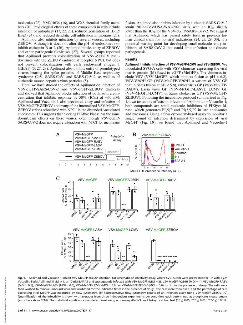
ResultsApilimod Inhibits Infection of VSV-MeGFP-LCMV and VSV-ZEBOV. Weinoculated SVG-A cells with VSV chimeras expressing the viralmatrix protein (M) fused to eGFP (MeGFP). The chimeras in-clude VSV (VSV-MeGFP, which initiates fusion at pH < 6.2),VSV-V269H GP (VSV-MeGFP-V269H, a variant of VSV GPthat initiates fusion at pH < 5.8), rabies virus GP (VSV-MeGFP-RABV), Lassa virus GP (VSV-MeGFP-LASV), LCMV GP(VSV-MeGFP-LCMV), or Zaire ebolavirus GP (VSV-MeGFP-ZEBOV). Following the incubation protocol summarized in Fig.1A, we tested the effects on infection of Apilimod or Vacuolin-1;both compounds are small-molecule inhibitors of PIKfyve ki-nase, which generates PI(5)P and PI(3,5)P2 in late endosomesand lysosomes. Using a flow cytometry-based assay to monitor asingle round of infection determined by expression of viralMeGFP (Fig. 1B), we found that Apilimod and Vacuolin-1
A
C
B
DMSOVacuolin-1ApilimodIN1BAF A1
% o
f Cel
ls
MeGFP fluorescence intensity (a.u.)
VSV-MeGFP-ZEBOV
0 1 2 3 4-1 5 6 7 8 9 hpi
VSV-MeGFP VSV-MeGFP-V269H VSV-MeGFP-RABV VSV-MeGFP-LASV VSV-MeGFP-LCMV
VSV-MeGFP-ZEBOV
Infectivity Assay
Inhibitor
Inhibitor
VSV-MeGFP-ZEBOVVSV-MeGFP-LCMVVSV-MeGFP-LASV
VSV-MeGFP-V269HVSV-MeGFP VSV-MeGFP-RABV
** ***
*****
** *** *** ******
Fig. 1. Apilimod and Vacuolin-1 inhibit VSV-MeGFP-ZEBOV infection. (A) Schematic of infectivity assay, where SVG-A cells were pretreated for 1 h with 5 μMVacuolin, 5 μM Apilimod, 5 μM IN1, or 10 nM BAF A1 and subsequently infected with VSV-MeGFP (MOI = 2), VSV-MeGFP-V269H (MOI = 1), VSV-MeGFP-RABV(MOI = 0.6), VSV-MeGFP-LASV (MOI = 0.6), VSV-MeGFP-LCMV (MOI = 0.6), or VSV-MeGFP-ZEBOV (MOI = 0.6) for 1 h in the presence of drugs. The cells werethen washed to remove unbound virus and incubated for the indicated times in the presence of drugs. The cells were then fixed, and the percentage of cellsexpressing viral MeGFP was measured by flow cytometry. (B) Representative flow cytometry results of an infection assay using VSV-MeGFP-ZEBOV. (C)Quantification of the infectivity is shown with averages from three independent experiments per condition, each determined as a duplicate measurement(error bars show SEM). The statistical significance was determined using a one-way ANOVA and Tukey post hoc test (*P ≤ 0.05; **P ≤ 0.01; ***P ≤ 0.001).
2 of 11 | www.pnas.org/cgi/doi/10.1073/pnas.2007837117 Kang et al.
Dow
nloa
ded
at L
IBR
AR
Y F
EG
AN
PLA
ZA
on
Aug
ust 2
4, 2
020
potently inhibit VSV-MeGFP-ZEBOV infection (Fig. 1C).These results agree with results obtained by others with Apili-mod (26, 31) in different cell types infected with murine leuke-mia virus (MLV) virus pseudotyped with ZEBOV GP or withEbola virus itself (26, 27, 32). Apilimod was a less effective in-hibitor of VSV-MeGFP-LCMV infection, and Vacuolin-1 hadno effect at the concentration used. In contrast, Apilimod andVacuolin-1 failed to prevent infection by VSV-MeGFP, VSV-MeGFP-V269H, VSV-MeGFP-RABV, or VSV-MeGFP-LASV(Fig. 1C). IN1 (33), an inhibitor of the phosphoinositide kinase
Vps34, the main endosomal generator of PI3P, also interferedwith VSV-MeGFP-LCMV and VSV-MeGFP-ZEBOV infection(Fig. 1C). All of these viruses require low pH to trigger viral mem-brane fusion with the endosomal membranes, and, as expected, in-fection was fully blocked by Bafilomycin A1, which inhibits thevacuolar type H+-ATPase (V-ATPase) acidification activity (Fig. 1C).
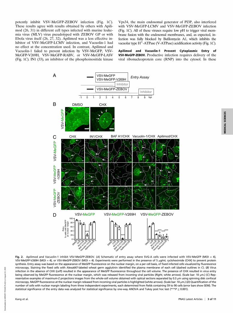
Apilimod and Vacuolin-1 Prevent Cytoplasmic Entry ofVSV-MeGFP-ZEBOV. Productive infection requires delivery of theviral ribonucleoprotein core (RNP) into the cytosol. In these
A
0 1 2 3 4 5 6 7 8 9 hpi
VSV-MeGFP VSV-MeGFP-V269H
VSV-MeGFP-ZEBOV
Entry Assay
Inhibitor
B
C
-1
DMSO
VS
V-M
eGFP
CHX
D
Apilimod/CHXVacuolin-1/CHX
VS
V-M
eGFP
-V26
9H
BAF A1/CHXCHX IN1/CHX
VS
V-M
eGFP
-ZE
BO
VV
SV-
MeG
FP
Inhibitor
VSV-MeGFP-V269H VSV-MeGFP-ZEBOVVSV-MeGFP
******
************
Fig. 2. Apilimod and Vacuolin-1 inhibit VSV-MeGFP-ZEBOV. (A) Schematic of entry assay where SVG-A cells were infected with VSV-MeGFP (MOI = 4),VSV-MeGFP-V269H (MOI = 4), or VSV-MeGFP-ZEBOV (MOI = 4). Experiments were performed in the presence of 5 μg/mL cycloheximide (CHX) to prevent proteinsynthesis. Entry assay was based on the appearance of MeGFP fluorescence on the nuclear margin, on a per cell basis, of fixed infected cells visualized by fluorescencemicroscopy. Staining the fixed cells with Alexa647-labeled wheat germ agglutinin identified the plasma membrane of each cell (dashed outlines in C). (B) Virusinfection in the absence of CHX (Left) resulted in the appearance of MeGFP fluorescence throughout the cell volume. The presence of CHX resulted in virus entrybeing observed by MeGFP fluorescence at the nuclear margin, which was released from incoming viral particles (Right, white arrows). (Scale bar: 10 μm.) (C) Rep-resentative examples of maximum-Z projections images from the whole-cell volume obtained with optical sections separated by 0.3 μm using spinning disk confocalmicroscopy. MeGFP fluorescence at the nuclear margin released from incoming viral particles is highlighted (white arrows). (Scale bar: 10 μm.) (D) Quantification of thenumber of cells with nuclear margin labeling from three independent experiments, each determined from fields containing 59 to 90 cells (error bars show SEM). Thestatistical significance of the entry data was analyzed for statistical significance by one-way ANOVA and Tukey post hoc test (***P ≤ 0.001).
Kang et al. PNAS Latest Articles | 3 of 11
MED
ICALSC
IENCE
S
Dow
nloa
ded
at L
IBR
AR
Y F
EG
AN
PLA
ZA
on
Aug
ust 2
4, 2
020
experiments, we deemed RNP delivery, as monitored by single-cellfluorescence microscopy imaging (experimental protocol summa-rized in Figs. 2A and 3A), to be successful when fluorescentMeGFP encapsulated in the incoming virus appeared at the nu-clear margin of infected cells. The representative examples of VSVinfection and RNP core release shown in Fig. 2B were obtained inthe absence or presence of cycloheximide, which prevents viralprotein expression. In the absence of cycloheximide (Fig. 2 B,Left), large amounts of newly synthesized MeGFP are present
throughout the cell. In the presence of cycloheximide (Fig. 2 B,Right), we observed MeGFP in virions (fluorescent spots) as well asreleased MeGFP concentrated at the nuclear margin. We scoredthe effect of Apilimod, Vacuolin-1, or IN1 on RNP delivery byVSV-MeGFP, VSV-MeGFP-V269H, and VSV-MeGFP-ZEBOVby determining the appearance of MeGFP at the nuclear margin incycloheximide-treated cells. Consistent with the infection results,Apilimod, Vacuolin-1, and IN1 prevented entry of VSV-MeGFP-ZEBOV but not of VSV-MeGFP or VSV-MeGFP-V269H. As
H VSV-MeGFP VSV-MeGFP-ZEBOVCHX Ap/CHX CHXAp/CHX
Edited
TagRFP-Rab5c-/- +/+
Endogenous
A B
C
CHX Apilimod/CHXIN1/CHX
Rab5c MeGFP
E
VS
V-M
eGFP
-V
269H
VS
V-M
eGFP
-Z
EB
OV
VS
V-M
eGFP
Control0 1 2 3 4-1 5 6 7 8 hpi
VSV-MeGFP VSV-MeGFP-V269H
VSV-MeGFP-ZEBOV
Live-cell imaging
Inhibitor
Inhibitor
D
Apip limod/CHHH
VSV-MeGFP VSV-MeGFP-ZEBOVCHXAp/CHX CHXAp/CHX
TagR
FP-R
ab5c
V
SV-
MeG
FP
DMSO CHX F
I
Edited
TagRFP-Rab7a-/- +/+
Endogenous
G
CHX Apilimod/CHXVacuolin-1/CHXIN1/CHX
Rab7a MeGFP
VS
V-M
eGFP
-V
269H
VS
V-M
eGFP
-Z
EB
OV
VS
V-M
eGFP
TagR
FP-R
ab7a
V
SV-
MeG
FP
DMSO CHX
Vacuolin-1/CHX Apilimod/CHX
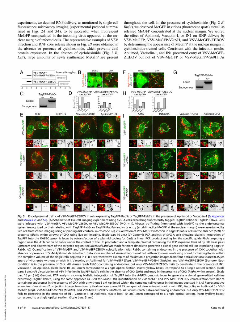
Fig. 3. Endolysosomal traffic of VSV-MeGFP-ZEBOV in cells expressing TagRFP-Rab5c or TagRFP-Rab7a in the presence of Apilimod or Vacuolin-1 (SI Appendixand Movies S1 and S2). (A) Schematic of live cell imaging experiment using SVG-A cells expressing fluorescently tagged TagRFP-Rab5c or TagRFP-Rab7a. Cellswere infected with VSV-MeGFP, VSV-MeGFP-V269H, or VSV-MeGFP-ZEBOV (MOI = 4). Viruses trafficking (monitored with MeGFP) to the endolysosomalsystem (recognized by their labeling with TagRFP-Rab5c or TagRFP-Rab7a) and virus entry (established by MeGFP at the nuclear margin) were ascertained bylive-cell florescence imaging using a spinning disk confocal microscope. (B) Visualization of VSV-MeGFP infection in TagRFP-Rab5c cells in the absence (Left) orpresence (Right, white arrows) of CHX using live-cell imaging. (Scale bar: 10 μm.) (C) Genomic PCR analysis of SVG-A cells showing biallelic integration ofTagRFP into the RAB5C genomic locus by cotransfection of a plasmid coding for Cas9, a linear PCR product coding for the specific guide RNAstargeting aregion near the ATG codon of Rab5c under the control of the U6 promoter, and a template plasmid containing the RFP sequence flanked by 800 base pairsupstream and downstream of the targeted region (see Materials and Methods for more details) to generate a clonal gene-edited cell line expressing TagRFP-Rab5c. (D) Quantification of VSV-MeGFP and VSV-MeGFP-ZEBOV colocalization with Rab5c containing endosomes in the presence of CHX together withabsence or presence of 5 μMApilimod depicted in E. Data show number of viruses that colocalized with endosomes containing or not containing Rab5c withinthe complete volume of the single cells depicted in E. (E) Representative examples of maximum-Z projection images from four optical sections spaced 0.35 μmapart of virus entry without or with IN1, Vacuolin, or Apilimod for VSV-MeGFP (Top), VSV-Me-GFP-V269H (Middle), and VSV-MeGFP-ZEBOV (Bottom). Eachcondition is in the presence of CHX. All viruses reach Rab5c-containing endosomes, but only VSV-MeGFP-ZEBOV fails to penetrate in the presence of IN1,Vacuolin-1, or Apilimod. (Scale bars: 10 μm.) Insets correspond to a single optical section. Insets (yellow boxes) correspond to a single optical section. (Scalebars: 3 μm.) (F) Visualization of VSV infection in TagRFP-Rab7a cells in the absence of CHX (Left) and entry in the presence of CHX (Right, white arrows). (Scalebar: 10 μm.) (G) Genomic PCR analysis showing biallelic integration of TagRFP into the RAB7A genomic locus to generate a clonal gene-edited cell-lineexpressing TagRFP-Rab7a, using the same approach as used for RAB5C. (H) Quantification of VSV-MeGFP and VSV-MeGFP-ZEBOV colocalization with Rab7acontaining endosomes in the presence of CHX with or without 5 μM Apilimod within the complete cell volumes in the images depicted in I. (I) Representativeexamples of maximum-Z projection images from four optical sections spaced 0.35 μm apart of virus entry without or with IN1, Vacuolin, or Apilimod for VSV-MeGFP (Top), VSV-Me-GFP-V269H (Middle), and VSV-MeGFP-ZEBOV (Bottom). All viruses reach Rab7a-containing endosomes, but only VSV-MeGFP-ZEBOVfails to penetrate in the presence of IN1, Vacuolin-1, or Apilimod. (Scale bars: 10 μm.) Insets correspond to a single optical section. Insets (yellow boxes)correspond to a single optical section. (Scale bars: 3 μm.)
4 of 11 | www.pnas.org/cgi/doi/10.1073/pnas.2007837117 Kang et al.
Dow
nloa
ded
at L
IBR
AR
Y F
EG
AN
PLA
ZA
on
Aug
ust 2
4, 2
020
expected, Bafilomycin A1 blocked entry of all viruses (images inFig. 2C and quantification in Fig. 2D).
Intracellular Trafficking of Virus Particles in the Presence of Apilimodor Vacuolin-1. Internalized virus particles traffic along the endo-cytic pathway to reach the endosomal compartment(s) fromwhich membrane fusion and genome entry into the cytosol occur.To establish the identity of the endosomal compartments, we
used genome editing in SVG-A cells (Figs. 3 C and G and 4 Band D) to replace expression of a subset of proteins enriched indifferent endosomal compartments (the small GTPases Rab5cand Rab7a, EEA1, or NPC1) with their corresponding fluores-cent chimeras obtained by fusion with TagRFP, mScarlet, orHalo (Figs. 3 B, E, F, and I and 4 C and E). The lack of fluo-rescently tagged filipin (a cholesterol binder) in the endolyso-somal compartment in the absence but not in the presence of
A
Control0 1 2 3 4-1 5 6 hpi
VSV-MeGFP-ZEBOV
B
NPC1-Halo-/- +/+
Edited
Endogenous
ApilimodDMSO
NP
C1-
Hal
o V
SV-
MeG
FP-Z
EB
OV
NPC1 MeGFP
C
D
NPC1-Halo-/- +/+
Edited
mScarlet-EEA1-/- +/+
Endogenous
EEA1 NPC1 MeGFPmS
carle
t-EE
A1
NP
C1-
Hal
o V
SV-
MeG
FP-Z
EB
OV
ApilimodDMSOE
NP
C1-
Hal
o fil
ipin
pare
ntal
fil
ipin
- U18666A +U18666AF
Live-cell imaging
Inhibitor
Fig. 4. Endolysosomal traffic of VSV-MeGFP-ZEBOV in cells expressing NPC1-Halo or coexpressing mScarlet-EEA1 and NPC1-Halo in the presence of Apilimod(SI Appendix and Movie S3). (A) Schematic of live-cell imaging experiment with gene-edited SVG-A cells expressing NPC1-Halo or NPC1-Halo together withmScarlet-EEA1. Halo was labeled with either JF549 or JF647. Cells were infected with VSV-MeGFP-ZEBOV (MOI = 3). (B) Genomic PCR analysis showing biallelicintegration of Halo into the NPC1 genomic locus to generate a clonal gene-edited cell line expressing NPC1-Halo, using the same approach as for RAB5C andRAB7A. (C) Representative examples of maximum-Z projection images from four optical sections spaced 0.25 μm apart in the absence (Left) and presence(Right) of Apilimod, showing that VSV-MeGFP-ZEBOV reached NPC1-Halo−containing endosomes even in the presence of Apilimod, while failing to penetrateand infect. (Scale bar: 10 μm.) Insets correspond to a single optical section. (Scale bar: 3 μm.) (D) SVG-A cells with genomic NPC1-Halo were further gene editedto contain EEA1 tagged with mScarlet. Genomic PCR analysis shows biallelic integration into the EEA1 locus of mScarlet-EEA1 (Left) and into the NPC1 locus ofNPC1-Halo (Right). (E) Representative examples of maximum-Z projection images in the absence (Left) and presence (Right) of Apilimod, showing thatVSV-MeGFP-ZEBOV reached endosomes containing mScarlet-EEA1 and endosomes containing both mScarlet-EEA1 and NPC1-Halo in the presence of Apili-mod, while failing to penetrate and infect. (Scale bar: 10 μm.) Insets correspond to a single optical section. (Scale bar: 3 μm.) (F) Representative images ofparental cells (Top) and gene-edited SVG-A cells expressing NPC1-Halo (Bottom) incubated with filipin III (naturally fluorescent polyene antibiotic that bindsto cholesterol) in the absence (Left) and presence (Right, NPC1 inhibitor of cholesterol export) of U18666A, showing NPC1-Halo is a functional cholesteroltransporter. (Scale bar: 10 μm.)
Kang et al. PNAS Latest Articles | 5 of 11
MED
ICALSC
IENCE
S
Dow
nloa
ded
at L
IBR
AR
Y F
EG
AN
PLA
ZA
on
Aug
ust 2
4, 2
020
U18666A, a potent inhibitor of NPC1 (Fig. 4F), showed thatNPC1-Halo remained active as a cholesterol transporter.Using live-cell spinning disk confocal microscopy (Figs. 3 and
4), we monitored the presence of virus particles in the fluo-rescently tagged endosomes by colocalization with the fluores-cent spots from the virus-incorporated MeGFP. We monitoredentry by carrying out the experiments in the presence of cyclo-heximide, thus ensuring that any MeGFP fluorescent signal atthe nuclear margin originated only from MeGFP moleculescarried by incoming viral particles (Fig. 3 B and F). All cells weremaintained at 37 °C throughout all phases of the experiment toensure normal and undisturbed intracellular trafficking. All con-trol experiments performed in the absence of inhibitors showedarrival of VSV-MeGFP, VSV-MeGFP-V269H, or VSV-MeGFP-ZEBOV virus particles to early (Rab5c and EEA1) (Figs. 3E and4E) or late (Rab7a or NPC1) endosomes and lysosomes (Figs. 3Iand 4 C and E). MeGFP released from all viruses appeared at thenuclear margin, showing effective RNP release. NPC1, the re-ceptor for VSV-MeGFP-ZEBOV entry, is required for fusionfrom endosomes (2). The successful VSV-MeGFP-ZEBOV in-fection observed in the absence of drug in cells expressing NPC1-
Halo alone or in combination with mScarlet-EEA1 indicates thatNPC1-Halo is capable of facilitating infection and that VSV-MeGFP-ZEBOV trafficked to NPC1-Halo−containing endosomes.Apilimod and Vacuolin-1 treatment of the SVG-A cells led to
enlargement and vacuolization of their endosomes and lyso-somes tagged with fluorescent EEA1, Rab5c, Rab7a, or NPC1(Figs. 3–5), in agreement with earlier PIKfyve ablation studies(13, 21). VSV-MeGFP and VSV-MeGFP-V269H (fluorescentdots, white) reached all tagged species of enlarged endolyso-somes and successfully penetrated into the cytosol, as indicatedby MeGFP at the nuclear margin (Fig. 3 E and I). VSV-MeGFP-ZEBOV also trafficked to all tagged species of en-larged endolysosomes (Fig. 3 E and I), often reaching one of thenumerous NPC1-containing vacuoles enriched in EEA1 (Figs.4E and 5 B and C). VSV-MeGFP-ZEBOV in EEA1-containingendosomes increased in the presence of Apilimod, as alsoreported for VLP ZEBOV (27). While able to reach NPC1-containing functional endosomes in cells treated with Apilimod(Figs. 4 C and E and 5 B and C), VSV-MeGFP-ZEBOV failed topenetrate into the cytoplasm, as reflected by absence of MeGFPin the nuclear margin (Figs. 2C, 3 E and I, 4 C and E, and 5B).
C
A
Control0 1 2 3 4-1 5 6 hpi
VSV-MeGFP-ZEBOV
Imaging
Inhibitor
ApilimodDMSO ApilimodDMSO
2 hrs 4 hrs
EEA1 NPC1 MeGFP
B
mS
carle
t-EE
A1
NP
C1-
Hal
o V
SV-
MeG
FP-Z
EB
OV
EEA1 9 11 0 0
EEA1/NPC1 2 3 19 16
NPC1 22 25 23 52
# cells 7 6 4 9
# of endosomes containing VSV-MeGFP-ZEBOVDMSO Apilimod
Fig. 5. Extent of VSV-MeGFP-ZEBOV traffic into endosomes enriched in NPC1-Halo or NPC1-Halo and mScarlet-EEA1. (A) Schematic of imaging experiment ofVSV-MeGFP-ZEBOV trafficking in NPC1-Halo or NPC1-Halo and mScarlet-EEA1 gene-edited SVG-A cells. (B) Representative examples of maximum-Z projectionimages from four optical sections spaced 0.25 μm apart in the absence and presence of Apilimod 2 h or 4 h postinfection. A large number ofVSV-MeGFP-ZEBOV but not of VSV-MeGFP particles accumulated in the endosomes enlarged upon Apilimod treatment. (Scale bar: 10 μm.) (C) Quantificationof VSV-MeGFP-ZEBOV colocalization with mScarlet-EEA1 alone, both mScarlet-EEA1 and NPC1-Halo, or NPC1-Halo alone 2 h and 4 h postinfection, in theabsence or presence of 5 μM Apilimod. Data obtained from complete cell volumes are presented as numbers and corresponding percent colocalizations ofVSV-MeGFP-ZEBOV particles associated with a given type of endosome.
6 of 11 | www.pnas.org/cgi/doi/10.1073/pnas.2007837117 Kang et al.
Dow
nloa
ded
at L
IBR
AR
Y F
EG
AN
PLA
ZA
on
Aug
ust 2
4, 2
020
Apilimod Blocks Infection of VSV SARS-CoV-2. Using a recombinantVSV expressing soluble eGFP (VSV-eGFP) where the glycopro-tein (GP) was replaced with that of ZEBOV GP (VSV-eGFP-ZEBOV) or SARS-CoV-2 S (VSV-eGFP-SARS-Cov2), we inoc-ulated MA104 cells with these chimera viruses and tested the ef-fects of Apilimod on infection by flow cytometry (Fig. 6A). Wefound potent inhibition of VSV-eGFP-SARS-CoV-2 infection byApilimod and confirmed that the compound also inhibits VSV-eGFP-ZEBOV infection (Fig. 6B). The dose–response curvesindicated similar effects for VSV-eGFP-ZEBOV and VSV-eGFP-SARS-CoV-2 (IC50s of ∼50 nM), in contrast to the absence of anydetectable inhibition of VSV-eGFP infection, used here as anegative control.
Apilimod Blocks Infection of SARS-CoV-2 Virus. To test the effect ofApilimod on bona fide SARS-CoV-2 infection, we exposed VeroE6 cells to fully infectious SARS-CoV-2 (strain 2019-nCoV/USA-WA1/2020); after 24-h incubation, supernatants were har-vested and titered by focus-forming assay on a separate set ofVero E6 cells (Fig. 7A). Apilimod strongly inhibited SARS-CoV-2 infection, and the dose–response curve was similar or morepotent than those observed for VSV-eGFP-ZEBOV or VSV-eGFP-SARS-CoV-2 (IC50s of ∼10 nM) (Fig. 7B).
DiscussionCoronaviruses, filoviruses, and arenaviruses have different rep-lication strategies and unrelated surface GPs that engage dif-ferent receptor molecules during entry (1, 2, 5–8). Coronavirus
and filovirus surface GPs share a requirement for entry-associatedproteolytic processing for activation as fusogens (1). Filovirusesrequire passage through low-pH compartments where cathepsinsare active. Coronaviruses may enter directly by fusion at theplasma membrane or following receptor-mediated endocytosis.Cell entry of SARS-CoV and SARS-CoV-2 depends on the pro-tease TMPRSS2 in conjunction with ACE2 (34–37), and, whenTMPRSS2 is present, the entry pathway becomes insensitive tocathepsin inhibition (34, 37, 38).The common inhibition of viruses from all three groups by
Apilimod is a consequence of perturbing their shared entrypathway. Moreover, it is not the cathepsin activity itself thatthese compounds affect, judging from the outcome of the assayswith Apilimod and Vacuolin-1 showing they inhibit VSV chi-meras bearing the surface GPs of ZEBOV and LCMV and, to alesser extent, LASV. Apilimod also inhibits infection of cells byVSV-SARS-CoV-2 as well as by authentic SARS-CoV-2; neithercompound blocks infection by wild-type VSV. For VSV-ZEBOV,we have shown that the virus reaches a compartment enriched inNPC1, the ZEBOV coreceptor, and often also enriched in EEA1,but that it nonetheless fails to release internal proteins into thecytosol. Apilimod does not inhibit cathepsin (26), but Apilimod(39) and Vacuolin-1 (17, 23) can interfere with cathepsin matu-ration, as evidenced by an increase in procathepsin in treated cells;they do not influence endosomal pH (18, 26, 40), although otherstudies report that Apilimod decreases cathepsin activity (41) andVacuolin-1 increases pH (17, 23). Irrespective of this discrep-ancy, both Apilimod and Vacuolin-1 inhibit PIKfyve (17, 19), a
B
0 1 2 3 4-1 5 6 hpi
VSV-eGFP VSV-eGFP-ZEBOV VSV-eGFP-SARS-CoV-2
Inhibitor
Infectivity AssayA
B
Apilimod (uM)
DMSO 0.001 0.01 0.05 0.1 1
Infe
cted
cel
ls
% o
f eG
FP c
ells
VSV-eGFP VSV-eGFP-ZEBOV VSV-eGFP-SARS-CoV-2
Fig. 6. Apilimod and Vacuolin-1 inhibit infection of VSV-eGFP-SARS-CoV-2. (A) Schematic of infectivity assay of VSV-eGFP, VSV-eGFP-ZEBOV, andVSV-eGFP-SARS-CoV-2 in MA104 cells. MA104 cells were pretreated for 1 h with the indicated concentration of Apilimod. Pretreated cells were inoculatedwith the indicated virus (MOI = 1) for 1 h at 37 °C. At 6 h postinfection cells were harvested, and the fraction of cell expressing eGFP cells was quantified byflow cytometry. (B) Quantification of the infectivity is shown with averages ± SEM from three independent experiments. Statistical significance was deter-mined using a t test (*P ≤ 0.05; **P ≤ 0.01).
Kang et al. PNAS Latest Articles | 7 of 11
MED
ICALSC
IENCE
S
Dow
nloa
ded
at L
IBR
AR
Y F
EG
AN
PLA
ZA
on
Aug
ust 2
4, 2
020
three-subunit complex (14) with a PI-3P−binding FYVE domain(10, 11) that recognizes the endosomal marker, PI-3-P. Functionalablation of this enzyme by genetic means (12, 15) gives rise to thesame cellular phenotype as treatment with either compound(17–19). The similar dose–response curves for Apilimod inhibitionof the ZEBOV and SARS-CoV-2 chimeras (IC50 of ∼50 nM) andof authentic SARS-CoV-2 virus (IC50 of ∼10 nM) are in goodagreement with the IC50 of ∼15 nM for Apilimod inhibition ofPIKfyve in vitro (19). Thus, perturbing normal endosomal traf-ficking by inhibiting PIKfyve activity suggests it is the mechanismby which Apilimod and Vacuolin-1 block entry of such a diverseset of viral pathogens.One of the most striking consequence of PIKfyve inhibition,
and hence of PI-3,5-P2 restriction in endosomal membranes, isthe swelling of endosomes into small, spherical vacuoles—thephenomenon that gave Vacuolin-1 its name (18). Our imagingdata with VSV-MeGFP-ZEBOV chimeras show that the virusparticles accumulating in these structures, many of which alsocontain the NPC1 coreceptor (2, 42), often appear to be rela-tively immobile and adjacent to the endosomal limiting mem-brane. One possible explanation is that, when a virion reachesthese distended endosomes, it can bind or remain bound to thelimiting membrane, but not fuse. Another is that virions may fusewith smaller intraluminal vesicles in the endosomal lumen (43),but that PI-3,5-P2 depletion prevents back-fusion of these vesi-cles with the endosomal limiting membrane and inhibits releaseinto the cytosol of the viral genome.
Inhibition of infection by authentic SARS-CoV-2 shows thatthe blocked release of the viral genome from a vacuolated endo-some is independent of the shape, size, and distribution of spikeprotein on the virion. The assay we used to determine effects oninfectivity of authentic virus measured release of virions aftermultiple rounds of infection, rather than entry, which we moni-tored in the VSV-SARS-CoV-2 experiments by detecting eGFPsynthesis in the cytosol. Nevertheless, the IC50 of Apilimod in ex-periments with authentic virus is remarkably similar to (or evenmore potent than) that obtained with chimeric VSV-SARS-CoV-2.Although cathepsin L inhibitors block SARS-CoV and SARS-
CoV-2 infection in cell culture (4, 5), they have less pronouncedeffects when tested in animals (44). This may because anotherprotease, TMPRSS2 on the surface of cells in relevant tissues,appears to prime SARS-CoV (44) and SARS-CoV-2 (37) spikeproteins for efficient entry. As the effectiveness of Apilimod andVacuolin-1 does not depend on cathepsin inhibition, their ca-pacity to block entry of several distinct families of viruses is likelyto be independent and downstream of the protease that primestheir surface GP for fusion. Phase I and phase II clinical trialshave shown that Apilimod is safe and well tolerated (24, 25, 29,30). The trials were discontinued because of lack of effectivenessagainst the autoimmune condition for which the drug was tested.We suggest that one of these compounds, or a potential deriv-ative, could be a candidate broad-spectrum therapeutic for sev-eral emerging human viral pathogens, including SARS-CoV-2.
Material and MethodsCell Culture. Human astroglial SVG-A derived cells (a kind gift from WalterJ. Atwood, Brown University, Providence, RI) were grown at 37 °C and 5%CO2 in Minimum Essential Medium (MEM) (10-010-CV; Corning) supple-mented with 10% heat-inactivated fetal bovine serum (FBS, S11150; AtlantaBiologicals), penicillin, and streptomycin (1406-05-9; VWR International).African Green Monkey kidney epithelial MA104 cells (a kind gift from SiyuanDing, Washington University in St. Louis, St. Louis, MO) were grown at 37 °Cand 5% CO2 in Medium 199 supplemented with 10% heat-inactivated FBS.Vero C1008 (Vero 76, clone E6, Vero E6) [American Type Culture Collection(ATCC) CRL-1586] cells were cultured in Dulbecco’s Modified Eagle Medium(DMEM) supplemented with 10% FBS, and penicillin and streptomycin. VeroCCL-81 (ATCC CCL-81) cells were maintained in DMEM supplemented with10% FBS, 10 mM Hepes pH 7.4, 1% Glutamax, and penicillin/streptomycin.
Reagents. Vacuolin-1 (18) was custom synthesized; Apilimod (HY-14644) wasfrom MedChem Express, IN1 was a kind gift from N. Gray, Dana-FarberCancer Institute and Harvard Medical School, Boston, MA (33), U-18666A(10009085) and Filipin III (70440) were from Cayman Chemical, BafilomycinA1 (B1793-2UG) was from Sigma-Aldrich, Cycloheximide (239764) was fromCalbiochem, and wheat germ agglutinin conjugated with Alexa Fluor-647(W32466) was from ThermoFisher.
Viruses. Recombinant VSV (Indiana serotype) expressing MeGFP alone whichinitiates fusion at pH < 6.2 (VSV-MeGFP) (45) (or in combination with V269HGP, VSV-MeGFP-V269H), RABV GP (VSV-MeGFP-RABV) (46), LASV GP (VSV-MeGFP-LASV) (7), LCMV GP (VSV-MeGFP-LCMV), Zaire EBOV GP (VSV-MeGFP-ZEBOV) (47), or SARS-CoV-2 S Wuhan-Hu-1 strain (VSV-eGFP-SARS-CoV-2—description to be published elsewhere) were used for infection, entry, and live-cell imaging assays. All viruses were generated and recovered according toref. 48.
SARS-CoV-2 strain 2019-nCoV/USA-WA1/2020 was obtained from theCenters for Disease Control and Prevention (gift of Natalie Thornburg,Centers for Disease Control, Atlanta, GA). Virus was passaged once in VeroCCL81 cells (ATCC) and titrated by focus-forming assay also on Vero E6 cells.
Genome Editing. Individual cell lines of SVG-A were gene edited in both al-leles using the CRISPR-Cas9 system to incorporate fluorescent tags into the Nterminus of Rab5c (TagRFP), Rab7a (TagRFP), EEA1 (mScarlet), or the C ter-minus of NPC1 (Halo). The NPC1-Halo expressing cells were further geneedited to incorporate mScarlet-EEA1 creating SVG-A cells simultaneouslyexpressing mScarlet-EEA1 and NPC1-Halo.
A free PCR strategy (49, 50) was used to generate small guide RNAs (sgRNA)with target sequences for either Rab5c, Rab7a, NPC1, or EEA1 (Table 1).
0 1 2 3 4-1 5 6 24 hip
Infectivity Assay
2019-nCoV/USA-WA1/2020
25
Inhibitor
Mock
Vehicl
e0.0
10.0
5 0.1 0.5 1 50
20
40
60
80
100
% In
hibi
tion
of in
fect
ion
DMSO 0.10.05
0.01 5.01.0
Mock 0.5
A
B
Fig. 7. Apilimod inhibits infection of SARS-CoV-2 virus. (A) Schematic ofinfectivity assay of fully infectious Sars-CoV-2 (strain 2019-nCoV/USA-WA1/2020). Vero E6 cell monolayers were pretreated with medium containingDMSO or serial dilutions of Apilimod at the indicated concentrations.SARS-CoV-2 was diluted (MOI = 0.01) in Apilimod-containing medium andadded to Vero E6 cells for 1 h at 37 °C. After adsorption, the viral inoculawere removed, and medium containing the respective concentration ofApilimod was reapplied. After 24-h incubation, supernatants were harvestedand titrated by focus-forming assay on a separate set of Vero E6 cells. (B)Quantification of the infectivity is shown with averages ± SEM from threeindependent experiments per condition and expressed as the percent in-fection relative to mock-treated SARS-CoV-2 infected cells.
8 of 11 | www.pnas.org/cgi/doi/10.1073/pnas.2007837117 Kang et al.
Dow
nloa
ded
at L
IBR
AR
Y F
EG
AN
PLA
ZA
on
Aug
ust 2
4, 2
020
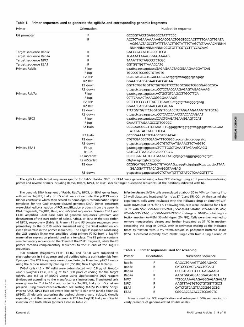
The genomic DNA fragment of Rab5c, Rab7a, NPC1, or EEA1 genes fusedwith either TagRFP, Halo, or mScarlet were cloned into the pUC19 vector(donor constructs) which then served as homologous recombination repairtemplates for the Cas9 enzyme-cleaved genomic DNA. Donor constructswere obtained by a ligation of PCR amplification products from the genomicDNA fragments, TagRFP, Halo, and mScarlet sequences. Primers F1-R1 andF3-R3 amplified ∼800 base pairs of genomic sequences upstream anddownstream of the start codon of Rab5c, Rab7a, or EEA1 or the stop codonof NPC1, respectively (Table 1). Primers F1 and R3 contain sequences com-plementary to the pUC19 vector linearized using the SmaI restriction en-zyme (lowercase in the primer sequences). The TagRFP sequence containingthe GGS peptide linker was amplified using primers F2-R2 from a TagRFPmammalian expression plasmid used as a template. The F2 primer containscomplementary sequences to the 3′ end of the F1-R1 fragment, while the F3primer contains complementary sequences to the 3′ end of the TagRFPsequences.
PCR products (fragments F1-R1, F2-R2, and F3-R3) were subjected toelectrophoresis in 1% agarose and gel purified using a purification kit fromZymogen. The PCR fragments were cloned into the linearized pUC19 vectorusing the Gibson Assembly Cloning Kit (E5510S; New England Biolabs).
SVG-A cells (1.5 × 10⁵ cells) were cotransfected with 0.8 μg of Strepto-coccus pyogenes Cas9, 0.8 μg of free PCR product coding for the targetsgRNA, and 0.8 μg of pUC19 vector using Lipofectamine 2000 reagent(Invitrogen) according to the manufacturer’s instructions. Transfected cellswere grown for 7 d to 10 d and sorted for TagRFP, Halo, or mScarlet ex-pression using fluorescence-activated cell sorting (FACS) (SH-800S; Sony).Prior to FACS, NPC1-Halo cells were labeled for 15 min with Janelia Fluor 647(JF647). Single cells expressing the desired chimera were isolated, clonallyexpanded, and then screened by genomic PCR for TagRFP, Halo, or mScarletinsertion into both alleles (primers listed in Table 2).
Infection Assays. SVG-A cells were plated at about 30 to 40% confluency into24-well plates and incubated for 1 d at 37 °C and 5% CO2. At the start of theexperiment, cells were incubated with the indicated drug or dimethyl sulf-oxide (DMSO) at 37 °C for 1 h. Following this, cells were incubated for 1 h at37 °C with VSV, VSV-MeGFP-V269H, VSV-MeGFP-RABV, VSV-MeGFP-LASV,VSV-MeGFP-LCMV, or VSV-MeGFP-ZEBOV in drug- or DMSO-containing in-fection medium (α-MEM, 50 mM Hepes, 2% FBS). Cells were then washed toremove nonadsorbed viruses and further incubated at 37 °C in mediumcontaining the drug or DMSO, with experiments ending at the indicatedtimes by fixation with 3.7% formaldehyde in phosphate-buffered saline(PBS). Fluorescent intensity from 20,000 single cells from a single round of
Table 1. Primer sequences used to generate the sgRNAs and corresponding genomic fragments
Primer Orientation Nucleotide sequence
U6 promoter F GCCGGTACCTGAGGGCCTATTTCCCR ACCTCTAGAAAAAAAGCACCGACTCGGTGCCACTTTTTCAAGTTGATA
ACGGACTAGCCTTATTTTAACTTGCTATTTCTAGCTCTAAAACNNNNNNNNNNNNNNNNNNNNCGGTGTTTCGTCCTTTCCACAAG
Target sequence Rab5c R GACCCGCCATTGCCCGTCCATarget sequence Rab7a R TCAAACTAAAGGGGGAAAAGTarget sequence NPC1 R TAAATTTCTAGCCCTCTCGCTarget sequence EEA1 R GGTGGTGGTTAAACCATGPrimers Rab5c F1up gaattcgagctcggtacccGAGAGAACTAGGGAAGAAGGATCAG
R1up TGCCCGTCCAGCTGTAGTGF2 RFP CCACTACAGCTGGACGGGCAatggtgtctaagggcgaagagcR2 RFP GGAACCACCAGAACCACCAGAAF3 down GGTTCTGGTGGTTCTGGTGGTTCCCTGGCGGGTCGGGGAGGCGCAR3 down gtcgactctagaggatccccCCTCCTACCAAGAGAGTAGAGAAAG
Primers Rab7a F1up gaattcgagctcggtacccACTGCTGTCAGCCTTGCCTTCAR1up CCTTCAAACTAAAGGGGGAAAAGGF2 RFP CCTTTTCCCCCTTTAGTTTGAAGGatggtgtctaagggcgaagR2 RFP GGAACCACCAGAACCACCAGAAF3 down TTCTGGTGGTTCTGGTGGTTCCACCTCTAGGAAGAAAGTGTTGCTGR3 down gtcgactctagaggatccccCCTCACCCAACCTACCACAGAAT
Primers NPC1 F1up gaattcgagctcggtacccCCACTGAGATGAAGGAGTCCATR1up GAAATTTAGAAGCCGTTCGCGC
F2 Halo CGCGAACGGCTTCTAAATTTCggaggttctggtggttctggtggttccGCAGAAATCGGTACTGGCTTTCCA
R2 Halo GCCGGAAATCTCGAGCGTCGACAGF3 down CTGTCGACGCTCGAGATTTCCGGCtagccctctcgcagggcatccR3 down gtcgactctagaggatccccGCTGTCTAATGAAACTTCTAGGTC
Primers EEA1 F1 up gaattcgagctcggtacccCTCTTTGGCTGAAATTAGAAGCAGGR1 up CATGGTTTAACCACCACCCGGCG
F2 mScarlet CGCCGGGTGGTGGTTAAACCATGgtgagcaagggcgaggcagtgatR2 mScarlet cttgtacagctcgtccatgccgcF3 down GCGGCATGGACGAGCTGTACAAGggaggttctggtggttctggtggttccTTAA
GGAGGATTTTACAGAGGGTAAGAGR3 down gtcgactctagaggatccccGCTCTAATCTTTCTATCCTCAAGGTTTTC
The sgRNAs with target sequences specific for Rab5c, Rab7a, NPC1, or EEA1 were generated using a free PCR strategy using a U6 promoter-containingprimer and reverse primers including Rab5c, Rab7a, NPC1, or EEA1 specific target nucleotide sequences (at the positions indicated with N).
Table 2. Primer sequences used for screening
Primer Orientation Nucleotide sequence
Rab5c F GAGCCTGAAGTTGGGAGACCRab5c R CATGCCCACTCACCTCCAATRab7a F GCGGTCACTTCTTTGAGAAAGTRab7a R AAGTGGCAGCACGGACAGTGTNPC1 F TCTCCAAAAGAGAGGGAGAGAGATNPC1 R AAGTTTAGTGTCCTGTGGTTGCCTEEA1 F CATCTGTCAGTTACGGGGGCTGEEA1 R CGGCACCACACCCTCCAGCTC
Primers used for PCR amplification and subsequent DNA sequencing toverify presence of genome-edited double alleles.
Kang et al. PNAS Latest Articles | 9 of 11
MED
ICALSC
IENCE
S
Dow
nloa
ded
at L
IBR
AR
Y F
EG
AN
PLA
ZA
on
Aug
ust 2
4, 2
020
infection was determined by flow cytometry using a BD FACSCanto IIequipped with DIVA software package.
MA104 cells were pretreated for 1 h with the indicated concentration ofApilimod or DMSO. Pretreated cells were inoculated with VSV-eGFP,VSV-eGFP-ZEBOV, or VSV-eGFP-SARS-CoV-2 at a multiplicity of infection(MOI) = 1 (based on titers in MA104 cells) in the presence of Apilimod orDMSO for 1 h at 37 °C. At 6 to 8 h postinfection, cells were collected andfixed in 2% paraformaldehyde (PFA) and then subjected to flow cytometry.The percentage of GFP cells was determined using FlowJo software (TreeStar Industries).
Vero E6 cell monolayers were pretreated for 1 h at 37 °C with serial di-lutions of Apilimod at the indicated concentrations. Next, SARS-CoV-2 wasdiluted to an MOI of 0.01 focus-forming units per cell in Apilimod-containingmedium and added to Vero E6 cells for 1 h at 37 °C. After adsorption, cellswere washed once with PBS, and medium containing the respective con-centration of Apilimod was added. Cells were incubated for 24 h at 37 °C, atwhich time cell culture supernatants were removed and used for determi-nation of viral titer by focus forming assay.
SARS-CoV-2 Focus-Forming Assay. Cell culture supernatants from virus-infected cells were diluted serially 10-fold, added to Vero E6 cell mono-layers in 96-well plates, and incubated at 37 °C for 1 h. Subsequently, cellswere overlaid with 1% (wt/vol) methylcellulose in MEM supplemented with2% FBS. Plates were harvested 30 h later by removing overlays and fixedwith 4% paraformaldehdye in PBS for 20 min at room temperature. Plateswere washed and sequentially incubated with 1 μg/mL CR3022 anti-spikeantibody (51) and HRP-conjugated goat anti-human IgG in PBS supple-mented with 0.1% saponin and 0.1% bovine serum albumin (BSA).SARS-CoV-2−infected cell foci were visualized using TrueBlue peroxidasesubstrate (KPL) and quantitated on an ImmunoSpot microanalyzer (CellularTechnologies). Data were processed using Prism software (GraphPad Prism8.0), and viral titers are reported as percent inhibition relative to mock-treated SARS-CoV-2−infected cells.
Entry Assay and Intracellular Traffic. SVG-A cells plated on glass #1.5 coverslipsat about 30 to 40% confluency 1 d prior to experiment were treated withdrug or DMSO for 1 h at 37 °C. Following this, cells were incubated at 37 °Cwith VSV, VSV-MeGFP-V269H, VSV-MeGFP-RABV, VSV-MeGFP-LASV, VSV-MeGFP-LCMV, or VSV-MeGFP-ZEBOV in drug- or DMSO-containing infec-tion medium. After this, cells were washed, then further incubated in me-dium containing the drug or DMSO at 37 °C, with the experiment ending at
the indicated time by fixation for 20 min at room temperature with 3.7%formaldehyde in PBS. This was followed with a 10-min incubation of 5 μg/mLAlexa647-labeled wheat germ agglutinin in PBS to label the outline ofthe cells.
Cells were imaged using a spinning disk confocal microscope with opticalplanes spaced 0.3 μm apart (52). The entry assay scored the presence ofMeGFP at the nuclear margin in each cell. Trafficking of viruses to endo-somal compartments was observed using live-cell imaging using the spinningdisk confocal microscope. Chemical fixation tends to eliminate the largeendolysosomal vacuoles generated by Vacuolin-1 or Apilimod and reducesthe colocalization with viral particles contained within. Time series withimages taken every 3 s for 3 min in a single optical plane with the appro-priate fluorescent channels (52) were acquired from nonfixed samples im-aged at the end of the experimental period. For experiments containingNPC1-Halo, the Halo-tagged cells were labeled with either 250 nM JF549or JF647 dye in media for 30 min at 37 °C. Following labeling, cells werewashed three times with media. The microscope was operated using theSlidebook 6.4 software package (3I), and images were displayed also usingthis software.
Statistical Tests. To compare the means from cells with different treatments,one-way ANOVA and post hoc Tukey test analysis were used to take intoaccount unequal sample sizes as indicated in the Figs. 1, 2, and 6 figurelegends.
Data Availability. The VSV virus chimeras are available from the corre-sponding author S.P.W. upon request.
All study data are included in the article and SI Appendix and MoviesS1–S3.
ACKNOWLEDGMENTS. We thank Walter J. Atwood for providing theparental SVG-A cells; Eric Marino, Justin H. Houser, and Tegy John Vadakkanfor maintaining the spinning disc confocal microscope (T.K. Laboratory);Marina Cella and Erica Lantelme from the flow cytometry facility, Depart-ment of Pathology and Immunology at WUSM for help with flow cytometry;Stephen C. Harrison for helpful discussions and editorial assistance; and AlexJ. B. Kreutzberger for editorial help. This research was supported by NIHfunding (Grant AI109740) to S.P.J.W. and T.K., by NIH Maximizing Investi-gators’ Research Award (MIRA) GM130386 to T.K., by NIH funding (AI059371)and unrestricted funds from WUSM to S.P.J.W., and by NIH Contract75N93019C00062 and NIH Grant R01 AI127828 to M.S.D.
1. K. Chandran, N. J. Sullivan, U. Felbor, S. P. Whelan, J. M. Cunningham, Endosomalproteolysis of the Ebola virus glycoprotein is necessary for infection. Science 308,1643–1645 (2005).
2. J. E. Carette et al., Ebola virus entry requires the cholesterol transporter Niemann-PickC1. Nature 477, 340–343 (2011).
3. I.-C. Huang et al., SARS coronavirus, but not human coronavirus NL63, utilizes ca-thepsin L to infect ACE2-expressing cells. J. Biol. Chem. 281, 3198–3203 (2006).
4. G. Simmons et al., Inhibitors of cathepsin L prevent severe acute respiratory syndromecoronavirus entry. Proc. Natl. Acad. Sci. U.S.A. 102, 11876–11881 (2005).
5. X. Ou et al., Characterization of spike glycoprotein of SARS-CoV-2 on virus entry andits immune cross-reactivity with SARS-CoV. Nat. Commun. 11, 1620 (2020).
6. W. Cao et al., Identification of alpha-dystroglycan as a receptor for lymphocyticchoriomeningitis virus and Lassa fever virus. Science 282, 2079–2081 (1998).
7. L. T. Jae et al., Virus entry. Lassa virus entry requires a trigger-induced receptor switch.Science 344, 1506–1510 (2014).
8. G. Pasqual, J. M. Rojek, M. Masin, J.-Y. Chatton, S. Kunz, Old world arenaviruses enterthe host cell via the multivesicular body and depend on the endosomal sortingcomplex required for transport. PLoS Pathog. 7, e1002232 (2011).
9. J. G. Carlton, P. J. Cullen, Coincidence detection in phosphoinositide signaling. TrendsCell Biol. 15, 540–547 (2005).
10. D. Sbrissa, O. C. Ikonomov, A. Shisheva, PIKfyve, a mammalian ortholog of yeastFab1p lipid kinase, synthesizes 5-phosphoinositides. Effect of insulin. J. Biol. Chem.274, 21589–21597 (1999).
11. A. Shisheva, D. Sbrissa, O. Ikonomov, Cloning, characterization, and expression of anovel Zn2+-binding FYVE finger-containing phosphoinositide kinase in insulin-sensitive cells. Mol. Cell. Biol. 19, 623–634 (1999).
12. O. C. Ikonomov, D. Sbrissa, A. Shisheva, Mammalian cell morphology and endocyticmembrane homeostasis require enzymatically active phosphoinositide 5-kinase PIK-fyve. J. Biol. Chem. 276, 26141–26147 (2001).
13. A. C. Rutherford et al., The mammalian phosphatidylinositol 3-phosphate 5-kinase(PIKfyve) regulates endosome-to-TGN retrograde transport. J. Cell Sci. 119, 3944–3957(2006).
14. D. Sbrissa et al., Core protein machinery for mammalian phosphatidylinositol 3,5-bi-sphosphate synthesis and turnover that regulates the progression of endosomaltransport. Novel Sac phosphatase joins the ArPIKfyve-PIKfyve complex. J. Biol. Chem.282, 23878–23891 (2007).
15. O. C. Ikonomov et al., Functional dissection of lipid and protein kinase signals of
PIKfyve reveals the role of PtdIns 3,5-P2 production for endomembrane integrity.
J. Biol. Chem. 277, 9206–9211 (2002).16. H. B. J. Jefferies et al., A selective PIKfyve inhibitor blocks PtdIns(3,5)P(2) production and
disrupts endomembrane transport and retroviral budding. EMBO Rep. 9, 164–170 (2008).17. O. Sano et al., Vacuolin-1 inhibits autophagy by impairing lysosomal maturation via
PIKfyve inhibition. FEBS Lett. 590, 1576–1585 (2016).18. J. Cerny et al., The small chemical vacuolin-1 inhibits Ca2+-dependent lysosomal
exocytosis but not cell resealing. EMBO Rep. 5, 883–888 (2004).19. X. Cai et al., PIKfyve, a class III PI kinase, is the target of the small molecular IL-12/IL-23 in-
hibitor apilimod and a player in Toll-like receptor signaling. Chem. Biol. 20, 912–921 (2013).20. G. Sharma et al., A family of PIKFYVE inhibitors with therapeutic potential against
autophagy-dependent cancer cells disrupt multiple events in lysosome homeostasis.
Autophagy 15, 1694–1718 (2019).21. O. C. Ikonomov et al., Active PIKfyve associates with and promotes the membrane
attachment of the late endosome-to-trans-Golgi network transport factor Rab9 ef-
fector p40. J. Biol. Chem. 278, 50863–50871 (2003).22. C. Chen et al., Identification of novel vacuolin-1 analogues as autophagy inhibitors by
virtual drug screening and chemical synthesis. Molecules 22, 891 (2017).23. Y. Lu et al., Vacuolin-1 potently and reversibly inhibits autophagosome-lysosome
fusion by activating RAB5A. Autophagy 10, 1895–1905 (2014).24. B. E. Sands et al., Randomized, double-blind, placebo-controlled trial of the oral in-
terleukin-12/23 inhibitor apilimod mesylate for treatment of active Crohn’s disease.
Inflamm. Bowel Dis. 16, 1209–1218 (2010).25. Y. Wada et al., Apilimod inhibits the production of IL-12 and IL-23 and reduces
dendritic cell infiltration in psoriasis. PLoS One 7, e35069 (2012).26. E. A. Nelson et al., The phosphatidylinositol-3-phosphate 5-kinase inhibitor apilimod
blocks filoviral entry and infection. PLoS Negl. Trop. Dis. 11, e0005540 (2017).27. S. Qiu et al., Ebola virus requires phosphatidylinositol (3,5) bisphosphate production
for efficient viral entry. Virology 513, 17–28 (2018).28. J. S. Spence, T. B. Krause, E. Mittler, R. K. Jangra, K. Chandran, Direct visualization of
Ebola virus fusion triggering in the endocytic pathway. mBio 7, e01857-15 (2016).29. R. Burakoff et al., A phase 1/2A trial of STA 5326, an oral interleukin-12/23 inhibitor,
in patients with active moderate to severe Crohn’s disease. Inflamm. Bowel Dis. 12,
558–565 (2006).
10 of 11 | www.pnas.org/cgi/doi/10.1073/pnas.2007837117 Kang et al.
Dow
nloa
ded
at L
IBR
AR
Y F
EG
AN
PLA
ZA
on
Aug
ust 2
4, 2
020

30. S. Krausz et al., Brief report: A phase IIa, randomized, double-blind, placebo-controlled trial of apilimod mesylate, an interleukin-12/interleukin-23 inhibitor, inpatients with rheumatoid arthritis. Arthritis Rheum. 64, 1750–1755 (2012).
31. C. E. Hulseberg et al., Arbidol and other low-molecular-weight drugs that inhibitLassa and Ebola viruses. J. Virol. 93, 11 (2019).
32. J. Dyall et al., Identification of combinations of approved drugs with synergistic ac-tivity against Ebola virus in cell cultures. J. Infect. Dis. 218 (suppl. 5), S672–S678 (2018).
33. R. Bago et al., Characterization of VPS34-IN1, a selective inhibitor of Vps34, revealsthat the phosphatidylinositol 3-phosphate-binding SGK3 protein kinase is a down-stream target of class III phosphoinositide 3-kinase. Biochem. J. 463, 413–427 (2014).
34. I. Glowacka et al., Evidence that TMPRSS2 activates the severe acute respiratorysyndrome coronavirus spike protein for membrane fusion and reduces viral control bythe humoral immune response. J. Virol. 85, 4122–4134 (2011).
35. A. Shulla et al., A transmembrane serine protease is linked to the severe acute re-spiratory syndrome coronavirus receptor and activates virus entry. J. Virol. 85,873–882 (2011).
36. S. Matsuyama et al., Efficient activation of the severe acute respiratory syndromecoronavirus spike protein by the transmembrane protease TMPRSS2. J. Virol. 84,12658–12664 (2010).
37. M. Hoffmann et al., SARS-CoV-2 cell entry depends on ACE2 and TMPRSS2 and isblocked by a clinically proven protease inhibitor. Cell 181, 271–280.e8 (2020).
38. G. Simmons et al., Characterization of severe acute respiratory syndrome-associatedcoronavirus (SARS-CoV) spike glycoprotein-mediated viral entry. Proc. Natl. Acad. Sci.U.S.A. 101, 4240–4245 (2004).
39. S. Gayle et al., Identification of apilimod as a first-in-class PIKfyve kinase inhibitor fortreatment of B-cell non-Hodgkin lymphoma. Blood 129, 1768–1778 (2017).
40. C. D. S. Nelson, A. Derdowski, M. S. Maginnis, B. A. O’Hara, W. J. Atwood, The VP1subunit of JC polyomavirus recapitulates early events in viral trafficking and is a noveltool to study polyomavirus entry. Virology 428, 30–40 (2012).
41. M. V. Baranov et al., The phosphoinositide kinase PIKfyve promotes cathepsin-S-mediated major histocompatibility complex class II antigen presentation. iScience11, 160–177 (2019).
42. M. Côté et al., Small molecule inhibitors reveal Niemann-Pick C1 is essential for Ebolavirus infection. Nature 477, 344–348 (2011).
43. I. Le Blanc et al., Endosome-to-cytosol transport of viral nucleocapsids. Nat. Cell Biol.7, 653–664 (2005).
44. Y. Zhou et al., Protease inhibitors targeting coronavirus and filovirus entry. AntiviralRes. 116, 76–84 (2015).
45. T. K. Soh, S. P. J. Whelan, Tracking the fate of genetically distinct vesicular stomatitisvirus matrix proteins highlights the role for late domains in assembly. J. Virol. 89,11750–11760 (2015). Erratum in: J. Virol. 90, 5847 (2016).
46. S. Piccinotti, T. Kirchhausen, S. P. J. Whelan, Uptake of rabies virus into epithelial cellsby clathrin-mediated endocytosis depends upon actin. J. Virol. 87, 11637–11647(2013).
47. A. C. Wong, R. G. Sandesara, N. Mulherkar, S. P. Whelan, K. Chandran, A forwardgenetic strategy reveals destabilizing mutations in the Ebolavirus glycoprotein thatalter its protease dependence during cell entry. J. Virol. 84, 163–175 (2010).
48. S. P. Whelan, L. A. Ball, J. N. Barr, G. T. Wertz, Efficient recovery of infectious vesicularstomatitis virus entirely from cDNA clones. Proc. Natl. Acad. Sci. U.S.A. 92, 8388–8392(1995).
49. F. A. Ran et al., Genome engineering using the CRISPR-Cas9 system. Nat. Protoc. 8,2281–2308 (2013).
50. Y.-Y. Chou et al., Identification and characterization of a novel broad spectrum virusentry inhibitor. J. Virol. 90, 4494–4510 (2016).
51. M. Yuan et al., A highly conserved cryptic epitope in the receptor-binding domains ofSARS-CoV-2 and SARS-CoV. Science 368, 630–633 (2020).
52. E. Cocucci, F. Aguet, S. Boulant, T. Kirchhausen, The first five seconds in the life of aclathrin-coated pit. Cell 150, 495–507 (2012).
Kang et al. PNAS Latest Articles | 11 of 11
MED
ICALSC
IENCE
S
Dow
nloa
ded
at L
IBR
AR
Y F
EG
AN
PLA
ZA
on
Aug
ust 2
4, 2
020