identification of tumor-initiating cells in a highly …identification of tumor-initiating cells in...
TRANSCRIPT
Identification of tumor-initiating cells in a highlyaggressive brain tumor using promoter activityof nucleosteminAkira Tamasea,b,1, Teruyuki Muraguchia,1, Kazuhito Nakaa,1, Shingo Tanakaa,b, Masashi Kinoshitab, Takayuki Hoshiia,Masako Ohmuraa, Haruhiko Shugoa, Takako Ooshioa,c, Mitsutoshi Nakadab, Kazunobu Sawamotod,Masafumi Onoderae, Kunio Matsumotof, Masanobu Oshimag, Masahide Asanoh, Hideyuki Sayai, Hideyuki Okanoj,Toshio Sudak, Jun-ichiro Hamadab, and Atsushi Hiraoa,c,2
aDivision of Molecular Genetics, Center for Cancer and Stem Cell Research, Cancer Research Institute, Kanazawa University, Kanazawa, Ishikawa 920-0934,Japan; bDepartment of Neurosurgery, Graduate School of Medical Science, Kanazawa University, Kanazawa, Ishikawa 920-8641, Japan; cCore Research forEvolutional Science and Technology, Japan Science and Technology Agency, Kawaguchi, Saitama 332-0012, Japan; dDepartment of Developmental andRegenerative Biology, Institute of Molecular Medicine, Nagoya City University Graduate School of Medical Sciences, Mizuho-ku, Nagoya 467-8601, Japan;eLaboratory of Genetic Diagnosis and Gene Therapy, Department of Genetics, National Research Institute for Child Health and Development, Setagaya-ku,Tokyo 157-8535, Japan; fDivision of Tumor Dynamics and Regulation and gDivision of Genetics, Cancer Research Institute and hDivision of Transgenic AnimalScience, Advanced Science Research Center, Kanazawa University, Kanazawa, Ishikawa, 920-8640, Japan; and iDivision of Gene Regulation, Institute forAdvanced Medical Research and Departments of jPhysiology and kCell Differentiation, The Sakaguchi Laboratory of Developmental Biology, Keio UniversitySchool of Medicine, Shinjuku-ku, Tokyo 160-8582, Japan
Edited by Tak Wah Mak, Princess Margaret Hospital, Toronto, Canada, and approved August 12, 2009 (received for review May 7, 2009)
Controversy remains over whether the cancer stem cell (CSC)theory applies to all tumors. To determine whether cells within ahighly aggressive solid tumor are stochastically or hierarchicallyorganized, we combined a reporter system where the nucleoste-min (NS) promoter drives GFP expression (termed NS-GFP) with amouse brain tumor model induced by retroviral Ras expression ona p16Ink4a/p19Arf-deficient background. The NS-GFP system al-lowed us to monitor the differentiation process of normal neuralstem/precursor cells by analyzing GFP fluorescence intensity. Intumor-bearing mice, despite the very high frequency of tumori-genic cells, we successfully identified the NS-GFP� cells as tumor-initiating cells (T-ICs). The clonal studies conclusively establishedthat phenotypical heterogeneity can exist among the cells com-prising a genetically homogeneous tumor, suggesting that thisaggressive brain tumor follows the CSC model. Detailed analysesof the NS-GFP� brain tumor cells revealed that T-ICs showedactivation of the receptor tyrosine kinase c-Met, which functions intumor invasiveness. Thus, the NS-GFP system provides a powerfultool to elucidate stem cell biology in normal and malignant tissues.
cancer stem cell � invasion
Recent improvements in cell purification and transplantationtechniques have contributed to the identification of cell pop-
ulations known as tumor-initiating cells (T-ICs). These findings ledto the idea that tumors are organized as hierarchies of cellssustained by such T-ICs, conceptually termed cancer stem cells(CSCs) (1, 2). Supporting this idea, in vivo models in whichleukemia is initiated from primary human hematopoietic cellsrevealed that disease is sustained by leukemia-initiating cells (L-ICs) and that the L-ICs retain both myeloid and lymphoid lineagepotential (3). Although many human malignancies appear to con-tain only rare tumorigenic cells or T-ICs when transplanted intoNOD/SCID mice (4–6), the question of whether NOD/SCID assaysunderestimate the frequency of human tumorigenic cells due todifferences between human and murine tissues has been raised.Recently, Quintana et al. (7) reported that �25% of unselectedhuman melanoma cells from patients formed tumors when trans-planted into highly immunocompromised NOD/SCID interleu-kin-2 receptor gamma chain null (Il2r��/�) mice, in contrast to thevery few T-ICs identified when the melanoma cells were trans-planted into NOD/SCID mice (6). These results suggest that cellscomprising human melanomas may constitute a homogeneouspopulation and that any melanoma cells can form a tumor, i.e., thata hierarchical organization of tumor cells does not exist. Alterna-
tively, it is also possible that, although T-IC frequency is very highin the melanoma, a true hierarchy exists in the aggressive tumor,because 75% of the tumor cells lack T-IC activity. It thereforeremains controversial whether the cancer stem cell theory appliesto all tumors (2). In our study, we have attempted to resolve thisissue by examining the frequency of tumorigenic cells present in ahighly aggressive murine solid tumor orthotopically transplantedinto recipient mice. Our approach thus avoids the underestimationof tumorigenic cell frequency that might arise due to environmentaldifferences between human and mouse tissues.
To investigate whether murine brain tumors exhibit cellularheterogeneity, we took advantage of our unique NS-GFP stemcell-marking system, in which the green fluorescent protein (GFP)is expressed under the control of promoter of the nucleostemin(NS) gene (8). The NS, a nucleolar GTPase, is found at high levelsin various tissue stem cells and cancer cells (9). Because NSexpression decreases rapidly in stem cells when these cells differ-entiate before cell cycle exit, it has been suggested that the NSprotein is a marker for proliferating cells in an early multipotentialstate (9, 10). In the regenerating newt lens, the NS protein rapidlyaccumulates in the nucleoli of dedifferentiating pigmented epithe-lial cells (11), suggesting that NS expression correlates with undif-ferentiated status of cells. Previously, we generated NS-GFP trans-genic (NS-GFP-Tg) mice and used these mice to identify a fractionof neonatal germ cells as spermatogonial stem cells (8). In thepresent study, we have combined our NS-GFP-Tg system with amurine brain tumor model to investigate whether aggressive solidtumors contain a distinct population of T-ICs.
ResultsHigh Frequency of Tumorigenic Cells in an Aggressive Murine BrainTumor. T-ICs have been identified in human high-grade gliomas(glioblastoma multiforme), which are very aggressive, invasive, and
Author contributions: A.H. designed research; A.T., T.M., K.N., S.T., M.K., T.H., M. Ohmura,H. Shugo, and T.O. performed research; M. Onodera, K.M., and M.A. contributed newreagents/analytic tools; M.N., K.S., M. Oshima, H. Saya, H.O., T.S., J.-i.H., and A.H. analyzeddata; and A.T. and A.H. wrote the paper.
The authors declare no conflict of interest.
This article is a PNAS Direct Submission.
1A.T., T.M., and K.N. contributed equally to this work.
2To whom correspondence should be addressed. E-mail: [email protected].
This article contains supporting information online at www.pnas.org/cgi/content/full/0905016106/DCSupplemental.
www.pnas.org�cgi�doi�10.1073�pnas.0905016106 PNAS � October 6, 2009 � vol. 106 � no. 40 � 17163–17168
MED
ICA
LSC
IEN
CES
Dow
nloa
ded
by g
uest
on
Feb
ruar
y 20
, 202
0

destructive brain tumors (12). Invasive tumor cells escape surgicalremoval and geographically dodge lethal radiation exposure andchemotherapy. A mouse brain tumor model of human glioblastomamultiforme can be generated by triggering Ras signaling down-stream of the epidermal growth factor (EGF) receptor in brain cellsof mice deficient for the tumor suppressors p16Ink4a/p19Arf (13–15).We modified the reported protocol (15) and constructed a vectorcontaining a constitutively active mutant K-ras gene (K-rasG12V)plus the humanized Kusabira-Orange (huKO) gene as a marker.We used retroviral infection to introduce this vector into culturedneurospheres composed of neural stem cells and precursor cells(NSC/NPCs) derived from the subventricular zone (SVZ) of brainsof neonatal p16Ink4a�/�/p19Arf�/� mice. The infected neurosphereswere then injected into the basal ganglia of wild type (WT) recipientmice (Fig. S1). Brain tumors developed as early as 20 days aftertransplantation, and most recipients died within 40 days of injec-tion. Consistent with previous reports (13–16), histological analysesof these tumors demonstrated that these tumors showed severalfeatures characteristic of human gliomas (17), including microvas-cular proliferation, the presence of giant cells, and/or areas of tumornecrosis bordered by dense palisades of viable tumor cells (necrosiswith pseudopalisading) (Fig. 1A).
To analyze the frequency of T-ICs within tumors, the malignan-cies were recovered from recipients and dissociated by collagenasetreatment. To eliminate any contaminating normal brain cells, flow
cytometry was used to collect huKO� cells (i.e., cells overexpressingK-rasG12V). Transplantation of 100 or 1,000 freshly isolated tumorcells into the brains of WT mice (8 weeks old) resulted in braintumor formation in 100% of recipients (Fig. 1B). Even when only10 tumor cells were injected, 50% of recipients developed braintumors, suggesting that the frequency of tumorigenic cells in theoriginal tumor was very high.
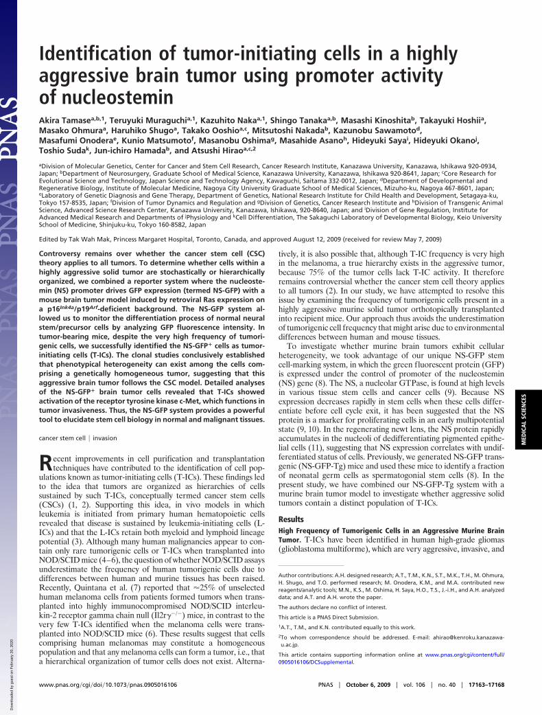
Correlation of In Vivo Differentiation Status of NSC/NPC and GFPFluorescence Intensity in NS-GFP-Tg Brain. Tsai and McKay havereported that NS is highly expressed in undifferentiated NSC/NPCsbut not in differentiating neurons (9). To determine whether theNS-GFP system marked NSC/NPCs, we evaluated GFP expressionin embryonic brains of our NS-GFP-Tg mice. In embryonic brainat E14.5, radial glial cells in the ventricular zone are NSC/NPCs. Atthis stage, the cortical plate is formed by active neurogenesisderived from NSC/NPCs (18). In the NS-GFP-Tg brain at E14.5,higher GFP expression was observed in ventricular zone cellsexpressing nestin (19) or musashi-1 (20), a protein enriched inNSC/NPCs, whereas TuJ1� neurons showed a lower level of GFP(Fig. 2A and Fig. S2). In neonatal brain (P3), GFP was highlyexpressed in SVZ, which are actively cycling, and down-regulatedin the striatum (Fig. S3). These analyses suggested that NSC/NPCsare included within the subpopulation of normal brain cells thatexpresses high levels of GFP.
To investigate the relationship between GFP fluorescence in-tensity and cellular properties, we used flow cytometry to divide thetotal cell population recovered from dissociated E14.5 NS-GFP-Tgbrains into four fractions according to GFP fluorescence intensity:GFP�/�, GFP��, GFP��� and GFP���� (Fig. 2B). Immuno-staining of the sorted cells with anti-nestin or anti-TuJ1 revealedthat most GFP���� cells expressed nestin but not TuJ1 (Fig. 2C),indicating that they were undifferentiated. In contrast, GFP�/� cellsexpressed TuJ1 but not nestin. Thus, our NS-GFP-Tg system allowsus to monitor stem cell differentiation during neurogenesis. Whenwe examined the capacity of our four GFP-expressing subpopula-tions to form neurospheres, we found that neurosphere-initiatingcells were most efficiently generated by cells with higher GFPexpression, whether these cells were derived from embryonic(E14.5) or neonatal (P3) NS-GFP-Tg brains (Fig. 2 D–F). Inparticular, half of the total neurosphere-initiating cells in neonatalbrain were enriched in the rare (10%) GFP���� cell population.Our findings support previous reports showing that neurosphere-initiating cells are enriched in sorted GFP-strong positive cellsisolated from the brains of nestin-EGFP mice (21). We concludethat brain cells in our NS-GFP system that express very high levelsof GFP exhibit a substantial capacity for both proliferation andNSC/NPC differentiation.
Identification of Brain T-ICs. Our success in monitoring normalNSC/NPC differentiation in NS-GFP-Tg mice prompted us to usethis system to analyze cellular heterogeneity in our brain tumormodel. We crossed our NS-GFP-Tg mice to p16Ink4a�/�/p19Arf�/�
mice and induced the generation of brain tumors as describedabove. Flow cytometric analyses of these huKO� tumors derivedfrom NS-GFP-Tg mice revealed that they contained both GFPhigh
and GFPlow populations. The ratio of GFPhigh to GFPlow cells washighly variable among individual tumors (Fig. 3A). Immunocyto-chemical analysis of freshly isolated tumor cells confirmed thatendogenous NS was highly expressed in GFPhigh cells but not inGFPlow cells (Fig. S4). CD133 (prominin 1) or nestin has beenreported to be a marker of T-ICs in human glioma. The expressionpattern of prominin 1 mRNA varied among tumors (Fig. S5);however, we found that GFPhigh cells primarily expressed nestin,whereas the GFPlow population showed nestin down-regulation(Fig. 3B). These data suggested that the GFPhigh tumor cells mightbe immature cells with the potential to differentiate into GFPlow
tumor cells in vivo.
Fig. 1. High frequency of tumorigenic cells in a murine brain tumor. (A)Development of brain tumors. (i) Gross appearance of a recipient brain. Amassive lesion can be seen in the cerebrum. (ii–iv) H&E staining of represen-tative sections of brain tumors. Regions of increased cell density, nuclearpleomorphism, and prominent mitotic figures can be seen. Arrows, giant cells;arrowheads, areas of necrosis with pseudopalisading; asterisks, invading tu-mor cells adjacent to blood vessels. (Scale bars: 200 �m.) (B) Survival ofrecipient mice after transplantation of 1,000, 100, or 10 huKO� tumor cells, asindicated.
17164 � www.pnas.org�cgi�doi�10.1073�pnas.0905016106 Tamase et al.
Dow
nloa
ded
by g
uest
on
Feb
ruar
y 20
, 202
0
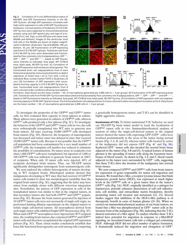
To investigate the properties of the GFPhigh and GFPlow tumorcells, we first evaluated their capacity to form spheres in culture.Many spheres were generated in cultures of GFPhigh cells, whereasGFPlow cells produced only a few spheres (Fig. 3C). To investigatethe behavior of the GFPhigh and GFPlow tumor cells in vivo, the cellswere orthotopically transplanted into WT mice to generate newbrain tumors. All mice receiving 10,000 GFPhigh cells developedbrain tumors (Fig. 3D). However, the frequency of tumorigenicitywas decreased and tumor onset was delayed in mice that received10,000 GFPlow tumor cells. Because it was possible that the GFPlow
cell population had been contaminated by a very small number ofGFPhigh cells, the transplant cell number was reduced to eliminatethe possibility of contamination. No tumor arose in recipients evenwhen 1,000 GFPlow cells were transplanted, but injection of 1,000 or100 GFPhigh cells was sufficient to generate brain tumors in 100%of recipients. When only 10 tumor cells were injected, 60% ofrecipients developed brain tumors. Thus, our NS-GFP systemsignificantly distinguishes tumorigenic from nontumorigenic cells.
We next explored the cellular heterogeneity of tumors develop-ing in WT recipient brains. Histological analysis showed thatmalignancies developing in WT mice that had received 10 GFPhigh
tumor cells tended to show less morphological variability than theoriginal brain tumors (Fig. 3E), suggesting that the latter may havearisen from multiple clones with different retrovirus integrationsites. Nonetheless, the pattern of GFP expression in cells of thetransplanted tumors was similar to that in the original tumors, inthat secondary tumors also had both GFPhigh and GFPlow popula-tions (Fig. 3E). Because the brain tumors generated by injection of10 GFPhigh tumor cells were not necessarily of single-cell origin, weperformed limiting dilution experiments on the original tumors toisolate single cell-derived tumors. All neurospheres derived fromsuch single tumor cells exhibited bright GFP fluorescence (Fig. 3F).When such GFPbright neurospheres were injected into WT recipientmice, the resulting brain tumors also contained GFPhigh and GFPlow
tumor cells and therefore recapitulated the original GFP expressionpattern (Fig. 3G). These clonal studies conclusively establish thatbona fide functional heterogeneity can exist among cells comprising
a genetically homogeneous tumor, and T-ICs can be identified inhighly aggressive tumors.
Invasiveness of T-ICs. To better understand T-IC behavior, we usedour NS-GFP-Tg brain tumor model to track the localization ofGFPhigh tumor cells in vivo. Immunohistochemical analyses ofsections of either the single-cell-derived tumors or the originaltumors showed the tumor cells expressing GFP (GFP� cells) werelocalized predominantly in the areas of the tumor facing normaltissues (Fig. 4 A, B, and D), whereas tumor cells located in centerof the malignancy did not express GFP (Fig. 4C and Fig. S6).Scattered GFP� tumor cells also invaded the normal brain tissueadjacent to the tumor (Fig. 4 B and D). A typical feature of humangliomas is the spreading of tumor cells along the basement mem-branes of blood vessels. As shown in Fig. 4 E and F, blood vesselsadjacent to the tumor were surrounded by GFP� cells, suggestingthat these T-ICs drive the invasion of the malignancy into normalbrain tissues.
We next compared GFPhigh and GFPlow cells from brain tumorsfor expression of genes responsible for tumor cell migration andinvasion. We found that c-Met, a receptor tyrosine kinase that bindshepatocyte growth factor (HGF), was expressed at much higherlevels in the GFPhigh cells of a given tumor than in the tumor’sGFPlow cells (Fig. 5A). HGF, originally identified as a mitogen forhepatocytes, potently enhances dissociation of cell–cell adhesive-ness, cell motility, and extracellular matrix breakdown, therebypromoting tumor cell invasion and metastasis (22, 23). Severalstudies suggest that inhibition of the HGF/c-Met pathway is oftherapeutic benefit in cases of human glioma (24–26). When wecarried out immunohistochemical analyses of our brain tumors, wefound that c-Met was phosphorylated in GFP� tumor cells at theinvasion front of the malignancy (Fig. 5B), indicating that the T-ICsshowed activation of c-Met signal. To analyze whether these T-ICsindeed have potential for migration in response to c-Met/HGFsignaling, we inoculated tumor cells into a collagen gel containingHGF. Although HGF did not affect number of sphere formation,it dramatically induced the migration and elongation of GFP�
Fig. 2. Correlation of in vivo differentiation status ofNSC/NPC and GFP fluorescence intensity in the NS-GFP-Tg brain. (A) High GFP expression correlates withhigh nestin expression in cells of NS-GFP-Tg embryonicforebrains. Coronal sections of forebrains of E14.5 NS-GFP-Tg mice were subjected to immunohistochemicalanalysis using anti-GFP (green) plus anti-type III �-tu-bulin (TuJ1; red, Top), or anti-GFP plus anti-nestin (red,Middle and Bottom). Images of the ventricular zone(VZ) cells in the Middle are shown at higher magnifi-cation in Bottom. (Scale bars: Top and Middle, 100 �m;Bottom, 10 �m). (B) Fractionation of GFP-expressingcells from E14.5 NS-GFP-Tg brain. Total brain cells fromE14.5 NS-GFP-Tg mice were dissociated and fraction-ated by flow cytometry into 4 subpopulations, GFP�/�,GFP��, GFP��� and GFP����, based on GFP fluores-cence intensity as indicated. Gray peak, WT C57BL/6mice; black peak, NS-GFP-Tg mice. (C) Correlation ofhigh GFP expression with immature cell properties. Thefractionated brain cell subpopulations from B werefixed and analyzed by immunocytochemistry to detectexpression of nestin (red, i–iv) or TuJ1 (red, v–viii) asindicated. Blue, nuclear marker TOTO-3. (Scale bars, 20�m.) (D) Correlation of GFP expression with neuro-sphere-forming capacity in E14.5 NS-GFP-Tg brain tis-sues. Fractionated brain cell subpopulations from Bwere cultured under conditions allowing neurosphereformation. Data shown are the mean number � SD. of neurospheres generated per 2,000 cells (n � 3 per group). (E) Fractionation of P3 GFP-expressing braincells. Total brain cells from P3 NS-GFP-Tg mice were dissociated and fractionated by flow cytometry into 4 subpopulations, GFP�/�, GFP��, GFP��� and GFP����,based on GFP fluorescence intensity as indicated. Gray peak, WT C57BL/6 mice; black peak, NS-GFP-Tg mice. (F) Correlation of GFP expression with neurosphere-forming capacity in P3 NS-GFP-Tg brain tissues. The fractionated brain cell subpopulations in E were cultured to allow neurosphere formation as for D. Data shownare the mean number � SD. of neurospheres generated per 2,000 cells (n � 3 per group).
Tamase et al. PNAS � October 6, 2009 � vol. 106 � no. 40 � 17165
MED
ICA
LSC
IEN
CES
Dow
nloa
ded
by g
uest
on
Feb
ruar
y 20
, 202
0
T-ICs placed in a collagen gel (Fig. 5C). These T-ICs therefore havethe potential to respond to HGF and migrate through a matrix withcollagenase activity. These data suggest that the T-ICs identified byour NS-GFP-Tg system lead the invasion of the malignancy intonormal brain tissue.
DiscussionIn this study, we used a murine brain tumor model to establish thateven in aggressive brain tumors that contain a high frequency ofT-ICs, T-ICs represent a distinct cell type capable of generatingnon-T-ICs, which comprise the bulk of the tumor tissue. The use ofa syngenic and orthotopic system overcomes limitations of xeno-transplant systems, which typically show relatively low frequenciesof T-ICs in human brain tumors (5). The concern that environ-mental differences existing between human and mouse tissuesmight lead to an underestimation of tumorigenic cell frequency may
be supported by our data. It could be argued that our tumor modelis more aggressive than a spontaneous tumor because we generatemalignancies by oncogene overexpression. However, although thetumors were generated experimentally, histological analyses dem-onstrate that these tumors exhibit apparent heterogeneity, similarto human gliomas. Our results clearly demonstrate that, if theappropriate marker is available, T-ICs can be identified in anaggressive solid tumor regardless of their frequency.
Optimization of xenotransplantation assays by using highly im-munocompromised NOD/SCID/Il2r��/� mice dramatically im-proved the transplantation efficiency of human melanoma cells,compared with traditional NOD/SCID mice (7), suggesting that thehuman melanoma randomly form tumor in the mice or geneticheterogeneity might be related to ability to form tumors, but notdue to hierarchical organization. However, examinations of humanacute myeloid leukemia (AML) have demonstrated that L-ICs are
Fig. 3. Identification of T-ICs. (A) Detection of GFPhigh and GFPlow subpopulations among brain tumor cells derived from NS-GFP-Tg mice. Tumors weredissociated with collagenase and GFP expression was analyzed in huKO� tumor cells by flow cytometry. Tumors contained variable percentages of GFPhigh andGFPlow cells. Three representative samples showing the GFP expression pattern in huKO� tumor cells are shown. (B) GFPhigh tumor cells exhibit immature cellproperties. Cytospin smears of sorted GFPhigh and GFPlow cells in A were fixed and immunostained to detect nestin (red). Blue, nuclear marker DAPI. Threerepresentative samples of 5 independent experiments are shown. (Scale bars: 20 �m.) (C) GFPhigh tumor cells can generate spheres. GFPhigh and GFPlow cells frombrain tumors in A were cultured for 14 days under sphere formation conditions. Data shown are the mean number � SD. of spheres per indicated number ofinoculated cells (n � 4 per group; *, P � 0.01). (D) Transplantation of GFPhigh brain tumor cells curtails survival. The percentage survival of WT recipient miceinjected with 10,000, 1,000, 100, or 10 GFPhigh or GFPlow brain tumor cells is shown as indicated. (E) Maintenance of the GFP expression pattern between originaland transplanted brain tumors. Analysis of histology (by H&E staining, Left) and GFP expression (by flow cytometry, Right) of brain tumors isolated from twoWT recipient mice transplanted with 10 GFPhigh brain tumor cells (as from D, Right). (Scale bars: 200 �m.) (F) A neurosphere derived from a single tumor cell bylimiting dilution. (Left Upper) Bright field. (Left Lower) GFP fluorescence. (Scale bars: 200 �m.) Right, GFP fluorescence as determined by flow cytometry. (Upper)Neurospheres from control mice (P3). (Lower) Single cell-derived spheres from NS-GFP tumor. Representative data are shown. (G) Maintenance of GFP expressionpattern between original tumors and tumors arising from transplantation of single-cell-derived neurospheres. Neurospheres derived in culture from single cellsof an original tumor were transplanted into WT recipient mice. Tumors arising in these recipients were dissociated and the expression of their component cellsanalyzed. Data representative of 4 independent experiments are shown.
17166 � www.pnas.org�cgi�doi�10.1073�pnas.0905016106 Tamase et al.
Dow
nloa
ded
by g
uest
on
Feb
ruar
y 20
, 202
0

rare even when analyzed in NOD/SCID/Il2r��/� mice (27). Arecent study of syngenic transplantation using mouse mammarytumor models also identified relatively rare T-ICs (28), suggestingthat the frequencies of T-ICs may depend on type of cancer. Indeed,in our study, the frequency of GFPhigh T-ICs varied greatly amongindividual brain tumors. In the case of a malignancy with a very highfrequency of T-ICs (e.g., Fig. 3A, case 3), we surmise that most ofthe brain tumor cells present were ‘‘CSCs,’’ as occurs in humanmelanomas. In other cases (e.g., Fig. 3A, case 1), the percentage ofT-ICs was relatively low. Although we cannot yet be sure preciselywhat factors determine the frequency of T-ICs in a given tumor, wesuspect that the stage of cancer progression (e.g., early versus latestage) is also important. Furthermore, we assume that the cell cyclestatus of T-ICs may also vary depending on the nature of the tumor.It has been previously reported that human leukemias include rareL-ICs, which showed slow cycling (27). In contrast, the GFPhigh
T-ICs in brain tumors analyzed in this study were actively cycling(Fig. S7). We hypothesize that, in the case of tumors showing a highfrequency of T-ICs like our brain tumor model or human mela-noma, T-ICs are actively cycling, resulting in very aggressive tumors.
Our NS-GFP-Tg system has allowed us to identify normalstem/progenitor cell populations in several different mouse tissues,including those responsible for neurogenesis and spermatogenesis(8). Although the population of cells strongly expressing GFP ineach NS-GFP-Tg tissue may include progenitors, not only stemcells, we have observed a consistent relationship between GFP
fluorescence intensity and degree of cellular maturation that hasheld across tissues. In addition, we also found that brain T-ICs wereenriched by the NS-GFP system. Thus, both normal and tumor cellsmay use the same ‘‘stemness’’ programs. To investigate whether theNS-GFP system identify the ‘‘stemness’’ programs in tumor celllines, we transfected the C6 glioma cell line with the NS-GFPconstruct and established stable clones. Although we found thatmost NS-GFP C6 cells in a single clone expressed GFP, as was thecase in NS-GFP E14.5 embryonic brain, we did not observedifferences in colony forming ability among subpopulations in C6cells (Fig. S8). The inability to distinguish C6 T-ICs based on theNS-GFP expression is possibly due to that the fact that cultured C6cells are more homogeneous than tumor tissue in vivo.
A key characteristic of human gliomas is their ability to invadenormal brain tissue. As shown in Fig. 4, some tumor cells adjacentto blood vessels may lose GFP, suggesting that NS-GFPhigh T-ICsmay not be the only cells responsible for tumor invasion. However,we do not believe that NS-GFPlow cells adjacent to blood vesselscontribute to tumor progression or expansion, because they lose thecapacity for tumor-initiation, as shown in Fig. 3D. Glioma inva-siveness is of clinical relevance, because brain tumor recurrenceoccurs most often within the surgical resection margin (12). There-fore, we believe that NS-GFPhigh cells are primarily responsible forin invasion. The use of our stem cell concept-based system to furtherinvestigate tumor organization may increase our understanding ofthe nature of cancer and lead to the development of novel cancertherapies.
Fig. 4. Localization of T-ICs. Serial sections of a single-cell-derived tumor were stained with H&E (A and F) oranti-GFP (B–E) to identify T-ICs. (A) H&E staining of atumor invading normal brain tissue. (B) Anti-GFP stain-ing of the tissue in A. (a and b) Low-power views ofareas shown at high power in C and D, respectively. (E)High-power view of area c in D. (F) H&E staining of E.(Scale bars: A and B, 1 mm; C and D, 200 �m; E and F,50 �m.)
Fig. 5. Migration potential of T-ICs. (A) High expres-sion of c-Met in GFPhigh cells. Total RNA was purifiedfrom GFPhigh and GFPlow cells isolated from three inde-pendent original tumors and c-Met mRNA levels wereevaluated by RT-PCR. �-actin, control. (B) c-Met is phos-phorylated in T-ICs. Serial sections of one of the singlecell-derived tumors were subjected to H&E (i and ii);anti-GFP (iii and iv); and anti-phospho-c-Met (v and vi)staining. Magnified views of the areas indicated by thesquares in i, iii and v are shown in ii, iv, and vi, respec-tively. (Scale bars: i, iii, v, 1 mm; ii, iv, vi, 200 �m.) (C)Migration of T-ICs in a collagen gel. Dissociated cellsfrom a tumor were inoculated into either a plain col-lagen gel (control) or a collagen gel containing HGF (10ng/mL) and incubated for 4 days. Representative GFP�
tumor cells with and without HGF in a collagen gel areshown. (Scale bars: 50 �m.)
Tamase et al. PNAS � October 6, 2009 � vol. 106 � no. 40 � 17167
MED
ICA
LSC
IEN
CES
Dow
nloa
ded
by g
uest
on
Feb
ruar
y 20
, 202
0

MethodsMice. All data presented in this study were obtained from experiments usingheterozygous NS-GFP-Tg mice as described in ref. 8. p16Ink4a�/�/p19Arf�/� micewereobtainedfromtheMouseModelsofHumanCancersConsortium(MMHCC),National Cancer Institute-Frederick (29). All mice are of the C57BL/6 background.All animal procedures were performed in accordance with the animal careguidelines of Kanazawa University.
Generation of the Brain Tumor Model. A mutant K-rasG12V gene was cloned intothe retroviral vector pGCDN sap IRES huKO (30). Using Plat-E with LipofectinReagent (Invitrogen) (31), this vector was transfected into cells from the subven-tricular zone of NS-GFP-Tg/p16Ink4a�/�/p19Arf�/� neonates (P4–5) that had beenmaintained under neurosphere culture conditions for 7days. The infected neu-rospherecellsweretransplantedintothebasalgangliaof8–10-week-oldC57BL/6mice to generate brain tumors containing NS-GFP-Tg tumor cells.
Sphere Formation. Brain tumor cells or normal cells isolated from the brains ofNS-GFP-Tg embryos or neonates and fractionated according to GFP fluorescenceintensity. Cells from each fraction (1 � 103 cells per 100 �L) were cultured asdescribed in ref. 32 in DMEM/F12-based serum-free growth medium containinginsulin (25 �g/mL), transferrin (100 �g/mL), progesterone (20 nM), sodiumselenate (30 nM), EGF (20 ng/mL), and bFGF (20 ng/mL). All reagents were fromSigma except for EGF, which was obtained from Stem Cell Technologies. On day7 or 14, the number of spheres of diameter �50 �m was counted under aphase-contrast microscope.
Immunohistochemistry. Tumor or normal embryonic brain tissues were fixed in4% paraformaldehyde and sections were immunostained with the followingprimary antibodies: mouse anti-nestin (BD), mouse anti-type III �-tubulin (TuJ1,Sigma), goat-anti-nucleostemin (R&D Systems), rabbit-anit-nucleostemin(Novus), rabbit-anti-GFP (Invitrogen), rabbit-anti-GFAP (Dakocytomation), andrabbit-anti-phosphorylated c-Met (Invitrogen). The staining signals for paraffin-embedded sections were visualized with peroxidase-conjugated secondary anti-body (Amersham Biosciences), and counterstained with hematoxylin using theDAB Peroxidase Substrate Kit (VECTOR). The staining signals for frozen sectionswere visualized with the Alexa Fluor dye-conjugated secondary antibody: anti-mouse IgG, anti-rabbit IgG, or anti-goat IgG (Molecular Probes). Completedimmunostainingwasvisualizedusingconfocalmicroscopy (OlympusFV1000). Forimmunocytochemistry, cells were collected by flow cytometry and cytospinsmears were prepared. Immunostaining was visualized using confocal micros-
copy. For visualization of nuclei, specimens were stained with DAPI or TOTO-3(Molecular Probes).
Flow Cytometry. Tumor tissues were dissociated with 1 mg/mL collagenase(Sigma), whereas normal brain tissues were dissociated using a pipetting proce-dure. Cell sorting and flow cytometric analyses were performed using JSAN (BayBioscience).SortedcellswereresuspendedinDMEMcontaining10%FBS,washedonce with medium, and prepared for further analysis. For transplantation orsphere formation experiments, we sorted subpopulations twice by flow cytom-etry.Forsomeexperiments, cytospinsmearsofthesortedcellswerefixedwith4%paraformaldehyde.
Collagen Gel Invasiveness Assay. Freshly isolated tumor cells were suspended at1 � 103 cells in 40 �L of ice-cold neutralized collagen type I from rat tail (2.4mg/mL; BD) and incubated at 37 °C for 30 min. The resulting cell aggregates werefurtherembedded in500 �Lofcollagentype I solution (2.4mg/mL)andsolidified.The gels were floated on 500 �L of sphere formation medium containing EGF (20ng/mL) and bFGF (20 ng/mL), with or without human recombinant HGF (10ng/mL). This HGF was purifed from the conditioned medium of Chinese hamsterovary cells transfected with human HGF cDNA (22). The purity of the HGF was�98% as determined by SDS/PAGE and protein staining.
RT-PCR Analysis. RNA samples were purified from fractionated tumor cells (1 �105) using the RNeasy kit (QIAGEN) and reverse-transcribed using the AdvantageRT-for-PCRkit (Clontech).PCRwasperformedusingaGeneAmpPCRsystem9,700(PE Applied Biosystems). The following primers were used: 5-AGCATTTCTC-CGAGGTACGG-3 and 5-CATTGAGATCATTACTGGCT-3 for c-Met; 5-GTACCT-CAGATCCAGCCAGCAA-3 and 5-ATTCTTCCAGCTTGGGCAGC-3 for prominin 1;5-AGGTCATCACTATTGGCAACGA-3 and 5-CACTTCATGATGGAATTGAATG-TAGTT-3 for �-actin.
Statistical Analyses. P values were calculated using the unpaired Student’s t test.
ACKNOWLEDGMENTS. We thank Dr. John E. Dick for helpful suggestions andcritical reading of the manuscript, Miyako Takegami and Akiko Imamura fortechnical assistance, Dr. Toshio Kitamura for providing Plat-E, and Dr. YoshinoriSuzuki for help on the collagen gel invasiveness assay. This work was supportedby Ministry of Education, Culture, Sports, Science and Technology, Japan Grant-in-Aid for Scientific Research on Priority Areas and Creative Scientific Researchand for Cancer Research 17GS0419 (to A.H.) and a grant from the Ministry ofHealth, Labour and Welfare, Japan, for the Third-Term Comprehensive 10-yearStrategy for Cancer Control (to A.H.) and in part by Kyowa Hakko Kirin Co. Ltd.
1. Pardal R, Clarke MF, Morrison SJ (2003) Applying the principles of stem-cell biology tocancer. Nat Rev Cancer 3:895–902.
2. Dick JE (2009) Looking ahead in cancer stem cell research. Nat Biotechnol 27:44–46.3. Barabe F, Kennedy JA, Hope KJ, Dick JE (2007) Modeling the initiation and progression
of human acute leukemia in mice. Science 316:600–604.4. Al-Hajj M, Wicha MS, Benito-Hernandez A, Morrison SJ, Clarke MF (2003) Prospective
identification of tumorigenic breast cancer cells. Proc Natl Acad Sci USA 100:3983–3988.5. Singh SK, et al. (2004) Identification of human brain tumour initiating cells. Nature
432:396–401.6. Schatton T, et al. (2008) Identification of cells initiating human melanomas. Nature
451:345–349.7. Quintana E, et al. (2008) Efficient tumour formation by single human melanoma cells.
Nature 456:593–598.8. Ohmura M, et al. (2008) Identification of stem cells during prepubertal spermatogen-
esis via monitoring of nucleostemin promoter activity. Stem Cells 26:3237–3246.9. Tsai RY, McKay RD (2002) A nucleolar mechanism controlling cell proliferation in stem
cells and cancer cells. Genes Dev 16:2991–3003.10. Beekman C, et al. (2006) Evolutionarily conserved role of nucleostemin: Controlling
proliferation of stem/progenitor cells during early vertebrate development. Mol CellBiol 26:9291–9301.
11. Maki N, et al. (2007) Rapid accumulation of nucleostemin in nucleolus during newtregeneration. Dev Dyn 236:941–950.
12. Nakada M, et al. (2007) Molecular targets of glioma invasion. Cell Mol Life Sci64:458–478.
13. Holland EC, et al. (2000) Combined activation of Ras and Akt in neural progenitorsinduces glioblastoma formation in mice. Nat Genet 25:55–57.
14. Uhrbom L, et al. (2002) Ink4a-Arf loss cooperates with KRas activation in astrocytes andneural progenitors to generate glioblastomas of various morphologies depending onactivated Akt. Cancer Res 62:5551–5558.
15. Bachoo RM, et al. (2002) Epidermal growth factor receptor and Ink4a/Arf: Convergentmechanisms governing terminal differentiation and transformation along the neuralstem cell to astrocyte axis. Cancer Cell 1:269–277.
16. Marumoto T, et al. (2009) Development of a novel mouse glioma model using lentiviralvectors. Nat Med 15:110–116.
17. Louis DN, et al. (2007) The 2007 WHO classification of tumours of the central nervoussystem. Acta Neuropathol 114:97–109.
18. Merkle FT, Alvarez-Buylla A (2006) Neural stem cells in mammalian development. CurrOpin Cell Biol 18:704–709.
19. Lendahl U, Zimmerman LB, McKay RD (1990) CNS stem cells express a new class ofintermediate filament protein. Cell 60:585–595.
20. Sakakibara S, et al. (1996) Mouse-Musashi-1, a neural RNA-binding protein highlyenriched in the mammalian CNS stem cell. Dev Biol 176:230–242.
21. Kawaguchi A, et al. (2001) Nestin-EGFP transgenic mice: Visualization of the self-renewal and multipotency of CNS stem cells. Mol Cell Neurosci 17:259–273.
22. Nakamura T, et al. (1989) Molecular cloning and expression of human hepatocytegrowth factor. Nature 342:440–443.
23. Matsumoto K, Nakamura T (2006) Hepatocyte growth factor and the Met system as amediator of tumor-stromal interactions. Int J Cancer 119:477–483.
24. Brockmann MA, et al. (2003) Inhibition of intracerebral glioblastoma growth by localtreatment with the scatter factor/hepatocyte growth factor-antagonist NK4. ClinCancer Res 9:4578–4585.
25. Martens T, et al. (2006) A novel one-armed anti-c-Met antibody inhibits glioblastomagrowth in vivo. Clin Cancer Res 12:6144–6152.
26. Tseng JR, et al. (2008) Preclinical efficacy of the c-Met inhibitor CE-355621 in a U87 MGmouse xenograft model evaluated by 18F-FDG small-animal PET. J Nucl Med 49:129–134.
27. Ishikawa F, et al. (2007) Chemotherapy-resistant human AML stem cells home to andengraft within the bone-marrow endosteal region. Nat Biotechnol 25:1315–1321.
28. Vaillant F, et al. (2008) The mammary progenitor marker CD61/beta3 integrin identifiescancer stem cells in mouse models of mammary tumorigenesis. Cancer Res 68:7711–7717.
29. Serrano M, et al. (1996) Role of the INK4a locus in tumor suppression and cell mortality.Cell 85:27–37.
30. Sanuki S, et al. (2008) A new red fluorescent protein that allows efficient marking ofmurine hematopoietic stem cells. J Gene Med 10:965–971.
31. Morita S, Kojima T, Kitamura T (2000) Plat-E: An efficient and stable system fortransient packaging of retroviruses. Gene Ther 7:1063–1066.
32. Reynolds BA, Weiss S (1996) Clonal and population analyses demonstrate that an EGF-responsive mammalian embryonic CNS precursor is a stem cell. Dev Biol 175:1–13.
17168 � www.pnas.org�cgi�doi�10.1073�pnas.0905016106 Tamase et al.
Dow
nloa
ded
by g
uest
on
Feb
ruar
y 20
, 202
0