clinical significance of lair1 (cd305) as assessed by …€¦ · reliable endpoint to assess tumor...
TRANSCRIPT

Clinical significance of LAIR1 (CD305) as assessed by flow cytometry in a prospective series of patients with chronic lymphocytic leukemia
by Omar Perbellini, Erika Falisi, Ilaria Giaretta, Elisa Boscaro, Elisabetta Novella, Monica Facco, Stefania Fortuna, Silvia Finotto, Eliana Amati, Francesco Maniscalco, Anna Montaldi, Alberta Alghisi, Fiorenza Aprili, Laura Bonaldi, Rossella Paolini, Maria Teresa Scupoli, Livio Trentin, Achille Ambrosetti, Gianpietro Semenzato, Giovanni Pizzolo, Francesco Rodeghiero, and Carlo Visco
Haematologica 2013 [Epub ahead of print]
Citation: Perbellini O, Falisi E, Giaretta I, Boscaro E, Novella E, Facco M, Fortuna S, Finotto S, Amati E, Maniscalco F, Montaldi A, Alghisi A, ApriliF, Bonaldi L, Paolini R, Scupoli MT, Trentin L,Ambrosetti A, Semenzato G, Pizzolo G, Rodeghiero F, and Visco C. Clinical significance of LAIR1(CD305) as assessed by flow cytometry in a prospective series of patients with chronic lymphocytic leukemia. Haematologica. 2013; 98:xxxdoi:10.3324/haematol.2013.096362
Publisher's Disclaimer.E-publishing ahead of print is increasingly important for the rapid dissemination of science.Haematologica is, therefore, E-publishing PDF files of an early version of manuscripts thathave completed a regular peer review and have been accepted for publication. E-publishingof this PDF file has been approved by the authors. After having E-published Ahead of Print,manuscripts will then undergo technical and English editing, typesetting, proof correction andbe presented for the authors' final approval; the final version of the manuscript will thenappear in print on a regular issue of the journal. All legal disclaimers that apply to thejournal also pertain to this production process.
Copyright 2014 Ferrata Storti Foundation.Published Ahead of Print on January 10, 2014, as doi:10.3324/haematol.2013.096362.

1
Clinical significance of LAIR1 (CD305) as assessed by flow cytometry in a prospective
series of patients with chronic lymphocytic leukemia
Omar Perbellini1, Erika Falisi2, Ilaria Giaretta2, Elisa Boscaro3, Elisabetta Novella2, Monica Facco3,
Stefania Fortuna4, Silvia Finotto2, Eliana Amati1, Francesco Maniscalco1, Anna Montaldi5,
Alberta Alghisi5, Fiorenza Aprili6, Laura Bonaldi7, Rossella Paolini8, Maria Teresa Scupoli1,
Livio Trentin3, Achille Ambrosetti1, Gianpietro Semenzato3, Giovanni Pizzolo1,
Francesco Rodeghiero2, and Carlo Visco2
Running heads: LAIR1 (CD305) and chronic lymphocytic leukemia
1Section of Hematology, Department of Medicine, University of Verona, Italy
2 Department of Hematology and Cell Therapy, San Bortolo Hospital, Vicenza, Italy
3Department of Medicine, Hematology and Clinical Immunology Branch, University of Padua, Italy.
4 Department of Medicine, Azienda ULSS N°4 Alto Vicentino, Santorso, Italy.
5Department of Immunohematology, Genetics and Transfusional Medicine, San Bortolo Hospital,
Vicenza, Italy.
6Laboratory of Cytogenetics, Department of Pathology and Diagnostics, Azienda Ospedaliera
Universitaria Integrata Verona, Verona, Italy
7Istituto Oncologico Veneto-IRCCS, Padova
8Department of Oncohaematology, Santa Maria della Misericordia Hospital, Rovigo, Italy.
Correspondence
Carlo Visco, MD, Department of Hematology, San Bortolo Hospital, 36100 Vicenza, Italy.
E-mail: [email protected]

2
Acknowledgments
The authors would like to thank Francesca Zampieri and Francesca Zoppi for the essential
technical support.
Funding
This study was supported in part by grants of AViLL/AIL (Associazione Vicentina per le Leucemie, i
Linfomi e il Mieloma/Associazione Italiana Leucemie) (Vicenza, Italy); the Hematology Project
Foundation (HPF, Fondazione Progetto Ematologia, Vicenza, Italy); Fondazione Berlucchi
(Brescia, Italy); Regione Veneto, Italy, through the “Ricerca Sanitaria Finalizzata 2006” program;
and Fondazione Cassa di Risparmio di Verona, Vicenza, Belluno e Ancona and Associazione
Italiana Ricerca sul Cancro (AIRC) (grant #6599);

3
Abstract Most patients affected by chronic lymphocytic leukemia are diagnosed by flow cytometry. Several
immunophenotypic markers have been identified as significant and independent prognostic
variables, especially from retrospective cohorts. However, while attractive because inexpensive
and feasible in most laboratories, only few were validated by independent series.
The expression of LAIR1 (also known as LAIR-1 or CD305), an inhibitor of B-cell receptor -
mediated signaling, has been reported to lack in high-risk chronic lymphocytic leukemia. However,
its correlation with biological variables and its prognostic significance remain unknown.
We investigated 311 consecutive patients, prospectively enrolled since 2007. Methods for studying
patients were standardized and included clinical assessment, immunophenotype, fluorescence in
situ hybridization, and immunoglobulin heavy chain variable region genes status. Overall, 22.1% of
patients had Binet B or C stage, 38.5% were unmutated, 15.1% had high risk cytogenetic
abnormalities, 23.4% were CD38+, 37.8% CD49d+, and 59.8% LAIR1+. Expression of LAIR1 was
inversely related to CD38 (p=0.0005), but was not associated to CD49d expression (p=0.96). A
significantly lower expression of LAIR1 was observed in patients with B or C Binet stage (p=0.023),
and in the presence of high risk cytogenetic abnormalities (p=0.048) or unmutated immunoglobulin
heavy chain variable region (p<0.0001). At univariate analysis LAIR1+ was significantly associated
with longer time to first treatment (p=0.0002). This favorable effect of LAIR1+ was confirmed by
multivariate analysis (Hazard Ratio=2.1, p=0.03 for LAIR1).
Our results support LAIR1 expression as a reliable and inexpensive marker to independently
predict time to first treatment in newly diagnosed unselected patients with chronic lymphocytic
leukemia.

4
Introduction
Chronic lymphocytic leukemia (CLL) is a heterogeneous disease with highly variable clinical
course. Some patients have a life expectancy which resembles the age-matched general
population, while others progress and need treatment within few months from diagnosis. Several
clinical and biological variables, some of which validated in prospective studies1 have been
reported to predict outcome of CLL patients when assessed at CLL presentation. Among them, the
old fashioned but still widely used clinical staging initially proposed by RAI and/or Binet,2,3 or the
more demanding mutational status of the variable region of the heavy-chain locus of the
immunoglobulin genes (IGHV) and fluorescent in situ hybridization (FISH) represent the hallmark
to discriminate patients with aggressive or indolent clinical course.4,5 Although the latter methods
have been standardized, tests are still expensive and cannot be provided by all laboratories. For
this reason, the search of new cytofluorimetric markers is still of great interest, especially after
CD38 and ZAP-70 have manifested major limitation, the first for its low prognostic power, the
second for its well recognized technical problems.1,6,7 Recently, CD49d and other markers, such as
CD25, CD26 and CD69 have been advocated as being predictive and reliable in identifying
patients with peculiar molecular characteristics of the disease and different prognosis.8-12
LAIR1 (Leukocyte-Associated Immunoglobulin-like Receptor-1), also known as CD305, is a
transmembrane glycoprotein acting as inhibitory receptor, which is expressed by most immune
cells. The known LAIR1 ligands are the extracellular matrix collagen and C1q, the first component
of the complement.13,14 LAIR1 expression varies during B-cell differentiation and has been recently
demonstrated in patients with CLL.15,16 In B-cells, the in-vivo role of LAIR1 consists in inhibiting B-
cell receptor (BCR)-mediated signaling15 and in controlling kinase pathways involved in cell
proliferation.13 Recently, two studies performed in CLL patients showed that LAIR1 is more
expressed in early than in advanced CLL stages17 and that its expression is lower in high risk CLL
patients.16
The association of LAIR1 expression with commonly recognized clinical and biological variables,
and its prognostic role in patients with CLL is still unknown. With this study we analyzed LAIR1
expression in a prospective cohort of 311 unselected CLL patients consecutively enrolled in the
5
“CLL Veneto” registry, showing that the expression of this molecule has a relevant impact on
disease progression.
Methods
Patients, follow-up and end-points
Patients with a new diagnosis of CLL presenting to three major Institutions of the Veneto region
were enrolled in a regional prospective registry since 2007, which included biological and clinical
investigations (CLL Veneto project). Three hundred and eleven patients affected by CLL were
enrolled in the present study from the Hematology Departments of Vicenza, Verona and Padova.
The registry and this study have been approved by the Institutional Review Boards of each
participating Institution and all patients signed informed consent before any study procedure. All
patients met the International Workshop CLL (IWCLL) criteria for the diagnosis of CLL and were
treated following standard criteria for treatment onset in CLL.18
All samples for immunophenotypic, cytogenetic and molecular analysis were obtained from
peripheral blood or bone marrow specimens at patient presentation and therefore before the
administration of any cytotoxic treatment.
Patients were prospectively accrued between february 2007 and april 2013. Median follow-up from
CLL diagnosis was 32 months (range 1-75.1). Time to first treatment (TTFT) was chosen as main
parameter to ascertain the tumor aggressiveness. As a matter of fact, 15 of the 311 patients died
so far, which prevented the use of overall survival as study end-point. Furthermore, TTFT was a
reliable endpoint to assess tumor progression because reasons and timing for initiating cytotoxic
treatment were standardized among centers by the “CLL Veneto” project, resembling what
previously reported by international guidelines.18
Immunophenotypic analysis
Anticoagulated peripheral blood samples were used for immunophenotypic analysis. After
collection, the samples were analyzed within 24 hours at the flow-cytometry (FC) labs of the three
6
hematologic Centers. Immunophenotyping was performed according to well-established
techniques,19 and briefly described in Supplementary Methods.
Besides standard immunophenotypic markers required for CLL diagnosis,18 all patients were
analyzed for the expression of CD38 (HB7 clone), CD49d (9F10 clone), CD305 (DX26 clone). The
cut-off value for LAIR1 positivity was 30%, as reported in Supplementary Figure 1 and methods.
Fluorescence in situ hybridization and IGHV mutation analysis
Cytogenetic abnormalities involving deletions in chromosomes 11q23, 13q14, 17p12, and trisomy
12 were evaluated by interphase fluorescence in situ hybridization (FISH) as previously
described.20, 21 Complex karyotype was defined as the presence of 3 or more abnormalities.
Total RNA was obtained from peripheral blood specimens and the IGHV mutation analysis was
performed as previously described.20, 21
Statistical methods
Relative frequencies or mean/median values and their standard deviation, as well as range, for
each categorical or continuous variable under study, the differences between groups, the
correlations between variables, and the survival statistics were calculated using the SPSS 20
software (IBM Corp, Armonk NY USA) as described in supplementary methods.
Results
Clinical and biological features according to LAIR1 expression
Overall, LAIR1 was positive in 186 (59.8%) of the 311 CLL patients. The LAIR1 positive (LAIR1+)
and negative (LAIR1-) patients were associated with significant different distribution of some
clinical and biological features, as reported in Table 1 and Figure 1. In particular, LAIR1+ patients
presented with significantly lower clinical stage, had inferior rate of IGHV-unmutated cases, and
more favorable cytogenetic lesions as compared to LAIR1- patients. Moreover, a positive direct
antiglobulin test (DAT) was detected more frequently in LAIR1- patients, which was consistent with
7
the finding of a significant higher occurrence of autoimmune hemolytic anemia (AHA) at CLL
presentation in LAIR1- patients.
LAIR1 expression and other immunophenotypic prognostic markers
Overall, CD38 and CD49d were positive in 23.4% and in 37.8% of patients, respectively (Table 1).
When we analyzed the different expression of CD38 and CD49d in LAIR1+ and LAIR1- groups, we
observed a significant association between LAIR1+ and CD38- patients (p=0.019), while no
significant difference in CD49d expression was observed between the two groups (p=0.13),
although CD49d was slightly less expressed in LAIR1+ patients. Similarly, when immunophenotypic
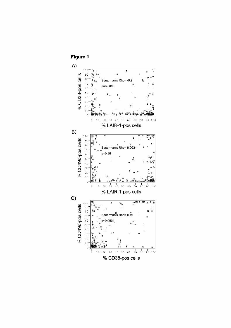
expression was computed as continuous variable, the expression of LAIR1 was inversely related to
CD38 but not to CD49d expression (p=0.0005 and p=0.96, respectively), as shown in Figure 1.
Conversely, CD49d expression was strongly associated with CD38 expression (p<0.0001; Figure
1C). The analysis of median fluorescence intensity (MFI) of LAIR1, CD38, and CD49d confirmed
the strong relation between CD38 and CD49d (p<0.0001), while no relation emerged between
LAIR1 and CD38 or CD49d using MFI values (Supplementary Figure 3).
Nineteen patients were analyzed for LAIR1 expression over time. As shown in supplementary
figure 2, no significant variation (p=0.64 by Wilcoxon test) was observed both among initially
positive or negative cases. Median time from the diagnosis to second analysis was 48 months (9-
71) with half of the patients that had received immunochemotherapy during this period.
Prognostic relevance of LAIR1 expression and univariate analysis
A significant lower fraction of LAIR1+ patients initiated cytotoxic treatment during follow-up (42 of
183, 22.9%) compared to LAIR1- patients (53 of 124, 42.7%, p<0.001). This translated in a
significant difference in terms of TTFT according to LAIR1 expression (p=0.0002), as shown in
Figure 2. Univariate analysis also identified high expression of CD38 (p=0.00003) and of CD49d
(p=0.00002), unmutated IGHV (p<0.00001), high-risk cytogenetic lesions, defined as del17p,
del11q or complex karyotype (p=0.0003), and Binet stage (p<0.00001) as significantly predictors of
a shorter TTFT, as shown in Figure 2.
8
Multivariate analysis
Since the three phenotypic markers showed significant correlations in their expression and were all
predictive for TTFT, LAIR1, CD38, and CD49d were included in a Cox’s proportional hazard
regression model to test their strength as independent prognostic factors in terms of TTFT in our
cohort of CLL patients. As shown in table 2A, LAIR1- (HR 2.269, p=0.002) and CD49d+ (HR 2.232
p=0.008) maintained an independent significant association with shorter TTFT, while CD38 did not.
The multivariate analysis was then extended to other clinical and biological significant variables in
univariate analysis. As shown in table 2B, LAIR1 expression was again significantly associated
with TTFT (HR 2.047, p=0.037) together with IGHV status (HR 2.881, p=0.011) and Binet stage
(HR 6.457, p<0.0001), while CD49d expression lost its predictive value (HR 1.668, p=0.178).
In order to visualize their additive prognostic value in terms of TTFT, LAIR1 expression, IGHV
status and Binet stage were then computed together in a Kaplan-Meyer curve, as shown in Figure
3.
Discussion
With the present study we report for the first time that LAIR1 expression, although related to
commonly recognized risk factors, has a significant and independent impact on time to tumor
progression in patients with CLL, supporting LAIR1 as an easily applicable and inexpensive marker
to predict TTFT in patients presenting with CLL.
Importantly, our results were obtained from a prospective cohort of newly diagnosed unselected
patients with CLL that were standardized in terms of phenotypical, biological and cytogenetic
characterization, as part of the “CLL Veneto” project. The short follow-up of recruited patients
prevented us to investigate the role of LAIR1 expression in terms of survival. However, when
addressing the clinical behavior and aggressiveness of the disease, TTFT is a reliable prognostic
marker strictly related to tumor progression, especially in our prospective cohort where criteria for
treatment initiation were standardized between centers before patients’ enrollment. A merit of our
study is that our population reflects real-life unselected patients with CLL that routinely present to
our Institutions, making our results readily useful to the clinician.

9
Immunophenotypic analysis was performed by multicolor flow cytometry evaluating the percentage
of CD19+CD5+ cells expressing LAIR1, CD38 or CD49d. We observed a significant inverse
correlation between the expression of LAIR1 and CD38 and we confirmed the strong correlation of
expression between CD38 and CD49d already observed by others.22 Further specific studies will
need to confirm these findings and analyze cell biology according to single molecules pattern of
expression. Furthermore, consistently with our findings, Poggi et al have recently observed that the
expression of LAIR1 is higher in patients with low risk CLL.16 In our series, LAIR1 expression
allowed us to discriminate different set of patients with significant biological and clinical differences,
confirming that LAIR1- patients were associated with worse clinical stage at diagnosis and adverse
biological factors, like unmutated IGHV status, CD38+, or high risk cytogenetic lesions.16,17
In last years several studies have focused on the identification of biological markers that could be
easily applicable in order to foresee the prognosis of CLL patients. Many new immunophenotypic
markers have been proposed for identifying high risk patients,11 in addition to historical ones, such
as CD38 and ZAP-70. Indeed, the prognostic power of CD38 expression has been questioned by
several studies.1,6 On the other hand, technical problems have been raised by many experts for the
study of ZAP-70 expression.1,7 Our previous results confirmed these technical issues showing high
discordance in the quantification of ZAP-70 expression either by using different monoclonal
antibodies or different approaches for the analysis.23 For these reasons we did not consider ZAP-
70 expression in our study. Recently, CD49d has been advocated as independent prognostic
marker from retrospective cohort.8-10 However, The expression of LAIR1 in our series seemed to
overcome the prognostic power of CD49d, at least in terms of TTFT. In line with our findings, Del
Poeta et al. recently reported a reduced prognostic power of CD49d when all significant prognostic
factors for CLL were included in the multivariate analysis.12
The cut-off for positivity of LAIR1 was set at 30%. This was assessed considering the distribution
of positive cells frequencies in our cohort of patients, as previously done by Damle et al. for CD384
(Supplementary Methods and Figure 1). Differently from others, this unsupervised procedure did
not consider any clinical parameter to set the cut-off, obviating selection bias.8,9,12

10
Interestingly, LAIR1 expression was related to the occurrence of autoimmune phenomena. Both
DAT and AHA were more frequently observed in LAIR1- patients, with a quite high percentage of
AHA occurrence at CLL presentation for LAIR1- (7.2%). Since it is well known that both DAT and
AHA are associated with IGHV status in patients with CLL,24 our findings might be a consequence
of the association between unmutated IGHV and lower LAIR expression, or, more intriguingly,
might reflect the biological activity of LAIR1 and its role in switching-off B-cells and their potential
antibody-producing activity. A larger number of patients and specific studies will be needed to
address this point.
The independent prognostic power of LAIR1 expression may be connected to its peculiar biological
characteristics and function. Its activity on BCR activation pathway makes LAIR1 very attractive,
since drugs targeting essential components of the BCR signaling (i.e. Bruton tyrosine kinase
inhibitors) have recently shown impressive activity in patients with CLL.25 LAIR1, now designed as
CD305, is an inhibitory receptor expressed on almost all hematopoietic cells, particularly on
immune system cells. After the binding of its known ligands,13,14 LAIR1 inhibits the activation of
immune cells using two immunoreceptor tyrosine-based inhibitory motifs (ITIMs) located in the
cytoplasmic tail of the receptor. LAIR1 is expressed during B cell ontogenesis, but is lost on a
subset of memory B cells, in all germinal center B cells, in plasmablast, and in plasma cells.15 From
a functional point of view, LAIR1 cross-linking results in inhibition of Ca2+ mobilization induced by
BCR-triggering. On the other hand, a prolonged BCR- or CD40-stimulation induces a
downregulation of LAIR1 on naive B cells in vitro,15 suggesting an inhibitory role for LAIR1 on BCR
signaling, as for other B cell inhibitory receptors, like CD22, FcgRIIb.15 Poggi A et al demonstrated
the role of LAIR1 in modulation of BCR signaling pathways implied in the CLL-cell activation, with
its inhibitor capacity that was completely lost or significantly reduced when CLL-cells did not
express LAIR1.16 Furthermore, in vitro studies confirmed that collagen produced by lymph nodes-
derived mesenchymal stromal cells was able to inhibit B cell functions through LAIR1
engagement.26 Altogether these data strengthen and increase the appeal of LAIR1 active role in
the pathophysiology of CLL with special emphasis on microenvironmental interactions inducing
11
BCR activation.25,27 The relationship between LAIR1 expression and therapy with BCR signaling
inhibitors certainly deserves investigation in the future.
The information about LAIR1 expression and function on neoplastic B cells are scanty so far, but
for several reasons CLL represents the right setting for characterizing this molecule. Our data
show an independent role of LAIR1 in predicting TTFT of patients with CLL, while longer follow-up
is needed to establish its predictivity in terms of survival. Its role in-vivo in these patients is also
substantiated by the association between its low expression on B-cell surface and aggressive
clinical presentation of the disease, which is consistent among different studies. Finally, the
influence of LAIR1 on B-cells activation pathways requires further studies that will establish the role
of this molecule in the context of the new target therapies.
Authorship and Disclosures OP and CV were the principal investigators and take primary responsibility for the paper; SFO, SFI,
EA, FM, RP, LT, AA recruited the patients; EF, IG, EB, EN, MF, EA, MAM, FA, LB performed the
laboratory work; OP, EF, CV performed the statistical analysis; AA, MTS, GS, GP, FR revised the
paper and co-ordinated the research; OP, EF, IG, and CV wrote the paper. The authors declare no
potential conflicts of interest.

12
References
1. Rosenquist R, Cortese D, Bhoi S, Mansouri L, Gunnarsson R. Prognostic markers and their
clinical applicability in chronic lymphocytic leukemia: where do we stand? Leuk Lymphoma.
2013;54(11):2351-64.
2. Rai KR, Sawitsky A, Cronkite EP, Chanana AD, Levy RN, Pasternack BS. Clinical staging
of chronic lymphocytic leukemia. Blood. 1975;46(2):219-34.
3. Binet JL, Auquier A, Dighiero G, Chastang C, Piguet H, Goasguen J, et al. A new
prognostic classification of chronic lymphocytic leukemia derived from a multivariate
survival analysis. Cancer. 1981;48(1):198-206.
4. Damle RN, Wasil T, Fais F, Ghiotto F, Valetto A, Allen SL, et al. Ig V gene mutation status
and CD38 expression as novel prognostic indicators in chronic lymphocytic leukemia.
Blood. 1999;94(6):1840-7.
5. Hamblin TJ, Davis Z, Gardiner A, Oscier DG, Stevenson FK. Unmutated Ig V-H genes are
associated with a more aggressive form of chronic lymphocytic leukemia. Blood.
1999;94(6):1848-54.
6. Hamblin TJ, Orchard JA, Gardiner A, Oscier DG, Davis Z, Stevenson FK. Immunoglobulin
V genes and CD38 expression in CLL. Blood. 2000;95(7):2455-7.
7. Chiorazzi N. Implications of new prognostic markers in chronic lymphocytic leukemia.
Hematology Am Soc Hematol Educ Program. 2012:76-87.
8. Gattei V, Bulian P, Del Principe MI, Zucchetto A, Maurillo L, Buccisano F, et al. Relevance
of CD49d protein expression as overall survival and progressive disease prognosticator in
chronic lymphocytic leukemia. Blood. 2008;111(2):865-73.
9. Shanafelt TD, Geyer SM, Bone ND, Tschumper RC, Witzig TE, Nowakowski GS, et al.
CD49d expression is an independent predictor of overall survival in patients with chronic
lymphocytic leukaemia: a prognostic parameter with therapeutic potential. Br J Haematol.
2008;140(5):537-46.
13
10. Rossi D, Zucchetto A, Rossi FM, Capello D, Cerri M, Deambrogi C, et al. CD49d
expression is an independent risk factor of progressive disease in early stage chronic
lymphocytic leukemia. Haematologica. 2008;93:163.
11. Huang PY, Best OG, Belov L, Mulligan SP, Christopherson RI. Surface profiles for
subclassification of chronic lymphocytic leukemia. Leuk Lymphoma. 2012;53(6):1046-56.
12. Del Poeta G, Del Principe MI, Zucchetto A, Luciano F, Buccisano F, Rossi FM, et al. CD69
is independently prognostic in chronic lymphocytic leukemia: a comprehensive clinical and
biological profiling study. Haematologica. 2012;97(2):279-87.
13. Meyaard L. The inhibitory collagen receptor LAIR1 (CD305). J Leukoc Biol. 2008;83(4):799-
803.
14. Son M, Santiago-Schwarz F, Al-Abed Y, Diamond B. C1q limits dendritic cell differentiation
and activation by engaging LAIR1. Proc Natl Acad Sci U S A. 2012;109(46):E3160-E7.
15. van der Vuurst de Vries AR, Clevers H, Logtenberg T, Meyaard L. Leukocyte-associated
immunoglobulin-like receptor-1 (LAIR1) is differentially expressed during human B cell
differentiation and inhibits B cell receptor-mediated signaling. Eur J Immunol.
1999;29(10):3160-7.
16. Poggi A, Catellani S, Bruzzone A, Caligaris-Cappio F, Gobbi M, Zocchi MR. Lack of the
leukocyte-associated Ig-like receptor-1 expression in high-risk chronic lymphocytic
leukaemia results in the absence of a negative signal regulating kinase activation and cell
division. Leukemia. 2008;22(5):980-8.
17. Rawstron AC, Shingles J, de Tute R, Bennett F, Jack AS, Hillmen P. Chronic Lymphocytic
Leukaemia (CLL) and CLL-Type Monoclonal B-Cell Lymphocytosis (MBL) Show Differential
Expression of Molecules Involved in Lymphoid Tissue Homing. Cytometry B Clin Cytom.
2010;78B:S42-S6.
18. Hallek M, Cheson BD, Catovsky D, Caligaris-Cappio F, Dighiero G, Doehner H, et al.
Guidelines for the diagnosis and treatment of chronic lymphocytic leukemia: a report from
the International Workshop on Chronic Lymphocytic Leukemia updating the National
Cancer Institute-Working Group 1996 guidelines. Blood. 2008;111(12):5446-56.

14
19. Clinical and Laboratory Standards Institute. Clinical Flow Cytometric Analysis of Neoplastic
Hematolymphoid Cells; Approved Guideline—Second Edition. CLSI document H43-A2
[ISBN 1-56238- 635-2]. Clinical and Laboratory Standards Institute, Wayne, PA, USA,
2007.
20. Visco C, Moretta F, Falisi E, Facco M, Maura F, Novella E, et al. Double productive
immunoglobulin sequence rearrangements in patients with chronic lymphocytic leukemia.
Am J Hematol. 2013;88(4):277-82.
21. Falisi E, Novella E, Visco C, Guercini N, Maura F, Giaretta I, et al. B-cell receptor
configuration and mutational analysis of patients with chronic lymphocytic leukaemia and
trisomy 12 reveal recurrent molecular abnormalities. Hematol Oncol. 2013 Jul 17. [Epub
ahead of print]
22. Zucchetto A, Bomben R, Dal Bo M, Bulian P, Benedetti D, Nanni P, et al. CD49d in B-cell
chronic lymphocytic leukemia: correlated expression with CD38 and prognostic relevance.
Leukemia. 2006;20(3):523-5.
23. Perbellini O, Zampieri F, Vincenzi C, Perantoni P, Mosna F, Boscaro E, et al. Regione
Veneto study group on B-CLL: standardization of ZAP-70 evaluation by flow cytometry.
Haematologica. 2008;93:S97-S.
24. Visco C, Novella E, Peotta E, Paolini R, Giaretta I, Rodeghiero F. Autoimmune hemolytic
anemia in patients with chronic lymphocytic leukemia is associated with IgVH status.
Haematologica. 2010;95(7):1230-2.
25. Byrd JC, Furman RR, Coutre SE, Flinn IW, Burger JA, Blum KA, et al. Targeting BTK with
Ibrutinib in Relapsed Chronic Lymphocytic Leukemia. N Engl J Med. 2013;369(1):32-42.
26. Colombo BM, Canevali P, Magnani O, Rossi E, Puppo F, Zocchi MR, et al. Defective
Expression and Function of the Leukocyte Associated Ig-like Receptor 1 in B Lymphocytes
from Systemic Lupus Erythematosus Patients. PloS One. 2012;7(2).
27. Wiestner A. Emerging role of kinase-targeted strategies in chronic lymphocytic leukemia.
Blood. 2012;120(24):4684-91.

15
Tables
Table 1. Clinical and biological characteristics of 311 patients with chronic lymphocytic
leukemia at disease presentation, then divided according to LAIR1 expression.
All pts (n. 311) LAIR1+ (n. 186) LAIR1- (n. 125) p. value°
Median Age, years (range) 66 (30.6-90) 66 (36-90) 67 (30.6-87) 0.59§
Female gender 121/311 (38.9%) 69/186 (37.1%) 52/125 (41.6%) 0.48
Median lymphocyte count, x103/mmc (range) 9.7 (2.1-656) 9.4 (2.1-270) 11 (2.1-656) 0.13*
AHA 11/311 (35.4%) 2/186 (1.1%) 9/125 (7.2%) 0.008
ITP 7/311 (2.2%) 5/186 (2.7%) 2/125 (1.6%) 0.70
DAT 13/145 (8.9%) 3/86 (3.5%) 10/59 (16.9) 0.007
BINET stage
A 233/299 (77.9%)
147/178 (82.6%) 86/121 (71.1%) 0.023
B 47/299 (15.7%) 23/178 (12.9%) 24/121 (19.8%) 0.145
C 19/299 (6.4%) 8/178 (4.5%) 11/121 (9.1%) 0.146
FISH
Normal 77/211 (36.5%) 39/124 (31.4%) 38/87 (43.7%) 0.081
del13q 84/211 (39.8%) 57/124 (46.0%) 27/87 (31.0%) 0.033
12+ 18/211 (8.5%) 14/124 (11.3%) 4/87 (4.6%) 0.131
del11q 17/211 (8.1%) 10/124 (8.1%) 7/87 (8.0%) 1.00
del17p 9/211 (4.3%) 1/124 (0.8%) 8/87 (9.2%) 0.004
3 or more alterations 6/211 (2.8%) 3/124 (2.4%) 3/87 (3.5%) 0.692
IGHV mutational status
Unmutated IGHV 77/200 (38.5%) 34/126 (27.0%) 43/74 (58.1%) <0.001
Immunophenotype

16
CD38 64/274 (23.4%) 30/165 (18.2%) 34/109 (31.2%) 0.019
CD49d 90/238 (37.8%) 51/150 (34.7%) 39/88 (44.3%) 0.129
Treatment, Survival and Follow-up
Had cytotoxic treatment 95/307 (30.9%) 42/183 (22.9%) 53/124 (42.7%) <0.001
Median TTFT, months (range) 23.5 (0-75) 25.1 (0-75.1) 18.9 (0-74.7) 0.023*
Dead patients 15/311 (4.8%) 7/186 (3.8%) 8/125 (6.4%) 0.295
Median OS, months (range) 30.7 (0.5-75.1) 29.5 (0.5-75.1) 33.9 (1-75) 0.44
Abbreviations: AHA: autoimmune hemolytic anemia; ITP: Immune thrombocytopenic purpura; DAT:
direct antiglobulin test; FISH: fluorescence in situ hybridisation; del13q: deletion in chromosome
13q14; del11q: deletion in chromosome 11q23; del17p: deletion in chromosome 17p12; +12:
trisomy 12; IGHV: immunoglobulin heavy chain variable region genes; TTFT: Time to First
Treatment; OS: Overall Survival. Statistical tests: °the differences between the categorical
variables were computed by Fisher exact test; § t-test; * Mann-Whitney U test.

17
Table 2. Multivariate Cox’s regression analysis for time to first treatment (TTFT).
A
Parameter HR (CI 95%) p. value
LAIR1- 2.269 (1.355-3.800) 0.002
CD49d+ 2.232 (1.234-4.037) 0.008
CD38+ 1.646 (0.909-2.981) 0.100
B
Parameter HR (IC 95%) p. value
LAIR1- 2.077 (1.057-4.081) 0.034
CD49d+ 1.575 (0.771-3.128) 0.213
unmutated IGHV status 2.675 (1.232-5.804) 0.0013
high risk FISH (del11q/del17p/complex) 2.041 (0.943-4.416) 0.070
Binet B or C 6.285 (3.071-12.816) <0.0001
Abbreviations: HR: Hazard Ratio; CI: Confidence Interval; IGHV: immunoglobulin heavy chain
variable region genes; FISH: fluorescence in situ hybridisation; del11q: deletion in chromosome
11q23; del17p: deletion in chromosome 17p12;
18
Figure Legends
Figure 1: Correlation between LAIR1 and other immunophenotypic prognostic markers.
Scatter plots for percentage of positive cells for LAIR1 and CD38 (A), LAIR1 and CD49d (B), and
for CD49d and CD38 (C). The two-tail Spearman test for nonparametric data was performed. P
value <0.05 was considered associated with a statistical significant correlation.
Figure 2: Time to first treatment (TTFT) according to identified prognostic variables.
Kaplan Meier plot for TTFT according to: phenotypical expression of LAIR1 (A), CD49d (B), and
CD38 (C); IGVH status (D); cytogenetic lesions (E); and Binet Stage (F). Log-rank test was
performed to compare the curves. P values <0.05 were considered statistically significant.
Figure 3: Time to first treatment (TTFT) curves based on the combination of LAIR1, IGHV
status and Binet stage.
Kaplan Meier plot for TTFT showing the additive prognostic value of LAIR1-, unmutated IGHV
status, and B or C Binet stages. Attributing to each variable a score of 1, the curves represent the
sum of adverse variables (0, 1, 2, or 3). Log-rank test was performed to compare different curves.
P values were as follows: p=0.471 between 0 and 1; p<0.0001 between 1 and 2; p=0.06 between
2 and 3.


1
Supplementary 1
Supplementary Methods 2
Immunophenotypic analysis 3
CD38 (HB7 clone), CD49d (9F10 clone), CD305 (DX26 clone) monoclonal antibodies (mAb) were 4
combined with CD19 and CD5 to perform the analysis of expression on CD19+/CD5+ CLL cells 5
(Supplementary figure 1A-F). The three mAb were PE-conjugate and purchased from BD 6
Biosciences (Milan, Italy). After the staining and red blood cell lysis (ammonium chloride solution), 7
the samples were washed twice and then acquired with FACSCanto I cytometers.19 The data were 8
analyzed by DIVA (BD Bioscience) or FlowJo (Tree Star, Inc. Ashland, OR, USA) softwares. The 9
expression data were reported as percentage of CD19+/CD5+ CLL cells. The threshold of positivity 10
was set at over 30% for CD38 and CD49d, as reported in the literature. 4,8 Regarding the LAIR1 11
expression, the cut-off at 30% was empirically chosen by observing the distribution of positive cells 12
frequencies in our cohort of patients (Supplementary Fig. 1G). This cut-off was subsequently 13
validated by computing time-dependent ROC curve and by calculating the Youden Index (YI= 14
sensitivity + specificity - 1) for each cut-off value in the ROC curve (Supplementary Fig. 1H-I). As 15
shown in fig. 1I, the highest YI value was obtained for a cut-off of LAIR1 positivity at 31%. Sup. Ref. 1 16
All these analysis and graphics were performed by using R software and the “survivalROC” 17
package. 18
19
Statistical methods 20
The Kolmogorov-Smirnov and the Shapiro-Wilk tests were used to verify for the normal distribution 21
of each continuous variable. The differences between the continuous variables were computed by 22
t-test or Mann-Whitney-Wilcoxon test as appropriate. The differences between categorical 23
variables were computed by Fisher exact test. Spearman test was used to analyze the 24
2
relationships between immunophenotypical variables. Time to first treatment (TTFT) was 1
calculated from the time of diagnosis to the time of first cytotoxic treatment received by the patient. 2
Curves for TTFT curves were constructed with the method of Kaplan and Meier using SPSS, and 3
the comparison between curves was performed using the log-rank test. P < 0.05 was considered 4
associated with statistical significance. Multivariate analysis was performed with SPSS according 5
to the Cox’s model. 6
7

3
Supplementary figure 1: Flow cytometry analysis. 1
CLL cells were selected by drawing a gate around CD19+/CD5+ cells (A); the percentage of 2
positive cells was recorded by setting the control markers on internal negative control cells (B-F). 3
Representative cases for CD49d (B), CD38 (C), and LAIR1 (D-F) are shown. The distribution of 4
LAIR1+ cells frequencies in our cohort of CLL patients was constructed to set the cut-off of 5
positivity for LAIR1 (chosen cut-off at 30% as shown by the dashed line (G). Time-dependent ROC 6
curve for different cut-off values of LAIR1 positivity computed by survivalROC package in R 7
software (H). Youden Index values computed for each cut-off value of the ROC curve. Dashed line 8
shows the empirically chosen cut-off for LAIR1 positivity (I). 9

4 1

5
1
Supplementary figure 2: LAIR1 expression over time. 2
Each horizontal line corresponds to a single patient. The left initial LAIR1 value refers to the 3
diagnostic sample. Solid line: patients followed-up that received no treatment. Dashed line: 4
patients treated with immunochemotherapy during observation time. Median time of observation (x-5
axis) was 48 months (range 9-71). P-value was calculated with the Wilcoxon test. 6
7
8
9
10

6
1
Supplementary figure 3: Correlation between MFI of LAIR1 and other immunophenotypic 2 markers. 3
Scatter plots of MFI values (log-scale) for LAIR-1 and CD38 (A), LAIR1 and CD49d (B), and for 4
CD49d and CD38 (C). 5
6

7
Supplementary References: 1
1. Heagerty PJ, Lumley T, Pepe MS. Time-dependent ROC curves for censored survival data 2
and a diagnostic marker. Biometrics. 2000;56(2):337-44. 3
4
5