histone deacetylase inhibitor trichostatin a represses...
TRANSCRIPT
Histone Deacetylase Inhibitor Trichostatin A Represses EstrogenReceptor �-Dependent Transcription and Promotes ProteasomalDegradation of Cyclin D1 in Human Breast CarcinomaCell Lines
John Patrick Alao, 1 Eric W-F. Lam, 1
Simak Ali, 1 Laki Buluwela, 1 Walter Bordogna, 2
Peter Lockey, 2 Rana Varshochi, 1
Alexandra V. Stavropoulou, 1
R. Charles Coombes, 1 and David M. Vigushin1
1Department of Cancer Medicine, Imperial College London,Hammersmith Hospital Campus, London, United Kingdom; and2Argenta Discovery Ltd., Harlow, United Kingdom
ABSTRACTPurpose: Estrogen receptor � (ER�)-positive breast
cancer cell lines are up to 10 times more sensitive thanER�-negative cell lines to the antiproliferative activity of thehistone deacetylase inhibitor trichostatin A (TSA). The pur-pose of the study was to investigate the mechanisms under-lying this differential response.
Experimental Design and Results: In the ER�-positiveMCF-7 cell line, TSA repressed ER� and cyclin D1 tran-scription and induced ubiquitin dependent proteasomal deg-radation of cyclin D1, leading primarily to G1-S-phase cellcycle arrest. By contrast, cyclin D1 degradation was en-hanced but its transcription unaffected by TSA in the ER�-negative MDA-MB-231 cell line, which arrested in G2-Mphase. Cyclin D1 degradation involved Skp2/p45, a regula-tory component of the Skp1/Cullin/F-box complex; silencingSKP2 gene expression by RNA interference stabilized cyclinD1 and abrogated the cyclin D1 down-regulation response toTSA.
Conclusions: Tamoxifen has been shown to inhibitER�-mediated cyclin D1 transcription, and acquired resist-ance to tamoxifen is associated with a shift to ER�-inde-pendent cyclin D1 up-regulation. Taken together, our datashow that TSA effectively induces cyclin D1 down-regula-
tion through both ER�-dependent and ER�-independentmechanisms, providing an important new strategy for com-bating resistance to antiestrogens.
INTRODUCTIONHistone deacetylase (HDAC) inhibitors such as the natural
antifungal antibiotic trichostatin A (TSA; refs. 1, 2) inhibit theproliferation of tumor cells in culture and in vivo by inducingcell cycle arrest, differentiation and/or apoptosis (3); reviewedin (4). In response to HDAC inhibition, accumulation of hyper-acetylated core histones in chromatin leads to transcriptionalactivation of certain genes such as the CDKN1A gene, whichencodes the p21WAF1/CIP1 cyclin-dependent kinase inhibitor (5),but leads to transcriptional repression of other genes includingthe CCND1 gene encoding cyclin D1 (6).
D-type cyclins collectively control progression through thecell cycle by activating their cyclin-dependent kinase partnersCDK4 and CDK6, which leads to phosphorylation of the reti-noblastoma protein and release of the E2F family of transcrip-tion factors, which, in turn, leads to advancing through G1 intoS phase of the cell cycle (7). Cyclin D1 is strongly implicated inmammary oncogenesis. Cyclin D1 accumulation is normallytightly regulated, but overexpression of the cyclin occurs insome 50% of human breast cancers (8, 9). Cyclin D1 overex-pression is seen in all histologic types of breast cancer and at allstages from carcinoma in situ through metastatic disease but notin premalignant lesions (10, 11). Transgenic mice that overex-press cyclin D1 in mammary tissue develop breast cancers (12)and cyclin D1-deficient mice are resistant to breast cancersinduced by neu and ras oncogenes (13).
Cyclin D1 may be overexpressed as a result of CCND1gene amplification or chromosomal translocation (14–16), sta-bilization of cyclin D1 mRNA (17), or defective degradation ofcyclin D1 protein (18). D-type cyclins as well as cyclin E, p21,p27, and E2F-1 are ubiquitinated and targeted for degradationby the 26S proteasome. Phosphorylation of cyclin D1 on thre-onine 286 by glycogen synthase kinase 3� (GSK-3�) targetscyclin D1 for ubiquitination (19). Skp2, a regulatory componentof the Skp1/Cullin/F-box complex, is implicated in the ubiquiti-nation of cyclin D1; cyclin D1 levels are modestly elevated inSkp2�/� mouse embryo fibroblasts (20), expression of Skp2antisense induces accumulation of cyclin D1 (20), and defectivecyclin D1 degradation in the SK-UT-1B uterine tumor cell linecan be rescued by stable transfection of Skp2 but not by a splicevariant of Skp2 that does not bind Skp1 and remains in thecytoplasm (21). However, a direct association between Skp2and cyclin D1 has yet to be confirmed.
Estrogen receptor � (ER�) is a member of the nuclearreceptor superfamily of transcription factors that have highly
Received 5/25/04; revised 8/31/04; accepted 9/9/04.Grant support: Supported by Argenta Discovery Ltd.The costs of publication of this article were defrayed in part by thepayment of page charges. This article must therefore be hereby markedadvertisement in accordance with 18 U.S.C. Section 1734 solely toindicate this fact.Note: Presented at the 95th Annual Meeting of the American Associa-tion for Cancer Research in Orlando, Florida, March 27–31, 2004.Requests for reprints: David Vigushin, Department of Cancer Medi-cine, 6th Floor MRC Cyclotron Building, Imperial College London,Hammersmith Hospital Campus, Du Cane Road, London W12 0NN,United Kingdom. Phone: 44-20-8383-8370; Fax: 44-20-8383-5830; E-mail: [email protected].
©2004 American Association for Cancer Research.
8094 Vol. 10, 8094–8104, December 1, 2004 Clinical Cancer Research
Cancer Research. on January 13, 2020. © 2004 American Association forclincancerres.aacrjournals.org Downloaded from
conserved DNA and ligand-binding domains (LBDs) and regu-late gene expression in a ligand-dependent fashion. ER� stim-ulates transcription on binding to cis-acting estrogen responseelements (EREs) in the promoters of estrogen-regulated targetgenes (22–24). The proliferative response of mammary epithe-lial cells to estrogen is, in part, due to ER�-mediated transientactivation of CCND1 gene transcription (25). There is no clas-sical ERE in the CCND1 promoter (26). Instead, ER� up-regulation of the CCND1 promoter is mediated mainly by cyclicAMP response element (CRE) but also by AP-1 and Sp1 sites(27, 28). Reduction in cyclin D1 mRNA and protein expressionis an early and critical event in antiestrogen action (29, 30), andinducible or constitutive cyclin D1 overexpression can conferresistance to antiestrogens (31, 32).
Cyclin D1 can activate ER� transcription by promoting thebinding of both ligand-bound and unliganded ER� to EREsequences in estrogen-regulated genes (33). Cyclin D1 canthereby enhance transcription of ERE-containing genes in boththe presence and the absence of estrogen. This effect of cyclinD1 makes it possible for ER�-positive breast cancer cells tobypass the requirement for estrogen and provides a mechanismfor estrogen-independent proliferation of cyclin D1-overex-pressing breast cancer cells (8) that cannot be inhibited byantiestrogens (8).
We previously observed that ER�-positive breast cancercell lines are highly sensitive to the antiproliferative activity ofTSA compared with ER�-negative cell lines (34). Here we showthat ER� and cyclin D1 down-regulation play a central role inthis differential response. For the first time, we demonstrate thatTSA not only inhibits ER� mediated cyclin D1 transcription inan ER�-positive breast cancer cell line but also induces ubiq-uitin-dependent proteasomal degradation of cyclin D1 in bothER�-positive and ER�-negative breast cancer cell lines. Silenc-ing of SKP2 gene expression by RNA interference stabilizescyclin D1 and abrogates the cyclin D1 down-regulation re-sponse to TSA. Cyclin D1 degradation is, therefore, a criticalevent in the cytostatic action of TSA. Previous studies haveshown that antiestrogens inhibit ER� mediated cyclin D1 tran-scription and acquired resistance to antiestrogens is associatedwith a shift toward ER�-independent cyclin D1 up-regulation.Taken together, our results demonstrate that HDAC inhibitioncan effectively inhibit cyclin D1 expression through both ER�-dependent and ER�-independent mechanisms, providing an im-portant new strategy for overcoming endocrine resistance inbreast cancer.
MATERIALS AND METHODSChemicals. Stock solutions of trichostatin A (TSA) and
HC-toxin (Sigma-Aldrich, Dorset, United Kingdom) 2 mmol/Lin dimethyl sulfoxide (DMSO), MG-132 (Merck BiosciencesLtd., Nottingham, United Kingdom) 10 mmol/L in DMSO, andleptomycin B 5 �g/ml in 70% (v/v) methanol (Sigma-Aldrich)were stored at �20°C until use. All other chemicals and bio-chemicals were the highest quality available from commercialsources.
Cell Lines. MCF-7 and MDA-MB-231 human breastcarcinoma and SK-UT-1B uterine cancer cell lines (AmericanType Culture Collection, Rockville, MD) were cultured in
DMEM containing 10% (v/v) fetal calf serum, 2 mmol/L L-glutamine, 100 units/mL penicillin and 100 �g/mL streptomy-cin. For estrogen depleted conditions, cells were grown inphenol red-free DMEM supplemented with 5% (v/v) dextran-coated charcoal-stripped fetal calf serum, 2 mmol/L L-gluta-mine, 100 units/mL penicillin, and 100 �g/mL streptomycin asdescribed previously (35). Cells were cultured at 37°C in 5%CO2 humidified atmosphere.
Cell Proliferation Assay. Breast cancer cell lines werecounted in a hemocytometer after detachment with 0.25% (w/v)trypsin in Dulbecco’s PBS without Ca2� or Mg2� (DPBS,Sigma-Aldrich) containing 0.02% (w/v) EDTA. Viability wasdetermined by trypan blue exclusion. For each cell line, cellswere seeded in 96-well microtiter plates at optimal densitiesdetermined in prior experiments to ensure exponential growthfor the duration of the assay. After a 24-h incubation, growthmedium was replaced with experimental medium. For determin-ing the concentration of TSA that inhibited cell proliferation by50% (IC50), the experimental medium contained TSA at finalconcentrations ranging from 10�10 mol/L to 10�5 mol/L in logdilutions and 0.1% (v/v) DMSO or growth medium containing0.1% (v/v) DMSO as a vehicle control. To determine the con-centration of 17�-estradiol that stimulated MCF-7 cell prolifer-ation by 50% (EC50), experimental medium contained 17�-estradiol at final concentrations ranging from 10�14 mol/L to10�8 mol/L in log dilutions and 0.1% (v/v) EtOH or growthmedium containing 0.1% (v/v) EtOH as a vehicle control. Cellproliferation at various time points was determined with thesulforhodamine B colorimetric assay (36), and the results ex-pressed as the mean for six replicates as a percentage of vehiclecontrol (taken as 100%).
Flow Cytometry. MCF-7 and MDA-MB-231 cells weretreated as indicated. Floating and adherent cells were collectedby centrifugation (500 � g for 5 minutes at 4°C) and washedtwice with PBS. Cells were then fixed in 90% EtOH and storedat 4°C. For analysis, the samples were washed once in PBS andstained by resuspension in PBS containing propidium iodide(PI; 50 �g/mL) and RNase A (2 �g/mL) for 30 minutes at 4°C.Single cell suspensions were analyzed on a FACSCalibur flowcytometer (BD Biosciences Immunocytometry Systems, SanJose, CA) with CellQuest (BD Biosciences) acquisition soft-ware. PI fluorescence was measured through a 585/42 nm bandpass filter, and list mode data were acquired on a minimum of12,000 single cells defined by a dot plot of PI width versus PIarea. Data analysis was done in ModFit LT (Verity SoftwareHouse, Topsham, ME) with PI width versus PI area to excludecell aggregates.
Immunoprecipitation and Immunoblot Analysis. Cellstreated as indicated were harvested in 5 mL of medium, pelletedby centrifugation (1,000 � g for 5 minutes at 4°C), washedtwice with ice-cold PBS and lysed in ice-cold HEPES lysisbuffer [50 mmol/L HEPES (pH 7.5), 10 mmol/L NaCl, 5mmol/L MgCl2, 1 mmol/L EDTA, 10% (v/v) glycerol, 1% (v/v)Triton X-100 and a cocktail of protease inhibitors] on ice for 30minutes. Lysates were clarified by centrifugation (15,000 � gfor 10 minutes at 4°C). The supernatants were then separatedand either analyzed immediately or stored at �80°C. Equivalentamounts of protein (20–50 �g) from total cell lysates wereresolved on precast 4–12% Bis-Tris gradient gels (Invitrogen
8095Clinical Cancer Research
Cancer Research. on January 13, 2020. © 2004 American Association forclincancerres.aacrjournals.org Downloaded from
Ltd., Paisley, United Kingdom) and transferred onto polyvinyli-dene difluoride membranes (Hybond P; Amersham BiosciencesUnited Kingdom Limited, Little Chalfont, United Kingdom)with a Novex XCell system (Invitrogen). Membranes wereblocked overnight at 4°C in blocking buffer [5% (w/v) nonfatdried milk, 150 mmol/L NaCl, 10 mmol/L Tris (pH 8.0) and0.05% (v/v) Tween 20]. Proteins were detected by incubationwith primary antibodies diluted in blocking buffer at roomtemperature for 1 hour. Rabbit polyclonal anti-HDAC1 (ab7028;Abcam Ltd., Cambridge, United Kingdom), mouse monoclonalanti-ER� (NCL-L-ER-6F11/2, Novocastra Ltd., Newcastle uponTyne, United Kingdom), rabbit polyclonal anti-CDK2 (sc-163,Santa Cruz Biotechnology, Inc., Santa Cruz, CA), rabbit poly-clonal anti-CDK4 (sc-260, Santa Cruz Biotechnology), rabbitpolyclonal anti-CDK6 (sc-177, Santa Cruz Biotechnology),mouse monoclonal anti-cyclin D1 (sc-20044, Santa Cruz Bio-technology), rabbit polyclonal anti-Skp2/p45 (sc-7164, SantaCruz Biotechnology), mouse monoclonal anti-CUL-1 (sc-12761,Santa Cruz Biotechnology), rabbit polyclonal anti-� tubulin(sc-5546, Santa Cruz Biotechnology), goat polyclonal anti-actin(sc-1615, Santa Cruz Biotechnology), mouse monoclonal anti-p21 and mouse monoclonal anti-cyclin E antibodies (both kindgifts from Dr. E. W-F. Lam, Imperial College London, London,United Kingdom) were used. Blots were then incubated at roomtemperature with horseradish peroxidase-conjugated secondaryantibody. Bands were visualized by enhanced chemilumines-cence (Supersignal West Pico, Perbio Science, United KingdomLtd., Cheshire, United Kingdom) followed by exposure to auto-radiography film (Kodak BioMax ML-light or MR-1).
For immunoprecipitations, rabbit polyclonal anti-green flu-orescent protein (anti-GFP) antibody (sc-8334, Santa Cruz Bio-technology) was added to lysate at a concentration of 2 �g/mL.After incubation for 2 hours at 4°C, 50 �L of Protein A/Gagarose (Santa Cruz Biotechnology) 50% (v/v) slurry in lysisbuffer was added, and the incubation continued for an additional60 minutes. After washing four times with ice-cold HEPES lysisbuffer, immunoprecipitated proteins were resolved by SDS-PAGE and analyzed by immunoblotting with mouse mono-clonal anti-GFP (sc-9996, Santa Cruz Biotechnology) and anti-ubiquitin antibodies (sc-8017, Santa Cruz Biotechnology).
Reverse Transcription-Polymerase Chain Reaction.Total RNA was prepared from breast cancer cell lines (�2 �107 cells) by guanidine isothiocyanate lysis followed by silicagel membrane purification (RNeasy mini kit; Qiagen Ltd., WestSussex, United Kingdom). The concentration and purity of RNAwere determined by measuring the spectrophotometric absorp-tion at 260 nm to 280 nm. Integrity of the RNA was confirmedby 1.2% denaturing formaldehyde agarose gel electrophoresiswith ethidium bromide staining. Reverse transcription (RT)-PCR reactions were done with a OneStep RT-PCR kit accordingto the manufacturer’s instructions (Qiagen). Primer pairs usedfor ER� (ESR1) RT-PCR were forward, 5�-CAGATGGCCA-CAGTTTCC-3�, and reverse, 5�-CCAAGAGCAAGTTAGGA-GCAAACAG-3�, and for cyclin D1 (CCND1) forward, 5�-AA-CAGAAGTGCGAGGAGGAG-3�, and reverse, 5�-CTGGCAT-TTTGGAGAGGAAG-3�.
Expression Plasmids. CCND1 was amplified from hu-man breast carcinoma cDNA with the primers forward, 5�-GGAATTCCCCAGCCATGGAA-3�, and reverse, 5�-CGGG-
ATCCCGCCCTCAGAT-3�. PCR products were digested andcloned into the EcoRI and BamHI sites of the pEGFP-C1mammalian expression vector (BD Biosciences).
Transfections. Small interfering RNA (siRNA) technol-ogy was used to silence expression of specific genes. Synthetic21-nt RNA duplexes (Dharmacon Inc., Lafayette, CO) weretransfected into breast cancer cell lines with Oligofectaminereagent according to the manufacturer’s instructions (Invitro-gen). Control scrambled siRNA, ESR1 siRNA, CCND1 siRNAor SKP2 siRNA duplexes were used. For plasmids, Fugene 6transfection reagent (Roche Diagnostics Ltd., East Sussex,United Kingdom) was used according to the manufacturer’sprotocol.
Fluorescence Microscopy. MCF-7 and SK-UT-1B cellsstably transfected to express GFP-tagged cyclin D1 were grownon coverslips and treated as indicated. Coverslips were washedgently with PBS and fixed in methanol at �20°C for 10 min-utes. The cells were subsequently mounted in Vectashieldmounting medium containing 4�,6�-diamidino-2-phenylindoledihydrochloride (DAPI; Vector Laboratories Ltd., Peterbor-ough, United Kingdom) to counterstain DNA and were observedwith an Olympus BX60 fluorescent microscope equipped withthe Micrometastatic Detection System (Applied Imaging Inter-national Ltd., Newcastle upon Tyne, United Kingdom) for anal-ysis of fluorescent images.
Statistical Analysis. The concentration of TSA that in-hibited cell proliferation by 50% (IC50) or 90% (IC90) and theconcentration of 17�-estradiol that effectively stimulated cellproliferation by 50% (EC50) was determined graphically in eachcase with nonlinear regression analysis to fit data to the appro-priate dose-response curve (GraphPad Prism version 4.0; Graph-Pad Software Inc., San Diego, CA). The extra sum of squares Ftest was used for comparative statistical analysis of best fitlogIC50 values. Cell proliferation responses to siRNA treatmentsat various time points were compared with two-way ANOVAwith Bonferroni after tests for multiple comparisons. Statisticalsignificance was set at the 5% level (� 0.05).
RESULTSDifferential Antiproliferative Potency of TSA in ER�-
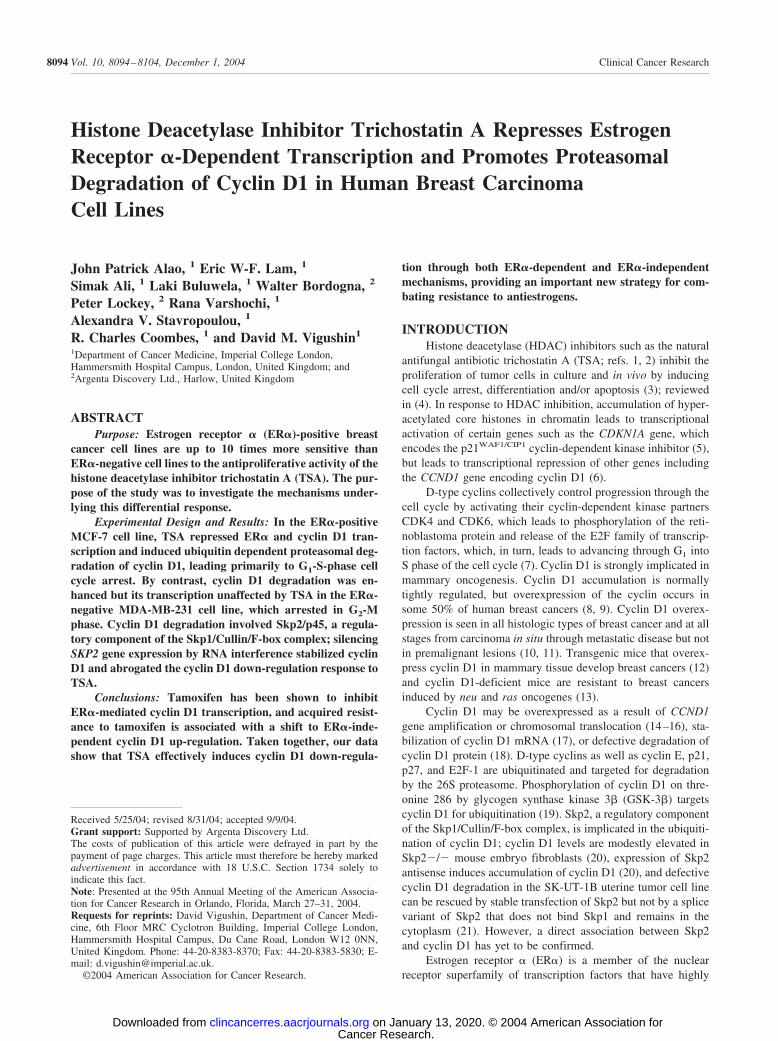
Positive and ER�-Negative Breast Cancer Cell Lines. TSAwas a potent inhibitor of breast cancer cell proliferation. TheER�-positive MCF-7 cell line was highly sensitive to this actionof TSA (IC50, 36 nmol/L; 95% confidence interval, 20–65nmol/L) compared with the ER�-negative MDA-MB-231 cellline (IC50, 242 nmol/L; 95% confidence interval, 182–320nmol/L; P 0.0002, F 178.9; Fig. 1A). ER� mRNA expres-sion was confirmed by RT-PCR and ER� protein expression byimmunocytochemistry and immunoblot experiments. The dose-dependent proliferative response of MCF-7 cells to the ER�ligand 17�-estradiol (EC50 11 pmol/L; 95% confidence interval,6–21 pmol/L) indicated that the expressed ER� was function-ally active (Fig. 1B).
Cell Cycle Response of MCF-7 and MDA-MB-231 Cellsto TSA. For these experiments, we initially used TSA at aconcentration (1 �mol/L) determined in prior experiments toinduce hyperacetylation of histone H3 and H4 and to inhibitproliferation of breast cancer cell lines by 90% (IC90) at 48
8096 ER� and Cyclin D1 Down-Regulation by Trichostatin A
Cancer Research. on January 13, 2020. © 2004 American Association forclincancerres.aacrjournals.org Downloaded from
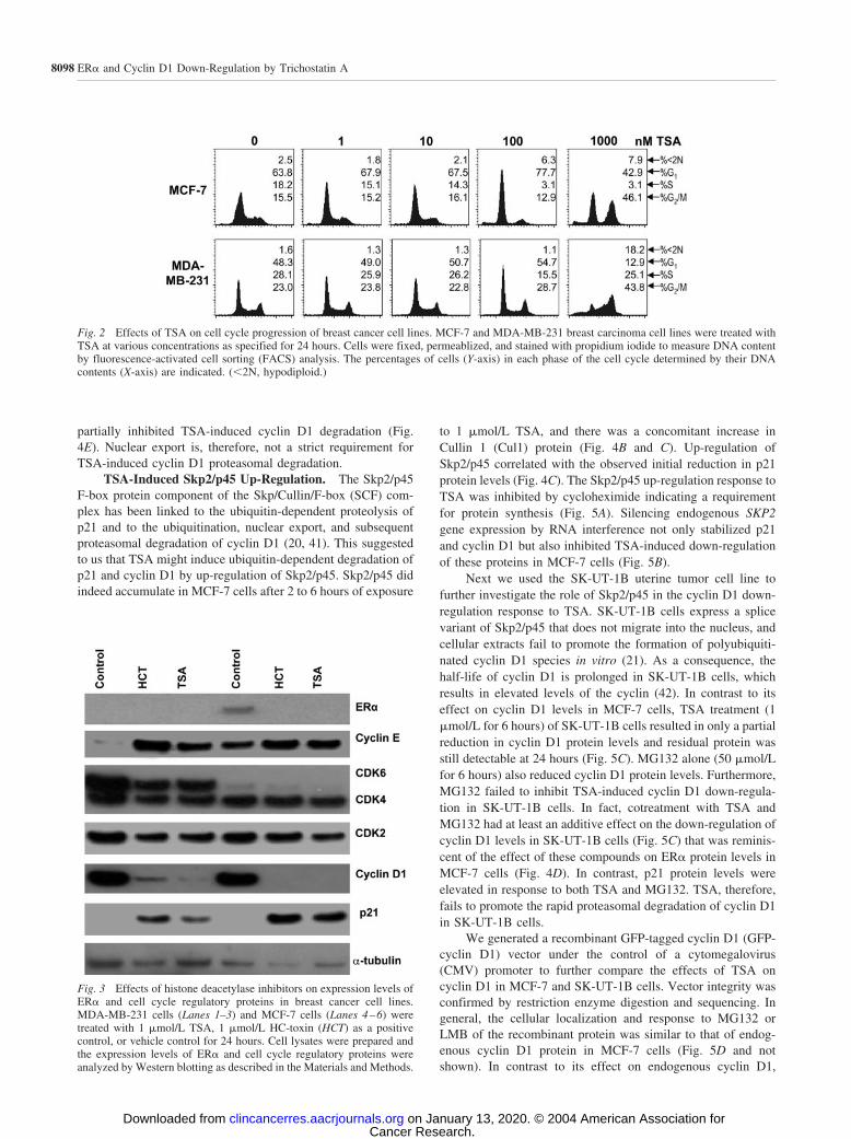
hours. There were striking differences in the cell cycle distri-bution of MCF-7 and MDA-MB-231 cells treated with 1�mol/L TSA for 24 hours (Fig. 2). MCF-7 cells arrested in theG1-S phase and G2-M phases whereas MDA-MB-231 cellsarrested primarily in G2-M phase of the cell cycle. In both celllines, a small population of apoptotic or necrotic cells accumu-lated in sub-G1. The observed effects of TSA on the cell cyclewere concentration dependent; 1 nmol/L or 10 nmol/L TSA for24 hours did not affect the cell cycle distribution of MCF-7 orMDA-MB-231 cells, 100 nmol/L TSA arrested MCF-7 cells inG1-S phase but had little effect on the cell cycle of MDA-MB-231 cells, and at 1 �mol/L, TSA MCF-7 cells arrested in bothG1-S phase and G2-M, whereas MDA-MB-231 cells arrestedmainly in G2-M (Fig. 2). Consistent differences in the cell cycledistribution of MCF-7 and MDA-MB-231 cells exposed to 1�mol/L TSA prompted us to use this concentration of TSA insubsequent experiments.
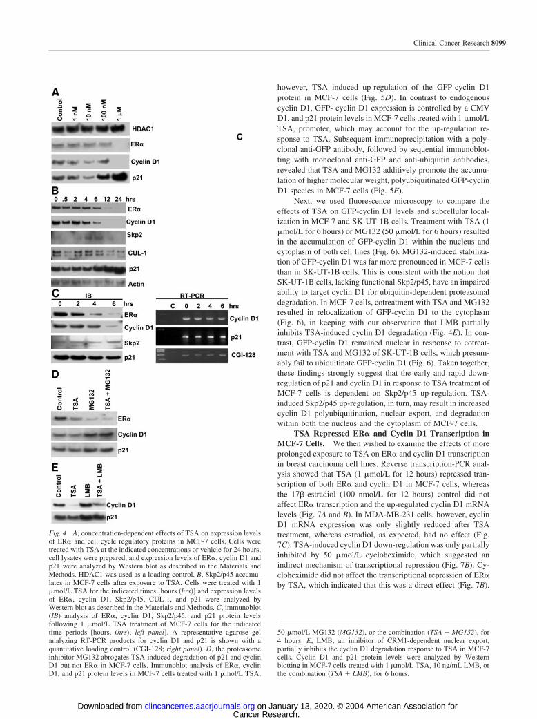
TSA Depleted ER� and Cyclin D1 in MCF-7 Cells.Immunoblot analysis of MCF-7 and MDA-MB-231 cellsshowed that TSA treatment (1 �mol/L for 24 hours) resulted ina marked down-regulation of cyclin D1 levels (Fig. 3). Theeffects of 1 �mol/L HC-toxin, a cyclic tetrapeptide HDACinhibitor used as a positive control, were indistinguishable fromTSA. Cellular levels of CDK4 and CDK6 fell sharply in MDA-MB-231 cells but were either unchanged or only marginallyreduced in MCF-7 cells, although p21 was up-regulated in bothcell types. It is notable that the cyclin E level was substantiallyinduced by TSA or HC-toxin in both cell lines, but the basallevel of the cyclin was lower in MDA-MB-231 cells. ER� wasstrikingly down-regulated in MCF-7 cells treated with TSA orHC-toxin.
TSA Lowered ER� Expression and Promoted Protea-somal Degradation of Cyclin D1 in MCF-7 Cells. ER� andcyclin D1 are important regulators of MCF-7 cell proliferationand we, therefore, sought to investigate further the effect of TSAon stability of these proteins. ER� and cyclin D1 were partiallydown-regulated in response to 10 nmol/L TSA but exposure to1 �mol/L TSA induced complete degradation of both proteins(Fig. 4A). p21 up-regulation (Fig. 4A) and G1-S-phase cell cyclearrest (Fig. 2) both occurred at 100 nmol/L TSA. In MCF-7 cellscultured for 24 hours in estrogen-depleted medium, 1 �mol/LTSA induced a rapid reduction in ER� and cyclin D1 proteinlevels within 2 to 4 hours, followed by a progressive decline toalmost undetectable levels by 12 hours, which persisted for atleast up to 24 hours (Fig. 4B). ER� down-regulation was accel-erated in response to TSA treatment of MCF-7 cells cultured inestrogen-containing complete medium (Fig. 4C), an effect thatmay be attributable to concomitant ligand-induced proteasomaldegradation of the receptor (37). Interestingly, p21 was consis-tently down-regulated at 2 to 6 hours, with subsequent stabili-zation and up-regulation occurring between 6 and 24 hours (Fig.4B and C). The rapid fall in cyclin D1 protein was accompaniedby only partial down-regulation of cyclin D1 mRNA levels,which suggested that the initial effects of TSA on the stability ofthis protein occur mainly at the posttranslational level (Fig. 4C).In contrast, there was a steady accumulation of p21 mRNAduring the first 6 hours of TSA treatment. p21 protein levels fellduring the first 6 hours of TSA treatment (Fig. 4C) but thenincreased progressively during the subsequent 18-hour period(Fig. 4B). Treatment of MCF-7 cells with the proteasome in-hibitor MG132 (50 �mol/L for 4 hours) resulted in elevated p21and cyclin D1 levels, confirming the rapid turnover of theseproteins via the ubiquitin-dependent degradation pathway (Fig.4D; refs. 18, 38). Importantly, cotreatment with MG132 com-pletely abolished TSA-induced p21 and cyclin D1 degradation,indicating that TSA promotes proteasomal degradation of theseproteins in MCF-7 cells. Both TSA and MG132 down-regulatedER� protein levels, and cotreatment had an additive effect (Fig.4D). In contrast to p21 and cyclin D1, TSA-induced down-regulation of ER� does not, therefore, occur via ubiquitin-dependent proteasomal degradation. Leptomycin B (LMB; 10ng/mL for 6 hours), a potent inhibitor of CRM1-dependentnuclear export (39), also induced elevated p21 and cyclin D1levels in MCF-7 cells (Fig. 4E). This observation is in keepingwith previous reports linking cyclin D1 degradation and nuclearexport (40, 41). In contrast to MG132, however, LMB only
Fig. 1 A, effects of TSA on proliferation of breast cancer cell lines.ER�-positive MCF-7 (f) and ER�-negative MDA-MB-231 (Œ) breastcancer cell lines were treated with the indicated concentrations of TSAfor 48 hours. Cell proliferation was then quantified with the sulforho-damine B colorimetric assay as described in Materials and Methods.Results are the mean SD of six replicates expressed as a percentageof the control value in vehicle treated cells. B, proliferative response ofMCF-7 cells to ER� ligand. Cells were treated with 17�-estradiol at arange of concentrations as indicated for 96 hours. Cell proliferation wasthen measured as described in A.
8097Clinical Cancer Research
Cancer Research. on January 13, 2020. © 2004 American Association forclincancerres.aacrjournals.org Downloaded from
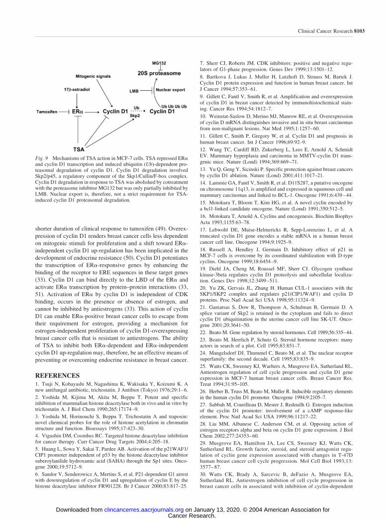
partially inhibited TSA-induced cyclin D1 degradation (Fig.4E). Nuclear export is, therefore, not a strict requirement forTSA-induced cyclin D1 proteasomal degradation.
TSA-Induced Skp2/p45 Up-Regulation. The Skp2/p45F-box protein component of the Skp/Cullin/F-box (SCF) com-plex has been linked to the ubiquitin-dependent proteolysis ofp21 and to the ubiquitination, nuclear export, and subsequentproteasomal degradation of cyclin D1 (20, 41). This suggestedto us that TSA might induce ubiquitin-dependent degradation ofp21 and cyclin D1 by up-regulation of Skp2/p45. Skp2/p45 didindeed accumulate in MCF-7 cells after 2 to 6 hours of exposure
to 1 �mol/L TSA, and there was a concomitant increase inCullin 1 (Cul1) protein (Fig. 4B and C). Up-regulation ofSkp2/p45 correlated with the observed initial reduction in p21protein levels (Fig. 4C). The Skp2/p45 up-regulation response toTSA was inhibited by cycloheximide indicating a requirementfor protein synthesis (Fig. 5A). Silencing endogenous SKP2gene expression by RNA interference not only stabilized p21and cyclin D1 but also inhibited TSA-induced down-regulationof these proteins in MCF-7 cells (Fig. 5B).
Next we used the SK-UT-1B uterine tumor cell line tofurther investigate the role of Skp2/p45 in the cyclin D1 down-regulation response to TSA. SK-UT-1B cells express a splicevariant of Skp2/p45 that does not migrate into the nucleus, andcellular extracts fail to promote the formation of polyubiquiti-nated cyclin D1 species in vitro (21). As a consequence, thehalf-life of cyclin D1 is prolonged in SK-UT-1B cells, whichresults in elevated levels of the cyclin (42). In contrast to itseffect on cyclin D1 levels in MCF-7 cells, TSA treatment (1�mol/L for 6 hours) of SK-UT-1B cells resulted in only a partialreduction in cyclin D1 protein levels and residual protein wasstill detectable at 24 hours (Fig. 5C). MG132 alone (50 �mol/Lfor 6 hours) also reduced cyclin D1 protein levels. Furthermore,MG132 failed to inhibit TSA-induced cyclin D1 down-regula-tion in SK-UT-1B cells. In fact, cotreatment with TSA andMG132 had at least an additive effect on the down-regulation ofcyclin D1 levels in SK-UT-1B cells (Fig. 5C) that was reminis-cent of the effect of these compounds on ER� protein levels inMCF-7 cells (Fig. 4D). In contrast, p21 protein levels wereelevated in response to both TSA and MG132. TSA, therefore,fails to promote the rapid proteasomal degradation of cyclin D1in SK-UT-1B cells.
We generated a recombinant GFP-tagged cyclin D1 (GFP-cyclin D1) vector under the control of a cytomegalovirus(CMV) promoter to further compare the effects of TSA oncyclin D1 in MCF-7 and SK-UT-1B cells. Vector integrity wasconfirmed by restriction enzyme digestion and sequencing. Ingeneral, the cellular localization and response to MG132 orLMB of the recombinant protein was similar to that of endog-enous cyclin D1 protein in MCF-7 cells (Fig. 5D and notshown). In contrast to its effect on endogenous cyclin D1,
Fig. 2 Effects of TSA on cell cycle progression of breast cancer cell lines. MCF-7 and MDA-MB-231 breast carcinoma cell lines were treated withTSA at various concentrations as specified for 24 hours. Cells were fixed, permeablized, and stained with propidium iodide to measure DNA contentby fluorescence-activated cell sorting (FACS) analysis. The percentages of cells (Y-axis) in each phase of the cell cycle determined by their DNAcontents (X-axis) are indicated. (�2N, hypodiploid.)
Fig. 3 Effects of histone deacetylase inhibitors on expression levels ofER� and cell cycle regulatory proteins in breast cancer cell lines.MDA-MB-231 cells (Lanes 1–3) and MCF-7 cells (Lanes 4–6) weretreated with 1 �mol/L TSA, 1 �mol/L HC-toxin (HCT) as a positivecontrol, or vehicle control for 24 hours. Cell lysates were prepared andthe expression levels of ER� and cell cycle regulatory proteins wereanalyzed by Western blotting as described in the Materials and Methods.
8098 ER� and Cyclin D1 Down-Regulation by Trichostatin A
Cancer Research. on January 13, 2020. © 2004 American Association forclincancerres.aacrjournals.org Downloaded from
however, TSA induced up-regulation of the GFP-cyclin D1protein in MCF-7 cells (Fig. 5D). In contrast to endogenouscyclin D1, GFP- cyclin D1 expression is controlled by a CMVD1, and p21 protein levels in MCF-7 cells treated with 1 �mol/LTSA, promoter, which may account for the up-regulation re-sponse to TSA. Subsequent immunoprecipitation with a poly-clonal anti-GFP antibody, followed by sequential immunoblot-ting with monoclonal anti-GFP and anti-ubiquitin antibodies,revealed that TSA and MG132 additively promote the accumu-lation of higher molecular weight, polyubiquitinated GFP-cyclinD1 species in MCF-7 cells (Fig. 5E).
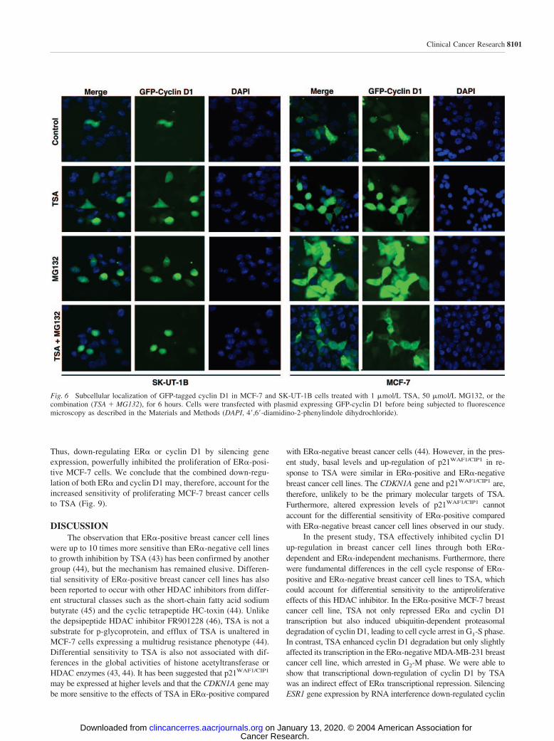
Next, we used fluorescence microscopy to compare theeffects of TSA on GFP-cyclin D1 levels and subcellular local-ization in MCF-7 and SK-UT-1B cells. Treatment with TSA (1�mol/L for 6 hours) or MG132 (50 �mol/L for 6 hours) resultedin the accumulation of GFP-cyclin D1 within the nucleus andcytoplasm of both cell lines (Fig. 6). MG132-induced stabiliza-tion of GFP-cyclin D1 was far more pronounced in MCF-7 cellsthan in SK-UT-1B cells. This is consistent with the notion thatSK-UT-1B cells, lacking functional Skp2/p45, have an impairedability to target cyclin D1 for ubiquitin-dependent proteasomaldegradation. In MCF-7 cells, cotreatment with TSA and MG132resulted in relocalization of GFP-cyclin D1 to the cytoplasm(Fig. 6), in keeping with our observation that LMB partiallyinhibits TSA-induced cyclin D1 degradation (Fig. 4E). In con-trast, GFP-cyclin D1 remained nuclear in response to cotreat-ment with TSA and MG132 of SK-UT-1B cells, which presum-ably fail to ubiquitinate GFP-cyclin D1 (Fig. 6). Taken together,these findings strongly suggest that the early and rapid down-regulation of p21 and cyclin D1 in response to TSA treatment ofMCF-7 cells is dependent on Skp2/p45 up-regulation. TSA-induced Skp2/p45 up-regulation, in turn, may result in increasedcyclin D1 polyubiquitination, nuclear export, and degradationwithin both the nucleus and the cytoplasm of MCF-7 cells.
TSA Repressed ER� and Cyclin D1 Transcription inMCF-7 Cells. We then wished to examine the effects of moreprolonged exposure to TSA on ER� and cyclin D1 transcriptionin breast carcinoma cell lines. Reverse transcription-PCR anal-ysis showed that TSA (1 �mol/L for 12 hours) repressed tran-scription of both ER� and cyclin D1 in MCF-7 cells, whereasthe 17�-estradiol (100 nmol/L for 12 hours) control did notaffect ER� transcription and the up-regulated cyclin D1 mRNAlevels (Fig. 7A and B). In MDA-MB-231 cells, however, cyclinD1 mRNA expression was only slightly reduced after TSAtreatment, whereas estradiol, as expected, had no effect (Fig.7C). TSA-induced cyclin D1 down-regulation was only partiallyinhibited by 50 �mol/L cycloheximide, which suggested anindirect mechanism of transcriptional repression (Fig. 7B). Cy-cloheximide did not affect the transcriptional repression of ER�by TSA, which indicated that this was a direct effect (Fig. 7B).
Fig. 4 A, concentration-dependent effects of TSA on expression levelsof ER� and cell cycle regulatory proteins in MCF-7 cells. Cells weretreated with TSA at the indicated concentrations or vehicle for 24 hours,cell lysates were prepared, and expression levels of ER�, cyclin D1 andp21 were analyzed by Western blot as described in the Materials andMethods. HDAC1 was used as a loading control. B, Skp2/p45 accumu-lates in MCF-7 cells after exposure to TSA. Cells were treated with 1�mol/L TSA for the indicated times [hours (hrs)] and expression levelsof ER�, cyclin D1, Skp2/p45, CUL-1, and p21 were analyzed byWestern blot as described in the Materials and Methods. C, immunoblot(IB) analysis of ER�, cyclin D1, Skp2/p45, and p21 protein levelsfollowing 1 �mol/L TSA treatment of MCF-7 cells for the indicatedtime periods [hours, (hrs); left panel]. A representative agarose gelanalyzing RT-PCR products for cyclin D1 and p21 is shown with aquantitative loading control (CGI-128; right panel). D, the proteasomeinhibitor MG132 abrogates TSA-induced degradation of p21 and cyclinD1 but not ER� in MCF-7 cells. Immunoblot analysis of ER�, cyclinD1, and p21 protein levels in MCF-7 cells treated with 1 �mol/L TSA,
50 �mol/L MG132 (MG132), or the combination (TSA � MG132), for4 hours. E, LMB, an inhibitor of CRM1-dependent nuclear export,partially inhibits the cyclin D1 degradation response to TSA in MCF-7cells. Cyclin D1 and p21 protein levels were analyzed by Westernblotting in MCF-7 cells treated with 1 �mol/L TSA, 10 ng/mL LMB, orthe combination (TSA � LMB), for 6 hours.
8099Clinical Cancer Research
Cancer Research. on January 13, 2020. © 2004 American Association forclincancerres.aacrjournals.org Downloaded from

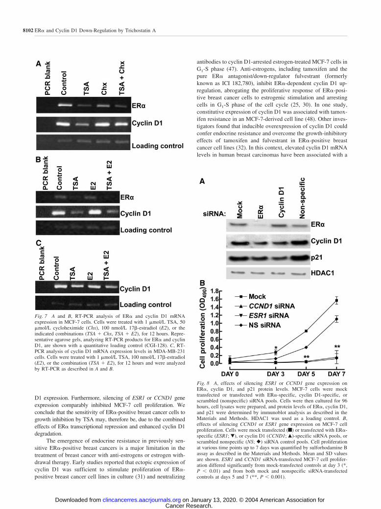
Silencing ESR1 And CCND1 Gene Expression InhibitedMCF-7 Cell Proliferation. To investigate the mechanismwhereby TSA repressed transcription of cyclin D1 in MCF-7cells and to further characterize the role of ER� in this action,we transfected subconfluent MCF-7 cells with pooled ESR1siRNA duplexes to silence ER� gene (ESR1) expressionthrough RNA interference. The resultant effect on expression ofcyclin D1 was examined by immunoblot analysis after 96 hours(Fig. 8A). ER� protein expression was effectively abolished,leading to reduction in cyclin D1 levels at 96 hours and aparallel, down-regulation of p21 expression. We, therefore, con-clude that in MCF-7 cells, TSA-induced transcriptional repres-sion of cyclin D1 results in part from the effect of TSA on ER�expression.
Next we examined the functional effects of silencing ESR1and CCND1 gene expression on MCF-7 cell proliferation. ER�-or cyclin D1-specific siRNA pools or scrambled siRNA controlpool duplexes were transfected into MCF-7 cells, and cell pro-liferation was then quantified by sulforhodamine B assay atvarious time points for up to 7 days. Immunoblot analysisconfirmed that CCND1 gene expression had been successfullysilenced in cells transfected with cyclin D1-specific siRNA;cyclin D1 protein levels could not be detected at the time ofseeding and remained undetectable for 7 days, whereas cyclinD1 levels were unaffected in control siRNA-transfected cells(Fig. 8A and data not shown). Specific ESR1 gene expressionwas successfully silenced in parallel experiments (Fig. 8A anddata not shown). Specific silencing of either ESR1 or CCND1gene expression had a profound inhibitory effect on the prolif-eration of MCF-7 cells (Fig. 8B). Although the mock andnonspecific siRNA control cells proliferated rapidly, the ER�-or cyclin D1-siRNA–transfected MCF-7 cells remained almostquiescent throughout the 7-day period of analysis (Fig. 8B).
Fig. 5 A, immunoblot analysis of Skp2/p45 protein levels in MCF-7cells treated with 1 �mol/L TSA, 50 �mol/L cycloheximide (Chx), orthe combination (TSA � Chx) for 12 hours. Actin was used as a quan-titative loading control. B, effects of silencing SKP2 gene expression onSkp2, cyclin D1, and p21 protein levels. MCF-7 cells treated with 1�mol/L TSA (�) or vehicle control (�) for 2 hours were transfectedwith SKP2 siRNA duplex (Skp2) or a scrambled siRNA control (non-specific). Expression levels of Skp2, cyclin D1, and p21 protein werethen analyzed by Western blotting as described in the Materials andMethods. C, immunoblot analysis of cyclin D1 and p21 protein levels inSK-UT-1B cells treated with 1 �mol/L TSA, 50 �mol/L MG132(MG132), or the combination (TSA � MG132) for 6 hours (left panel) or24 hours (right panel). D, MCF-7 cells that had been stably transfectedto express GFP-tagged cyclin D1 were treated with 1 �mol/L TSA, 50�mol/L MG132 (MG132), or the combination (TSA � MG132), for 6hours. Cellular levels of recombinant GFP-tagged cyclin D1 (GFP-Cyclin D1), endogenous cyclin D1, and p21 proteins were compared byimmunoblot analysis along with actin as a quantitative loading control.E, TSA and the proteasome inhibitor MG132 additively promote theaccumulation of polyubiquitinated GFP-cyclin D1 [GFP-cyclinD1(Ub)n] species in MCF-7 cells. MCF-7 cells were stably transfectedwith a plasmid expressing GFP-tagged cyclin D1. Cells were treatedwith 1 �mol/L TSA (T), 50 �mol/L MG132 (M), the combination(T�M), or control (C) for 6 hours. Proteins were then extracted, andrecombinant GFP-cyclin D1 was immunoprecipitated with a polyclonalanti-GFP antibody (IP: anti-GFP) and analyzed by immunoblottingwith monoclonal anti-GFP (IB: anti-GFP) and anti-ubiquitin (IB: anti-Ubiquitin) antibodies.
8100 ER� and Cyclin D1 Down-Regulation by Trichostatin A
Cancer Research. on January 13, 2020. © 2004 American Association forclincancerres.aacrjournals.org Downloaded from
Thus, down-regulating ER� or cyclin D1 by silencing geneexpression, powerfully inhibited the proliferation of ER�-posi-tive MCF-7 cells. We conclude that the combined down-regu-lation of both ER� and cyclin D1 may, therefore, account for theincreased sensitivity of proliferating MCF-7 breast cancer cellsto TSA (Fig. 9).
DISCUSSIONThe observation that ER�-positive breast cancer cell lines
were up to 10 times more sensitive than ER�-negative cell linesto growth inhibition by TSA (43) has been confirmed by anothergroup (44), but the mechanism has remained elusive. Differen-tial sensitivity of ER�-positive breast cancer cell lines has alsobeen reported to occur with other HDAC inhibitors from differ-ent structural classes such as the short-chain fatty acid sodiumbutyrate (45) and the cyclic tetrapeptide HC-toxin (44). Unlikethe depsipeptide HDAC inhibitor FR901228 (46), TSA is not asubstrate for p-glycoprotein, and efflux of TSA is unaltered inMCF-7 cells expressing a multidrug resistance phenotype (44).Differential sensitivity to TSA is also not associated with dif-ferences in the global activities of histone acetyltransferase orHDAC enzymes (43, 44). It has been suggested that p21WAF1/CIP1
may be expressed at higher levels and that the CDKN1A gene maybe more sensitive to the effects of TSA in ER�-positive compared
with ER�-negative breast cancer cells (44). However, in the pres-ent study, basal levels and up-regulation of p21WAF1/CIP1 in re-sponse to TSA were similar in ER�-positive and ER�-negativebreast cancer cell lines. The CDKN1A gene and p21WAF1/CIP1 are,therefore, unlikely to be the primary molecular targets of TSA.Furthermore, altered expression levels of p21WAF1/CIP1 cannotaccount for the differential sensitivity of ER�-positive comparedwith ER�-negative breast cancer cell lines observed in our study.
In the present study, TSA effectively inhibited cyclin D1up-regulation in breast cancer cell lines through both ER�-dependent and ER�-independent mechanisms. Furthermore, therewere fundamental differences in the cell cycle response of ER�-positive and ER�-negative breast cancer cell lines to TSA, whichcould account for differential sensitivity to the antiproliferativeeffects of this HDAC inhibitor. In the ER�-positive MCF-7 breastcancer cell line, TSA not only repressed ER� and cyclin D1transcription but also induced ubiquitin-dependent proteasomaldegradation of cyclin D1, leading to cell cycle arrest in G1-S phase.In contrast, TSA enhanced cyclin D1 degradation but only slightlyaffected its transcription in the ER�-negative MDA-MB-231 breastcancer cell line, which arrested in G2-M phase. We were able toshow that transcriptional down-regulation of cyclin D1 by TSAwas an indirect effect of ER� transcriptional repression. SilencingESR1 gene expression by RNA interference down-regulated cyclin
Fig. 6 Subcellular localization of GFP-tagged cyclin D1 in MCF-7 and SK-UT-1B cells treated with 1 �mol/L TSA, 50 �mol/L MG132, or thecombination (TSA � MG132), for 6 hours. Cells were transfected with plasmid expressing GFP-cyclin D1 before being subjected to fluorescencemicroscopy as described in the Materials and Methods (DAPI, 4�,6�-diamidino-2-phenylindole dihydrochloride).
8101Clinical Cancer Research
Cancer Research. on January 13, 2020. © 2004 American Association forclincancerres.aacrjournals.org Downloaded from
D1 expression. Furthermore, silencing of ESR1 or CCND1 geneexpression comparably inhibited MCF-7 cell proliferation. Weconclude that the sensitivity of ER�-positive breast cancer cells togrowth inhibition by TSA may, therefore be, due to the combinedeffects of ER� transcriptional repression and enhanced cyclin D1degradation.
The emergence of endocrine resistance in previously sen-sitive ER�-positive breast cancers is a major limitation in thetreatment of breast cancer with anti-estrogens or estrogen with-drawal therapy. Early studies reported that ectopic expression ofcyclin D1 was sufficient to stimulate proliferation of ER�-positive breast cancer cell lines in culture (31) and neutralizing
antibodies to cyclin D1-arrested estrogen-treated MCF-7 cells inG1-S phase (47). Anti-estrogens, including tamoxifen and thepure ER� antagonist/down-regulator fulvestrant (formerlyknown as ICI 182,780), inhibit ER�-dependent cyclin D1 up-regulation, abrogating the proliferative response of ER�-posi-tive breast cancer cells to estrogenic stimulation and arrestingcells in G1-S phase of the cell cycle (25, 30). In one study,constitutive expression of cyclin D1 was associated with tamox-ifen resistance in an MCF-7-derived cell line (48). Other inves-tigators found that inducible overexpression of cyclin D1 couldconfer endocrine resistance and overcome the growth-inhibitoryeffects of tamoxifen and fulvestrant in ER�-positive breastcancer cell lines (32). In this context, elevated cyclin D1 mRNAlevels in human breast carcinomas have been associated with a
Fig. 8 A, effects of silencing ESR1 or CCND1 gene expression onER�, cyclin D1, and p21 protein levels. MCF-7 cells were mocktransfected or transfected with ER�-specific, cyclin D1-specific, orscrambled (nonspecific) siRNA pools. Cells were then cultured for 96hours, cell lysates were prepared, and protein levels of ER�, cyclin D1,and p21 were determined by immunoblot analysis as described in theMaterials and Methods. HDAC1 was used as a loading control. B,effects of silencing CCND1 or ESR1 gene expression on MCF-7 cellproliferation. Cells were mock transfected (f) or transfected with ER�-specific (ESR1; �), or cyclin D1 (CCND1; Œ)-specific siRNA pools, orscrambled nonspecific (NS; �) siRNA control pools. Cell proliferationat various time points up to 7 days was quantified by sulforhodamine Bassay as described in the Materials and Methods. Mean and SD valuesare shown. ESR1 and CCND1 siRNA-transfected MCF-7 cell prolifer-ation differed significantly from mock-transfected controls at day 3 (*,P � 0.01) and from both mock and nonspecific siRNA-transfectedcontrols at days 5 and 7 (**, P � 0.001).
Fig. 7 A and B, RT-PCR analysis of ER� and cyclin D1 mRNAexpression in MCF-7 cells. Cells were treated with 1 �mol/L TSA, 50�mol/L cycloheximide (Chx), 100 nmol/L 17�-estradiol (E2), or theindicated combinations (TSA � Chx, TSA � E2), for 12 hours. Repre-sentative agarose gels, analyzing RT-PCR products for ER� and cyclinD1, are shown with a quantitative loading control (CGI-128). C, RT-PCR analysis of cyclin D1 mRNA expression levels in MDA-MB-231cells. Cells were treated with 1 �mol/L TSA, 100 nmol/L 17�-estradiol(E2), or the combination (TSA � E2), for 12 hours and were analyzedby RT-PCR as described in A and B.
8102 ER� and Cyclin D1 Down-Regulation by Trichostatin A
Cancer Research. on January 13, 2020. © 2004 American Association forclincancerres.aacrjournals.org Downloaded from
shorter duration of clinical response to tamoxifen (49). Overex-pression of cyclin D1 renders breast cancer cells less dependenton mitogenic stimuli for proliferation and a shift toward ER�-independent cyclin D1 up-regulation has been implicated in thedevelopment of endocrine resistance (50). Cyclin D1 potentiatesthe transcription of ER�-responsive genes by enhancing thebinding of the receptor to ERE sequences in these target genes(33). Cyclin D1 can bind directly to the LBD of the ER� andactivate ER� transcription by protein–protein interactions (33,51). Activation of ER� by cyclin D1 is independent of CDKbinding, occurs in the presence or absence of estrogen, andcannot be inhibited by antiestrogens (33). This action of cyclinD1 can enable ER�-positive breast cancer cells to escape fromtheir requirement for estrogen, providing a mechanism forestrogen-independent proliferation of cyclin D1-overexpressingbreast cancer cells that is resistant to antiestrogens. The abilityof TSA to inhibit both ER�-dependent and ER�-independentcyclin D1 up-regulation may, therefore, be an effective means ofpreventing or overcoming endocrine resistance in breast cancer.
REFERENCES1. Tsuji N, Kobayashi M, Nagashima K, Wakisaka Y, Koizumi K. Anew antifungal antibiotic, trichostatin. J Antibiot (Tokyo) 1976;29:1–6.2. Yoshida M, Kijima M, Akita M, Beppu T. Potent and specificinhibition of mammalian histone deacetylase both in vivo and in vitro bytrichostatin A. J Biol Chem 1990;265:17174–9.3. Yoshida M, Horinouchi S, Beppu T. Trichostatin A and trapoxin:novel chemical probes for the role of histone acetylation in chromatinstructure and function. Bioessays 1995;17:423–30.4. Vigushin DM, Coombes RC. Targeted histone deacetylase inhibitionfor cancer therapy. Curr Cancer Drug Targets 2004;4:205–18.5. Huang L, Sowa Y, Sakai T, Pardee AB. Activation of the p21WAF1/CIP1 promoter independent of p53 by the histone deacetylase inhibitorsuberoylanilide hydroxamic acid (SAHA) through the Sp1 sites. Onco-gene 2000;19:5712–9.6. Sandor V, Senderowicz A, Mertins S, et al. P21-dependent G1 arrestwith downregulation of cyclin D1 and upregulation of cyclin E by thehistone deacetylase inhibitor FR901228. Br J Cancer 2000;83:817–25.
7. Sherr CJ, Roberts JM. CDK inhibitors: positive and negative regu-lators of G1-phase progression. Genes Dev 1999;13:1501–12.
8. Bartkova J, Lukas J, Muller H, Lutzhoft D, Strauss M, Bartek J.Cyclin D1 protein expression and function in human breast cancer. IntJ Cancer 1994;57:353–61.
9. Gillett C, Fantl V, Smith R, et al. Amplification and overexpressionof cyclin D1 in breast cancer detected by immunohistochemical stain-ing. Cancer Res 1994;54:1812–7.
10. Weinstat-Saslow D, Merino MJ, Manrow RE, et al. Overexpressionof cyclin D mRNA distinguishes invasive and in situ breast carcinomasfrom non-malignant lesions. Nat Med 1995;1:1257–60.
11. Gillett C, Smith P, Gregory W, et al. Cyclin D1 and prognosis inhuman breast cancer. Int J Cancer 1996;69:92–9.
12. Wang TC, Cardiff RD, Zukerberg L, Lees E, Arnold A, SchmidtEV. Mammary hyperplasia and carcinoma in MMTV-cyclin D1 trans-genic mice. Nature (Lond) 1994;369:669–71.
13. Yu Q, Geng Y, Sicinski P. Specific protection against breast cancersby cyclin D1 ablation. Nature (Lond) 2001;411:1017–21.
14. Lammie GA, Fantl V, Smith R, et al. D11S287, a putative oncogeneon chromosome 11q13, is amplified and expressed in squamous cell andmammary carcinomas and linked to BCL-1. Oncogene 1991;6:439–44.
15. Motokura T, Bloom T, Kim HG, et al. A novel cyclin encoded bya bcl1-linked candidate oncogene. Nature (Lond) 1991;350:512–5.
16. Motokura T, Arnold A. Cyclins and oncogenesis. Biochim BiophysActa 1993;1155:63–78.
17. Lebwohl DE, Muise-Helmericks R, Sepp-Lorenzino L, et al. Atruncated cyclin D1 gene encodes a stable mRNA in a human breastcancer cell line. Oncogene 1994;9:1925–9.
18. Russell A, Hendley J, Germain D. Inhibitory effect of p21 inMCF-7 cells is overcome by its coordinated stabilization with D-typecyclins. Oncogene 1999;18:6454–9.19. Diehl JA, Cheng M, Roussel MF, Sherr CJ. Glycogen synthasekinase-3beta regulates cyclin D1 proteolysis and subcellular localiza-tion. Genes Dev 1998;12:3499–511.20. Yu ZK, Gervais JL, Zhang H. Human CUL-1 associates with theSKP1/SKP2 complex and regulates p21(CIP1/WAF1) and cyclin Dproteins. Proc Natl Acad Sci USA 1998;95:11324–9.21. Ganiatsas S, Dow R, Thompson A, Schulman B, Germain D. Asplice variant of Skp2 is retained in the cytoplasm and fails to directcyclin D1 ubiquitination in the uterine cancer cell line SK-UT. Onco-gene 2001;20:3641–50.22. Beato M. Gene regulation by steroid hormones. Cell 1989;56:335–44.23. Beato M, Herrlich P, Schutz G. Steroid hormone receptors: manyactors in search of a plot. Cell 1995;83:851–7.24. Mangelsdorf DJ, Thummel C, Beato M, et al. The nuclear receptorsuperfamily: the second decade. Cell 1995;83:835–9.25. Watts CK, Sweeney KJ, Warlters A, Musgrove EA, Sutherland RL.Antiestrogen regulation of cell cycle progression and cyclin D1 geneexpression in MCF-7 human breast cancer cells. Breast Cancer Res.Treat 1994;31:95–105.26. Herber B, Truss M, Beato M, Muller R. Inducible regulatory elementsin the human cyclin D1 promoter. Oncogene 1994;9:2105–7.27. Sabbah M, Courilleau D, Mester J, Redeuilh G. Estrogen inductionof the cyclin D1 promoter: involvement of a cAMP response-likeelement. Proc Natl Acad Sci USA 1999;96:11217–22.28. Liu MM, Albanese C, Anderson CM, et al. Opposing action ofestrogen receptors alpha and beta on cyclin D1 gene expression. J BiolChem 2002;277:24353–60.29. Musgrove EA, Hamilton JA, Lee CS, Sweeney KJ, Watts CK,Sutherland RL. Growth factor, steroid, and steroid antagonist regu-lation of cyclin gene expression associated with changes in T-47Dhuman breast cancer cell cycle progression. Mol Cell Biol 1993;13:3577– 87.30. Watts CK, Brady A, Sarcevic B, deFazio A, Musgrove EA,Sutherland RL. Antiestrogen inhibition of cell cycle progression inbreast cancer cells in associated with inhibition of cyclin-dependent
Fig. 9 Mechanisms of TSA action in MCF-7 cells. TSA repressed ER�and cyclin D1 transcription and induced ubiquitin (Ub)-dependent pro-teasomal degradation of cyclin D1. Cyclin D1 degradation involvedSkp2/p45, a regulatory component of the Skp1/Cullin/F-box complex.Cyclin D1 degradation in response to TSA was abolished by cotreatmentwith the proteasome inhibitor MG132 but was only partially inhibited byLMB. Nuclear export is, therefore, not a strict requirement for TSA-induced cyclin D1 proteasomal degradation.
8103Clinical Cancer Research
Cancer Research. on January 13, 2020. © 2004 American Association forclincancerres.aacrjournals.org Downloaded from
kinase activity and decreased retinoblastoma protein phosphoryla-tion. Mol Endocrinol 1995;9:1804 –13.
31. Wilcken NR, Prall OW, Musgrove EA, Sutherland RL. Inducibleoverexpression of cyclin D1 in breast cancer cells reverses the growth-inhibitory effects of antiestrogens. Clin Cancer Res 1997;3:849–54.
32. Hui R, Finney GL, Carroll JS, Lee CS, Musgrove EA, SutherlandRL. Constitutive overexpression of cyclin D1 but not cyclin E confersacute resistance to antiestrogens in T-47D breast cancer cells. CancerRes 2002;62:6916–23.
33. Zwijsen RM, Wientjens E, Klompmaker R, van der Sman J, Ber-nards R, Michalides RJ. CDK-independent activation of estrogen recep-tor by cyclin D1. Cell 1997;88:405–15.
34. Vigushin DM, Ali S, Pace PE, et al. Trichostatin A is a histonedeacetylase inhibitor with potent antitumor activity against breast cancerin vivo. Clin Cancer Res 2001;7:971–6.
35. Horwitz KB, Koseki Y, McGuire WL. Estrogen control of proges-terone receptor in human breast cancer: role of estradiol and antiestro-gen. Endocrinology 1978;103:1742–51.
36. Skehan P, Storeng R, Scudiero D, et al. New colorimetric cytotox-icity assay for anticancer-drug screening. J Natl Cancer Inst (Bethesda)1990;82:1107–12.
37. Reid G, Hubner MR, Metivier R, et al. Cyclic, proteasome-medi-ated turnover of unliganded and liganded ERalpha on responsive pro-moters is an integral feature of estrogen signaling. Mol Cell 2003;11:695–707.
38. Coleman ML, Marshall CJ, Olson MF. Ras promotes p21(Waf1/Cip1) protein stability via a cyclin D1-imposed block in proteasome-mediated degradation. EMBO J 2003;22:2036–46.
39. Fukuda M, Asano S, Nakamura T, et al. CRM1 is responsible forintracellular transport mediated by the nuclear export signal. Nature1997;390:308–11.
40. Alt JR, Gladden AB, Diehl JA. p21(Cip1) Promotes cyclin D1nuclear accumulation via direct inhibition of nuclear export. J BiolChem 2002;277:8517–23.
41. Alt JR, Cleveland JL, Hannink M, Diehl JA. Phosphorylation-dependent regulation of cyclin D1 nuclear export and cyclin D1-dependent cellular transformation. Genes Dev 2000;14:3102–14.42. Welcker M, Lukas J, Strauss M, Bartek J. Enhanced protein stabil-ity: a novel mechanism of D-type cyclin over-abundance identified inhuman sarcoma cells. Oncogene 1996;13:419–25.43. Vigushin DM, Ali S, Pace PE, et al. Trichostatin A is a histonedeacetylase inhibitor with potent antitumor activity against breast cancerin vivo. Clin Cancer Res 2001;7:971–6.44. Margueron R, Licznar A, Lazennec G, Vignon F, Cavailles V.Oestrogen receptor alpha increases p21(WAF1/CIP1) gene expressionand the antiproliferative activity of histone deacetylase inhibitors inhuman breast cancer cells. J Endocrinol 2003;179:41–53.45. Davis T, Kennedy C, Chiew YE, Clarke CL, deFazio A. Histonedeacetylase inhibitors decrease proliferation and modulate cell cyclegene expression in normal mammary epithelial cells. Clin Cancer Res2000;6:4334–42.46. Lee JS, Paull K, Alvarez M, et al. Rhodamine efflux patterns predictP-glycoprotein substrates in the National Cancer Institute drug screen.Mol Pharmacol 1994;46:627–38.47. Lukas J, Bartkova J, Bartek J. Convergence of mitogenic signallingcascades from diverse classes of receptors at the cyclin D-cyclin-dependent kinase-pRb-controlled G1 checkpoint. Mol Cell Biol 1996;16:6917–25.48. Hodges LC, Cook JD, Lobenhofer EK, et al. Tamoxifen functionsas a molecular agonist inducing cell cycle-associated genes in breastcancer cells. Mol Cancer Res 2003;1:300–11.49. Kenny FS, Hui R, Musgrove EA, et al. Overexpression of cyclin D1messenger RNA predicts for poor prognosis in estrogen receptor-posi-tive breast cancer. Clin Cancer Res 1999;5:2069–76.50. Ali S, Coombes RC. Endocrine-responsive breast cancer and strat-egies for combating resistance. Nat Rev Cancer 2002;2:101–12.51. Neuman E, Ladha MH, Lin N, et al. Cyclin D1 stimulation ofestrogen receptor transcriptional activity independent of cdk4. Mol CellBiol 1997;17:5338–47.
8104 ER� and Cyclin D1 Down-Regulation by Trichostatin A
Cancer Research. on January 13, 2020. © 2004 American Association forclincancerres.aacrjournals.org Downloaded from
2004;10:8094-8104. Clin Cancer Res John Patrick Alao, Eric W-F. Lam, Simak Ali, et al. Breast Carcinoma Cell LinesPromotes Proteasomal Degradation of Cyclin D1 in Human
-Dependent Transcription andαEstrogen Receptor Histone Deacetylase Inhibitor Trichostatin A Represses
Updated version
http://clincancerres.aacrjournals.org/content/10/23/8094
Access the most recent version of this article at:
Cited articles
http://clincancerres.aacrjournals.org/content/10/23/8094.full#ref-list-1
This article cites 50 articles, 21 of which you can access for free at:
Citing articles
http://clincancerres.aacrjournals.org/content/10/23/8094.full#related-urls
This article has been cited by 14 HighWire-hosted articles. Access the articles at:
E-mail alerts related to this article or journal.Sign up to receive free email-alerts
SubscriptionsReprints and
To order reprints of this article or to subscribe to the journal, contact the AACR Publications
Permissions
Rightslink site. (CCC)Click on "Request Permissions" which will take you to the Copyright Clearance Center's
.http://clincancerres.aacrjournals.org/content/10/23/8094To request permission to re-use all or part of this article, use this link
Cancer Research. on January 13, 2020. © 2004 American Association forclincancerres.aacrjournals.org Downloaded from