histone deacetylase inhibition suppresses myogenin-dependent
TRANSCRIPT
Histone deacetylase inhibition suppressesmyogenin-dependent atrogene activationin spinal muscular atrophy mice
Katherine V. Bricceno1,2, Paul J. Sampognaro3, James P. Van Meerbeke3,
Charlotte J. Sumner3,4, Kenneth H. Fischbeck1 and Barrington G. Burnett1,∗
1Neurogenetics Branch, National Institute of Neurological Disorders and Stroke, National Institutes of Health,
Bethesda, MD, USA, 2Institute of Biomedical Sciences, The George Washington University, Washington, DC, USA,3Department of Neurology and 4Department of Neuroscience, Johns Hopkins University, Baltimore, MD, USA
Received April 17, 2012; Revised June 15, 2012; Accepted July 10, 2012
Spinal muscular atrophy (SMA) is an autosomal recessive neuromuscular disease caused by mutations in thesurvival of motor neuron 1 (SMN1) gene and deficient expression of the ubiquitously expressed SMN protein.Pathologically, SMA is characterized by motor neuron loss and severe muscle atrophy. During muscle atro-phy, the E3 ligase atrogenes, atrogin-1 and muscle ring finger 1 (MuRF1), mediate muscle protein breakdownthrough the ubiquitin proteasome system. Atrogene expression can be induced by various upstream regula-tors. During acute denervation, they are activated by myogenin, which is in turn regulated by histone deace-tylases 4 and 5. Here we show that atrogenes are induced in SMA model mice and in SMA patient muscle inassociation with increased myogenin and histone deacetylase-4 (HDAC4) expression. This activation duringboth acute denervation and SMA disease progression is suppressed by treatment with a histone deacetylaseinhibitor; however, this treatment has no effect when atrogene induction occurs independently of myogenin.These results indicate that myogenin-dependent atrogene induction is amenable to pharmacological inter-vention with histone deacetylase inhibitors and help to explain the beneficial effects of these agents onSMA and other denervating diseases.
INTRODUCTION
Spinal muscular atrophy (SMA) is a neuromuscular disordercharacterized by loss of motor neurons and skeletal muscleweakness and atrophy. SMA is caused by deficient expressionof the survival motor of neuron (SMN) protein due to a dele-tion or other mutations in the SMN1 gene (1). A nearly iden-tical gene, SMN2, is retained in SMA patients. SMN1primarily produces a full-length SMN transcript, whileSMN2 primarily produces an alternatively spliced isoformlacking exon 7 (2,3). Nonetheless, increased SMN2 copynumber ameliorates SMA disease severity in a dose-dependentfashion (4–6). SMA model mice, which are null for the en-dogenous mouse Smn gene, but express the human SMN2transgene (‘delta 7’ mice) show progressive weakness,decreased motor neuron number, low body weight and an
average lifespan of just 13 days (7). Treatment of SMA micewith the histone deacetylase (HDAC) inhibitor trichostatin A(TSA) improves body weight, life span, myofiber numberand myofiber cross-sectional area (8). Beneficial effects ofTSA on survival, muscle mass and motor function havealso been shown in a mouse model of amyotrophic lateralsclerosis (9). TSA has been shown to modestly increaseSMN expression (8,10,11), but as an HDAC inhibitor it mayalso inhibit HDAC4 activity, which has been shown to modu-late the expression of genes involved in muscle atrophy. Wehypothesized that TSA improves SMA muscle pathology, inpart, by inhibiting this muscle atrophy pathway.
Muscle atrophy occurs in a variety of denervating diseases(12) and with disuse (13). During atrophy, muscle protein isdegraded through the ubiquitin proteasome pathway (14). Ex-pression of genes encoding the skeletal muscle-specific E3
∗To whom correspondence should be addressed at: 35 Convent Dr, Bldg 35, Room 2A1008, Bethesda, MD 20892, USA. Tel: +1 301 435 9288;Fax: +1 301 480 3365; Email: [email protected].
Published by Oxford University Press 2012.
Human Molecular Genetics, 2012, Vol. 21, No. 20 4448–4459doi:10.1093/hmg/dds286Advance Access published on July 13, 2012
Dow
nloaded from https://academ
ic.oup.com/hm
g/article/21/20/4448/655746 by guest on 24 February 2022
ligases atrogin-1 and muscle ring finger 1 (MuRF1), referredto as atrogenes, is increased in atrophy models, includingfasting and glucocorticoid treatment, as well as denervation(15). MuRF1 and atrogin-1 null mice are resistant to loss ofmuscle mass after denervation (15).
Upregulation of the atrogenes is mediated by multiple path-ways, including reduction in the activity of the PI3K/Akt sig-naling cascade, which activates the FoxO1/3 transcriptionfactors (16,17). MuRF1 upregulation in tumor-bearing miceis mediated by the transcription factor NF-kB, which is acti-vated by tumor necrosis factor alpha (TNF-a) and otherpro-inflammatory cytokines (18). In atrophy due to acute de-nervation, atrogin-1 and MuRF1 expression is regulated inpart by HDACs and myogenin. Following denervation,HDAC4 is upregulated and suppresses the expression ofDach2, a transcription factor and a repressor of the myogeninpromoter (19,20). Myogenin is a muscle-specific transcriptionfactor essential for muscle development (21). Myogeninexpression increases following denervation via increasedHDAC4 and decreased Dach2 expression (19). Myogenin, inturn, positively regulates HDAC4 expression followingdenervation, creating a positive feedback loop. Atrogin-1and MuRF1 expression and muscle atrophy following denerv-ation are attenuated in myogenin-null mice (22,23). Whilethese studies modeled neurogenic atrophy by severing thesciatic nerve, it remains to be determined whether (i) thispathway is conserved in a chronic motor neuron diseasesuch as SMA and (ii) myogenin expression can be pharmaco-logically modulated to abrogate atrogene induction.
In this study, we demonstrate that the muscle-specific E3ligases atrogin-1 and MuRF1 are upregulated in a cellularmodel of muscle atrophy and in the muscle of SMA miceand patients. We provide evidence that myogenin mediates in-duction of these atrogenes, and that TSA inhibits their upregu-lation. Thus, it is likely that this HDAC inhibitor mitigatesSMA disease manifestations by inhibition of atrogene upregu-lation in addition to upregulation of SMN expression.
RESULTS
Atrogin-1 and MuRF1 are upregulated in SMA mousemuscle and reduced by TSA treatment
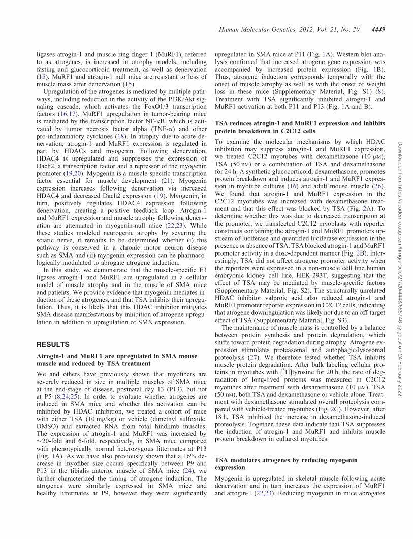
We and others have previously shown that myofibers areseverely reduced in size in multiple muscles of SMA miceat the end-stage of disease, postnatal day 13 (P13), but notat P5 (8,24,25). In order to evaluate whether atrogenes areinduced in SMA mice and whether this activation can beinhibited by HDAC inhibition, we treated a cohort of micewith either TSA (10 mg/kg) or vehicle (dimethyl sulfoxide,DMSO) and extracted RNA from total hindlimb muscles.The expression of atrogin-1 and MuRF1 was increased by�20-fold and 6-fold, respectively, in SMA mice comparedwith phenotypically normal heterozygous littermates at P13(Fig. 1A). As we have also previously shown that a 16% de-crease in myofiber size occurs specifically between P9 andP13 in the tibialis anterior muscle of SMA mice (24), wefurther characterized the timing of atrogene induction. Theatrogenes were similarly expressed in SMA mice andhealthy littermates at P9, however they were significantly
upregulated in SMA mice at P11 (Fig. 1A). Western blot ana-lysis confirmed that increased atrogene gene expression wasaccompanied by increased protein expression (Fig. 1B).Thus, atrogene induction corresponds temporally with theonset of muscle atrophy as well as with the onset of weightloss in these mice (Supplementary Material, Fig. S1) (8).Treatment with TSA significantly inhibited atrogin-1 andMuRF1 activation at both P11 and P13 (Fig. 1A and B).
TSA reduces atrogin-1 and MuRF1 expression and inhibitsprotein breakdown in C2C12 cells
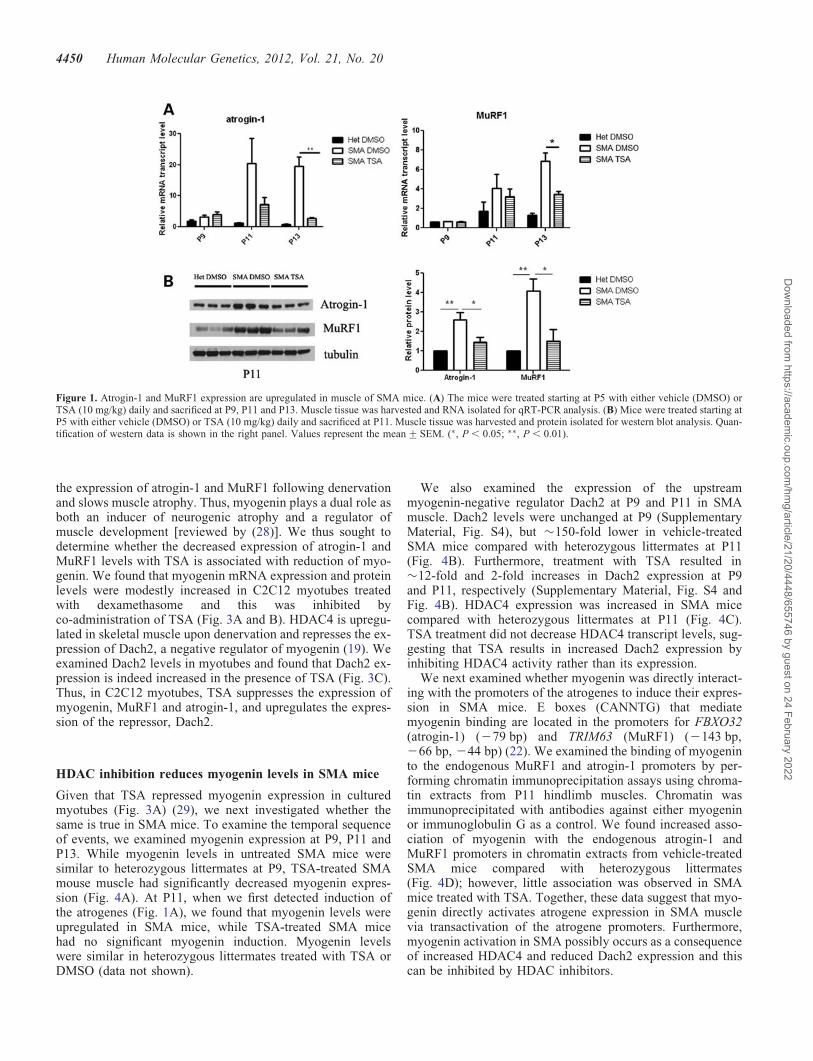
To examine the molecular mechanisms by which HDACinhibition may suppress atrogin-1 and MuRF1 expression,we treated C2C12 myotubes with dexamethasone (10 mM),TSA (50 nM) or a combination of TSA and dexamethasonefor 24 h. A synthetic glucocorticoid, dexamethasone, promotesprotein breakdown and induces atrogin-1 and MuRF1 expres-sion in myotube cultures (16) and adult mouse muscle (26).We found that atrogin-1 and MuRF1 expression in theC2C12 myotubes was increased with dexamethasone treat-ment and that this effect was blocked by TSA (Fig. 2A). Todetermine whether this was due to decreased transcription atthe promoter, we transfected C2C12 myoblasts with reporterconstructs containing the atrogin-1 and MuRF1 promoters up-stream of luciferase and quantified luciferase expression in thepresence or absence of TSA. TSA blocked atrogin-1 and MuRF1promoter activity in a dose-dependent manner (Fig. 2B). Inter-estingly, TSA did not affect atrogene promoter activity whenthe reporters were expressed in a non-muscle cell line humanembryonic kidney cell line, HEK-293T, suggesting that theeffect of TSA may be mediated by muscle-specific factors(Supplementary Material, Fig. S2). The structurally unrelatedHDAC inhibitor valproic acid also reduced atrogin-1 andMuRF1 promoter reporter expression in C2C12 cells, indicatingthat atrogene downregulation was likely not due to an off-targeteffect of TSA (Supplementary Material, Fig. S3).
The maintenance of muscle mass is controlled by a balancebetween protein synthesis and protein degradation, whichshifts toward protein degradation during atrophy. Atrogene ex-pression stimulates proteasomal and autophagic/lysosomalproteolysis (27). We therefore tested whether TSA inhibitsmuscle protein degradation. After bulk labeling cellular pro-teins in myotubes with [3H]tyrosine for 20 h, the rate of deg-radation of long-lived proteins was measured in C2C12myotubes after treatment with dexamethasone (10 mM), TSA(50 nM), both TSA and dexamethasone or vehicle alone. Treat-ment with dexamethasone stimulated overall proteolysis com-pared with vehicle-treated myotubes (Fig. 2C). However, after18 h, TSA inhibited the increase in dexamethasone-inducedproteolysis. Together, these data indicate that TSA suppressesthe induction of atrogin-1 and MuRF1 and inhibits muscleprotein breakdown in cultured myotubes.
TSA modulates atrogenes by reducing myogeninexpression
Myogenin is upregulated in skeletal muscle following acutedenervation and in turn increases the expression of MuRF1and atrogin-1 (22,23). Reducing myogenin in mice abrogates
Human Molecular Genetics, 2012, Vol. 21, No. 20 4449
Dow
nloaded from https://academ
ic.oup.com/hm
g/article/21/20/4448/655746 by guest on 24 February 2022
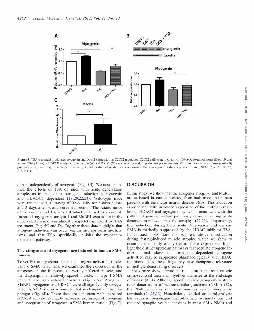
the expression of atrogin-1 and MuRF1 following denervationand slows muscle atrophy. Thus, myogenin plays a dual role asboth an inducer of neurogenic atrophy and a regulator ofmuscle development [reviewed by (28)]. We thus sought todetermine whether the decreased expression of atrogin-1 andMuRF1 levels with TSA is associated with reduction of myo-genin. We found that myogenin mRNA expression and proteinlevels were modestly increased in C2C12 myotubes treatedwith dexamethasome and this was inhibited byco-administration of TSA (Fig. 3A and B). HDAC4 is upregu-lated in skeletal muscle upon denervation and represses the ex-pression of Dach2, a negative regulator of myogenin (19). Weexamined Dach2 levels in myotubes and found that Dach2 ex-pression is indeed increased in the presence of TSA (Fig. 3C).Thus, in C2C12 myotubes, TSA suppresses the expression ofmyogenin, MuRF1 and atrogin-1, and upregulates the expres-sion of the repressor, Dach2.
HDAC inhibition reduces myogenin levels in SMA mice
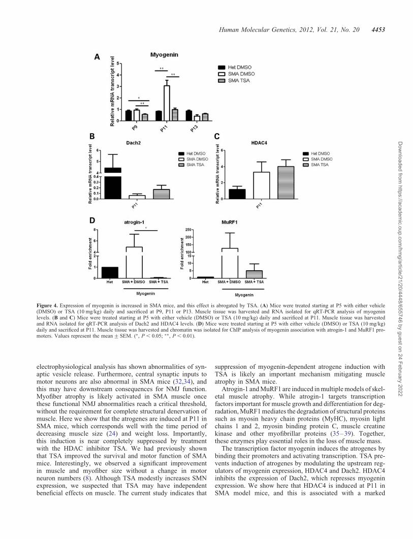
Given that TSA repressed myogenin expression in culturedmyotubes (Fig. 3A) (29), we next investigated whether thesame is true in SMA mice. To examine the temporal sequenceof events, we examined myogenin expression at P9, P11 andP13. While myogenin levels in untreated SMA mice weresimilar to heterozygous littermates at P9, TSA-treated SMAmouse muscle had significantly decreased myogenin expres-sion (Fig. 4A). At P11, when we first detected induction ofthe atrogenes (Fig. 1A), we found that myogenin levels wereupregulated in SMA mice, while TSA-treated SMA micehad no significant myogenin induction. Myogenin levelswere similar in heterozygous littermates treated with TSA orDMSO (data not shown).
We also examined the expression of the upstreammyogenin-negative regulator Dach2 at P9 and P11 in SMAmuscle. Dach2 levels were unchanged at P9 (SupplementaryMaterial, Fig. S4), but �150-fold lower in vehicle-treatedSMA mice compared with heterozygous littermates at P11(Fig. 4B). Furthermore, treatment with TSA resulted in�12-fold and 2-fold increases in Dach2 expression at P9and P11, respectively (Supplementary Material, Fig. S4 andFig. 4B). HDAC4 expression was increased in SMA micecompared with heterozygous littermates at P11 (Fig. 4C).TSA treatment did not decrease HDAC4 transcript levels, sug-gesting that TSA results in increased Dach2 expression byinhibiting HDAC4 activity rather than its expression.
We next examined whether myogenin was directly interact-ing with the promoters of the atrogenes to induce their expres-sion in SMA mice. E boxes (CANNTG) that mediatemyogenin binding are located in the promoters for FBXO32(atrogin-1) (279 bp) and TRIM63 (MuRF1) (2143 bp,266 bp, 244 bp) (22). We examined the binding of myogeninto the endogenous MuRF1 and atrogin-1 promoters by per-forming chromatin immunoprecipitation assays using chroma-tin extracts from P11 hindlimb muscles. Chromatin wasimmunoprecipitated with antibodies against either myogeninor immunoglobulin G as a control. We found increased asso-ciation of myogenin with the endogenous atrogin-1 andMuRF1 promoters in chromatin extracts from vehicle-treatedSMA mice compared with heterozygous littermates(Fig. 4D); however, little association was observed in SMAmice treated with TSA. Together, these data suggest that myo-genin directly activates atrogene expression in SMA musclevia transactivation of the atrogene promoters. Furthermore,myogenin activation in SMA possibly occurs as a consequenceof increased HDAC4 and reduced Dach2 expression and thiscan be inhibited by HDAC inhibitors.
Figure 1. Atrogin-1 and MuRF1 expression are upregulated in muscle of SMA mice. (A) The mice were treated starting at P5 with either vehicle (DMSO) orTSA (10 mg/kg) daily and sacrificed at P9, P11 and P13. Muscle tissue was harvested and RNA isolated for qRT-PCR analysis. (B) Mice were treated starting atP5 with either vehicle (DMSO) or TSA (10 mg/kg) daily and sacrificed at P11. Muscle tissue was harvested and protein isolated for western blot analysis. Quan-tification of western data is shown in the right panel. Values represent the mean+SEM. (∗, P , 0.05; ∗∗, P , 0.01).
4450 Human Molecular Genetics, 2012, Vol. 21, No. 20
Dow
nloaded from https://academ
ic.oup.com/hm
g/article/21/20/4448/655746 by guest on 24 February 2022
HDAC inhibition suppresses myogenin-dependent, but notmyogenin-independent atrogene induction
Because the atrogenes can be induced by fasting, and SMAmice likely have inadequate nutrition at the end-stage ofdisease (11,30,31), we examined whether TSA is capableof repressing atrogene induction that occurs during fasting.
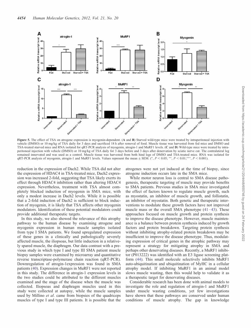
Wild-type mice were treated with 10 mg/kg of TSA dailyfor 3 days, and then food was removed and the animalswere sacrificed 18 h later. Atrogin-1 and MuRF1 expressionwas markedly increased in the fasting mice, but this effectwas not blocked by TSA treatment (Fig. 5A). Importantly,myogenin expression was also not induced in the fastingmice, indicating that during starvation atrogene activation
Figure 2. TSA inhibits atrogin-1 and MuRF1 induction and muscle protein breakdown in C2C12 myotubes. (A) qRT-PCR analysis of atrogin-1 and MuRF1expression in C2C12 cells after 24-h treatment with dexamethasone (Dex, 10 mM), and/or TSA (50 nM). The expression level is represented relative to thevehicle control (n ¼ 4, experiments per treatment). (B) Transfection of C2C12 myotubes with either an atrogin-1 or a MuRF1 luciferase reporter and treatmentwith DMSO, 10 nM, 50 nM, 100 nM or 200 nM TSA. Luciferase activity is represented relative to DMSO control (n ¼ 4, experiments per dose). (C) The rate ofprotein breakdown in C2C12 myotubes labeled with L-[3,5-3H]tyrosine. Myotubes were treated with vehicle, Dex (10 mM) and/or TSA (50 nM), and release ofL-[3,5-3H]tyrosine from cell proteins was used as an index of the breakdown of long-lived myofibrillar proteins. Dex treatment compared with Dex and TSAtreatment (n ¼ 3, experiments per treatment). Values represent the mean+SEM (∗, P , 0.05; ∗∗, P , 0.01;∗∗∗, P , 0.001).
Human Molecular Genetics, 2012, Vol. 21, No. 20 4451
Dow
nloaded from https://academ
ic.oup.com/hm
g/article/21/20/4448/655746 by guest on 24 February 2022
occurs independently of myogenin (Fig. 5B). We next exam-ined the effects of TSA on mice with acute denervationatrophy as in this context atrogene induction is myogeninand HDAC4/5 dependent (19,20,22,23). Wild-type micewere treated with 10 mg/kg of TSA daily for 3 days beforeand 3 days after sciatic nerve transection. The sciatic nerveof the conralateral leg was left intact and used as a control.Increased myogenin, atrogin-1 and MuRF1 expression in thedenervated muscle was almost completely inhibited by TSAtreatment (Fig. 5C and D). Together these data highlight thatatrogene induction can occur via distinct upstream mechan-isms, and that TSA specifically inhibits the myogenin-dependent pathway.
The atrogenes and myogenin are induced in human SMAmuscle
To verify that myogenin-dependent atrogene activation is rele-vant to SMA in humans, we examined the expression of theatrogenes in the iliopsoas, a severely affected muscle, andthe diaphragm, a relatively spared muscle, in type I SMApatients and age-matched controls (Fig. 6A). Atrogin-1,MuRF1, myogenin and HDAC4 were all significantly upregu-lated in SMA iliopsoas muscle, but unchanged in the dia-phragm (Fig. 6B). These data are consistent with increasedHDAC4 activity leading to increased expression of myogeninand upregulation of atrogenes in SMA human muscle (Fig. 7).
DISCUSSION
In this study, we show that the atrogenes atrogin-1 and MuRF1are activated in muscle isolated from both mice and humanpatients with the motor neuron disease SMA. This inductionis associated with increased expression of the upstream regu-lators, HDAC4 and myogenin, which is consistent with thepattern of gene activation previously observed during acutedenervation-induced muscle atrophy (22,23). Importantly,this induction during both acute denervation and chronicSMA is markedly suppressed by the HDAC inhibitor TSA.In contrast, TSA does not suppress atrogene activationduring fasting-induced muscle atrophy, which we show tooccur independently of myogenin. These experiments high-light the distinct upstream pathways that regulate atrogene in-duction and show that myogenin-dependent atrogeneactivation may be suppressed pharmacologically with HDACinhibitors. Thus, these drugs may have therapeutic relevancein multiple denervating disorders.
SMA mice show a profound reduction in the total musclecross-sectional area and myofiber diameter at the end-stageof disease (8,24). Although specific muscle groups show struc-tural denervation of neuromuscular junctions (NMJs) (32),the NMJ endplates of many muscles retain presynapticterminals (24,25,33). Nonetheless, detailed structural analysishas revealed presynaptic neurofilament accumulations andreduced synaptic vesicle densities in most SMA NMJs and
Figure 3. TSA treatment modulates myogenin and Dach2 expression in C2C12 myotubes. C2C12 cells were treated with DMSO, dexamethasone (Dex, 10 mM)and/or TSA (50 nM). qRT-PCR analysis of myogenin (A) and Dach2 (C) expression (n ¼ 4, experiments per treatment). Western blot analysis of myogenin (B)protein levels (n ¼ 3, experiments per treatment). Quantification of western data is shown in the lower panel. Values represent mean+SEM. (∗, P , 0.05; ∗∗,P , 0.01).
4452 Human Molecular Genetics, 2012, Vol. 21, No. 20
Dow
nloaded from https://academ
ic.oup.com/hm
g/article/21/20/4448/655746 by guest on 24 February 2022
electrophysiological analysis has shown abnormalities of syn-aptic vesicle release. Furthermore, central synaptic inputs tomotor neurons are also abnormal in SMA mice (32,34), andthis may have downstream consequences for NMJ function.Myofiber atrophy is likely activated in SMA muscle oncethese functional NMJ abnormalities reach a critical threshold,without the requirement for complete structural denervation ofmuscle. Here we show that the atrogenes are induced at P11 inSMA mice, which corresponds well with the time period ofdecreasing muscle size (24) and weight loss. Importantly,this induction is near completely suppressed by treatmentwith the HDAC inhibitor TSA. We had previously shownthat TSA improved the survival and motor function of SMAmice. Interestingly, we observed a significant improvementin muscle and myofiber size without a change in motorneuron numbers (8). Although TSA modestly increases SMNexpression, we suspected that TSA may have independentbeneficial effects on muscle. The current study indicates that
suppression of myogenin-dependent atrogene induction withTSA is likely an important mechanism mitigating muscleatrophy in SMA mice.
Atrogin-1 and MuRF1 are induced in multiple models of skel-etal muscle atrophy. While atrogin-1 targets transcriptionfactors important for muscle growth and differentiation for deg-radation, MuRF1 mediates the degradation of structural proteinssuch as myosin heavy chain proteins (MyHC), myosin lightchains 1 and 2, myosin binding protein C, muscle creatinekinase and other myofibrillar proteins (35–39). Together,these enzymes play essential roles in the loss of muscle mass.
The transcription factor myogenin induces the atrogenes bybinding their promoters and activating transcription. TSA pre-vents induction of atrogenes by modulating the upstream reg-ulators of myogenin expression, HDAC4 and Dach2. HDAC4inhibits the expression of Dach2, which represses myogeninexpression. We show here that HDAC4 is induced at P11 inSMA model mice, and this is associated with a marked
Figure 4. Expression of myogenin is increased in SMA mice, and this effect is abrogated by TSA. (A) Mice were treated starting at P5 with either vehicle(DMSO) or TSA (10 mg/kg) daily and sacrificed at P9, P11 or P13. Muscle tissue was harvested and RNA isolated for qRT-PCR analysis of myogeninlevels. (B and C) Mice were treated starting at P5 with either vehicle (DMSO) or TSA (10 mg/kg) daily and sacrificed at P11. Muscle tissue was harvestedand RNA isolated for qRT-PCR analysis of Dach2 and HDAC4 levels. (D) Mice were treated starting at P5 with either vehicle (DMSO) or TSA (10 mg/kg)daily and sacrificed at P11. Muscle tissue was harvested and chromatin was isolated for ChIP analysis of myogenin association with atrogin-1 and MuRF1 pro-moters. Values represent the mean+SEM. (∗, P , 0.05; ∗∗, P , 0.01).
Human Molecular Genetics, 2012, Vol. 21, No. 20 4453
Dow
nloaded from https://academ
ic.oup.com/hm
g/article/21/20/4448/655746 by guest on 24 February 2022
reduction in the expression of Dach2. While TSA did not alterthe expression of HDAC4 in TSA-treated mice, Dach2 expres-sion was increased 2-fold, suggesting that TSA likely exerts itseffect through HDAC4 inhibition rather than altering HDAC4expression. Nevertheless, treatment with TSA almost com-pletely blocked induction of myogenin in SMA mice, withonly a modest increase in Dach2 levels. While it is possiblethat a 2-fold induction of Dach2 is sufficient to block induc-tion of myogenin, it is likely that TSA affects other myogeninmodulators. Identification of these potential modulators couldprovide additional therapeutic targets.
In this study, we also showed the relevance of this atrophypathway to the human disease by examining atrogene andmyogenin expression in human muscle samples isolatedfrom type I SMA patients. We found upregulated expressionof these genes in a clinically and pathologically severelyaffected muscle, the iliopsoas, but little induction in a relative-ly spared muscle, the diaphragm. Our data contrast with a pre-vious study in which type I and type III SMA patient musclebiopsy samples were examined by microarray and quantitativereverse transcriptase-polymerase chain reaction (qRT-PCR).Downregulation of atrogin-1expression was found in SMApatients (40). Expression changes in MuRF1 were not reportedin this study. The difference in atrogin-1 expression levels inthe two studies could be attributed to the different musclesexamined and the stage of the disease when the muscle wascollected. Iliopsoas and diaphragm muscles used in thisstudy were collected at autopsy, while the muscle samplesused by Millino et al. came from biopsies of the quadricepsmuscles of type I and type III patients. It is possible that the
atrogenes were not yet induced at the time of biopsy, sinceatrogene induction occurs late in the SMA mice.
While motor neuron loss is central to SMA disease patho-genesis, therapeutic targeting of muscle may provide benefitsto SMA patients. Previous studies in SMA mice investigatedthe effect of factors known to regulate muscle growth, suchas myostatin, an inhibitor of muscle growth, and follistatin,an inhibitor of myostatin. Both genetic and therapeutic inter-ventions to modulate these growth factors have not improvedmuscle mass or the overall SMA phenotype (41–43). Theseapproaches focused on muscle growth and protein synthesisto improve the disease phenotype. However, muscle mainten-ance is a balance between protein synthesis induced by growthfactors and protein breakdown. Targeting protein synthesiswithout inhibiting atrophy-related protein breakdown may beinsufficient to improve the disease phenotype. Thus, modulat-ing expression of critical genes in the atrophic pathway mayrepresent a strategy for mitigating atrophy in SMA andperhaps other denervating diseases. Recently, a MuRF1 inhibi-tor (P013222) was identified with an E3 ligase screening plat-form (44). This small molecule selectively inhibits MuRF1auto-ubiquitination and ubiquitination of MyHC in a cellularatrophy model. If inhibiting MuRF1 in an animal modelslows muscle wasting, then this would help to validate it asa therapeutic target for denervating diseases.
Considerable research has been done with animal models toinvestigate the role and regulation of atrogin-1 and MuRF1under muscle wasting conditions, yet few investigationshave shown that these pathways are conserved under humanconditions of muscle atrophy. The gap in knowledge
Figure 5. The effect of TSA on atrogene expression is myogenin-dependent. (A and B) Starved wild-type mice were treated by intraperitoneal injection withvehicle (DMSO) or 10 mg/kg of TSA daily for 3 days and sacrificed 18 h after removal of food. Muscle tissue was harvested from fed mice and DMSO andTSA-treated starved mice and RNA isolated for qRT-PCR analysis of myogenin, atrogin-1 and MuRF1 levels. (C and D) Wild-type mice were treated by intra-peritoneal injection with vehicle (DMSO) or 10 mg/kg of TSA daily for 3 days before and 3 days after denervation by sciatic nerve cut. The contralateral legremained innervated and was used as a control. Muscle tissue was harvested from both hind legs of DMSO and TSA-treated mice. RNA was isolated forqRT-PCR analysis of myogenin, atrogin-1 and MuRF1 levels. Values represent the mean+SEM. (∗, P , 0.05; ∗∗, P , 0.01;∗∗∗, P , 0.001).
4454 Human Molecular Genetics, 2012, Vol. 21, No. 20
Dow
nloaded from https://academ
ic.oup.com/hm
g/article/21/20/4448/655746 by guest on 24 February 2022
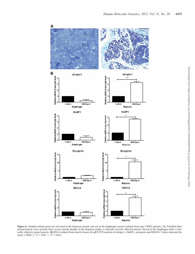
Figure 6. Atrophy-related genes are activated in the iliopsoas muscle, but not in the diaphragm muscle isolated from type I SMA patients. (A) Toluidine-bluestained muscle cross sections show severe muscle atrophy in the iliopsoas (right), a clinically severely affected muscle, but not in the diaphragm (left), a clin-ically relatively spared muscle. (B) RNA isolated from muscle tissues for qRT-PCR analysis of atrogin-1, MuRF1, myogenin and HDAC4. Values represent themean+SEM. (∗, P , 0.05; ∗∗, P , 0.01).
Human Molecular Genetics, 2012, Vol. 21, No. 20 4455
Dow
nloaded from https://academ
ic.oup.com/hm
g/article/21/20/4448/655746 by guest on 24 February 2022
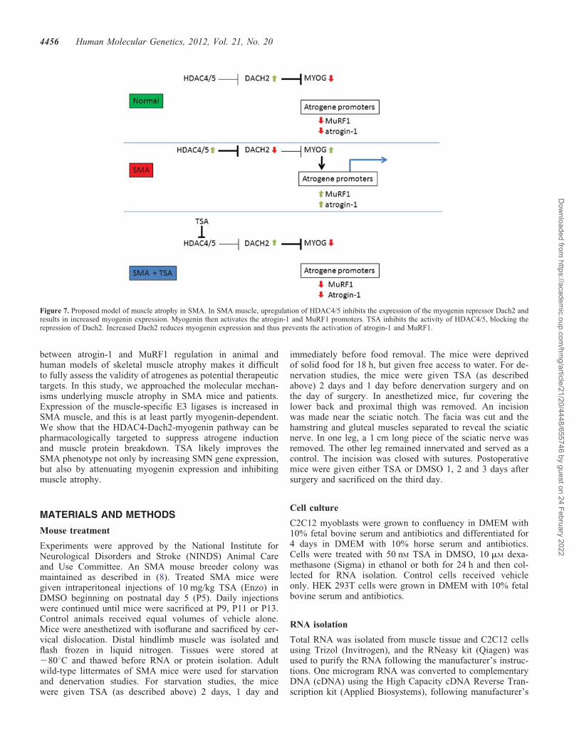
between atrogin-1 and MuRF1 regulation in animal andhuman models of skeletal muscle atrophy makes it difficultto fully assess the validity of atrogenes as potential therapeutictargets. In this study, we approached the molecular mechan-isms underlying muscle atrophy in SMA mice and patients.Expression of the muscle-specific E3 ligases is increased inSMA muscle, and this is at least partly myogenin-dependent.We show that the HDAC4-Dach2-myogenin pathway can bepharmacologically targeted to suppress atrogene inductionand muscle protein breakdown. TSA likely improves theSMA phenotype not only by increasing SMN gene expression,but also by attenuating myogenin expression and inhibitingmuscle atrophy.
MATERIALS AND METHODS
Mouse treatment
Experiments were approved by the National Institute forNeurological Disorders and Stroke (NINDS) Animal Careand Use Committee. An SMA mouse breeder colony wasmaintained as described in (8). Treated SMA mice weregiven intraperitoneal injections of 10 mg/kg TSA (Enzo) inDMSO beginning on postnatal day 5 (P5). Daily injectionswere continued until mice were sacrificed at P9, P11 or P13.Control animals received equal volumes of vehicle alone.Mice were anesthetized with isoflurane and sacrificed by cer-vical dislocation. Distal hindlimb muscle was isolated andflash frozen in liquid nitrogen. Tissues were stored at2808C and thawed before RNA or protein isolation. Adultwild-type littermates of SMA mice were used for starvationand denervation studies. For starvation studies, the micewere given TSA (as described above) 2 days, 1 day and
immediately before food removal. The mice were deprivedof solid food for 18 h, but given free access to water. For de-nervation studies, the mice were given TSA (as describedabove) 2 days and 1 day before denervation surgery and onthe day of surgery. In anesthetized mice, fur covering thelower back and proximal thigh was removed. An incisionwas made near the sciatic notch. The facia was cut and thehamstring and gluteal muscles separated to reveal the sciaticnerve. In one leg, a 1 cm long piece of the sciatic nerve wasremoved. The other leg remained innervated and served as acontrol. The incision was closed with sutures. Postoperativemice were given either TSA or DMSO 1, 2 and 3 days aftersurgery and sacrificed on the third day.
Cell culture
C2C12 myoblasts were grown to confluency in DMEM with10% fetal bovine serum and antibiotics and differentiated for4 days in DMEM with 10% horse serum and antibiotics.Cells were treated with 50 nM TSA in DMSO, 10 mM dexa-methasone (Sigma) in ethanol or both for 24 h and then col-lected for RNA isolation. Control cells received vehicleonly. HEK 293T cells were grown in DMEM with 10% fetalbovine serum and antibiotics.
RNA isolation
Total RNA was isolated from muscle tissue and C2C12 cellsusing Trizol (Invitrogen), and the RNeasy kit (Qiagen) wasused to purify the RNA following the manufacturer’s instruc-tions. One microgram RNA was converted to complementaryDNA (cDNA) using the High Capacity cDNA Reverse Tran-scription kit (Applied Biosystems), following manufacturer’s
Figure 7. Proposed model of muscle atrophy in SMA. In SMA muscle, upregulation of HDAC4/5 inhibits the expression of the myogenin repressor Dach2 andresults in increased myogenin expression. Myogenin then activates the atrogin-1 and MuRF1 promoters. TSA inhibits the activity of HDAC4/5, blocking therepression of Dach2. Increased Dach2 reduces myogenin expression and thus prevents the activation of atrogin-1 and MuRF1.
4456 Human Molecular Genetics, 2012, Vol. 21, No. 20
Dow
nloaded from https://academ
ic.oup.com/hm
g/article/21/20/4448/655746 by guest on 24 February 2022
instructions. Gene expression was determined by qRT-PCRusing TaqMan and SYBR Green reagents (Applied Biosys-tems). Primers are listed in the Supplementary Table. Tran-script levels were quantified by threshold cycle values usingphosphoglycerate kinase 1 as a control. For each gene, thevalues were normalized to vehicle-treated, unaffectedanimals or cells. For patient samples, the values were normal-ized to unaffected controls.
Western blot
C2C12 cells were lysed in 1% NP-40, 50 mM Tris–HCl (pH8), 150 mM NaCl and protease inhibitor cocktail (Roche) onice for 10 min. The lysates were centrifuged at 48C for10 min and the supernatants were collected. Protein concentra-tion was determined using the BCA Protein Assay kit (Pierce)according to the manufacturer’s protocol. Protein extractionfrom hindlimb muscle was done as previously described(10). Protein lysates (100 mg for C2C12 cells, 50 mg formouse muscle) were resolved by SDS PAGE (10%) and trans-ferred to polyvinylidene difluoride membranes. The mem-branes were blocked in 5% milk and probed with mouseanti-myogenin (1:500; clone F5D, Developmental StudiesHybridoma Bank), rabbit anti-atrogin 1 (1:1000, ECM Bios-ciences), rabbit anti-MuRF1 (1:500, Santa Cruz Biotechnol-ogy) and mouse anti-alpha tubulin (1:3000, Sigma).
Luciferase assay
C2C12 cell transfections were performed using GenJet Ver. III(SignaGen) following the manufacturer’s instructions. HEK293T cell transfections were performed using FuGENE(Roche) following the manufacturer’s directions. The reporterplasmid, either atrogin-1 (Dr Stewart Lecker, Beth Israel Dea-coness Medical Center) or MuRF1 (Dr. Eric Olson, Universityof Texas Southwestern Medical Center) was used in a ratio of5:1 with a pGL4-TK Renilla luciferase plasmid (Promega).Twenty-four hours after transfection, the cells were treatedfor an additional 24 h with 10 nM, 50 nM, 100 nM, 200 nM or500 nM TSA in DMSO or 0.5 mM, 2.5 mM or 12.5 mM valproicacid (Sigma) in water. Control cells received vehicle only.Following treatment, the cells were collected and luciferaseassays were performed using the Dual Luciferase ReporterAssay system (Promega) following the manufacturer’s instruc-tions.
Protein breakdown assay
C2C12 cells were grown for 3 days in differentiation media,DMEM, with 10% horse serum and antibiotics. The mediawas supplemented with L-[3,5-3H]tyrosine (5 mCi/ml, Perki-nElmer) for an additional 22 h. The cells were then washedwith phosphate-buffered saline (PBS) and returned to differen-tiation media with 50 nM TSA in DMSO, 10 mM dexametha-sone in ethanol or both. Control cells received vehicle only.200 ml aliquots of media were taken at 0, 1, 3, 9, 18 and24 h for quantification of L-[3,5-3H]tyrosine release. Proteinprecipitation and radioactivity measurement were done as pre-viously described (45).
Chromatin immunoprecipitation
Flash frozen hindlimb muscle was minced on ice and fixed in1% formaldehyde/PBS for 20 min and then quenched withglycine. Samples were centrifuged and washed twice in PBSwith protease inhibitors. The cells were lysed and chromatinisolated using the EZ Magna ChIP A kit (Millipore) followingthe manufacturer’s directions. Anti-myogenin antibody(M-225x, Santa Cruz) was used to immunoprecipitate thechromatin fragments. Primers for amplifying atrogin-1 andMuRF1 promoters were described previously (22).
Patient tissue
Diaphragm and iliopsoas muscle tissues were obtained fromautopsies of type I SMA patients (n ¼ 3) or age-matched con-trols (n ¼ 2) (NICHD Brain and Tissue Bank for Developmen-tal Disorders at the University of Maryland, Baltimore, MD,USA). Cross sections of muscle were stained with toluidine-blue.
Statistical analysis
Data are presented as the mean + the standard error of themean. t-Tests were performed using GraphPad Prism 5 soft-ware (GraphPad Software) with a significance set at P , 0.05.
SUPPLEMENTARY MATERIAL
Supplementary material is available at HMG online.
ACKNOWLEDGEMENTS
We thank Dr Stewart Lecker (Beth Israel Deaconess MedicalCenter) and Dr Eric Olson (University of Texas SouthwesternMedical Center) for providing the atrogin-1 and MuRF1reporter plasmids and Dr Peter Macpherson (University ofMichigan) for the Dach2 primer sequences. Human tissuewas obtained from the NICHD brain and Tissue Bank forDevelopmental Disorders at the University of Maryland, Bal-timore, MD. This work was supported by intramural NINDSresearch funds. C.J.S. was supported by NINDS grantR01NS062869 and funds from the Spinal Muscular AtrophyFoundation.
Conflict of Interest statement. None declared.
REFERENCES
1. Lefebvre, S., Burglen, L., Reboullet, S., Clermont, O., Burlet, P., Viollet,L., Benichou, B., Cruaud, C., Millasseau, P., Zeviani, M. et al. (1995)Identification of spinal muscular atrophy-determining gene. Cell, 80,155–165.
2. Monani, U.R., Lorson, C.L., Parsons, D.W., Prior, T.W., Androphy, E.J.,Burghes, A.H. and McPherson, J.D. (1999) A single nucleotide differencethat alters splicing patterns distinguishes the SMA gene SMN1 from thecopy gene SMN2. Hum. Mol. Genet., 8, 1177–1183.
3. Lorson, C.L., Hahnen, E., Androphy, E.J. and Wirth, B. (1999) A singlenucleotide in the SMN gene regulates splicing and is responsible forspinal muscular atrophy. Proc. Natl Acad. Sci. U S A, 96, 6307–6311.
Human Molecular Genetics, 2012, Vol. 21, No. 20 4457
Dow
nloaded from https://academ
ic.oup.com/hm
g/article/21/20/4448/655746 by guest on 24 February 2022

4. Hsieh-Li, H.M., Chang, J.G., Jong, Y.J., Wu, M.H., Wang, N.M., Tsai,C.H. and Li, H. (2000) A mouse model for spinal muscular atrophy. Nat.
Genet., 24, 66–70.5. Le, T.T., Coovert, D.D., Monani, U.R., Morris, G.E. and Burghes, A.H.
(2000) The survival motor neuron (SMN) protein: effect of exon loss andmutation on protein localization. Neurogenetics, 3, 7–16.
6. Prior, T.W., Swoboda, K.J., Scott, H.D. and Hejmanowski, A.Q. (2004)Homozygous SMN1 deletions in unaffected family members andmodification of the phenotype by SMN2. Am. J. Med. Genet. A, 130A,307–310.
7. Le, T.T., Pham, L.T., Butchbach, M.E., Zhang, H.L., Monani, U.R.,Coovert, D.D., Gavrilina, T.O., Xing, L., Bassell, G.J. and Burghes, A.H.(2005) SMNDelta7, the major product of the centromeric survival motorneuron (SMN2) gene, extends survival in mice with spinal muscularatrophy and associates with full-length SMN. Hum. Mol. Genet., 14,845–857.
8. Avila, A.M., Burnett, B.G., Taye, A.A., Gabanella, F., Knight, M.A.,Hartenstein, P., Cizman, Z., Di Prospero, N.A., Pellizzoni, L., Fischbeck,K.H. et al. (2007) Trichostatin A increases SMN expression and survivalin a mouse model of spinal muscular atrophy. J. Clin. Invest., 117, 659–671.
9. Yoo, Y.E. and Ko, C.P. (2011) Treatment with trichostatin A initiatedafter disease onset delays disease progression and increases survival in amouse model of amyotrophic lateral sclerosis. Exp. Neurol., 231,147–159.
10. Kwon, D.Y., Motley, W.W., Fischbeck, K.H. and Burnett, B.G. (2011)Increasing expression and decreasing degradation of SMN amelioratethe spinal muscular atrophy phenotype in mice. Hum. Mol. Genet., 20,3667–3677.
11. Narver, H.L., Kong, L., Burnett, B.G., Choe, D.W., Bosch-Marce, M.,Taye, A.A., Eckhaus, M.A. and Sumner, C.J. (2008) Sustainedimprovement of spinal muscular atrophy mice treated with trichostatin Aplus nutrition. Ann. Neurol., 64, 465–470.
12. Lecker, S.H., Solomon, V., Mitch, W.E. and Goldberg, A.L. (1999)Muscle protein breakdown and the critical role of theubiquitin-proteasome pathway in normal and disease states. J. Nutr., 129,227S–237S.
13. Tischler, M.E., Rosenberg, S., Satarug, S., Henriksen, E.J., Kirby, C.R.,Tome, M. and Chase, P. (1990) Different mechanisms of increasedproteolysis in atrophy induced by denervation or unweighting of rat soleusmuscle. Metabolism, 39, 756–763.
14. Solomon, V., Baracos, V., Sarraf, P. and Goldberg, A.L. (1998) Rates ofubiquitin conjugation increase when muscles atrophy, largely throughactivation of the N-end rule pathway. Proc. Natl Acad. Sci. U S A., 95,12602–12607.
15. Bodine, S.C., Latres, E., Baumhueter, S., Lai, V.K., Nunez, L., Clarke,B.A., Poueymirou, W.T., Panaro, F.J., Na, E., Dharmarajan, K. et al.
(2001) Identification of ubiquitin ligases required for skeletal muscleatrophy. Science, 294, 1704–1708.
16. Sandri, M., Sandri, C., Gilbert, A., Skurk, C., Calabria, E., Picard, A.,Walsh, K., Schiaffino, S., Lecker, S.H. and Goldberg, A.L. (2004) Foxotranscription factors induce the atrophy-related ubiquitin ligase atrogin-1and cause skeletal muscle atrophy. Cell, 117, 399–412.
17. Stitt, T.N., Drujan, D., Clarke, B.A., Panaro, F., Timofeyva, Y., Kline,W.O., Gonzalez, M., Yancopoulos, G.D. and Glass, D.J. (2004) TheIGF-1/PI3K/Akt pathway prevents expression of muscle atrophy-inducedubiquitin ligases by inhibiting FOXO transcription factors. Mol. Cell, 14,395–403.
18. Cai, D., Frantz, J.D., Tawa, N.E. Jr., Melendez, P.A., Oh, B.C., Lidov,H.G., Hasselgren, P.O., Frontera, W.R., Lee, J., Glass, D.J. et al. (2004)IKKbeta/NF-kappaB activation causes severe muscle wasting in mice.Cell, 119, 285–298.
19. Tang, H., Macpherson, P., Marvin, M., Meadows, E., Klein, W.H., Yang,X.J. and Goldman, D. (2009) A histone deacetylase 4/myogenin positivefeedback loop coordinates denervation-dependent gene induction andsuppression. Mol. Biol. Cell, 20, 1120–1131.
20. Tang, H. and Goldman, D. (2006) Activity-dependent gene regulation inskeletal muscle is mediated by a histone deacetylase(HDAC)-Dach2-myogenin signal transduction cascade. Proc. Natl Acad.
Sci. U S A, 103, 16977–16982.
21. Hasty, P., Bradley, A., Morris, J.H., Edmondson, D.G., Venuti, J.M.,Olson, E.N. and Klein, W.H. (1993) Muscle deficiency and neonatal death
in mice with a targeted mutation in the myogenin gene. Nature, 364,501–506.
22. Moresi, V., Williams, A.H., Meadows, E., Flynn, J.M., Potthoff, M.J.,McAnally, J., Shelton, J.M., Backs, J., Klein, W.H., Richardson, J.A. et al.
(2010) Myogenin and class II HDACs control neurogenic muscle atrophyby inducing E3 ubiquitin ligases. Cell, 143, 35–45.
23. Macpherson, P.C., Wang, X. and Goldman, D. (2011) Myogenin regulatesdenervation-dependent muscle atrophy in mouse soleus muscle. J. Cell
Biochem., 112, 2149–2159.24. Kong, L., Wang, X., Choe, D.W., Polley, M., Burnett, B.G., Bosch-Marce,
M., Griffin, J.W., Rich, M.M. and Sumner, C.J. (2009) Impaired synapticvesicle release and immaturity of neuromuscular junctions in spinalmuscular atrophy mice. J. Neurosci., 29, 842–851.
25. Lee, Y.I., Mikesh, M., Smith, I., Rimer, M. and Thompson, W. (2011)Muscles in a mouse model of spinal muscular atrophy show profounddefects in neuromuscular development even in the absence of failure inneuromuscular transmission or loss of motor neurons. Dev. Biol., 356,432–444.
26. Baehr, L.M., Furlow, J.D. and Bodine, S.C. (2011) Muscle sparing inmuscle RING finger 1 null mice: response to synthetic glucocorticoids.J. Physiol., 589, 4759–4776.
27. Mammucari, C., Milan, G., Romanello, V., Masiero, E., Rudolf, R., DelPiccolo, P., Burden, S.J., Di Lisi, R., Sandri, C., Zhao, J. et al. (2007)FoxO3 controls autophagy in skeletal muscle in vivo. Cell Metab., 6,458–471.
28. Rudnicki, M.A., Le Grand, F., McKinnell, I. and Kuang, S. (2008) Themolecular regulation of muscle stem cell function. Cold Spring Harb.
Symp. Quant. Biol., 73, 323–331.29. Hagiwara, H., Saito, F., Masaki, T., Ikeda, M., Nakamura-Ohkuma, A.,
Shimizu, T. and Matsumura, K. (2011) Histone deacetylase inhibitortrichostatin A enhances myogenesis by coordinating muscle regulatoryfactors and myogenic repressors. Biochem. Biophys. Res. Commun., 414,826–831.
30. Bowerman, M., Swoboda, K.J., Michalski, J.-P., Wang, G.-S., Reeks, C.,Beauvais, A., Murphy, K., Woulfe, J., Screaton, R.A., Scott, F.W. et al.
(2012) Glucose metabolism and pancreatic defects in spinal muscularatrophy. Ann. Neurol., doi: 10.1002/ana.23582. [Epub ahead of print].
31. Butchbach, M.E., Rose, F.F. Jr., Rhoades, S., Marston, J., McCrone, J.T.,Sinnott, R. and Lorson, C.L. (2010) Effect of diet on the survival andphenotype of a mouse model for spinal muscular atrophy. Biochem.
Biophys. Res. Commun., 391, 835–840.
32. Ling, K.K., Gibbs, R.M., Feng, Z. and Ko, C.P. (2012) Severeneuromuscular denervation of clinically relevant muscles in amouse model of spinal muscular atrophy. Hum. Mol. Genet., 21,185–195.
33. Kariya, S., Park, G.H., Maeno-Hikichi, Y., Leykekhman, O., Lutz, C.,Arkovitz, M.S., Landmesser, L.T. and Monani, U.R. (2008) ReducedSMN protein impairs maturation of the neuromuscular junctions inmouse models of spinal muscular atrophy. Hum. Mol. Genet., 17,2552–2569.
34. Mentis, G.Z., Blivis, D., Liu, W., Drobac, E., Crowder, M.E., Kong, L.,Alvarez, F.J., Sumner, C.J. and O’Donovan, M.J. (2011) Early functionalimpairment of sensory-motor connectivity in a mouse model of spinalmuscular atrophy. Neuron, 69, 453–467.
35. Clarke, B.A., Drujan, D., Willis, M.S., Murphy, L.O., Corpina, R.A.,Burova, E., Rakhilin, S.V., Stitt, T.N., Patterson, C., Latres, E. et al.
(2007) The E3 Ligase MuRF1 degrades myosin heavy chain protein indexamethasone-treated skeletal muscle. Cell Metab., 6, 376–385.
36. Cohen, S., Brault, J.J., Gygi, S.P., Glass, D.J., Valenzuela, D.M., Gartner,C., Latres, E. and Goldberg, A.L. (2009) During muscle atrophy, thick,but not thin, filament components are degraded by MuRF1-dependentubiquitylation. J. Cell Biol., 185, 1083–1095.
37. Koyama, S., Hata, S., Witt, C.C., Ono, Y., Lerche, S., Ojima, K., Chiba,T., Doi, N., Kitamura, F., Tanaka, K. et al. (2008) Muscle RING-fingerprotein-1 (MuRF1) as a connector of muscle energy metabolism andprotein synthesis. J. Mol. Biol., 376, 1224–1236.
38. Mearini, G., Gedicke, C., Schlossarek, S., Witt, C.C., Kramer, E., Cao, P.,Gomes, M.D., Lecker, S.H., Labeit, S., Willis, M.S. et al. (2010)Atrogin-1 and MuRF1 regulate cardiac MyBP-C levels via differentmechanisms. Cardiovasc. Res., 85, 357–366.
39. Witt, S.H., Granzier, H., Witt, C.C. and Labeit, S. (2005) MURF-1 andMURF-2 target a specific subset of myofibrillar proteins redundantly:
4458 Human Molecular Genetics, 2012, Vol. 21, No. 20
Dow
nloaded from https://academ
ic.oup.com/hm
g/article/21/20/4448/655746 by guest on 24 February 2022
towards understanding MURF-dependent muscle ubiquitination. J. Mol.Biol., 350, 713–722.
40. Millino, C., Fanin, M., Vettori, A., Laveder, P., Mostacciuolo, M.L.,Angelini, C. and Lanfranchi, G. (2009) Different atrophy-hypertrophytranscription pathways in muscles affected by severe and mild spinalmuscular atrophy. BMC Med., 7, 14.
41. Rindt, H., Buckley, D.M., Vale, S.M., Krogman, M., Rose, F.F. Jr.,Garcia, M.L. and Lorson, C.L. (2012) Transgenic inactivation of murinemyostatin does not decrease the severity of disease in a model of SpinalMuscular Atrophy. Neuromuscul. Disord., 22, 277–285.
42. Rose, F.F. Jr., Mattis, V.B., Rindt, H. and Lorson, C.L. (2009) Delivery ofrecombinant follistatin lessens disease severity in a mouse model of spinalmuscular atrophy. Hum. Mol. Genet., 18, 997–1005.
43. Sumner, C.J., Wee, C.D., Warsing, L.C., Choe, D.W., Ng, A.S., Lutz, C.and Wagner, K.R. (2009) Inhibition of myostatin does not amelioratedisease features of severe spinal muscular atrophy mice. Hum. Mol.Genet., 18, 3145–3152.
44. Eddins, M.J., Marblestone, J.G., Suresh Kumar, K.G., Leach, C.A.,Sterner, D.E., Mattern, M.R. and Nicholson, B. (2011) Targeting theubiquitin E3 ligase MuRF1 to inhibit muscle atrophy. Cell Biochem.Biophys., 60, 113–118.
45. Sacheck, J.M., Ohtsuka, A., McLary, S.C. and Goldberg, A.L.(2004) IGF-I stimulates muscle growth by suppressing proteinbreakdown and expression of atrophy-related ubiquitin ligases,atrogin-1 and MuRF1. Am. J. Physiol. Endocrinol. Metab., 287,E591–E601.
Human Molecular Genetics, 2012, Vol. 21, No. 20 4459
Dow
nloaded from https://academ
ic.oup.com/hm
g/article/21/20/4448/655746 by guest on 24 February 2022