estrogen represses hepatocellular carcinoma (hcc) growth via
TRANSCRIPT

Estrogen Represses Hepatocellular Carcinoma (HCC) Growthvia Inhibiting Alternative Activation of Tumor-associatedMacrophages (TAMs)*□S
Received for publication, February 5, 2012, and in revised form, August 6, 2012 Published, JBC Papers in Press, August 20, 2012, DOI 10.1074/jbc.M112.348763
Weiwei Yang‡, Yan Lu‡, Yichen Xu‡, Lizhi Xu§, Wei Zheng‡, Yuanyuan Wu‡, Long Li‡, and Pingping Shen‡1
From the ‡State Key Laboratory of Pharmaceutical Biotechnology and Model Animal Research Center, Nanjing University, Nanjing210093, China and the §Jiangsu Key Laboratory of Molecular Medicine, Nanjing University School of Medicine,Nanjing 210093, China
Background: Hepatocarcinoma cancer (HCC) occurs more often in men than in women, and little is known about itsunderlying molecular mechanisms.Results:We identify that 17�-estradiol (E2) could suppress tumor growth via regulating the polarization of macrophages.Conclusion: Estrogen functions as a suppressor for macrophage alternative activation.Significance: These studies introduce a novel mechanism for suppressing male-predominant HCC.
Hepatocarcinoma cancer (HCC), one of the most malignantcancers, occurs significantly more often in men than in women;however, little is known about its underlying molecular mecha-nisms.Herewe identified that 17�-estradiol (E2) could suppresstumor growth via regulating the polarization of macrophages.We showed that E2 re-administration reduced tumor growth inorthotopic and ectopic mice HCC models. E2 functioned as asuppressor for macrophage alternative activation and tumorprogression by keeping estrogen receptor � (ER�) away frominteracting with ATP5J (also known as ATPase-coupling factor6), a part of ATPase, thus inhibiting the JAK1-STAT6 signalingpathway. These studies introduce a novel mechanism for sup-pressing male-predominant HCC.
Hepatocelluar carcinoma (HCC)2 is the most common pri-mary malignancy of the liver in adults and the third most com-mon cause of cancer deathworldwide (1). Previous reports haveshown that HCC is amale-predominant cancer associated withchronic hepatitis (2). Men are about 3–5 times more likely todevelop HCC than women; a similar pronounced gender dis-parity can be seen in rodent HCC models (3–5). In addition,males have a worse prognosis in comparison with females (6).Because the liver is a sexually dimorphic organ, sex hor-
mones are potential agents for bringing about the genderdisparity seen in HCC progression. Studies showed that 17�-
estradiol (E2) administration markedly diminished the inflam-mation and injury associated with a chemical carcinogen ofdiethylnitrosamine (7). Administration of E2 to male mice alsoinhibited the development of chemically diethylnitrosamine-induced HCC (8), although several preliminary clinical trialstargeting sex hormone pathways had proved disappointing (9,10). Meanwhile, some studies have shown that proinflamma-tory IFN� cytokinesmay promoteHCC in amale-predominantfashion due to high sensitivity of the masculinized liver to lossof sex-specific transcriptional balance (2), and Naugler et al.proposed that gender disparity in liver cancer was due to sexdifferences in MyD88-dependent interleukin-6 (IL-6) produc-tion by Kupffer cells (7). Furthermore, Naugler et al. (7) discov-ered that estrogen-mediated inhibition of IL-6 production byKupffer cells, which reduced liver cancer risk in females, mayalso be used to prevent HCC in males. These results may intro-duce a novel mechanism of inflammation-associated carcino-genesis consistent with male-predominant HCC risk (9).Tumor-associated macrophages (TAMs), macrophages
within the tumor microenvironment facilitate tumor angio-genesis, promote extracellular matrix degradation and itsremodeling and promote tumor cell motility (11). Recentstudies have revealed a direct communication betweenmacrophages and tumor cells that leads to the invasion andegress of tumor cells into blood vessels (12); a high density ofthese tumor-associated macrophages correlates with poorprognosis in over 80% of studies published (13). Overexpres-sion of colony-stimulating factor 1 (Csf-1) accelerated tumorprogression and metastasis in a mouse model of breast cancer,and these effects were induced by expression of the polyomamiddle T oncoprotein inmammary epithelial cells (14). In con-trast, blocking expression of mouse Csf-1 in a xenograft model(mice engrafted with human tumor cells) reduced the growthand metastatic capacity of the tumor cells. This was associatedwith reduced invasion by host tumor-associated macrophages(15) and demonstrated that macrophages appeared to bedirectly involved in tumor progression and metastasis. Indeed,in human breast cancers, there is a positive correlation between
* This work was supported by National Natural Science Foundation of ChinaProjects 31070764, 91013015, and 81121062 and National Key BasicResearch Program of China Project 2010CB912203.
□S This article contains supplemental Figs. 1–5.1 To whom correspondence should be addressed. Tel.: 86-25-83686635; Fax:
86-25-83594060; E-mail: [email protected] The abbreviations used are: HCC, hepatocellular carcinoma; ER, estrogen
receptor; E2, 17�-estradiol; OVX, ovariectomized; Shp, SH2-containingphosphatase; Socs, suppressor of cytokine signaling; M1, classically acti-vated macrophage(s); M2, alternatively activated macrophages(s); TAM,tumor-associated macrophage; OVX, ovariectomized; PE, phycoerythrin;p-Jak1 and p-Stat6, phosphorylated Jak1 and Stat6, respectively; DPN, 2,3-bis(4-hydroxyphenyl)-propionitrile; PPT, 4,4�,4�-(4-propyl-c-pyrazole-1,3,5-triyl)tris-phenol.
THE JOURNAL OF BIOLOGICAL CHEMISTRY VOL. 287, NO. 48, pp. 40140 –40149, November 23, 2012© 2012 by The American Society for Biochemistry and Molecular Biology, Inc. Published in the U.S.A.
40140 JOURNAL OF BIOLOGICAL CHEMISTRY VOLUME 287 • NUMBER 48 • NOVEMBER 23, 2012
by guest on April 3, 2018
http://ww
w.jbc.org/
Dow
nloaded from
poor prognosis and the density of tumor-associated macro-phages (16).Zeisberger et al. (17) reported that clodronate-liposome-me-
diated depletion of tumor-associated macrophages combinedwith new antiangiogenic or cytotoxic therapies posed a prom-ising new approach with high clinical potential. Moreover,depletion of tumor-associated macrophages with zoledronicacid and clodrolip enhanced the effect of sorafenib in HCCmicemodels by antimetastatic and antiangiogenic effects. Zole-dronic acid is clinically available and promising when com-bined with sorafenib for HCC patients. Subsequently, TAMsrepresent amajor component of tumor-inflammatory infiltrate(18), and several lines of evidence suggest that these cells areimportant targets for cancer therapy (11).Macrophage origins, lineage, and regulation by growth fac-
tors have been recently reviewed (19). The “classically acti-vated” macrophages (M1) are involved in the responses of Th1cells to pathogens. The “alternatively activated” macrophages(M2) instead respond to Th2-type cytokines, such as IL-4 andIL-13, and are involved in fibrosis, tissue repair, and humoralimmunity (20). Mantovani et al. (21) have suggested thatmacrophages in tumors could convert from M1 to M2. Recentgene profiling experiments on TAMs supported this shift to animmunoregulatory type (22–24), indicating that the functionsof macrophages are converted by tumor environment frominhibiting tumor growth to tumor trophic roles.In this study, we used orthotopic and ectopic mouse models
with mouse HCC cell lines to investigate the role of M2 intumor progression, andwe showed a possible link among estro-gen, M2, and HCC gender disparity. We demonstrated thatestrogen could suppress HCC progression through inhibitingmacrophage alternative activation. Additionally, we showedthat estrogen exerted its repressive function by inhibiting theJak-Stat6 signaling pathway through estrogen receptor� (ER�),not estrogen receptor � (ER�). Taken together, our findingsindicated that M2 played a protumor role in HCC and alsoprovided a possible explanation for the disparity between maleand female HCC occurrence.
MATERIALS AND METHODS
Reagents—RPMI 1640 medium and DMEM were supplied byInvitrogen. DMEM high glucose medium was purchased fromInvitrogen. Except for PPT and DPN (Tocris, Bristol, UK), otherchemicals were purchased from Sigma unless otherwise stated.Mouse IL-4 was purchased from Peprotech (London, UK)
and resuspended in PBS before use. Final cytokine concentra-tions used are described in detail under “Results” (specific foreach experiment). CD206 (mousemacrophagemannose recep-tor) antibody was purchased from AbD serotec (Oxford, UK).Mice and Surgical Procedure—Male and female BALB/c
mice (6 weeks, 20–25 g) were purchased from the Model Ani-mal Research Center of Nanjing University (Nanjing, China)and bred in our animal facilities under specific pathogen-freeconditions. Animal welfare and experimental procedures werecarried out in strict compliance with the “Guide for the CareandUse of LaboratoryAnimals” (Ministry of Science andTech-nology of China, 2006) and the related ethical regulations ofNanjingUniversity. The heterotopic tumormodelwas set up by
injecting 1� 106 Heps cells under skin of BALB/cmice. Ortho-topic liver tumors were induced by introhepatic tumor cellinjection of Heps murine hematoma cells. Briefly, mice wereanesthetized with 70 mg/kg sodium pentobarbital injectedintraperitoneally and then placed in a supine position on theprocedure table. The liver was exposed through a small verticalincision made below the xyphoid, and then 1 � 106 Heps cellssuspended in 10�l of PBSwere slowly injected into the left lobeof the liver using a microinjector. After injection, a sterile cot-ton swabwas placed on the injection site, and light pressurewasapplied for 1min to ensure hemostasis. The pinprickwas sealedwith NEXABAN liquid topical tissue adhesive (Closure Medi-cal Corp., Raleigh, NC). The abdominal musculature and skinwere then closed.One week after tumor implantation, mice were distributed
into six groups: male control, female control, surgical castra-tion, surgically ovariectomized (OVX) (surgeries of castrationand ovariectomy were performed 2 weeks before tumorimplantation), surgically castrated treated with E2, and surgi-cally ovariectomized treated with E2. Mice were injected dailywith E2 (50 �g/kg) or vehicle (saline containing 0.1% ethanol)subcutaneously for 3 weeks and then sacrificed. Tumors wereweighed after removal of the livers and evaluated by histopa-thology at the conclusion of this study.Cell Culture and Estrogen Treatment—Heps cells (Jiangsu
Institute of Cancer Research), ANA-1 cells, and Hepa1-6 cells(Shanghai Institutes for Biological Sciences, Chinese Academyof Sciences, Shanghai, China), were cultured in RPMI 1640medium and Dulbecco’s modified Eagle’s medium, respectively,supplemented with 10% fetal bovine serum (PAA, Germany), 2mM L-glutamine, 100 units/ml penicillin, and 100 units/ml strep-tomycin in a humidified atmosphere of 5% CO2 at 37 °C.E2 was dissolved in ethanol and then diluted inmedium con-
taining 1% fetal bovine serum to a final ethanol concentration of0.1%. Control cultures were exposed to equal concentrations ofethanol with no estradiol. Cells were treated daily for 3 dayswith or without E2.Histology and Immunohistochemistry—Formalin-fixed par-
affin-embedded samples were sectioned and stained with H&Efor tumor histology analysis. Sections were blocked for 1 h with3%BSAand 0.05%Tween 20 diluted in 50mMTris-HCl, pH7.6,containing 150mMNaCl. Sections were then probed with anti-bodies against CD206 conjugated to PE (AbD Serotec, Kidling-ton, UK) and anti-mouse F4/80 antigen-purified (ebioscience,San Diego) followed by anti-rat IgG FITC (Boster, Wuhan,China).Western Blotting—Cells cultured were harvested and then
suspended in whole cell lysis buffer or by a nuclear proteinextract kit purchased from Beytotime (Haimen, Jiangsu,China). Protein concentration was determined by BCA reagentfrom Pierce. The immunoblot experiments were performed asdescribed previously (25). The protein (50�g/sample)was elec-trophoresed on 8, 10, 12, or 15% SDS-polyacrylamide gels andthen electrotransferred onto polyvinylidene fluoride mem-brane (Amersham Biosciences). Antibodies against the follow-ing proteins were used: p-Stat6, p-Jak1, ER� (Santa Cruz Bio-technology, Inc., Santa Cruz, CA), ATP5J (Boster), and Gapdh(Kangcheng, Shanghai, China).
Estrogen Represses HCC by Inhibiting Alternative Activation of TAMs
NOVEMBER 23, 2012 • VOLUME 287 • NUMBER 48 JOURNAL OF BIOLOGICAL CHEMISTRY 40141
by guest on April 3, 2018
http://ww
w.jbc.org/
Dow
nloaded from

Coomassie Brilliant Blue Staining—After electrophoresis,the gels were stained with Brilliant Blue G250. Following elec-trophoresis, the gel was placed in a solution of 40% methanol,10% acetic acid, 0.025% Coomassie Brilliant Blue R-250, whichhad been filtered throughWhatman 1 paper. The gel was incu-bated for 6 h to overnight in the staining solution. The gel wasdestained with several changes of distilled water until the back-ground was transparent. All steps were done on a rotary shakerwith gentle mixing.Flow Cytometry Analysis—PE-conjugated anti-mouse CD206
(Clone M1) and corresponding isotype controls were pur-chased from BD Pharmingen (San Diego, CA). 0.05% saponin(Sigma-Aldrich) diluted in phosphate-buffered salinewas used asamembranepermeabilization agent for innermembrane staining.Cells were analyzed on a FACSCalibur cytometer using Cellquestsoftware (BD Biosciences). Dead cells were excluded by forwardand side scatter characteristics. Statistics presented are based on10,000 events gated on the population of interest.Cytokine Assays by Specific Enzyme-linked Immunosorbent
Assay (ELISA)—IL-10 and IL-12 in supernatants were quanti-fied using standard sandwich ELISAs according to instructionmanuals. IL-10 and IL-12 concentrations are expressed inng/ml, as calculated from calibration curves from serial dilu-tions of murine recombinant standards (eBioscience, SanDiego) in each assay. The sensitivity of both IL-10 and IL-12assays was 20 pg/ml.Immunoprecipitation and LTQMass Spectra—Cell proteins
were extracted as described above. A sufficient amount of ER�(Santa Cruz Biotechnology, Inc.) or ATP5J (Boster) antibodywas added into 500 �g of proteins and gently rotated at 4 °Covernight. The immunocomplex was captured by adding 20 �lof protein A � G-agarose beads (Beyotime, Jiangsu, China) andgently rotating at 4 °C for 3 h. Then themixturewas centrifugedat 2500 rpm for 5 min at 4 °C, and the supernatant was dis-carded. The precipitate was washed three times with ice-coldPBS buffer, resuspended in 5� sample buffer, and boiled for 5min to dissociate the immunocomplex from the beads. Thesupernatant was collected by centrifugation and subjected toWestern blot (10% SDS-PAGE).Protein bands were excised from SDS-PAGE. The excised
bands were then transferred to 1.5-ml tubes loaded with 500 �lof 50% ACN, 25mM ammonium bicarbonate solution per tube.After being destained for 1 h, gel plugs were dehydrated with500 �l of 100% ACN for 30 min. The dried gel bands wererehydrated with 30 �l/well trypsin in 25 mM ammonium bicar-bonate with an approximately 1:50 enzyme/protein ratio. Afterstaying at 37 °C for 45min, the tubeswere incubated at 37 °C for8–10 h. After trypsin digestion, the peptide mixtures wereextracted with 1% TFA, 5 mM OGP(Octyl �-D-glucopyrano-side) at 37 °C for 1 h using 8 �l of the extraction solution pertube. Peptidemixtures were deposited onto an anchor and thenwere automatically analyzed in an Ultraflex MALDI TOF/TOFmass spectrometer (Bruker Daltonik) with an automated anal-ysis loop using external mass calibration, under the control ofFlexControlTM version 2.2 software (Bruker Daltonik GmbH).The protein identification was performed by PMF (peptidemass fingerprinting) and peptide fragment fingerprinting usingthe database search programMASCOT (MatrixScience).
Macrophage/Tumor Cell Coculture—A PET film 6-holehanging cell culture chamber (Millipore, Billerica, MA) wasused. The ANA-1 macrophages (1 � 105 cells/ml) were put inthewells of the 6-well plate (eachwell 2ml); the tumor cells (1�105 cells/ml) were put in the filter chamber (each hole 500 �l),allowing diffusion of secreted molecules between the two celltypes. The serum concentrations of the two cell types were keptconsistent.Small Interfering RNA (siRNA) Transfection—Cells were
plated in 6-well plates 12 h before transfection with siRNA oli-gonucleotides (Stealth siRNA duplex oligoribonucleotides,Invitrogen). We used LipofectamineTM 2000 reagent (Invitro-gen) andOpti-MEM (Invitrogen) according to themanufactur-ers’ recommendations. Treatments were taken 24 h aftersiRNA transfection.Cell Invasion Assay—The ability of liver cancer cells to
migrate through Matrigel-coated filters was measured usingTranswell chambers (Costar, Cambridge, MA) with polycar-bonate membranes (8.0-�m pore size) coated with 100 �l ofMatrigel (BD Biosciences) on the top side of the membrane.The upper surface of the matrix was challenged with 20,000Hepa1-6 cells, and cells were kept in serum-free medium con-taining 0.1% BSA. The lower chamber contained medium sup-plemented with 10% serum, vehicle, or 10 nM E2. After 6 h, thecells were stained with 0.1% crystal violet solution. Cells andMatrigel on the upper surface of the membrane were removedcarefully with a cotton swab.Arginase Activity Assay—Arginase activity was measured in
104 cell lysates, which were lysed with 25 �l of 0.5% TritonX-100, containing 1mM phenylmethylsulfonyl fluoride (PMSF)and 2 �g/ml aprotinin, leupeptin, and pepstatin (lysis buffer),respectively. We took 20 �l of this lysate and added 25 mM
Tris-HCl, pH7.5, and 5�l of 10mMMnCl2, and the enzymewasactivated by heating for 3 min at 56 °C. After arginine hydro-lyzes, the urea concentration is measured. One unit of enzymeactivity is defined as the amount of enzyme that catalyzes theformation of 1 �mol of urea/min.Gene Expression Analysis—TRIzol reagent (Invitrogen) was
used to prepare total RNA from macrophages or tissues. TotalRNA (1.5�g) was reverse transcribed using a first strand cDNAsynthesis kit (Bioteke, Beijing, China). Socs1, Socs3, Shp1, andShp3 were analyzed by a PCR kit (Bioteke). Arginase-1 (Arg1),found in inflammatory zone protein (Fizz1) and CD206 wereanalyzed by Q-PCR. Q-PCR assays were carried out on theCFX96 real-time PCR detection system (Bio-Rad), using theQ-PCR kit (Bio-Rad). The comparative threshold method forrelative quantification was used, and results are expressed as-fold change. The primers were synthesized by Invitrogen. Theentire list of primers can be found in supplemental Fig. 4.Statistical Analysis—Data were expressed as themean� S.E.
Statistical analysis was performed by Students’ t test when onlytwo value sets were compared and by one-way analysis of vari-ance followed byDunnett’s test when the data involved three ormore groups. p � 0.05, p � 0.01, or p � 0.001 was consideredstatistically significant and indicated by one, two, or three aster-isks, respectively.
Estrogen Represses HCC by Inhibiting Alternative Activation of TAMs
40142 JOURNAL OF BIOLOGICAL CHEMISTRY VOLUME 287 • NUMBER 48 • NOVEMBER 23, 2012
by guest on April 3, 2018
http://ww
w.jbc.org/
Dow
nloaded from
RESULTS
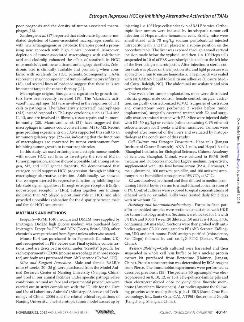
Estrogen Inhibits Macrophage Alternative Activation inMouse Liver TumorModels—Based on our preliminary results,we hypothesized that estrogen played an important role in thegender bias of HCC progression. To test this hypothesis, weexamined six groups of mice (described under “Materials andMethods”) in orthotopic liver tumors animalmodels (Fig. 1 andsupplemental Fig. 1) and ectopic mouse liver tumor models(supplemental Fig. 5). The tumor weight in OVX female micewas higher than that in female control; E2-treated OVX femalemice demonstrated a reduced tumor weight as compared withOVX femalemice. However, there was no significant differencebetween the weight of tumors that developed in the castratedmale mice compared with male control (Fig. 1B), suggestingthat androgen did not play a significant role in HCC progres-sion in our model. More importantly, after E2 supplement,tumor weight in castrated male mice was significantly reduced(Fig. 1A and supplemental Fig. 5). These results indicated thatestrogen rather than androgen contributed to the gender dis-parity in HCC progression.Then macrophages in the tumor sections were counted by
immunofluorescence staining for CD68 (cluster of differentia-tion 68) (FITC), CD206 (PE), and nucleus (DAPI) (supplemen-tal Fig. 2).We found that macrophages mainly exist in the form
of CD206�. CD206 high expression is an important character-ization of M2 macrophages. The CD206� density in OVXfemale mice (�72 cells/field of vision) was about 2-fold morethan that in the female control (�42 cells/field of vision). How-ever, after the administration of exogenous E2, the number ofCD206� cells in E2-treated OVX female mice was almostrestored to the level of female control, from 72 to 26 per field ofvision (supplemental Fig. 2, a–c). In male mice, there was nochange between male control and castration in CD206� num-ber. However, E2 treatment reduced the number of CD206�,from 65 to 32 per field of vision. The relative value of CD68 andCD206 in each group was shown in Fig. 1, C and D.
In conclusion, the macrophages inside tumor tissue in ourmodel primarily existed in the form of CD206�, and E2 pre-treatment reduced the number of CD206� macrophages.These findings represented a novel characterization of themolecular events underlying gender disparity in HCC progres-sion and may shed light on the pathological mechanisms ofestrogen in alternative activation of macrophages.Estrogen Inhibits Alternative Activation of Tumor-associated
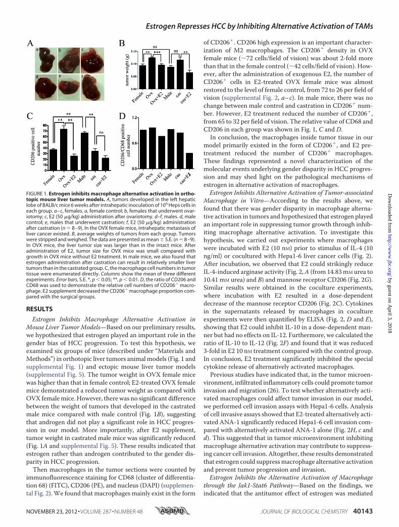
Macrophage in Vitro—According to the results above, wefound that there was gender disparity in macrophage alterna-tive activation in tumors and hypothesized that estrogen playedan important role in suppressing tumor growth through inhib-iting macrophage alternative activation. To investigate thishypothesis, we carried out experiments where macrophageswere incubated with E2 (10 nM) prior to stimulus of IL-4 (10ng/ml) or cocultured with Hepa1-6 liver cancer cells (Fig. 2).After incubation, we observed that E2 could strikingly reduceIL-4-induced arginase activity (Fig. 2,A (from 14.83mM urea to10.41 mM urea) and B) and mannose receptor CD206 (Fig. 2G).Similar results were obtained in the coculture experiments,where incubation with E2 resulted in a dose-dependentdecrease of the mannose receptor CD206 (Fig. 2C). Cytokinesin the supernatants released by macrophages in cocultureexperiments were then quantified by ELISA (Fig. 2, D and E),showing that E2 could inhibit IL-10 in a dose-dependent man-ner but had no effects on IL-12. Furthermore, we calculated theratio of IL-10 to IL-12 (Fig. 2F) and found that it was reduced3-fold in E2 10 nM treatment compared with the control group.In conclusion, E2 treatment significantly inhibited the specialcytokine release of alternatively activated macrophages.Previous studies have indicated that, in the tumor microen-
vironment, infiltrated inflammatory cells could promote tumorinvasion and migration (26). To test whether alternatively acti-vated macrophages could affect tumor invasion in our model,we performed cell invasion assays with Hepa1-6 cells. Analysisof cell invasive assays showed that E2-treated alternatively acti-vated ANA-1 significantly reduced Hepa1-6 cell invasion com-pared with alternatively activated ANA-1 alone (Fig. 2H, c andd). This suggested that in tumor microenvironment inhibitingmacrophage alternative activation may contribute to suppress-ing cancer cell invasion. Altogether, these results demonstratedthat estrogen could suppressmacrophage alternative activationand prevent tumor progression and invasion.Estrogen Inhibits the Alternative Activation of Macrophage
through the Jak1-Stat6 Pathway—Based on the findings, weindicated that the antitumor effect of estrogen was mediated
FIGURE 1. Estrogen inhibits macrophage alternative activation in ortho-topic mouse liver tumor models. A, tumors developed in the left hepaticlobe of BALB/c mice 6 weeks after intrahepatic inoculation of 106 Heps cells ineach group. a– c, females. a, female control; b, females that underwent ovar-iotomy; c, E2 (50 �g/kg) administration after ovariotomy. d–f, males. d, malecontrol; e, males that underwent castration; f, E2 (50 �g/kg) administrationafter castration (n � 8 –9). In the OVX female mice, intrahepatic metastasis ofliver cancer existed. B, average weights of tumors from each group. Tumorswere stripped and weighed. The data are presented as mean � S.E. (n � 8 –9).In OVX mice, the liver tumor size was larger than in the intact mice. Afteradministration of E2, tumor size for OVX mice was small compared withgrowth in OVX mice without E2 treatment. In male mice, we also found thatestrogen administration after castration can result in relatively smaller livertumors than in the castrated group. C, the macrophage cell numbers in tumortissue were enumerated directly. Columns show the mean of three differentexperiments. Error bars, S.E. *, p � 0.05; **, p � 0.01. D, the ratio of CD206 andCD68 was used to demonstrate the relative cell numbers of CD206� macro-phage. E2 supplement decreased the CD206� macrophage proportion com-pared with the surgical groups.
Estrogen Represses HCC by Inhibiting Alternative Activation of TAMs
NOVEMBER 23, 2012 • VOLUME 287 • NUMBER 48 JOURNAL OF BIOLOGICAL CHEMISTRY 40143
by guest on April 3, 2018
http://ww
w.jbc.org/
Dow
nloaded from
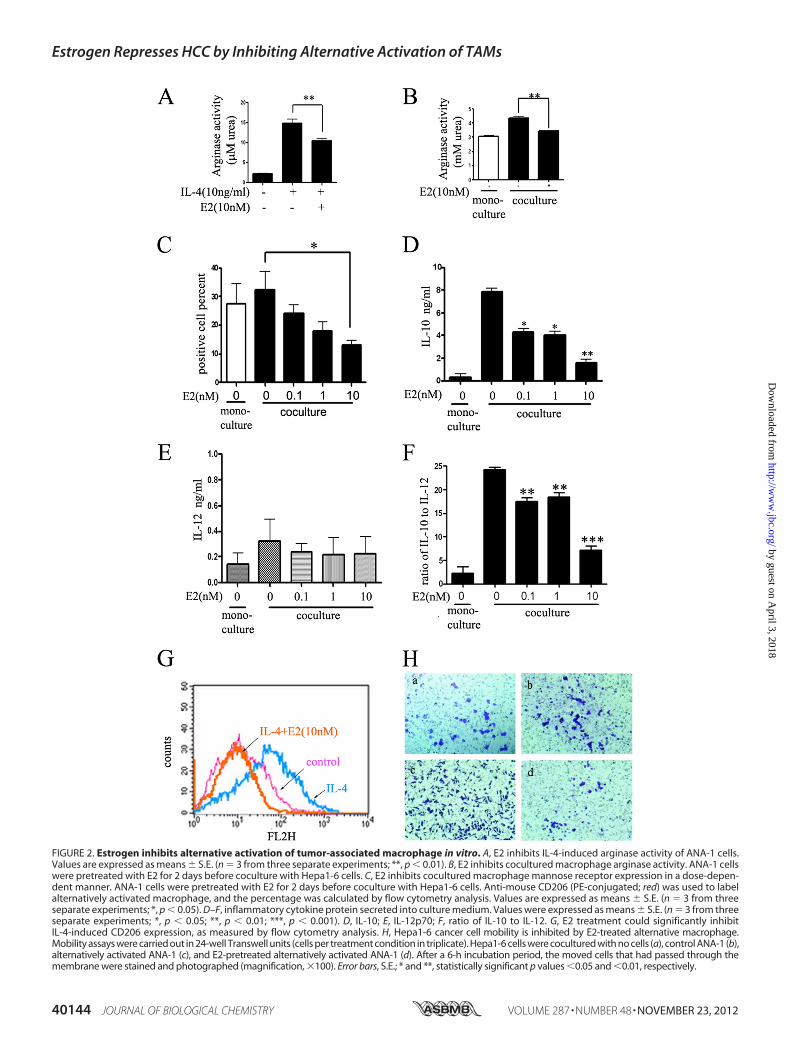
FIGURE 2. Estrogen inhibits alternative activation of tumor-associated macrophage in vitro. A, E2 inhibits IL-4-induced arginase activity of ANA-1 cells.Values are expressed as means � S.E. (n � 3 from three separate experiments; **, p � 0.01). B, E2 inhibits cocultured macrophage arginase activity. ANA-1 cellswere pretreated with E2 for 2 days before coculture with Hepa1-6 cells. C, E2 inhibits cocultured macrophage mannose receptor expression in a dose-depen-dent manner. ANA-1 cells were pretreated with E2 for 2 days before coculture with Hepa1-6 cells. Anti-mouse CD206 (PE-conjugated; red) was used to labelalternatively activated macrophage, and the percentage was calculated by flow cytometry analysis. Values are expressed as means � S.E. (n � 3 from threeseparate experiments; *, p � 0.05). D–F, inflammatory cytokine protein secreted into culture medium. Values were expressed as means � S.E. (n � 3 from threeseparate experiments; *, p � 0.05; **, p � 0.01; ***, p � 0.001). D, IL-10; E, IL-12p70; F, ratio of IL-10 to IL-12. G, E2 treatment could significantly inhibitIL-4-induced CD206 expression, as measured by flow cytometry analysis. H, Hepa1-6 cancer cell mobility is inhibited by E2-treated alternative macrophage.Mobility assays were carried out in 24-well Transwell units (cells per treatment condition in triplicate). Hepa1-6 cells were cocultured with no cells (a), control ANA-1 (b),alternatively activated ANA-1 (c), and E2-pretreated alternatively activated ANA-1 (d). After a 6-h incubation period, the moved cells that had passed through themembrane were stained and photographed (magnification, �100). Error bars, S.E.; * and **, statistically significant p values �0.05 and �0.01, respectively.
Estrogen Represses HCC by Inhibiting Alternative Activation of TAMs
40144 JOURNAL OF BIOLOGICAL CHEMISTRY VOLUME 287 • NUMBER 48 • NOVEMBER 23, 2012
by guest on April 3, 2018
http://ww
w.jbc.org/
Dow
nloaded from

through its inhibitory effect on macrophage alternative activa-tion in the tumor microenvironment. It is known that alterna-tively activated macrophage responded to Th2-type cytokines,such as IL-4. Recent studies have also identified IL-4 as a majorregulator of the phenotypes of TAMs (27, 28). IL-4 signaling isinitiated when this cytokine binds its cell surface receptor, acti-vating receptor-associated Jaks, followed by recruitment andphosphorylation of Stat6. Once phosphorylated, Stat6 pro-moted the expression of IL-4-responsive genes, such asCD206,Arg1, and Fizz1. Hence, we tested whether E2 prior incubationcould reverse IL-4-induced macrophage alternative activation.We first examined IL-4 receptor � (IL-4R�); the results indi-cated that IL-4R� expression had no changes before or aftercoculture (data not shown). Interestingly, we found that E2could significantly reduce the phosphorylation of Stat6 andJak1 (Fig. 3, A and B) by Western blotting. E2 inhibited phos-phorylation of Jak1 in a time-dependent manner in macro-phages by IL-4 treatment (Fig. 3B) and phosphorylation of Jak1and Stat6 in a dose-dependent manner in macrophages (Fig.3A). We also examined the downstream genes of Jak1-Stat6(Fig. 4), confirming the inhibitory effects of E2 on macrophagealternative activation.On the basis of Fig. 3, A and B, we observed that these E2
inhibitory effects on p-Jak1 occur after 12 h of E2 pretreatment.To examine whether some regulators influence the Jak1-Stat6pathway, many negative factors, such as suppressor of cytokinesignaling (SOCS) andSH2-containing phosphatase (SHP),weremeasured in RNA isolated from macrophages treated with E2.E2 treatment did not modify the expression of Socs3, Shp1, andShp3. However, the expression of Socs1was strikingly increasedin macrophages treated with E2 (supplemental Fig. 3).
We also investigated this result in protein levels (Fig. 3C).This result was in line with those obtained in mRNA levels.Furthermore, Socs1-specific RNA interference experiment wascarried out (Fig. 3, D and E), and the result confirmed theseobservations. Taken together, these results demonstrated thatE2 inhibited macrophage alternative activation by way ofdampening the phosphorylation of Jak1 and Stat6, throughinducing Jak1-Stat6 pathway negative regulator Socs1.Estrogen Exerts Its Inhibitory Effects via ER� but Not
ER�—Having confirmed the role of estrogen in HCC progres-sion, we needed to clearly define how estrogen exerted its func-tions. The biological actions of estrogen are mediated by bind-ing to one of two specific ERs, ER� and ER�, which belong tothe nuclear receptor superfamily, a family of ligand-regulatedtranscription factors. After binding to the ligand-bindingdomain of the ER, estrogen initiates a series ofmolecular eventsculminating in the activation or repression of target genes. Tostudy the specific role of these two types of ERs in the negativeregulation signaling pathway of Jak1-Stat6, we chose the selec-tive agonist PPT of ER� and selective agonist DPN of ER� tostimulate macrophages, respectively, and then analyzed themacrophage alternative activation state under these two ago-nists. As shown in Fig. 5,A and B, taking arginase activity as thedetection index, we found thatDPNexhibited inhibitory effectson the alternative activation of macrophages similar to E2, butthis effect was not obtained by PPT. Furthermore, a substantialincrease of Socs1 at the protein level (Fig. 5C) was observedwhen macrophages were treated with E2 and DPN. Theseresults suggested that ER� was responsible for interfering withthe Jak1-Stat6 signaling pathway. To confirm this conclusion,ER�-siRNA interference experimentswere performed (Fig. 5D)independently. The results showed that ER�-siRNA almostcompletely abolished the inhibitory effects initiated by E2 onIL-4-induced macrophage alternative activation, similar to theeffects of ER-specific antagonist ICI,182780 on macrophagealternative activation. These results confirmed that E2 inhib-ited macrophage alternative activation mainly through ER�.Because ER�-selective agonist DPN could enhance Socs1 pro-tein level, DPNwas thought to affect the phosphorylation statusof Stat6 by regulating Socs1. These data suggested that E2supressedmacrophage alternative activation and the Jak1-Stat6pathway via ER�, not ER�.EstrogenDamages the Interaction of ER� andATPase in IL-4-
stimulated Cells—To determine how ER�-mediated inhibitionof macrophage alternative activation is linked to the Jak1-Stat6pathway, we performed immunoprecipitation experimentswith anti-ER� under different treatment conditions and usedCoomassie Brilliant Blue staining. We detected a weak proteinband (black arrow) only in the IL-4-treated group immunopre-cipitants. Obviously, this weak band disappeared after pretreat-ment with E2, restored to a state similar with the control group(Fig. 6A). Then, by the mass spectrometry analysis, we identi-fied this protein as mitochondrial membrane protein ATPase,which catalyzes ATP synthesis and is composed of two linkedmultisubunit complexes, includingATP5J. Therefore, we chosethe subunit membrane protein ATP5J randomly to stand forATPase. To further define the interaction between ER� andATPase in the absence and presence of E2, we continued to
FIGURE 3. Estrogen inhibits the alternative activation of macrophagethrough the Jak1-Stat6 pathway. A, estrogen inhibits phosphorylation ofJak1-Stat6 in a dose-dependent manner in macrophages. ANA-1 macro-phage cell lines were cocultured with murine hepatocarcinoma cell lineHepa1-6, and different estrogen concentrations (0.1, 1.0, 5.0, and 10 nM) wereused to treat cells. Blots were probed with antibodies against p-Jak1 andp-Stat6. Blots were then stripped and reprobed with antibody against Gapdhas an internal control for equal loading. B, estrogen inhibits phosphorylationof Jak1 in a time-dependent manner in macrophages by IL-4 treatment. Thecytoplasm protein was extracted, and the protein level was detected by West-ern blot. Results shown are representative of three independent experiments.C, Western blot analysis of the protein level of Jak1-Stat6 signaling pathwaynegative regulators after estrogen administration. Results shown are repre-sentative of three independent experiments. D and E, ANA-1 cells were trans-fected with scrambled siRNA (20 pM) and Socs1 siRNA (20 pM), and thentreated with E2 (10 nM). The black arrow points to the position of the Socs1band. D, Western blot analysis of total extracts (E2 36-h treatment after trans-fection). E, p-Jak1, p-Stat6, and Socs1 from extracts (E2 36-h treatment aftertransfection and then IL-4 24-h treatment) were analyzed by Western blot.The black arrow points to the position of the p-Jak1 band.
Estrogen Represses HCC by Inhibiting Alternative Activation of TAMs
NOVEMBER 23, 2012 • VOLUME 287 • NUMBER 48 JOURNAL OF BIOLOGICAL CHEMISTRY 40145
by guest on April 3, 2018
http://ww
w.jbc.org/
Dow
nloaded from

perform immunoprecipitation with anti-ER� and anti-ATP5J(Fig. 6B). The results suggested that interaction between ER�and ATP5J was impaired after E2 treatment.Based on our collective results, we propose a cascade of
molecular events (Fig. 7) initiated by E2 as a mechanism toexplain its suppressive effects on tumor-associated alterna-tively activated macrophage cells. ER� mainly located in themitochondria (29), and it combined with the ATPase under thestimulus of IL-4. Therefore, ER� was involved in the regulationof ATP production and mitochondrial oxidative phosphoryla-tion in macrophages after IL-4 treatment. However, after E2activated ER�, the interaction between ER� and ATPase(ATP5J) weakened, and ER� separated fromATPase. ER� relo-cationmay induce a series of consequences, such as up-regulat-ing the expression of Jak1-Stat6 signaling pathway negative reg-ulator SOCS1, thereby attenuating the macrophage alternativeactivation and HCC tumor growth.
DISCUSSION
HCC is a male-predominant cancer, and the rates of HCC inmen are typically 2–4 times higher than in women (5, 30).Recent studies using mouse models show that sex hormonesplay a key role in hepatocarcinogenesis. However, the effects ofsex hormones on HCC progression have no consistent conclu-sion to date. Therefore, we focused on the gender disparity inHCC progression rather than on HCC carcinogenesis. Ourresults showed that E2 functioned as a suppressor for macro-phage alternative activation and tumor progression by keepingestrogen receptor � (ER�) away from interacting with ATPase-coupling factor 6 (ATP5J), a part of ATPase, thus inhibiting theJak1-Stat6 signaling pathway. These studies introduce a novelmechanism for suppressing male-predominant HCC.The Role of Estrogen in HCC Progression—Previous studies
on gender disparity in HCC have highlighted the linkagebetween estrogen and inflammation-induced carcinogenesis(2, 7). Our previous studies have shown that estrogen ratherthan androgenmay contribute to gender disparity in HCC pro-gression (31), and we obtained the same results here.In the current study, we explored the effects of estrogen on
the biological behavior of TAMs and their fate in different hor-mone environments.Results revealed that in comparisonwith the femalemice, the
male mice exhibited larger tumor weight (Fig. 1, A and B, andsupplemental Fig. S1), and the tumor weight in OVX female
FIGURE 4. Estrogen inhibits the downstream genes of the Jak1-Stat6 pathway. Total mRNA was extracted from ANA-1 cells treated with IL-4 and/or E2, anddownstream genes Arg1 (A), Fizz1 (B), and CD206 (C) were analyzed by quantitative PCR. Error bars, S.E.
FIGURE 5. Estrogen exerts its inhibitory effects via ER� but not ER�. Theeffects of estrogen receptor agonists on macrophage arginase activity wereanalyzed. Cells were precultured with E2 (nonspecific ER agonist), PPT (ER�-specific agonist) and DPN (ER�-specific agonist) at the indicated concentra-tion for 48 h and stimulated with IL-4 for another 24 h (A) or cocultured withHepa1-6 (B). Values were expressed as means � S.E. (error bars) (n � 6 fromthree separate experiments; *, p � 0.05 versus IL-4 treated group). C, Westernblot analysis of total extracts from macrophages that were exposed to E2 (10nM), ER�-specific agonist PPT (10 nM), and ER�-specific agonist DPN (100 nM)for 24 h. Socs1 level was analyzed by immunoblotting after treatment with E2,DPN, and PPT. D, extracellular surface receptor CD206 of ANA-1 macrophagewas detected by flow cytometry. Macrophages were pretreated with Estro-gen receptor antagonist (ICI182,780) or siRNA to knock down ER� (siESR2).The treatment in each group was independent from the others. siESR2 couldabolish the inhibitory effects of estrogen.
FIGURE 6. Estrogen damages the interaction of ER� and ATPase in IL-4-stimulated cells. A, whole cell lysates (WCL) were prepared for immunopre-cipitation with anti-ER�. ANA-1 macrophages were pretreated with E2 for48 h and stimulated by IL-14 for another 12 h. After SDS-PAGE, CoomassieG250/silver staining was carried out. A band (black arrow) was found with IL-4treatment, but it disappeared when pretreated with E2. B, coimmunoprecipi-tation (IP) of ATP5J and ER�. IB, immunoblot.
Estrogen Represses HCC by Inhibiting Alternative Activation of TAMs
40146 JOURNAL OF BIOLOGICAL CHEMISTRY VOLUME 287 • NUMBER 48 • NOVEMBER 23, 2012
by guest on April 3, 2018
http://ww
w.jbc.org/
Dow
nloaded from
mice significantly increased compared with female controlmice; however, castration had no impact on the HCC tumorvolume among tumor-bearing male mice. Meanwhile, E2 sup-plement reduced tumor volume in E2-treated OVX andE2-treated castrated mice with preestablished liver cancer (Fig.1B). These results demonstrated that gender disparity existed inHCC progression and that E2 played a critical role in this process.However, androgen has no effects on HCC progression.To fully appreciate the important role of sex hormones on
HCC progression and mimic the growth of human HCC com-prehensively, we developed orthotopic models by injectingHeps hepatocellular carcinoma cells directly into the liverparenchyma of BALB/c mice. Prior to ours, several orthotopictransplantation models using human liver cancer cell lines andathymic nude mice have been described (32, 33). However, oneobvious disadvantage of thesemodels is that they do not permitthe study of complex interactions between tumor cells and thehost immune system, which have been shown to significantlyinfluence tumor progression andmetastatic potential (34). Ourmouse models contended with this problem and improved onthis basis, providing us with a useful, reproducible, and consist-ent model wherein we could manipulate hormone levels toexamine their effect on HCC tumor progression.The Alteration of Macrophage Activation Status in HCC
Progression—Solid tumors consist of neoplastic cells, non-ma-lignant stromal cells, and migratory hematopoietic cells. Com-plex interaction between the cell types in this microenvironmentregulates tumorgrowth, progression, angiogenesis, andmetastasis(35). There is strong evidence that this microenvironment isinflammatory and that activation of the innate immune systemplays a role in the progression of cancer. One such inflammatorycell that has the potential to promote cancer progression is the
macrophage. There is a growing body of preclinical and clinicalevidence associating abundance of TAM with poor prognosis.According to Condeelis and Pollard (11), TAMs are obligate part-ners for malignant cell migration, invasion, and metastases inmany different cancers. In addition, these TAMs also enhancetumor progression by supporting tumor-associated angiogenesisand suppression of antitumor immune responses, such as impair-ing cytotoxic CD8�T cell immune responses (26, 36).
In our study, we found that the number of TAMs in malecontrol mouse models was significantly greater than that infemale control mice (Fig. 1, C and D, and supplemental Fig. 2).More importantly, TAMs have been shown to acquire the hall-mark properties of “alternatively activated” macrophages, suchas the ability to tune inflammatory and immune responses andto promote angiogenesis and tissue remodeling (20). Consist-ent with this notion, our murine results showed that TAMs inmouse model tumors existed mainly in the form of CD206�
status (Fig. 1, C and D, and supplemental Fig. S2). CD206 is animportant characterization ofM2macrophages.We also foundthat therewas a great gender disparity of CD206�macrophagesof TAMs between male control and female control mice inthe organization level in both orthotopic and ectopic mouseHCC models, and E2 administration resulted in a reductionof CD206� macrophages in both E2-treated castration andE2-treated OVX. Interestingly, similar findings were reportedby Ding et al. (37). In their study, they demonstrated that highmacrophage infiltration predicts poor prognosis in patientswith HCC. When macrophages infiltrated, tumor-derived fac-tors in the HCC microenvironment might divert macrophagefunctions toward an immunosuppressive and protumoral phe-notype,whichwas defined as TAMs (37). Ourwork in vitro alsosupported this notion. E2 treatment significantly reduced thearginase activity and the expression of CD206. Collectively,these results delineated the connections between E2, TAMs,and HCC cells and identified tumor-associated alternativelyactivated macrophages as a key factor in HCC progression.As mentioned above, our in vitro and in vivo data revealed
that E2 was able to suppress macrophage alternative activationand thus attenuate HCC progression significantly, indicatingthat with liver cancer deteriorating, estrogen may play a role ininhibiting alternative activation of macrophage and anti-in-flammatory factors, such as IL-10. However, Karin andco-workers (7) found that in early stages of hepatocarcinogen-esis, estrogen seemed to be able to suppressMyD88-dependentIL-6 production pathway in Kupffer cells to inhibit liver carci-nogenesis. Thus, our results may be a novel mechanism for thetumor-suppressing function of estrogen during the progressionofHCC, which seems not to exhibit an anti-inflammation func-tion but to change the activation patterns of macrophage.Estrogen Inhibits Macrophage Alternative Activation Mainly
through ER�—Previous studies indicated a critical role for theJak1-Stat6 signaling pathway in maturation of anti-inflamma-tory alternatively activated macrophages. Moreover, Ohmoriand Hamilton (38) confirmed that Stat6 was necessary for theIL-4-mediated inflammatory suppression. Meanwhile, the IL-4signaling pathways are subject to negative regulation by severalmechanisms, such as SHP proteins (39, 40) and the Socs family(41). In addition, in recent years, IL-4 has also been identified to
FIGURE 7. The speculative model of macrophage alternative activationregulation by estrogen. When estrogen interacts with ER�, it can keep ER�away from ATPase. Consequently, this process may enhance Socs1 expression, anegative regulator of the Jak1-Stat6 signaling pathway, to suppress the phos-phorylation of Stat6 and inhibit the macrophage alternative phenotype.
Estrogen Represses HCC by Inhibiting Alternative Activation of TAMs
NOVEMBER 23, 2012 • VOLUME 287 • NUMBER 48 JOURNAL OF BIOLOGICAL CHEMISTRY 40147
by guest on April 3, 2018
http://ww
w.jbc.org/
Dow
nloaded from

be a major regulator of the phenotypes of TAMs (27, 28). Wereported here that E2 pretreatment could inhibit the phospho-rylation of Jak1 and Stat6 in a dose- and time-dependent man-ner in vitro (Fig. 3, A and B).A previous study (42) reported that E2-liganded ER-� could
directlymodulate Socs1 expression by binding to the Socs1 pro-moter. These authors proposed that ER-� regulated threeclasses of genes (42). Socs1 genes belonged to the class II genesnot regulated by unliganded ER� but regulated by E2-boundER�. In our study, the effects of E2 on Socs1 expression areconsistent with theirs. The PCR (supplemental Fig. 3) andWestern blot results showed that the expression of Socs1 wasstrikingly increased in macrophages pretreated with E2 (Fig.3C), and Socs1-specific RNA interferences confirmed thisobservation. Taken together, the findings presented here dem-onstrated that E2 promoted the expression of negative regula-tor Socs1 of the Jak1-Stat6 signaling pathway.The biological functions of estrogen are mainly mediated by
estrogen binding to one of the two specific ERs, ER� and ER�(43). And our data demonstrated that E2 suppressed macro-phage alternative activation mainly via ER�.In addition, our work raised an intriguing question as to how
ER� exerts its function to inhibit the phosphorylation of Stat6.In the presence of IL-4, we found that ER� interacted withmembrane proteinATPase (Fig. 6A), which catalyzedATP syn-thesis fromADPand inorganic phosphate (Pi) by using the elec-trochemical potential of protons (or sodium ions in some bac-teria) across themembrane (44), whereas after pretreating withE2, the interaction was impaired (Fig. 6B). ER� was separatedfrom ATPase and turned to interacting with other mitochon-drial proteins or was translocated into the cytoplasm from thechondriosome to affect some other factors such as Socs1, exert-ing its inhibitory effect on macrophage alternative activation.However, after immunoprecipitation with ATP5J, ER� proteinexpression was not significantly different among the groups.This may be due to the fact that ATP5J was only one part of theATPase; ER� could interact with other parts. These results sug-gested that ER� was involved in the regulation of ATP produc-tion and mitochondrial oxidative phosphorylation in macro-phages. This hypothesis was supported by the literatureindicating that ER� is involved in mitochondrial membranepotential maintenance and mitochondrial vulnerability (29).Importantly, relocation of the ER� after separating fromATPase requires further study.The Significance of Our ResultsMay Suggest a New Insight for
HCC Therapy—Taken together, the findings presented heresupported the notion that estrogen could inhibit macrophagealternative activation, resulting in attenuated HCC progres-sion. Because HCC therapy has failed to improve overall sur-vival in patients with advanced HCC, efforts to develop newdrug treatments have shifted from systemic chemotherapy totargeted treatment against the tumor-stromal interaction.Therefore, targeting components of the tumor microenviron-ment will be a better auxiliary way to suppress HCC progres-sion. For example, sorafenib, an oral multikinase inhibitor, isthe most successful medication of this kind. It inhibits Vegfr-2/-3 (vascular endothelial growth factor receptor-2/3) andPdgfr (PDGF receptor) as well as Raf kinase, disrupting tumor-
stromal interactions and resulting in decreased cell prolifera-tion and angiogenesis. In addition, because tumor-associatedmacrophages accumulation correlates with tumor progression,it is feasible to convert tumor-associated macrophage polariza-tion in order to inhibit tumor growth. In T241 tumors, by skew-ing TAM polarization away from the M2 macrophage to atumor-inhibiting M1-like phenotype, Hrg (histidine-rich gly-coprotein) promotes antitumor immune responses and vesselnormalization, effects known to decrease tumor growth andmetastasis and to enhance chemotherapy (45). Therefore, con-sidering the important role of tumor-associated M2 in thetumor microenvironment, estrogen therapy may be feasible intreating HCC, especially for males. However, despite the disad-vantages of hormone replacement therapy, estrogen analoguesmay contribute as a complementary therapy in HCC treatmentand may also be a possible adjuvant therapy for controllingHCC. Our research has proposed that estrogen-attenuatedHCCprogression, through inhibiting tumor-associatedmacro-phage alternative activation, therefore selectively targetingmacrophages in combination with targeting the cancer cells,may be a promising trend for tumor cure.
Acknowledgments—We thank Chris T. Chen (Laboratory for Trans-lational Research, Harvard Medical School) and Nina Y. Xue (Uni-versity of California San Diego, La Jolla, CA) for critical commentsand grammatical correction of themanuscript.We thankDr. Ning Su(School of Basic Medical Sciences, Southeast University, China) forexcellent technical assistance.
REFERENCES1. Thomas, M. B., Jaffe, D., Choti, M. M., Belghiti, J., Curley, S., Fong, Y.,
Gores, G., Kerlan, R., Merle, P., O’Neil, B., Poon, R., Schwartz, L., Tepper,J., Yao, F., Haller, D., Mooney, M., and Venook, A. (2010) Hepatocellularcarcinoma. Consensus recommendations of theNational Cancer InstituteClinical Trials Planning Meeting. J. Clin. Oncol. 28, 3994–4005
2. Rogers, A. B., Theve, E. J., Feng, Y., Fry, R. C., Taghizadeh, K., Clapp, K.M.,Boussahmain, C., Cormier, K. S., and Fox, J. G. (2007) Hepatocellularcarcinoma associated with liver-gender disruption in male mice. CancerRes. 67, 11536–11546
3. Ghebranious, N., and Sell, S. (1998) Hepatitis B injury, male gender, afla-toxin, and p53 expression each contribute to hepatocarcinogenesis intransgenic mice. Hepatology 27, 383–391
4. Maeda, S., Kamata, H., Luo, J. L., Leffert, H., and Karin, M. (2005) IKK�couples hepatocyte death to cytokine-driven compensatory proliferationthat promotes chemical hepatocarcinogenesis. Cell 121, 977–990
5. Bosch, F. X., Ribes, J., Dıaz, M., and Cleries, R. (2004) Primary liver cancer.Worldwide incidence and trends. Gastroenterology 127, S5–S16
6. El-Serag, H. B., Mason, A. C., and Key, C. (2001) Trends in survival ofpatients with hepatocellular carcinoma between 1977 and 1996 in theUnited States. Hepatology 33, 62–65
7. Naugler,W. E., Sakurai, T., Kim, S., Maeda, S., Kim, K., Elsharkawy, A.M.,and Karin, M. (2007) Gender disparity in liver cancer due to sex differ-ences in MyD88-dependent IL-6 production. Science 317, 121–124
8. Nakatani, T., Roy, G., Fujimoto, N., Asahara, T., and Ito, A. (2001) Sexhormone dependency of diethylnitrosamine-induced liver tumors inmiceand chemoprevention by leuprorelin. Jpn. J. Cancer Res. 92, 249–256
9. CLIP Group (Cancer of the Liver Italian Programme). (1998) Tamoxifenin treatment of hepatocellular carcinoma. A randomized control trial.Lancet 352, 17–20
10. Liu, C. L., Fan, S. T., Ng, I. O., Lo, C. M., Poon, R. T., and Wong, J. (2000)Treatment of advanced hepatocellular carcinoma with tamoxifen and thecorrelationwith expression of hormone receptors. A prospective random-
Estrogen Represses HCC by Inhibiting Alternative Activation of TAMs
40148 JOURNAL OF BIOLOGICAL CHEMISTRY VOLUME 287 • NUMBER 48 • NOVEMBER 23, 2012
by guest on April 3, 2018
http://ww
w.jbc.org/
Dow
nloaded from
ized study. Am. J. Gastroenterol. 95, 218–22211. Condeelis, J., and Pollard, J.W. (2006)Macrophages. Obligate partners for
tumor cell migration, invasion, and metastasis. Cell 124, 263–26612. Goswami, S., Sahai, E., Wyckoff, J. B., Cammer, M., Cox, D., Pixley, F. J.,
Stanley, E. R., Segall, J. E., and Condeelis, J. S. (2005) Macrophages pro-mote the invasion of breast carcinoma cells via a colony-stimulating fac-tor-1/epidermal growth factor paracrine loop.Cancer Res. 65, 5278–5283
13. Bingle, L., Brown, N. J., and Lewis, C. E. (2002) The role of tumor-associ-ated macrophages in tumor progression. Implications for new anticancertherapies. J. Pathol. 196, 254–265
14. Lin, E. Y., Nguyen, A. V., Russell, R. G., and Pollard, J. W. (2001) Colony-stimulating factor 1 promotes progression of mammary tumors to malig-nancy. J. Exp. Med. 193, 727–740
15. Pollard, J. W. (2004) Tumor-educated macrophages promote tumor pro-gression and metastasis. Nat. Rev. Cancer 4, 71–78
16. Lin, E. Y., Gouon-Evans, V., Nguyen, A. V., and Pollard, J. W. (2002) Themacrophage growth factor CSF-1 in mammary gland development andtumor progression. J. Mammary Gland Biol. Neoplasia 7, 147–162
17. Zeisberger, S. M., Odermatt, B., Marty, C., Zehnder-Fjallman, A. H.,Ballmer-Hofer, K., and Schwendener, R. A. (2006) Clodronate-liposome-mediated depletion of tumor-associated macrophages. A new and highlyeffective antiangiogenic therapy approach. Br. J. Cancer 95, 272–281
18. Mantovani, A., Porta, C., Rubino, L., Allavena, P., and Sica, A. (2006)Tumor-associatedmacrophages (TAMs) as new target in anticancer ther-apy. Drug Discov. Today 3, 361–366
19. Pollard, J. W. (2009) Trophic macrophages in development and disease.Nat. Rev. Immunol. 9, 259–270
20. Mantovani, A., Sozzani, S., Locati, M., Allavena, P., and Sica, A. (2002)Macrophage polarization. Tumor-associatedmacrophages as a paradigm forpolarizedM2mononuclear phagocytes. Trends Immunol. 23, 549–555
21. Mantovani, A., and Sica, A. (2010) Macrophages, innate immunity, andcancer. Balance, tolerance, and diversity. Curr. Opin. Immunol. 22,231–237
22. Biswas, S. K., Gangi, L., Paul, S., Schioppa, T., Saccani, A., Sironi, M.,Bottazzi, B., Doni, A., Vincenzo, B., Pasqualini, F., Vago, L., Nebuloni, M.,Mantovani, A., and Sica, A. (2006) A distinct and unique transcriptionalprogram expressed by tumor-associated macrophages (defective NF-�Band enhanced IRF-3/STAT1 activation). Blood 107, 2112–2122
23. Pucci, F., Venneri, M. A., Biziato, D., Nonis, A., Moi, D., Sica, A., Di Serio,C., Naldini, L., and De Palma, M. (2009) A distinguishing gene signatureshared by tumor-infiltrating Tie2-expressingmonocytes, blood “resident”monocytes, and embryonicmacrophages suggests common functions anddevelopmental relationships. Blood 114, 901–914
24. Ojalvo, L. S., King,W., Cox, D., and Pollard, J.W. (2009)High density geneexpression analysis of tumor-associated macrophages from mouse mam-mary tumors. Am. J. Pathol. 174, 1048–1064
25. Bao, X., Cui, J., Wu, Y., Han, X., Gao, C., Hua, Z., and Shen, P. (2007) Theroles of endogenous reactive oxygen species and nitric oxide in triptolide-induced apoptotic cell death in macrophages. J. Mol. Med. 85, 85–98
26. Qian, B. Z., and Pollard, J. W. (2010) Macrophage diversity enhances tu-mor progression and metastasis. Cell 141, 39–51
27. DeNardo, D. G., Barreto, J. B., Andreu, P., Vasquez, L., Tawfik, D., Kolhat-kar, N., and Coussens, L. M. (2009) CD4� T cells regulate pulmonarymetastasis of mammary carcinomas by enhancing protumor properties ofmacrophages. Cancer Cell 16, 91–102
28. Gocheva, V., Wang, H. W., Gadea, B. B., Shree, T., Hunter, K. E., Garfall,A. L., Berman, T., and Joyce, J. A. (2010) IL-4 induces cathepsin proteaseactivity in tumor-associated macrophages to promote cancer growth andinvasion. Genes Dev. 24, 241–255
29. Yang, S. H., Sarkar, S. N., Liu, R., Perez, E. J., Wang, X., Wen, Y., Yan, L. J.,and Simpkins, J.W. (2009) Estrogen receptor� as amitochondrial vulner-
ability factor. J. Biol. Chem. 284, 9540–954830. Hoenerhoff, M. J., Pandiri, A. R., Lahousse, S. A., Hong, H. H., Ton, T. V.,
Masinde, T., Auerbach, S. S., Gerrish, K., Bushel, P. R., Shockley, K. R.,Peddada, S. D., and Sills, R. C. (2011) Global gene profiling of spontaneoushepatocellular carcinoma in B6C3F1 mice. Similarities in the molecularlandscape with human liver cancer. Toxicol. Pathol. 39, 678–699
31. Xu, H.,Wei, Y., Zhang, Y., Xu, Y., Li, F., Liu, J., Zhang,W., Han, X., Tan, R.,and Shen, P. (2012) Estrogen attenuates tumour progression in hepatocel-lular carcinoma. J Pathol. 228, 216–229
32. Yao, X., Hu, J. F., Daniels, M., Yien, H., Lu, H., Sharan, H., Zhou, X., Zeng,Z., Li, T., Yang, Y., and Hoffman, A. R. (2003) A novel orthotopic tumormodel to study growth factors and oncogenes in hepatocarcinogenesis.Clin. Cancer Res. 9, 2719–2726
33. Lu, Y. S., Kashida, Y., Kulp, S. K.,Wang, Y. C.,Wang, D., Hung, J. H., Tang,M., Lin, Z. Z., Chen, T. J., Cheng, A. L., and Chen, C. S. (2007) Efficacy ofa novel histone deacetylase inhibitor in murine models of hepatocellularcarcinoma. Hepatology 46, 1119–1130
34. de Visser, K. E., Eichten, A., and Coussens, L. M. (2006) Paradoxical roles ofthe immune system during cancer development.Nat. Rev. Cancer 6, 24–37
35. Albini, A., Magnani, E., and Noonan, D. M. (2010) The tumor microenvi-ronment. Biology of a complex cellular and tissue society.Q. J. Nucl. Med.Mol. Imaging 54, 244–248
36. Barajas,M.,Mazzolini, G., Genove, G., Bilbao, R., Narvaiza, I., Schmitz, V.,Sangro, B., Melero, I., Qian, C., and Prieto, J. (2001) Gene therapy oforthotopic hepatocellular carcinoma in rats using adenovirus coding forinterleukin 12. Hepatology 33, 52–61
37. Ding, T., Xu, J., Wang, F., Shi, M., Zhang, Y., Li, S. P., and Zheng, L. (2009)High tumor-infiltrating macrophage density predicts poor prognosis inpatients with primary hepatocellular carcinoma after resection. Hum.Pathol. 40, 381–389
38. Ohmori, Y., and Hamilton, T. A. (1998) STAT6 is required for the anti-inflammatory activity of interleukin-4 in mouse peritoneal macrophages.J. Biol. Chem. 273, 29202–29209
39. Lu, X., Malumbres, R., Shields, B., Jiang, X., Sarosiek, K. A., Natkunam, Y.,Tiganis, T., and Lossos, I. S. (2008) PTP1B is a negative regulator of inter-leukin 4-induced STAT6 signaling. Blood 112, 4098–4108
40. Myers, M. P., Andersen, J. N., Cheng, A., Tremblay, M. L., Horvath, C.M.,Parisien, J. P., Salmeen, A., Barford, D., and Tonks, N. K. (2001) TYK2 andJAK2 are substrates of protein-tyrosine phosphatase 1B. J. Biol. Chem.276, 47771–47774
41. Dickensheets, H., Vazquez, N., Sheikh, F., Gingras, S., Murray, P. J., Ryan,J. J., and Donnelly, R. P. (2007) Suppressor of cytokine signaling-1 is anIL-4-inducible gene in macrophages and feedback inhibits IL-4 signaling.Genes Immun. 8, 21–27
42. Vivar, O. I., Zhao, X., Saunier, E. F., Griffin, C., Mayba, O. S., Tagliaferri,M., Cohen, I., Speed, T. P., and Leitman, D. C. (2010) Estrogen receptor �
binds to and regulates three distinct classes of target genes. J. Biol. Chem.285, 22059–22066
43. Pettersson, K., and Gustafsson, J. A. (2001) Role of estrogen receptor � inestrogen action. Annu. Rev. Physiol. 63, 165–192
44. Okuno, D., Iino, R., and Noji, H. (2011) Rotation and structure of F0F1-ATP synthase. J. Biochem. 149, 655–664
45. Rolny, C., Mazzone, M., Tugues, S., Laoui, D., Johansson, I., Coulon, C.,Squadrito, M. L., Segura, I., Li, X., Knevels, E., Costa, S., Vinckier, S.,Dresselaer, T., Åkerud, P., DeMol,M., Salomaki, H., Phillipson,M.,Wyns,S., Larsson, E., Buysschaert, I., Botling, J., Himmelreich, U., Van Ginder-achter, J. A., De Palma, M., Dewerchin, M., Claesson-Welsh, L., and Car-meliet, P. (2011) HRG inhibits tumor growth and metastasis by inducingmacrophage polarization and vessel normalization through down-regula-tion of PlGF. Cancer Cell 19, 31–44
Estrogen Represses HCC by Inhibiting Alternative Activation of TAMs
NOVEMBER 23, 2012 • VOLUME 287 • NUMBER 48 JOURNAL OF BIOLOGICAL CHEMISTRY 40149
by guest on April 3, 2018
http://ww
w.jbc.org/
Dow
nloaded from
Pingping ShenWeiwei Yang, Yan Lu, Yichen Xu, Lizhi Xu, Wei Zheng, Yuanyuan Wu, Long Li and
Alternative Activation of Tumor-associated Macrophages (TAMs)Estrogen Represses Hepatocellular Carcinoma (HCC) Growth via Inhibiting
doi: 10.1074/jbc.M112.348763 originally published online August 20, 20122012, 287:40140-40149.J. Biol. Chem.
10.1074/jbc.M112.348763Access the most updated version of this article at doi:
Alerts:
When a correction for this article is posted•
When this article is cited•
to choose from all of JBC's e-mail alertsClick here
Supplemental material:
http://www.jbc.org/content/suppl/2012/08/20/M112.348763.DC1
http://www.jbc.org/content/287/48/40140.full.html#ref-list-1
This article cites 45 references, 14 of which can be accessed free at
by guest on April 3, 2018
http://ww
w.jbc.org/
Dow
nloaded from