hipk2 represses -catenin-mediated transcription, epidermal...
TRANSCRIPT

HIPK2 represses �-catenin-mediated transcription,epidermal stem cell expansion, and skintumorigenesisGuangwei Wei*, Stephen Ku*, Gene K. Ma*, Shin’ichi Saito†, Amy A. Tang*, Jiasheng Zhang*, Jian-Hua Mao‡,Ettore Appella†, Allan Balmain‡, and Eric J. Huang*§
*Department of Pathology, University of California and Pathology Service 113B, Veterans Affairs Medical Center, San Francisco, CA 94121; †Laboratory ofCell Biology, National Cancer Institute, Bethesda, MD 20892; and ‡Cancer Center, University of California, San Francisco, CA 94143
Communicated by James E. Cleaver, University of California, San Francisco, CA, April 10, 2007 (received for review January 9, 2007)
Transcriptional control by �-catenin and lymphoid enhancer-bindingfactor 1 (LEF1)/T cell factor regulates proliferation in stem cells andtumorigenesis. Here we provide evidence that transcriptional co-repressor homeodomain interacting protein kinase 2 (HIPK2) controlsthe number of stem and progenitor cells in the skin and the suscep-tibility to develop squamous cell carcinoma. Loss of HIPK2 leads toincreased proliferative potential, more rapid G1–S transition in cellcycle, and expansion of the epidermal stem cell compartment. Amongthe critical regulators of G1–S transition in the cell cycle, only cyclin D1is selectively up-regulated in cells lacking HIPK2. Conversely, overex-pression of HIPK2 suppresses LEF1/�-catenin-mediated transcrip-tional activation of cyclin D1 expression. However, deletion of theC-terminal YH domain of HIPK2 completely abolishes its ability torecruit another transcriptional corepressor CtBP and suppress LEF1/�-catenin-mediated transcription. To determine whether loss ofHIPK2 leads to increased susceptibility to tumorigenesis, we treatwild-type, Hipk2�/�, and Hipk2�/� mice with the two-stage carcino-genesis protocol. Our results indicate that more skin tumors areinduced in Hipk2�/� and Hipk2�/� mutants, with most of the tumorsshowing shortened incubation time and malignant progression. To-gether, our results indicate that HIPK2 is a tumor suppressor thatcontrols proliferation by antagonizing LEF1/�-catenin-mediated tran-scription. Loss of HIPK2 synergizes with activation of H-ras to inducetumorigenesis.
cell cycle � proliferation � Wnt � corepressor
The process of skin carcinogenesis involves a series of transitionalevents, ranging from hyperplasia, dysplasia, and papilloma to
invasive squamous cell carcinoma. The current model indicates thatthe progression of benign lesions to malignancy depends on the celltype targeted by these mutations (1, 2). Indeed, recent evidence hasindicated that the bulge region of the hair follicles contains self-renewing, slow-cycling stem cells, which give rise to transientamplifying cells and several differentiated cell lineages in theinterfollicular epidermis (3, 4). The progression of skin carcino-genesis can be modified by mutations involving Tgf�1, Smad3,ubiquitin ligase Fbxw7/Cdc4, or Pten (5–9), suggesting that a geneticnetwork synergizes with activated Ras mutation to promote theinitiation or malignant transformation of skin tumors (10). Anotherimportant regulator of skin carcinogenesis is cyclin D1. Overex-pression of cyclin D1 increases the propensity of skin carcinogen-esis, whereas loss of cyclin D1 reduces tumor formation (11, 12).One mechanism to control cyclin D1 expression is through tran-scriptional regulation via the Wnt/�-catenin signaling pathway (13,14), which consists of a core set of highly evolutionarily conservedproteins that have wide-ranging effects on gene expression affectingproliferation, migration, pluripotency, morphogenesis, and tumor-igenesis (15–17). In the epidermis, targeted deletion of �-cateninduring embryogenesis results in severe abnormalities in cell fatedetermination and maintenance of hair follicle formation (18). Incontrast, mutations that activate �-catenin function lead to skintumorigenesis in both humans and mice (19, 20).
Homeodomain interacting protein kinase (HIPK) contains threedistinct members that regulate apoptosis, cell growth, and prolif-eration. HIPK2 has been identified as a transcriptional corepressorfor homeodomain proteins NK3 and Brn3a (21, 22). Previousresults have indicated that Brn3a and HIPK2 regulate a delicatebalance of gene expression that controls programmed cell death insensory and dopamine neurons (22–24). Both HIPK1 and HIPK2can interact with human p53 and, under DNA damage conditions,activate p53 by promoting phosphorylation and acetylation (25–27).Contrary to the predicted function, HIPK1-deficient mice show ahigher resistance to the development of DMBA-induced skintumors (27). Recent evidence, however, has demonstrated a func-tional redundancy between HIPK1 and HIPK2 during develop-ment, which could compensate for the loss of HIPK1 (28). Indeed,HIPK2 has a wide range of functions, including promoting thedegradation of CtBP in response to UV-induced DNA damage (29)and regulating cell proliferation through the Wnt signaling pathway(30). Furthermore, HIPK2 can interact with several proteins con-taining the high-mobility group I (HMGI), a domain highly con-served in transcription factors in the lymphoid enhancer-bindingfactor 1/T cell factor (LEF1-TCF) family (31).
In this work, we show that loss of HIPK2 leads to an expansionof epidermal stem cells due to increased proliferation. Our resultsreveal a mechanism of HIPK2 as a transcriptional corepressor ofLEF1/�-catenin-mediated cyclin D1 expression. As a consequence,cells derived from Hipk2�/� mutants show increased cyclin D1 andan accelerated G1–S cell cycle transition. The mechanism by whichHIPK2 achieves this suppressive effect is by forming a proteincomplex with �-catenin and LEF1 and by recruiting anothercorepressor, CtBP. Deletion of the CtBP-interacting domain abol-ishes the suppressive effect of HIPK2 on �-catenin. Consistent withthese findings, both Hipk2�/� and Hipk2�/� mutants show in-creased propensity for tumor formation and malignant progressionunder the two-stage chemical carcinogenesis paradigm. Togetherthese results provide evidence that HIPK2-mediated transcriptionalprograms restrict cell growth in epidermal stem and progenitor cellsby suppressing cyclin D1 expression. Removal of such mechanismsleads to an expansion in the stem cell pool and increases thepropensity for tumorigenesis.
Author contributions: A.B. and E.J.H. designed research; G.W., S.K., G.K.M., S.S., A.A.T., andJ.Z. performed research; J.-H.M. and A.B. contributed new reagents/analytic tools; E.A.,A.B., and E.J.H. analyzed data; and E.J.H. wrote the paper.
The authors declare no conflict of interest.
Freely available online through the PNAS open access option.
Abbreviations: En, embryonic day n; Pn, postnatal day n; LRC, label-retaining cells; TA cells,transient amplifying cells; LEF1, lymphoid enhancer-binding factor 1; TCF, T cell factor; MEF,mouse embryonic fibroblast.
§To whom correspondence should be addressed. E-mail: [email protected].
This article contains supporting information online at www.pnas.org/cgi/content/full/0703213104/DC1.
© 2007 by The National Academy of Sciences of the USA
13040–13045 � PNAS � August 7, 2007 � vol. 104 � no. 32 www.pnas.org�cgi�doi�10.1073�pnas.0703213104
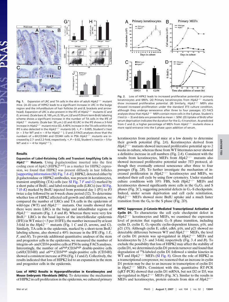
ResultsExpansion of Label-Retaining Cells and Transient Amplifying Cells inHipk2�/� Mutants. Using �-galactosidase inserted into the firstcoding exon of hipk2 (HIPK2LacZ) as a marker for HIPK2 expres-sion, we found that HIPK2 was present diffusely in hair follicles[supporting information (SI) Fig. 7 A–E]. HIPK2, detected either by�-galactosidase or HIPK2 antibodies, was present in keratinocytes,transient amplifying (TA) cells (see SI Fig. 7 F and G) marked bya short pulse of BrdU, and label-retaining cells (LRCs) (see SI Fig.7 H–K) marked by BrdU injected from postnatal day 1 (P1) to P3twice a day followed by an 8-week chase period (32). To determinewhether loss of HIPK2 affected cell growth and differentiation, wecompared the number of LRCs and TA cells in the epidermis ofwild-type (WT) and Hipk2�/� mutants. Our results showed thatthere were more LRCs in the bulge and infundibular regions ofHipk2�/� mutants (Fig. 1 A and B). Whereas there were very fewBrdU� LRCs in the basal layers of the interfollicular epidermis(IFE) in WT mice (�5 per 10 IFE), the number increased by at least3-fold in the Hipk2�/� mutants (Fig. 1 C and D, arrows, and G).Similarly, TA cells in the epidermis, marked by a short-term BrdUlabeling scheme, also showed a 40% increase in the IFE (Fig. 1 E,F, and H). To provide additional quantitative analyses of the stemand progenitor cells in the epidermis, we measured the number ofintegrin-�6- and CD34-positive cells at P56 by using FACS analyses.Interestingly, the number of �6high;CD34high double-positive cells(�6H;CD34H) and �6low;CD34high single-positive cells (CD34H)showed a consistent increase at P56 (Fig. 1 I and J). Collectively, theresults indicated that loss of HIPK2 led to an expansion in the stemand progenitor cells in the epidermis.
Loss of HIPK2 Results in Hyperproliferation in Keratinocytes andMouse Embryonic Fibroblasts (MEFs). To determine the mechanismof HIPK2 in cell proliferation in the epidermis, we cultured primary
keratinocytes from perinatal mice at a low density to determinetheir growth potential (Fig. 2A). Keratinocytes derived fromHipk2�/� mutants showed increased proliferative potential up to 2weeks in culture, whereas those from WT littermates never showeda definitive increase in cell numbers (Fig. 2A). Consistent with theresults from keratinocytes, MEFs from Hipk2�/� mutants alsoshowed increased proliferative potential under 3T3 protocol, al-though they eventually entered senescence after three to fourpassages (Fig. 2B). To further investigate the mechanism of in-creased proliferation in Hipk2�/� keratinocytes and MEFs, weanalyzed their cell cycle by using flow cytometry. Under standardculture conditions with 10% FBS, both Hipk2�/� MEFs andkeratinocytes showed significantly more cells in the G0/G1 and Sphases (Fig. 2C), suggesting potential defects in G1–S checkpoints.Indeed, under serum deprivation and cell cycle reinitiation,Hipk2�/� MEFs showed more BrdU uptake and a much fastertransition from the G0 to the S phase (Fig. 2D).
HIPK2 Suppresses �-Catenin-Mediated Transcriptional Activation ofCyclin D1. To characterize the cell cycle checkpoint defect inHipk2�/� keratinocytes and MEFs, we examined the expressionlevel of proteins that regulate G1–S phase transition, includingcyclin D, cyclin E, G1-specific cyclin-dependent kinases, p16, andp21 (33). Although cyclin E, cdk4, cdk6, p16, and p21 showed nodetectable difference between WT and Hipk2�/� MEFs, the levelof cyclin D1 protein was up-regulated in Hipk2�/� MEFs andkeratinocytes by 2.5- and 3-fold, respectively (Fig. 3 A and B). Toexclude the possibility that loss of HIPK2 may affect the stability ofcyclin D1, we determined cyclin D1 protein turnover and found thatdegradation of 35S-labeled cyclin D1 followed a similar kinetics inWT and Hipk2�/� MEFs (SI Fig. 8). Given the role of HIPK2 asa transcriptional corepressor, we reasoned that an increase in cyclinD1 protein may be due to an increase in transcription of cyclin D1in Hipk2�/� MEFs. Consistent with this, quantitative RT-PCR(qRT-PCR) showed that cyclin D1 mRNA, but not D2 or D3, wasup-regulated in Hipk2�/� MEFs (Fig. 3C). Similar to the results inMEFs and keratinocytes, protein extracts from skin of Hipk2�/�
Fig. 2. Loss of HIPK2 leads to increased proliferative potential in primarykeratinocytes and MEFs. (A) Primary keratinocytes from Hipk2�/� mutantsshow increased proliferative potential. (B) Similarly, Hipk2�/� MEFs alsoshowed increased proliferation under the standard 3T3 culture condition,although they undergo senescence after three to four passages. (C) FACSanalyses show that Hipk2�/� MEFs contain more cells in the S phase. Student’st test (n � 3) and data are presented as mean � SEM. (D) Uptake of BrdU afterserum deprivation indicates the duration for the G1–S transition. As predictedfrom C and D, a higher percentage of MEFs from Hipk2�/� mutants show amore rapid entrance into the S phase upon addition of serum.
Fig. 1. Expansion of LRC and TA cells in the skin of adult Hipk2�/� mutantmice. (A–D) Loss of HIPK2 leads to a significant increase in LRC in the bulgeregion and the infundibulum of hair follicles (A and B, brackets and arrow-head). Expansion of LRC is also present in the IFE of Hipk2�/� mutants (C andD, arrows). (Scale bars: B, 100 �m; D, 50 �m.) (E and F) Short-term BrdU labelingscheme shows a significant increase in the number of TA cells in the IFE ofHipk2�/� mutants. (Scale bar: 50 �m.) (G and H) LRC in the IFE shows a 3-foldincrease in Hipk2�/� mutant mice (G). A 40% increase in the TA cells within theIFE is also detected in the Hipk2�/� mutants (H). *, P � 0.005, Student’s t test(n � 3 for WT and n � 4 for Hipk2�/�). (I and J) FACS analyses show that thenumbers of �-6H;CD34H and CD34H cells in P56 Hipk2�/� mutants are in-creased by 2.7- and 2.2-fold, respectively. *, P � 0.02, Student’s t test (n � 5 forWT and n � 4 for Hipk2�/�).
Wei et al. PNAS � August 7, 2007 � vol. 104 � no. 32 � 13041
CELL
BIO
LOG
Y

and Hipk2�/� mice also showed significant increases in cyclin D1proteins (Fig. 3 D and E).
Our results indicated that HIPK2 suppressed �-catenin-mediatedactivation of cyclin D1 (Fig. 3F). The suppressive effect of HIPK2did not require phosphorylation on the critical Ser/Thr residues of�-catenin because HIPK2 suppressed the transcriptional activity ofa constitutively active form of �-catenin (CA-�-catenin) in whichamino acids 29–48 were deleted (Fig. 3F). Interestingly, the kinase-inactive form of HIPK2 (HIPK2K221A) showed a similar efficacy insuppressing cyclin D1 expression, suggesting that the kinase activityof HIPK2 was dispensable. To determine whether the effect ofHIPK2 was directly mediated through �-catenin and LEF1/TCF,we used the TOPFlash luciferase reporter that contained onlyLEF1/TCF binding sites as a highly specific reporter for Wnt/�-catenin signaling. Similar to the results with the cyclin D1 luciferasereporter, HIPK2 suppressed TOPFlash activity activated by �-cate-nin (Fig. 3G). The presence of HIPK2 did not alter the level of�-catenin, indicating that HIPK2 did not affect the stability orturnover of �-catenin (Fig. 3G and SI Fig. 9).
Transcriptional Repression of HIPK2 Requires Recruitment of Co-repressor CtBP via the C-Terminal YH Domain. To determine whethersuppression of �-catenin/LEF1-mediated gene expression byHIPK2 required the formation of a protein complex, we performeda series of coimmunoprecipitation (co-IP) assays to detect thepresence of HIPK2 with LEF1 and other known transcriptionalcorepressors. Interestingly, classical corepressors, such as HDAC1,were not detected in the HIPK2–�-catenin–LEF1 complex. Fur-thermore, HIPK2 continued to suppress �-catenin luciferase ac-tivity even in the presence of trichostatin A (TSA), a specificinhibitor for HDACs (G.W. and E.J.H., unpublished data). Incontrast, the transcriptional corepressor C-terminal binding protein(CtBP) was consistently detected in the complex with LEF1 andHIPK2 in the nuclear extracts, despite the overall reduction of CtBPlevel in the cytosol (Fig. 4A) (29). In the absence of HIPK2, CtBPcan be detected in a protein complex with LEF1, albeit at a much
lower affinity (Fig. 4A). The recruitment of CtBP to the HIPK2–LEF1 complex did not require the kinase activity of HIPK2. Incontrast, HIPK2 mutants, HIPK2-�969 and HIPK2-�898, whichlacked the C-terminus YH domain and the adjacent consensusCtBP binding motif PLNLS (amino acids 1031–1035), showed amarked reduction in their ability to form a protein complex withCtBP (Fig. 4 B and C). A series of luciferase assays confirmed thatHIPK2 mutants with deletions involving the YH domain or adja-cent regions, such as HIPK2-�1088, HIPK2-�969, and HIPK2-�898, failed to suppress the �-catenin-activated TOPFlash activity(Fig. 4D). In contrast, deletion of the kinase domain (HIPK2-�KD), the alternatively spliced exon 2B (HIPK2-�2B), Brn3a-interacting domain (HIPK2-�ID), or the speckle-retention signal(HIPK2-�SRS), did not affect the suppressive activity of HIPK2(Fig. 4D).
Two-Stage Skin Carcinogenesis Treatment Reveals Tumor SuppressorFunction of HIPK2. Given the important role of cyclin D1 as anoncogene, we reasoned that an increase in cyclin D1 mightpredispose Hipk2�/� mutants to tumor formation. Consistentwith this prediction, Hipk2�/� MEFs formed more colonies thanWT MEFs under clonogenic culture conditions (SI Fig. 10).Transfection with an activated form of H-ras (H-rasG12V) furtherinduced enlarged colonies in Hipk2�/� MEFs (SI Fig. 10),suggesting that Ras activation synergized with a loss of HIPK2to increase the proliferative potentials. Although neitherHipk2�/� nor Hipk2�/� mutant mice developed spontaneoustumors, when challenged with the two-stage skin carcinogenesisprotocol (34), both showed an �3-fold increase in tumor numbercompared with that in WT (Figs. 5A and 6 A–C). Consistent withprevious studies, all tumors, regardless of genotype, containedthe common CTA mutation in H-ras (SI Fig. 11). The tumor-freelatency was significantly shorter in Hipk2�/� and Hipk2�/�
mutants. By 24 weeks, essentially every Hipk2�/� mouse haddeveloped skin tumors (Fig. 5B). Whereas tumors in all WT micewere benign squamous papillomas, many Hipk2�/� and Hipk2�/�
Fig. 3. HIPK2 suppresses �-catenin-mediated tran-scription of cyclin D1. (A) Western blots showing in-crease of cyclin D1 in Hipk2�/� MEFs, whereas otherregulators of G1 cell cycle, e.g., cyclin E, cdk4, cdk6,p16, and p21, are not affected. (B) Similarly, Hipk2�/�
keratinocytes also show a 3-fold increase in cyclin D1protein, whereas p15 and p21 show no detectabledifference between WT and Hipk2�/� keratinocytes.(C) Selective increase of cyclin D1 mRNA in Hipk2�/�
mutant MEFs. (D and E) Cyclin D1 protein also is in-creased in skin from Hipk2�/� and Hipk2�/� mice. *, P �0.0189 between WT and Hipk2�/�; **, P � 0.00058between WT and Hipk2�/�; Student’s t test (n � 3). (F)HIPK2 suppresses the ability of �-catenin and consti-tutively active �-catenin (CA-�-catenin) to activate thecyclin D1 promoter. The kinase-inactive form of HIPK2,HIPK2K221A, continues to suppress cyclin D1 luciferaseactivity. (G) Similar to the cyclin D1 reporter, TOPFlashluciferase activity is inhibited by HIPK2 in a dose-dependent manner. HIPK2 has no effect on the stabil-ity of �-catenin protein.
13042 � www.pnas.org�cgi�doi�10.1073�pnas.0703213104 Wei et al.

mutants showed features of carcinoma in situ or invasive squa-mous cell carcinoma (Fig. 5C). Similar to the Hipk2�/� MEFsand keratinocytes in culture, tumor cells in Hipk2�/� mutantsshowed a higher labeling index for BrdU and phosphohistone 3(PH3) in the suprabasal layers of tumors (Fig. 6 D–G), a featureassociated with a high probability of malignant progression (35).Consistent with the results from keratinocytes and MEFs (Fig.3 A–C), tumors from Hipk2�/� and Hipk2�/� showed a higherlevel of cyclin D1 (Fig. 6 I and K–M). Although cyclin D1� cellsin WT tumors were restricted to the basal layer, many such cellsin Hipk2�/� tumors extended to the suprabasal layers (Fig. 6 Hand J, arrowheads). Together, these results supported the notionthat loss of HIPK2 and DMBA-induced H-ras activation actedsynergistically to promote skin tumorigenesis in Hipk2�/�
mutants.
DiscussionTranscriptional Regulation of Cyclin D1 by �-Catenin and HIPK2.Overexpression of cyclin D1 is commonly detected in colon, breast,and skin cancers (36). Several important regulatory elements havebeen identified in the promoter sequence of the cyclin D1 gene,suggesting that transcriptional control of cyclin D1 may harborpotential tumorigenic targets (13, 14). Results in this study indicatethat HIPK2 is an important component of an intrinsic transcrip-tional mechanism that negatively regulates cyclin D1 expression(Figs. 2, 4, and 6). Our results indicate that the C terminus of HIPK2is required to interact with the corepressor, CtBP, which in turnpromotes the suppressor function of HIPK2 (Fig. 4) (37). Deletionof the YH domain (HIPK2-�1088) and the putative CtBP bindingmotif (HIPK2-�969 and HIPK2-�898) completely abolishes therepressor effect of HIPK2 in TOPFlash reporter assays (Fig. 4).
Fig. 4. The YH domain in the Cterminus of HIPK2 is required to re-cruit transcriptional corepressorCtBP and suppress �-catenin-medi-ated transcription. (A) Co-IP showsthat HIPK2 can be detected in aprotein complex with LEF1 andtranscriptional corepressor CtBP. (Band C) The interaction betweenHIPK2 and CtBP requires the YH do-main in the C terminus of HIPK2because HIPK2 mutants lacking thisdomain, including HIPK2-�969 andHIPK2-�898, fail to coimmunopre-cipitate with CtBP. Quantificationof the interaction between HIPK2and CtBP is shown in C. (D) Deletionof the YH domain of HIPK2 abolishesthe ability of HIPK2 to suppress�-catenin. Truncation mutants in theHIPK2 C terminus (HIPK2-�1088,HIPK2-�969, and HIPK2-�898), thekinase domain (HIPK2-�KD), theBrn3a-interacting domain (HIPK2-�ID), and the speckle-retentionsignal (HIPK2-�SRS) are generatedto map the domain required forthe suppressor activity of HIPK2.Deletion of the YH domain in the Cterminus completely abolishesthe suppressor effect of HIPK2,whereas loss of the kinase domain,the Brn3a-interacting domain, orspeckle-retention signal has no de-tectable effect.
Fig. 5. Increased propensity of skin carcinogenesis in Hipk2�/� and Hipk2�/� mutant mice. (A) Two-stage skin carcinogenesis paradigm induces more tumorsin Hipk2�/� and Hipk2�/� mutants compared with WT littermates (P � 0.0001). (B) Incubation time for tumor formation is much shorter in Hipk2�/� and Hipk2�/�
mutants (P � 0.0151 for WT and Hipk2�/�; P � 0.004 for WT and Hipk2�/�). (C) A higher percentage of the skin tumors in Hipk2�/� and Hipk2�/� mutants developinto carcinoma (P � 0.0001). Data in B and C are presented by using the Kaplan–Meier method, and statistical analyses are performed by using Student’s t test.
Wei et al. PNAS � August 7, 2007 � vol. 104 � no. 32 � 13043
CELL
BIO
LOG
Y

Results obtained by using construct HIPK2-�1088 are particularlyintriguing because they suggest that, at least in the context of�-catenin-dependent activation of TOPFlash, the integrity of theC-terminal region of HIPK2 is important for the recruitment ofadditional transcriptional corepressors, such as CtBP.
Transcriptional Control of Proliferation in Epidermal Stem Cells.Lineage tracing by using genetic targeting, pulse chase with labelednucleotides, and transplantation experiments have provided solidevidence that epidermal stem cells contribute to the formation andhomeostasis of epidermis and its adnexal structures (3, 4, 38, 39).Within its niche, epidermal stem cells are subjected to a variety ofexogenous factors that control its growth, proliferation, migration,and differentiation (40). One prominent mechanism that controlsthe development and maintenance of epidermal stem cells is theWnt/�-catenin signaling pathway. Conditional deletion of �-cateninduring embryogenesis leads to a blockage of initial placode and hairfollicle formation during morphogenesis and a complete loss of haircycling during the first catagen phase in postnatal life (18). Theseresults underscore the dual roles of �-catenin in cell fate determi-nation and the maintenance of hair follicle formation, suggestingthat, in the absence of �-catenin, epidermal stem cells favor a fatetoward epidermis. Interestingly, a high level of LEF1 is expressedin the hair follicles during morphogenesis, and targeted deletion of
Lef1 also leads to phenotypes similar to �-catenin-conditionalmutants with a severe reduction of whiskers and hair follicles (41).Together these results indicate a cooperative role of �-catenin andLEF1 in the morphogenesis and fate determination in hair follicles.
Based on the characteristics of HIPK2 as a transcriptionalcorepressor of the �-catenin–LEF1 complex, one would predictthat a common set of target genes may be either up- or down-regulated in the epidermal stem cells of Hipk2�/� mutants andtransgenic mice that express a stabilized form of �-catenin (�N/�N). Indeed, both mice show a similar up-regulation in cyclin D1.However, several prominent differences are present. First, thenumber of the two distinct populations of stem cells is increased by2- to 3-fold in Hipk2�/� mutants (Fig. 1), but not in �N/�N miceat comparable ages (42). It is possible that loss of HIPK2 mayperturb the expression of additional target genes, which contributesto such differences. Alternatively, it is possible that the K14promoter, used to generate the �N/�N mice, selectively targets theconstitutively active form of �-catenin to a more restricted popu-lation of epidermal progenitor cells and preserves the microenvi-ronment in the stem cell niche. In contrast, loss of HIPK2 mayaffect a broader population of cells, including dermal fibroblasts,infiltrating T cells, and macrophages.
Tumor Suppressor Effects of HIPK2 in Skin Carcinogenesis. AlthoughHIPK2 has been implicated in regulating p53-dependent cellgrowth and apoptosis (25, 26), several lines of evidence indicate thatthis mechanism may not be conserved across different species. First,the patterns of cell growth in primary and E1A-transformedHipk2�/� MEFs are substantially different from that of p53�/�
mutants (SI Fig. 12). Second, the induction of total p53 protein andphosphorylation on several critical Ser and Thr residues in responseto various cell injury conditions remains intact in Hipk2�/� MEFs(SI Fig. 12). Finally, the pattern of tumor growth and the spectrumof differentiation in Hipk2�/� and Hipk2�/� mutants, such asincreased skin tumor formation and multilineage features, is verydifferent from the paradoxical reduction of papilloma number inp53�/� mutants (43). Together these results argue that HIPK2 andp53 have different, nonoverlapping functions in the control of cellgrowth and tumorigenesis. Indeed, preliminary results indicate thatpartial loss of both HIPK2 and p53 synergistically enhances radi-ation-induced thymic lymphoma (J.-H.M. and A.B., unpublisheddata), suggesting that HIPK2 and p53 act in trans, rather than in cis,to regulate cell growth and tumorigenesis.
Regulators of the G1 cell cycle progression play an important rolein skin carcinogenesis. For instance, conditional overexpression ofcyclin D1 using the Cre-lox approach increases carcinogenesis, evenin genetic backgrounds known to confer resistance to DMBA/TPAtreatment (11). Conversely, loss of cyclin D1 results in a markeddecrease in skin tumors in transplant recipients of keratinocytesinfected with a retrovirus carrying v-H-ras or transgenic miceexpressing v-H-ras (12). Our results indicate that loss of HIPK2‘‘de-represses’’ cyclin D1 transcription. It is most likely that thehigher level of cyclin D1 in keratinocytes and epidermal progenitorcells of Hipk2�/� mutants predisposes these mice to more skintumors with much shorter latency. Indeed, both Hipk2�/� andHipk2�/� mice are more prone to tumorigenesis (Figs. 5 and 6 andSI Fig. 13). Consistent with this observation, MEFs and keratino-cytes derived from Hipk2�/� mice showed faster growth rate, cellcycle abnormalities, and increase in cyclin D1, suggesting thathaploinsufficiency could induce these phenotypes.
The tumorigenic potentials of another member in the HIPKfamily, HIPK1, have been investigated by Kondo et al. (27).Unlike Hipk2-null cells, Hipk1�/� MEFs show a reduced prolif-erative potential and increased sensitivity to cell death underhigh-dose �-irradiation. Most intriguingly, mice lacking HIPK1have reduced tumor formation and malignant transformationunder the same carcinogenic conditions. Although these distinctdifferences raise the possibility that HIPK1 and HIPK2 may have
Fig. 6. Increased cell proliferation and cyclin D1 expression in tumors fromHipk2�/� and Hipk2�/� mutants. (A–C) Gross images of skin tumors in WT,Hipk2�/�, and Hipk2�/� mice. (D–I) Histopathological analyses of the squa-mous neoplasms show increased BrdU (arrowheads) and phosphohistone 3(PH3) (arrows) labeling indices in Hipk2�/� mutants. (H–K) In situ hybridizationand immunohistochemistry in adjacent sections show that cyclin D1 mRNAand protein levels (arrowheads) are significantly increased in the tumors fromHipk2�/� mutants. (Scale bars: 50 �m.) (L and M) Western blot analyses confirmincreased cyclin D1 protein in tumors from Hipk2�/� and Hipk2�/� mice. *, P �0.027 between WT and Hipk2�/�; **, P � 0.00358 between WT and Hipk2�/�;Student’s t test (n � 3).
13044 � www.pnas.org�cgi�doi�10.1073�pnas.0703213104 Wei et al.

opposite functions, it is possible that HIPK2 may compensate forthe loss of HIPK1. One unique feature of the skin tumors inHipk2�/� mutants is that the majority of tumors in Hipk2�/� andHipk2�/� mutants progress into carcinoma in situ or invasivecarcinoma (Figs. 5 and 6 and SI Fig. 13), raising the possibilitythat loss of HIPK2 may lead to dysregulation in these or otherunidentified genetic pathways, e.g., Tgf�1, Smad3, ubiquitinligase Fbxw7/Cdc4, or Pten (5–9).
Materials and MethodsCharacterization of Primary and Stable MEFs. Primary MEFs wereisolated from embryonic day (E) 13.5 embryos and maintained inDMEM and 10% FBS. All experiments were performed on MEFsat passage two to three. For establishment of stable MEF cell lines,primary MEFs were transfected with plasmid containing viraloncogene E1A. Positive clones were isolated, expanded, and main-tained under puromycin (2.5 �g/ml; Invitrogen, San Diego, CA).Standard 3T3 cell culture protocol was used to characterize theproliferative potentials of WT and Hipk2�/� MEFs. For FACSanalyses of cell cycle, MEFs were cultured in the presence of serum,trypsinized, washed twice with PBS, and fixed with ice-cold 70%ethanol for 3 h. After washing with PBS, cells were incubated in 0.25mg/ml of RNaseA in PBS for 30 min at 37°C and stained withpropidium iodide (Molecular Probes, Carlsbad, CA) at a finalconcentration of 20 �g/ml for 30 min at 4°C. DNA content wasmeasured by using FACSCalibur and analyzed by CellQuest soft-ware (Becton Dickinson, San Jose, CA).
To determine the G1–S transition of the cell cycle, MEFs wereplated on six-well plates at 2 � 105 cells per well. After 48 h, cellswere washed twice with DMEM and then incubated in a lowconcentration of serum (0.1%) for 72 h. Cells were allow toreenter the cell cycle by replacing the culture medium with thatcontaining 10% FCS. For each time point, cells were pulse-labeled with BrdU (50 mg/kg) for 1 h, harvested, and stainedwith BrdU antibody (Novocastra, Norwell, MA) and propidium
iodide. BrdU-positive cells were counted under microscope andrepresented as the percentage of the total number of cells.
Mouse Primary Keratinocyte Culture and FACS Analysis of EpidermalStem Cells. For primary keratinocyte culture, full-thickness skintaken from P1–3 mice was treated with dispase overnight at 4°C,and the epidermis was peeled off from the dermis andtrypsinized to create a single-cell suspension. Cells were platedin EMEM/EBSS medium (BioWhittaker, Walkersville, MD)supplemented with 10% chelexed FBS, 0.05 mM CaCl2,penicillin/streptomycin, and 0.2 �g/ml amphotericin B at 37°Cunder 5% CO2 in dishes precoated with fibronection and col-lagen solution (Sigma–Aldrich, St. Louis, MO) for 5 h. Unat-tached cells were removed by washing with PBS, and attachedcells were further cultured in the presence of EGF at 50 ng/ml.For FACS analyses, single-cell suspension of primary mousekeratinocytes was isolated from P1–3, P42, or P56 mouse dorsalskin as described above. One million cells were labeled withanti-�6-integrin antibody conjugated to FITC and anti-CD34conjugated to Phycoerithrin (PharMingen, San Diego, CA) for30 min at room temperature. Cells were then analyzed by usingFACSCalibur and analyzed by using CellQuest software (BectonDickinson).
See SI Materials and Methods for detailed experimental pro-cedures for histology, luciferase assays, qRT-PCR, and co-IP.
We thank Drs. W. Holleran and W. Lowry for the protocols to isolatekeratinocytes and epidermal stem cells; Drs. P. Leboit, B. Ruben, and S.Kakar for reviewing the skin tumor pathology; and Drs. R.T. Moon andO. Tetsu for valuable reagents. This work was supported by the Uni-versity of California, San Francisco, Medical Student Research Program(S.K.); National Institutes of Health Grants NS48393 (to E.J.H.),NS44223 (to E.J.H.), and CA84244 (to A.B.); Department of EnergyGrant DE-FG02-03ER63630 (to A.B.); and a Veterans Affairs MeritReview Award (to E.J.H.).
1. Owens DM, Watt FM (2003) Nat Rev Cancer 3:444–451.2. Perez-Losada J, Balmain A (2003) Nat Rev Cancer 3:434–443.3. Morris RJ, Liu Y, Marles L, Yang Z, Trempus C, Li S, Lin JS, Sawicki JA,
Cotsarelis G (2004) Nat Biotechnol 22:411–417.4. Blanpain C, Lowry WE, Geoghegan A, Polak L, Fuchs E (2004) Cell 118:635–648.5. Cui W, Fowlis DJ, Bryson S, Duffie E, Ireland H, Balmain A, Akhurst RJ
(1996) Cell 86:531–542.6. Glick AB, Lee MM, Darwiche N, Kulkarni AB, Karlsson S, Yuspa SH (1994)
Genes Dev 8:2429–2440.7. Mao JH, Perez-Losada J, Wu D, Delrosario R, Tsunematsu R, Nakayama KI,
Brown K, Bryson S, Balmain A (2004) Nature 432:775–779.8. Vijayachandra K, Lee J, Glick AB (2003) Cancer Res 63:3447–3452.9. Mao JH, To MD, Perez-Losada J, Wu D, Del Rosario R, Balmain A (2004)
Genes Dev 18:1800–1805.10. Mao JH, Balmain A (2003) Curr Opin Genet Dev 13:14–19.11. Yamamoto H, Ochiya T, Takeshita F, Toriyama-Baba H, Hirai K, Sasaki H,
Sasaki H, Sakamoto H, Yoshida T, Saito I, Terada M (2002) Cancer Res62:1641–1647.
12. Robles AI, Rodriguez-Puebla ML, Glick AB, Trempus C, Hansen L, Sicinski P,Tennant RW, Weinberg RA, Yuspa SH, Conti CJ (1998) Genes Dev 12:2469–2474.
13. Tetsu O, McCormick F (1999) Nature 398:422–426.14. Shtutman M, Zhurinsky J, Simcha I, Albanese C, D’Amico M, Pestell R,
Ben-Ze’ev A (1999) Proc Natl Acad Sci USA 96:5522–5527.15. Logan CY, Nusse R (2004) Annu Rev Cell Dev Biol 20:781–810.16. Moon RT, Kohn AD, De Ferrari GV, Kaykas A (2004) Nat Rev Genet
5:691–701.17. Reya T, Clevers H (2005) Nature 434:843–850.18. Huelsken J, Vogel R, Erdmann B, Cotsarelis G, Birchmeier W (2001) Cell
105:533–545.19. Chan EF, Gat U, McNiff JM, Fuchs E (1999) Nat Genet 21:410–413.20. Gat U, DasGupta R, Degenstein L, Fuchs E (1998) Cell 95:605–614.21. Kim YH, Choi CY, Lee SJ, Conti MA, Kim Y (1998) J Biol Chem 273:25875–
25879.22. Wiggins AK, Wei G, Doxakis E, Wong C, Tang AA, Zang K, Luo EJ, Neve RL,
Reichardt LF, Huang EJ (2004) J Cell Biol 167:257–267.23. Doxakis E, Huang EJ, Davies AM (2004) Curr Biol 14:1761–1765.
24. Zhang J, Pho V, Bonasera SJ, Holzmann J, Tang AT, Hellmuth J, Tang S, JanakPH, Tecott LH, Huang EJ (2007) Nat Neurosci 10:77–86.
25. D’Orazi G, Cecchinelli B, Bruno T, Manni I, Higashimoto Y, Saito S, GostissaM, Coen S, Marchetti A, Del Sal G, et al. (2002) Nat Cell Biol 4:11–19.
26. Hofmann TG, Moller A, Sirma H, Zentgraf H, Taya Y, Droge W, Will H,Schmitz ML (2002) Nat Cell Biol 4:1–10.
27. Kondo S, Lu Y, Debbas M, Lin AW, Sarosi I, Itie A, Wakeham A, Tuan J, SarisC, Elliott G, et al. (2003) Proc Natl Acad Sci USA 100:5431–5436.
28. Isono K, Nemoto K, Li Y, Takada Y, Suzuki R, Katsuki M, Nakagawara A,Koseki H (2006) Mol Cell Biol 26:2758–2771.
29. Zhang Q, Yoshimatsu Y, Hildebrand J, Frisch SM, Goodman RH (2003) Cell115:177–186.
30. Kanei-Ishii C, Ninomiya-Tsuji J, Tanikawa J, Nomura T, Ishitani T, Kishida S,Kokura K, Kurahashi T, Ichikawa-Iwata E, Kim Y, et al. (2004) Genes Dev18:816–829.
31. Pierantoni GM, Fedele M, Pentimalli F, Benvenuto G, Pero R, Viglietto G,Santoro M, Chiariotti L, Fusco A (2001) Oncogene 20:6132–6141.
32. Taylor G, Lehrer MS, Jensen PJ, Sun TT, Lavker RM (2000) Cell 102:451–461.
33. Massague J (2004) Nature 432:298–306.34. Balmain A, Ramsden M, Bowden GT, Smith J (1984) Nature 307:658–660.35. Hennings H, Shores R, Mitchell P, Spangler EF, Yuspa SH (1985) Carcino-
genesis 6:1607–1610.36. Hall M, Peters G (1996) Adv Cancer Res 68:67–108.37. Zhang Q, Nottke A, Goodman RH (2005) Proc Natl Acad Sci USA 102:2802–
2807.38. Tumbar T, Guasch G, Greco V, Blanpain C, Lowry WE, Rendl M, Fuchs E
(2004) Science 303:359–363.39. Ito M, Liu Y, Yang Z, Nguyen J, Liang F, Morris RJ, Cotsarelis G (2005) Nat
Med 11:1351–1354.40. Fuchs E, Tumbar T, Guasch G (2004) Cell 116:769–778.41. van Genderen C, Okamura RM, Farinas I, Quo RG, Parslow TG, Bruhn L,
Grosschedl R (1994) Genes Dev 8:2691–2703.42. Lowry WE, Blanpain C, Nowak JA, Guasch G, Lewis L, Fuchs E (2005) Genes
Dev 19:1596–1611.43. Kemp CJ, Donehower LA, Bradley A, Balmain A (1993) Cell 74:813–822.
Wei et al. PNAS � August 7, 2007 � vol. 104 � no. 32 � 13045
CELL
BIO
LOG
Y