histology of the skin of three limbless squamates dwelling...
TRANSCRIPT
Histology of the Skin of Three LimblessSquamates Dwelling in Mesic
and Arid EnvironmentsAHMED A. ALLAM,1,2* JUAN D. DAZA,3 AND RASHA E. ABO-ELENEEN1
1Department of Zoology, Faculty of Science, Beni-Suef University, Beni-Suef 65211, Egypt2College of Science, Zoology Department, King Saud University, Riyadh 11451,
Saudi Arabia3Department of Biological Sciences, Sam Houston State University, Huntsville, Texas
ABSTRACTThe skin of limbless squamates has an increased contact with the
substrate compared with limbed counterparts. Comparatively, the contactwith the substrate is intensified in fossorial species, where the whole cir-cumference of the body interacts with the soil during underground loco-motion. Although fossoriality in Squamata, specifically lizards andsnakes, has been studied ecologically and morphologically (e.g., osteologi-cal changes), not enough detail is yet available regarding changes inorgans critical for underground lifestyle such as the skin. Here we usedhistological and microscopical techniques (scanning electron microscopyand transmission electron microscopy) to uncover the structural detail ofthe epidermis and dermis in three limbless reptiles, the amphisbaenianDiplometopon zarudnyi, and two snakes, Indotyphlops braminus (Typhlo-pidae) and Cerastes cerastes (Viperidae). The skin of these taxa showspronounced morphological diversity, which is likely associated to differentenvironmental and functional demands upon these reptiles. Anat Rec,299:979–989, 2016. VC 2016 Wiley Periodicals, Inc.
Key words: dermis; epidermis; Diplometopon zarudnyi; Indoty-phlops braminus; Cerastes cerastes; amphisbaena;serpentes
Living non-avian reptiles include a very heterogene-ous vertebrate assemblage, as well as being one of themost species rich class of land vertebrates, surpassing10,200 described taxa today (Daza, 2014; Uetz, 2015).Reptiles are the most abundant vertebrates in deserts,where they occupy almost every conceivable habitatavailable (Pianka, 1986, 1989). Diversification of lizardsis high in arid tropical and subtropical regions world-wide, except in South America—Atacama and Patago-nian deserts, while snakes are more abundant in forestsof tropical latitudes, and extend into temperate latitudesnorth and south of the equator (Ditmars, 1931; Piankaand Vitt, 2003, Pincheira-Donoso et al., 2013).
There is a frequent misconception about the scaledintegument of reptiles, which is commonly referred asan adaptive trait for water retention that allows for rep-tiles to flourish and adapt to a terrestrial life (e.g.,Pough et al., 2013). Morphological studies considering
the scale morphology of squamates dwelling in differentenvironments have proposed that animals inhabitingwarm-dry and warm areas have larger scales in smaller
Grant sponsors: King Saud University; Deanship of ScientificResearch; College of Science Research Center; Department ofBiological Sciences; the Office of the Dean of the College of Sci-ence; Office of Research and Sponsored Programs at Sam Hous-ton State University.
*Correspondence to: Ahmed A. Allam, Department of Zoology,Faculty of Science, Beni-Suef University, Beni-Suef 65211,Egypt. Fax: 12-082-2334551. E-mail: [email protected] [email protected]
Received 27 January 2015; Revised 5 March 2016; Accepted 8March 2016.
DOI 10.1002/ar.23356Published online 25 April 2016 in Wiley Online Library(wileyonlinelibrary.com).
THE ANATOMICAL RECORD 299:979–989 (2016)
VVC 2016 WILEY PERIODICALS, INC.
numbers than those dwelling in colder regions (Calsbeeket al., 2006; Wegener et al., 2014; Tulli and Robles,2015), supporting the hypothesis that larger scales per-form better as a heat exchanger and water conservationstructure in arid environments. In contrast, it has beenargued that the integument of some reptiles is a signifi-cant path for evaporative water loss, which even exceedsrespiratory water loss (See revision in Lillywhite andMaderson, 1982). Consequently, reptile scales alone donot provide waterproofing; this quality is determinedalso by lipids, which provide an effective water barrier(Landmann, 1986; Lillywhite, 2006; Miller and Lut-terschmidtt, 2014; Pough et al., 2016). Recently it hasbeen found that some snakes also bear a microscopiclipid coating on both dorsal and ventral scales that func-tion as a lubricant and prevent scale wearing especiallyon the ventral surface (Baio et al., 2015). This conclusionis reinforced by empirical data derived from experimen-tal observations of one scaleless and one typical snakewith scales, where no difference in water loss wasobserved between individuals of comparable age and size(Licht and Bennett, 1972). Physiological studies on liz-ards and snakes concluded that lipid content in theintegument and differences in scale morphology are twononmutually exclusive mechanisms by which organismsfrom arid environments can limit rates of water loss(Gunderson et al., 2011).
In squamates, the ultrastructure of the skin mightplay an important physiological function for water bal-ance and protection. Early attempts to reveal the under-lying anatomy of the skin in reptiles dates back to the1800s (Dumeril et al., 1834), and although there aresome modern studies that have elaborated with betteroptical devices and provide a generalized idea of theintegument on squamates (e.g., Maderson, 1965; Gans,1974; Alibardi and Toni, 2006; Abo-Eleneen and Allam,2011), more anatomical detail is required to understandthe effect of the environment on the cellular structure ofthe skin.
In squamates, the epidermis is characterized by analternating vertical distribution of keratin types, thesuperficial part consists of cornified epidermis made ofb-type keratin, while the underlying part is made of a-type keratin (Lillywhite and Maderson, 1982). The cor-neous b-proteins layers are thick on the outer surfaceand are represented by only one cell layer (Ober-ha€utchen) in the hinge region. Conversely, the a-keratinlayer is fairly uniform in thickness over the entire bodysurface. This distribution of protein types can beexplained as follows: the outer scale surface which isexposed to mechanical stress is strengthened by the stiff,resistant corneous b-proteins (Spearman, 1969; Alibardi,2015), while the areas of the hinge regions, which facili-tate movement of the body regions relative to each other,are covered by the elastic and pliable a-type keratin(Spearman and Riley, 1969). Because of rectilinear loco-motion, the skin of limbless squamates has lost directnonelastic connections with the skeleton and developeddermal mobility; the so called “liberation of the skin”occurs around the whole body in amphisbaenians, whilein snakes only the ventral quadrants are detached(Gans, 1962).
The dermis of squamates (and other vertebrates) con-tains mainly fibrous collagen, secreted extracellularly byfibroblasts on connective tissue (Lange, 1931; Jones and
Boyde, 1974; Abo-Eleneen and Allam, 2011). The superfi-cial dermis is always relatively loosely packed with amuch denser-packed deep dermis. The latter is attachedto the muscle fascia by subcutaneous connective tissue(or hypodermis), the amount of which varies among bodyregions and species. In addition to fat cells, nerve axons,and blood vessels, the dermal matrix provides two celltypes of particular significance in reptiles: the chromato-phores and the scleroblasts. Chromatophores are promi-nent in anamniotes and lepidosaurians, where they formdermal chromatophore units that are responsible for bothpermanent and transient coloration patterns (Bagnaraet al., 1979). Chromatophores have been shown to derivefrom the neural crest (Noden, 1980). Pigment cells havealso been identified in the dermal skin of reptiles(Bagnara, 1998). Scleroblasts form a dermal skeleton inmany different vertebrates and may also originate fromthe neural crest (Hall, 1980).
Interactions of skin with the soil surface, such as fric-tional resistance and adhesive properties affect the rep-tile’s outer skin surface especially in limbless andstrictly fossorial animals such as the majority of amphis-baenians (Gans, 1960). Dietary preference for large preyalso can have some effect on the skin—the observabledistensibility of snake skin facilitates accommodatingand swallowing large food items (Gans, 1974). Likewise,morphological traits such as high numbers of scale rowsin the skin in snakes might assist distension duringswallowing (Pough and Groves, 1983).
In reptiles, the majority of scales are characterized byan expanded outer surface, a short inner surface, and ahinge region (Landmann, 1986). Scales also vary and dif-fer according to the location in the body region (Mader-son et al., 1998), or according to environmental impact(Allam and Abo-Eleneen, 2012). In snakes the scaledskin is formed by two regions that contrast drastically inhistology, a region formed by a series of elevated, thick-ened horny epidermal scales and another region formedby thin inter-scale hinge regions (Maderson, 1965; Gans,1974; Alibardi and Toni, 2006; Abo-Eleneen and Allam,2011). Anatomical details on the epidermal scales ofsnakes have been studied, such as the carbohydrate his-tochemical distribution (Natrix tessellata and Cerastesvipera; Abo-Eleneen and Allam, 2011), mucous cells inthe epidermal hinge region and carbohydrate distribu-tions in the epidermal cells (Xenochrophis piscator,Banerjee and Mittal, 1978).
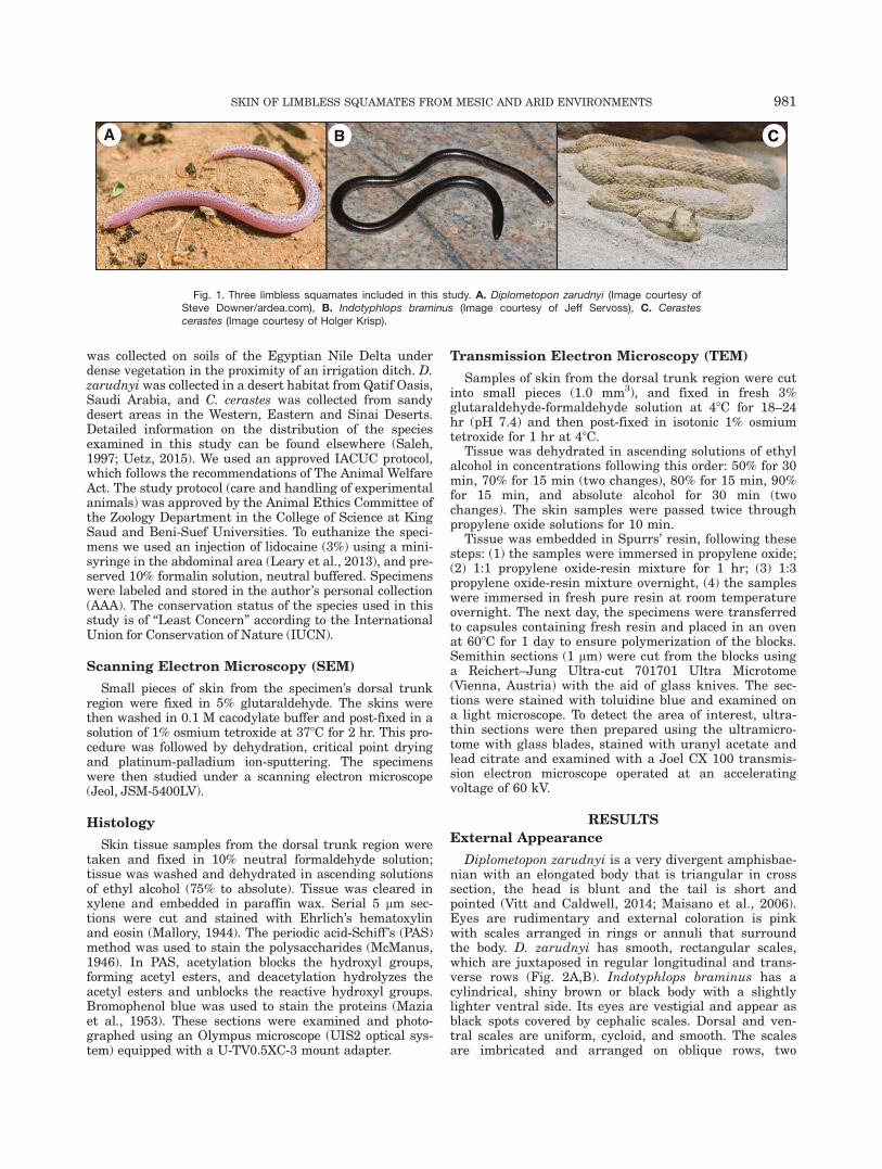
In this study we reviewed the histology of three limb-less, burrower squamates with different microhabitats inSaudi Arabia and Egypt—the introduced Indotyphlopsbraminus (Fig. 1A, Typhlopidae) which burrows onmesic soils, Diplometopon zarudnyi (Fig. 1B, Trogono-phiidae), and Cerastes cerastes (Fig. 1C, Viperidae)which burrow or dwell in dry and/or shifting sand envi-ronments (Said-Aliev, 1963). We focused on determiningadaptations to microhabitat selection of the surface andstructure of the skin using histological preparations,scanning electron microscope (SEM) and transmissionelectron microscope (TEM) images.
MATERIALS AND METHODS
Source of Specimens
Five adult specimens of each species were sampled indifferent localities in Egypt and Saudi Arabia. I. braminus
980 ALLAM ET AL.
was collected on soils of the Egyptian Nile Delta underdense vegetation in the proximity of an irrigation ditch. D.zarudnyi was collected in a desert habitat from Qatif Oasis,Saudi Arabia, and C. cerastes was collected from sandydesert areas in the Western, Eastern and Sinai Deserts.Detailed information on the distribution of the speciesexamined in this study can be found elsewhere (Saleh,1997; Uetz, 2015). We used an approved IACUC protocol,which follows the recommendations of The Animal WelfareAct. The study protocol (care and handling of experimentalanimals) was approved by the Animal Ethics Committee ofthe Zoology Department in the College of Science at KingSaud and Beni-Suef Universities. To euthanize the speci-mens we used an injection of lidocaine (3%) using a mini-syringe in the abdominal area (Leary et al., 2013), and pre-served 10% formalin solution, neutral buffered. Specimenswere labeled and stored in the author’s personal collection(AAA). The conservation status of the species used in thisstudy is of “Least Concern” according to the InternationalUnion for Conservation of Nature (IUCN).
Scanning Electron Microscopy (SEM)
Small pieces of skin from the specimen’s dorsal trunkregion were fixed in 5% glutaraldehyde. The skins werethen washed in 0.1 M cacodylate buffer and post-fixed in asolution of 1% osmium tetroxide at 378C for 2 hr. This pro-cedure was followed by dehydration, critical point dryingand platinum-palladium ion-sputtering. The specimenswere then studied under a scanning electron microscope(Jeol, JSM-5400LV).
Histology
Skin tissue samples from the dorsal trunk region weretaken and fixed in 10% neutral formaldehyde solution;tissue was washed and dehydrated in ascending solutionsof ethyl alcohol (75% to absolute). Tissue was cleared inxylene and embedded in paraffin wax. Serial 5 mm sec-tions were cut and stained with Ehrlich’s hematoxylinand eosin (Mallory, 1944). The periodic acid-Schiff ’s (PAS)method was used to stain the polysaccharides (McManus,1946). In PAS, acetylation blocks the hydroxyl groups,forming acetyl esters, and deacetylation hydrolyzes theacetyl esters and unblocks the reactive hydroxyl groups.Bromophenol blue was used to stain the proteins (Maziaet al., 1953). These sections were examined and photo-graphed using an Olympus microscope (UIS2 optical sys-tem) equipped with a U-TV0.5XC-3 mount adapter.
Transmission Electron Microscopy (TEM)
Samples of skin from the dorsal trunk region were cutinto small pieces (1.0 mm3), and fixed in fresh 3%glutaraldehyde-formaldehyde solution at 48C for 18–24hr (pH 7.4) and then post-fixed in isotonic 1% osmiumtetroxide for 1 hr at 48C.
Tissue was dehydrated in ascending solutions of ethylalcohol in concentrations following this order: 50% for 30min, 70% for 15 min (two changes), 80% for 15 min, 90%for 15 min, and absolute alcohol for 30 min (twochanges). The skin samples were passed twice throughpropylene oxide solutions for 10 min.
Tissue was embedded in Spurrs’ resin, following thesesteps: (1) the samples were immersed in propylene oxide;(2) 1:1 propylene oxide-resin mixture for 1 hr; (3) 1:3propylene oxide-resin mixture overnight, (4) the sampleswere immersed in fresh pure resin at room temperatureovernight. The next day, the specimens were transferredto capsules containing fresh resin and placed in an ovenat 608C for 1 day to ensure polymerization of the blocks.Semithin sections (1 mm) were cut from the blocks usinga Reichert–Jung Ultra-cut 701701 Ultra Microtome(Vienna, Austria) with the aid of glass knives. The sec-tions were stained with toluidine blue and examined ona light microscope. To detect the area of interest, ultra-thin sections were then prepared using the ultramicro-tome with glass blades, stained with uranyl acetate andlead citrate and examined with a Joel CX 100 transmis-sion electron microscope operated at an acceleratingvoltage of 60 kV.
RESULTS
External Appearance
Diplometopon zarudnyi is a very divergent amphisbae-nian with an elongated body that is triangular in crosssection, the head is blunt and the tail is short andpointed (Vitt and Caldwell, 2014; Maisano et al., 2006).Eyes are rudimentary and external coloration is pinkwith scales arranged in rings or annuli that surroundthe body. D. zarudnyi has smooth, rectangular scales,which are juxtaposed in regular longitudinal and trans-verse rows (Fig. 2A,B). Indotyphlops braminus has acylindrical, shiny brown or black body with a slightlylighter ventral side. Its eyes are vestigial and appear asblack spots covered by cephalic scales. Dorsal and ven-tral scales are uniform, cycloid, and smooth. The scalesare imbricated and arranged on oblique rows, two
Fig. 1. Three limbless squamates included in this study. A. Diplometopon zarudnyi (Image courtesy ofSteve Downer/ardea.com), B. Indotyphlops braminus (Image courtesy of Jeff Servoss), C. Cerastescerastes (Image courtesy of Holger Krisp).
SKIN OF LIMBLESS SQUAMATES FROM MESIC AND ARID ENVIRONMENTS 981
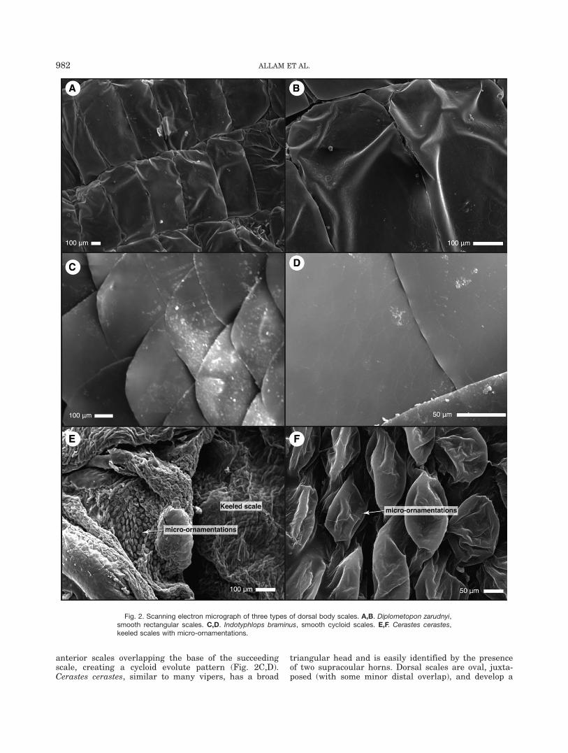
anterior scales overlapping the base of the succeedingscale, creating a cycloid evolute pattern (Fig. 2C,D).Cerastes cerastes, similar to many vipers, has a broad
triangular head and is easily identified by the presenceof two supracoular horns. Dorsal scales are oval, juxta-posed (with some minor distal overlap), and develop a
Fig. 2. Scanning electron micrograph of three types of dorsal body scales. A,B. Diplometopon zarudnyi,smooth rectangular scales. C,D. Indotyphlops braminus, smooth cycloid scales. E,F. Cerastes cerastes,keeled scales with micro-ornamentations.
982 ALLAM ET AL.
longitudinal keel. On the dorsal surface of the scale, itdevelops multiple micro-ornamentations (Fig. 2E,F).
Structure of the Skin
General structure of the epidermis of squamates fol-lows the known arrangement of several cell layers orstrata. One of the basal layers is the stratum germinati-vum, which is the precursor of the overlying epidermalcell layers. On top of this layer, there are a series oflayers, which in squamates alternate in the inner andouter generations (Landman, 1986). Overlying the stra-tum germinativum is the a-keratin layer, then a mucouslayer (meso), then the corneous b-protein layer, coveredby Oberha€utchen (which is generally the most superfi-cial layer of the epidermis). This layer, together with theouter keratinized layer of the epidermis tends to be lostin histological preparations (Lang, 1989). Pigment cellstermed melanocytes are located in the basal layer of theepidermis. These branched cells synthesize organelles(melanosomes) containing melanin pigment. In the
majority of lizards, melanosomes are found in the a-keratin and corneous b-protein layers (Vitt and Cald-well, 2014), while in snakes, melanosomes are only pres-ent in the corneous b-protein layer (Landman, 1986).
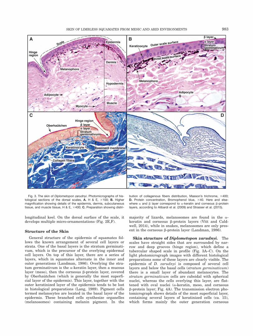
Skin structure of Diplometopon zarudnyi. Thescales have straight sides that are surrounded by nar-row and deep grooves (hinge region), which define atubercular shaped scale in profile (Fig. 3A–C). On thelight photomicrograph images with different histologicalpreparations some of these layers are clearly visible. Theepidermis of D. zarudnyi is composed of several celllayers and below the basal cells (stratum germinativum)there is a small layer of abundant melanocytes. Thestratum germinativum cells are cuboidal with sphericalnuclei, whereas the cells overlying this layer, are flat-tened with oval nuclei (a-keratin, meso, and corneousb-protein layer; Fig. 4A). The transmission electron pho-tomicrograph shows details of the most superficial layer,containing several layers of keratinized cells (ca. 15),which forms mainly the outer generation corneous
Fig. 3. The skin of Diplometopon zarudnyi. Photomicrographs of his-tological sections of the dorsal scales, A. H & E, 3100; B. Highermagnification showing details of the epidermis, dermis, subcutaneoustissue, and muscle tissue, H & E, 3400; C. Preparation showing distri-
bution of collagenous fibers distribution, Masson’s trichrome, 3400;D. Protein concentration, Bromophenol blue, 340. Here and else-where a and b layer correspond to a-keratin and corneous b-proteinlayers, according to Alibardi et al. (2009) and Strasser et al. (2015).
SKIN OF LIMBLESS SQUAMATES FROM MESIC AND ARID ENVIRONMENTS 983
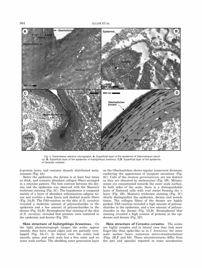
b-protein layer, and contains densely distributed mela-nosomes (Fig. 4A).
Below the epidermis, the dermis is at least four timesas thick, and contains abundant collagen fibers arrangedin a reticular pattern. The best contrast between the der-mis and the epidermis was observed with the Masson’strichrome staining (Fig. 3C). The hypodermis is composedmainly of a layer of abundant subcutaneous adipose tis-sue and overlies a deep fascia and skeletal muscle fibers(Fig. 3A,B). The PAS-reaction on the skin of D. zarudnyirevealed a moderate amount of polysaccharides in theepidermis and a low amount of polysaccharides in thedermis (Fig. 3A,B). Bromophenol blue staining of the skinof D. zarudnyi, revealed that proteins were scattered inthe epidermis and dermis (Fig. 3D).
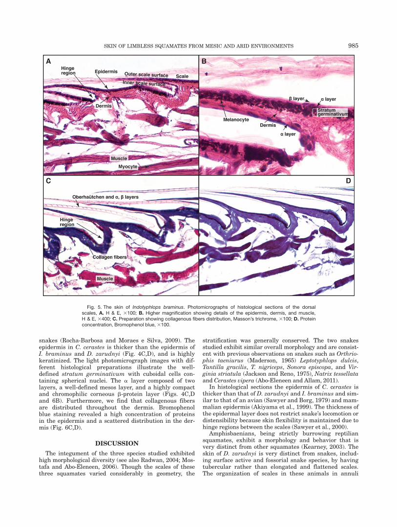
Skin structure of Indotyphlops braminus. Onthe light photomicrograph images the scales appearsmooth; they have round edges and are partially over-lapped (Fig. 5A–C). In lateral view the scales looksmooth, spiny, and each scale has a free outer and aninner scale surface. The shedding outer generation layer
on the Oberha€utchen shows regular transverse divisions,conferring the appearance of incipient serrations (Fig.5C). Cells of the stratum germinativum are not distinctas they are obscured by melanocytes (Fig. 5B). Melano-somes are concentrated towards the outer scale surface.In both sides of the scale, there is a distinguishablelayer of flattened cells with oval nuclei forming the alayer (Fig. 4B). Masson’s trichrome staining (Fig. 5C)clearly distinguishes the epidermis, dermis and muscletissue. The collagen fibers of the dermis are highlypacked. PAS reaction revealed a high amount of polysac-charides in the epidermis, and a low amount of polysac-charides in the dermis (Fig. 5A,B). Bromophenol bluestaining revealed a high content of proteins in the epi-dermis and dermis (Fig. 5D).
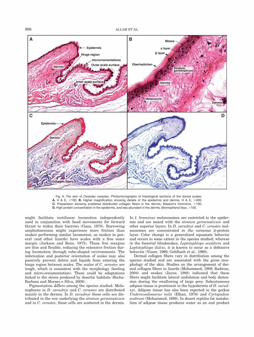
Skin structure of Cerastes cerastes. The scalesare highly complex and in lateral view they look morefinger-like than spike-like as in I. braminus; the outerscale surface bears numerous micro-ornamentations(Figs. 2E,F and 6A). These microstructures differ fromthe pits and spinules reported in some xenodontine
Fig. 4. Transmission electron micrographs. A. Superficial layer of the epidermis of Diplometopon zarud-nyi; B. Superficial layer of the epidermis of Indotyphlops braminus. C,D. Superficial layer of the epidermisof Cerastes cerastes.
984 ALLAM ET AL.
snakes (Rocha-Barbosa and Moraes e Silva, 2009). Theepidermis in C. cerastes is thicker than the epidermis ofI. braminus and D. zarudnyi (Fig. 4C,D), and is highlykeratinized. The light photomicrograph images with dif-ferent histological preparations illustrate the well-defined stratum germinativum with cuboidal cells con-taining spherical nuclei. The a layer composed of twolayers, a well-defined mesos layer, and a highly compactand chromophilic corneous b-protein layer (Figs. 4C,Dand 6B). Furthermore, we find that collagenous fibersare distributed throughout the dermis. Bromophenolblue staining revealed a high concentration of proteinsin the epidermis and a scattered distribution in the der-mis (Fig. 6C,D).
DISCUSSION
The integument of the three species studied exhibitedhigh morphological diversity (see also Radwan, 2004; Mos-tafa and Abo-Eleneen, 2006). Though the scales of thesethree squamates varied considerably in geometry, the
stratification was generally conserved. The two snakesstudied exhibit similar overall morphology and are consist-ent with previous observations on snakes such as Orthrio-phis taeniurus (Maderson, 1965) Leptotyphlops dulcis,Tantilla gracilis, T. nigriceps, Sonora episcopa, and Vir-ginia striatula (Jackson and Reno, 1975), Natrix tessellataand Cerastes vipera (Abo-Eleneen and Allam, 2011).
In histological sections the epidermis of C. cerastes isthicker than that of D. zarudnyi and I. braminus and sim-ilar to that of an avian (Sawyer and Borg, 1979) and mam-malian epidermis (Akiyama et al., 1999). The thickness ofthe epidermal layer does not restrict snake’s locomotion ordistensibility because skin flexibility is maintained due tohinge regions between the scales (Sawyer et al., 2000).
Amphisbaenians, being strictly burrowing reptiliansquamates, exhibit a morphology and behavior that isvery distinct from other squamates (Kearney, 2003). Theskin of D. zarudnyi is very distinct from snakes, includ-ing surface active and fossorial snake species, by havingtubercular rather than elongated and flattened scales.The organization of scales in these animals in annuli
Fig. 5. The skin of Indotyphlops braminus. Photomicrographs of histological sections of the dorsalscales, A. H & E, 3100; B. Higher magnification showing details of the epidermis, dermis, and muscle,H & E, 3400; C. Preparation showing collagenous fibers distribution, Masson’s trichrome, 3100; D. Proteinconcentration, Bromophenol blue, 3100.
SKIN OF LIMBLESS SQUAMATES FROM MESIC AND ARID ENVIRONMENTS 985
might facilitate rectilinear locomotion independentlyused in conjunction with head movements for forwardthrust to widen their burrows (Gans, 1978). Burrowingamphisbaenians might experience more friction thansnakes performing similar locomotion, as snakes in gen-eral (and other lizards) have scales with a free outermargin (Jackson and Reno, 1975). These free marginsare thin and flexible, reducing the extensive friction dur-ing locomotion through tube-shaped environments. Theimbrication and posterior orientation of scales may alsopassively prevent debris and liquids from entering thehinge region between scales. The scales of C. cerastes aretough, which is consistent with the morphology (keelingand micro-ornamentation). These could be adaptationslinked to the stress produced by desertic habitats (Rocha-Barbosa and Moraes e Silva, 2009).
Pigmentation differs among the species studied. Mela-nophores in D. zarudnyi and C. cerastes are distributedmainly in the dermis. In D. zarudnyi these cells are dis-tributed in the row underlying the stratum germinativumand in C. cerastes, these cells are scattered in the dermis.
In I. braminus melanosomes are restricted to the epider-mis and are mixed with the stratum germinativum andother superior layers. In D. zarudnyi and C. cerastes mel-anosomes are concentrated in the corneous b-proteinlayer. Color change is a generalized squamate behaviorand occurs to some extent in the species studied; whereasin the fossorial blindsnakes, Leptotyphlops scutiform andLeptotyphlops dulcis, it is known to occur as a defensivebehavior (Visser, 1966; Gehlbach et al., 1968).
Dermal collagen fibers vary in distribution among thespecies studied and are associated with the gross mor-phology of the skin. Studies on the arrangement of der-mal collagen fibers in lizards (Mohammed, 1989; Radwan,2004) and snakes (Jayne, 1988) indicated that thesefibers might facilitate lateral undulation and body disten-sion during the swallowing of large prey. Subcutaneousadipose tissue is prominent in the hypodermis of D. zarud-nyi. Adipose tissue has also been reported in the geckosUnderwoodisaurus milii (Elkan, 1976) and Cyrtopodionscabrum (Mohammed, 1989). In desert reptiles fat metabo-lism of adipose tissue produces water as an end product
Fig. 6. The skin of Cerastes cerastes. Photomicrographs of histological sections of the dorsal scales,A. H & E, 3100; B. Higher magnification showing details of the epidermis and dermis, H & E, 3400;C. Preparation showing scattered distributed collagen fibers in the dermis, Masson’s trichrome, 3100;D. High protein concentration in the epidermis, and less abundant in the dermis, Bromophenol blue, 3100.
986 ALLAM ET AL.
(Cloudsley-Thompson, 1971). This layer might also playa role in thermoregulation as an insulator or can serveas cushioning for the internal organs, especially consid-ering the external pressure consequence from burrowingthrough the soil.
The two snakes studied differed from the amphisbae-nian in the large amount of carbohydrates in the epider-mis and dermis. The epidermis of D. zarudnyi containedonly a moderate amount of polysaccharides and in thedermis just a small amount of PAS positive material wasobserved. This observation is consistent with previousreports in other amphisbaenians (Abo-Eleneen, 2008).
Polysaccharides were found distributed throughoutthe layers of the epidermis, except in the stratum germi-nativum, as well as in the dermis; thus, this observationis consistent with previous observations in lizards andsnakes (Mohammed, 1992). Polysaccharides are found inthe epidermis of metazoans and have been reported inthe integument of cephalopods (Srinivasan et al., 1969),echinoderms (Katzman and Jeanloz, 1969), fish (Senoet al., 1972; Banerjee 1980; Mittal and Banerjee, 1980),and snakes (Natrix tessellata and Cerastes vipera; Abo-Eleneen and Allam, 2011), these molecules have beenhypothesized to work as intercellular cement (Henkartet al., 1973). In some tetrapods, such as amphibians, rep-tiles, and birds, epithelial polysaccharides serve to defineintegumentary features (Sengel, 1976; Mohammed, 1984;Abdeen et al., 2008).
The bromophenol blue stain revealed different patternsof distribution in the skin of species studied, and I. bra-minus and C. cerastes resemble the distribution reportedin Natrix tessellata (Abo-Eleneen and Allam, 2011). As itis expected proteins are abundant in the scales; particu-larly where these molecules are needed to increasemechanical strength, hardness, and durability, whichseems to be advantageous especially in harsh environ-mental conditions (Abo-Eleneen and Allam, 2011).
The scales of D. zarudnyi are attached differentlyfrom the scales of snakes, which have an imbricationand a free edge. The integument plays an important rolein locomotion in limbless forms, especially in theamphisbaenian body, which is entirely in contact withthe substrate. The integument is associated withmuscles, and movement of the skin is determined by twosets of muscles in snakes and three sets in amphisbae-nians (Wiedemann, 1932; Gans, 1960), which explains inpart their distinct fossorial behavior. D. zarudnyi alsohas a regular arrangement of collagenous fibers and adense layer of subcutaneous adipose tissue, which canfunction as a source of energy and a water reservoir,and can also play a role in temperature insulation andas a cushion for the internal organs. The role of waterbalance of the skin in these animals cannot be addresseddirectly in this study. Considering the habitat selectionof these three species we can expect that D. zarudnyi ismore susceptible to water loss, especially in compactedsand dunes. I. braminus is also fossorial, but it dwells inmore mesic habitats or termite mounds where waterconditions might be more favorable. Although it can befound in sandy dry environments, C. cerastes is also sur-face active and can cover larger areas where it can finda balance between warm temperatures to bask, andhumidity to prevent dehydration (Johann, 1973; Mer-mod, 1970; Masood and Asiry, 2012).
In conclusion, the current study confirms that themorphological and histological configuration of the skinof D. zarudnyi, I. braminus and C. cerastes is adapted totheir habitat and lifestyle. The ultrastructure of the skinof more squamates dwelling in similar habitats needs tobe evaluated in order to confirm the observations of thisstudy.
ACKNOWLEDGEMENTS
The authors are very grateful with Elizabeth Glynneand Geneva E. Clark (Sam Houston State University),Alexandra Herrera (The George Washington University)and the Editorial Office from The Anatomical Record,especially to Cynthia Jensen, and two annonymousreviewers for their comments on the manuscript. Thisproject was supported by King Saud University, Dean-ship of Scientific Research, college of Science ResearchCenter.
LITERATURE CITED
Abdeen AM, Mostafa NA, Abo-Eleneen RE. 2008. Comparative his-tological and histochemical studies on the skin of some amphibianand reptilian species inhabiting Egypt. J Egypt Ger Soc Zool 56c:121–156.
Abo-Eleneen RE. 2008. Comparative studies on the functional mor-phology of the foot in some amphibian and reptilian species. PhDThesis, Beni-Suef University, Egypt.
Abo-Eleneen RE, Allam AA. 2011. Comparative morphology of theskin of Natrix tessellata (Family: Colubridae) and Cerastes vipera(Family: Viperidae). Zool Sci 28:743–748.
Akiyama M, Smith LT, Yoneda K, Holbrook KA, Hohl D, Shimizu H.1999. Peridermal cells form cornified cell envelope in their regres-sion process during human epidermal development. J InvestDermatol 112:903–909.
Allam AA, Abo-Eleneen RE. 2012. Scales microstructure of snakesfrom the Egyptian area. Zool Sci 29:770–775.
Alibardi L. Immunolocalization of large corneous beta-proteins inthe green anole lizard (Anolis carolinensis) suggests that theyform filaments that associate to the smaller beta-proteins in thebeta-layer of the epidermis. J Morphol 276:1244–1257.
Alibardi L, Toni M. 2006. Immunological characterization and finelocalization of a lizard beta-keratin. J Exp Zool B Mol Dev E 306:528–538.
Alibardi L, Valle LD, Nardi A, Toni M. 2009. Evolution of hard pro-teins in the sauropsid integument in relation to the cornificationof skin derivatives in amniotes. J Anat 214:560–586.
Bagnara JT. 1998. Comparative anatomy and physiology of pigmentcells in nonmammalian tissues. In: Nordlund JJ, Boissy RE,Hearing VJ, King RA, Ortonne J, editors. The pigmentary sys-tem. New York: Oxford University Press. p 9–40.
Bagnara JT, Matsumoto J, Frost SK, Turner WA, Tchen TT, TaylorJD. 1979. The common origin of pigment cells. Science 203:410–416.
Baio JE, Spinner M, Jaye C, Fischer DA, Gorb SN, Weidner T.2015. Evidence of a molecular boundary lubricant at snakeskinsurfaces. J R Soc Interface 12:20150817.
Banerjee TK. 1980. Histochemistry of snake epidermis. In: Spear-man RIC, Riley PA, editors. The skin of vertebrates. London: Aca-demic press. p 23–32.
Banerjee TK, Mittal AK. 1978. Epidermal mucous cell in the cheq-uered water snake Natrix piscator (Colubridae, Squamata). Acytochemical investigation. Zool J Linn Soc 63:289–293.
Calsbeek R, Knouft JH, Smith TB. 2006. Variation in scale numbersis consistent with ecologically based natural selection actingwithin and between lizard species. Evol Ecol 20:377–394.
Cloudsley-Thompson JL. 1971. The temperature and water rela-tions of reptiles. Watford: Merrow Technical Library.
SKIN OF LIMBLESS SQUAMATES FROM MESIC AND ARID ENVIRONMENTS 987
Daza JD. 2014. What’s so special about squamates? Anat Rec 297:341–343.
Ditmars RL. 1931. Snakes of the world. New York: The MacmillanCompany.
Elkan E. 1976. Mucopolysaccharides in reptilian skin. Israel J Zool25:73–94.
Gans C. 1960. Studies on amphisbaenids (Amphisbaenia, Reptilia).1, A taxonomic revision of the Trogonophinae, and a functionalinterpretation of the amphisbaenid adaptive pattern. Bull AmMus Nat Hist 119:129–204.
Gans C. 1962. Terrestrial locomotion without limbs. Am Zool 2:167–182.
Gans C. 1974. Biomechanics: an approach of vertebrate biology.Philadelphia and Toronto: JB Lippincott.
Gans C. 1978. The characteristics and affinities of the Amphisbae-nia. Trans Zool Soc Lond 34:347–416.
Gehlbach FR, Watkins JF, Reno HW. 1968. Blind snake defensivebehavior elicited by ant attacks. Bioscience 18:781–785.
Gunderson AR, Siegel J, Leal M. 2011. Tests of the contribution ofacclimation to geographic variation in water loss rates of the WestIndian lizard Anolis cristatellus. J Comp Physiol B 181:965–972.
Hall BK. 1980. Chondrogenesis and osteogenesis of cranial neuralcrest cells. In: Pratt RM, Christiansen RL, editors. Currentresearch trends in prenatal craniofacial development. New Yorkand Amsterdam: Elsevier/North Holland. p 47–64.
Henkart P, Humphreys S, Humphreys T. 1973. Characterization ofa sponge aggregation factor. A unique proteoglycan complex. Bio-chemistry 12:3045–3050.
Jackson MK, Reno HW. 1975. Comparative skin structure of somefossorial and subfossorial leptotyphlopid and colubrid snakes.J Herpetol 31:355–359.
Jayne BC. 1988. Mechanical behavior of snake skin. J Zool Lond214:125–140.
Johann H. 1973. A location of origin for Cerastes cerastes in north-ern Tunisia Serpentes Viperidae. Salamandra 9:160.
Jones SJ, Boyde A. 1974. The organization and gross mineralizationpatterns of the collagen fibrils in Sharpey fiber bone. Cell TissueRes 148:83–96.
Katzman RL, Jeanloz RW. 1969. Acid polysaccharides from inverte-brate connective tissue: phylogenetic aspects. Science 166:758–759.
Kearney M. 2003. Systematics of the Amphisbaenia (Lepidosauria:Squamata) based on morphological evidence from recent and fos-sil forms. Herpetol Monogr 17:1–74.
Landmann L. 1986. The skin of reptiles: epidermis and dermis. In:Bereiter-Hahn J, Matoltsy AG, Sylvia-Richards K, editors. Biologyof the integument. Vol. 2. Vertebrates. Berlin: Springer. p 150–187.
Lang M. 1989. The morphology of the Oberh€autchen with thedescription and distribution of scale organs in basiliscine igua-nians. Amphibia-Reptilia 10:423–434.
Lange B. 1931. Integument der sauropsiden. In: Bolk L, Kallius E,Lubosch W, editors. Handbuch der vergleichenden Anatomie derWirbeltiere. Vol. 2. No. 1. Berlin and Vienna: Urban und Schwar-zenberg. p 375–447.
Leary S, Underwood W, Anthony R, Cartner S, Corey D, Grandin T,Greenacre C, Gwaltney-Brant S, McCrackin MA, Meyer R, MillerD, Shearer J, Yanong R. 2013. AVMA guidelines for the euthana-sia of animals: 2013 Edition. Schaumburg, Illinois: American Vet-erinary Medical Association.
Licht P, Bennett AF. 1972. A scaleless snake: test of the role of rep-tilian scales in water loss and heat transfer. Copeia 1972:702–707.
Lillywhite HB. 2006. Water relations of tetrapod integument. J ExpBiol 209:202–226.
Lillywhite HB, Maderson PFA. 1982. Structure and permeability.In: Gans C, Pough FH, editors. Biology of the reptilia. Vol. 12.Physiology C. London: Academic Press. p 397–442.
Maderson PFA. 1965. Histological change in the epidermis ofsnakes during the sloughing cycle. J Zool Lond 146:98–113.
Maderson PFA, Rabinowitz T, Tandler B, Alibardi L. 1998. Ultra-structural contributions to an understanding of the cellular mech-
anisms involved in lizard skin shedding with comments on thefunction and evolution of a unique lepidosaurian phenomenon.J Morphol 236:1–24.
Maisano JA, Kearney M, Rowe T. 2006. Cranial anatomy of thespade-headed amphisbaenian Diplometopon zarudnyi (Squamata,Amphisbaenia) based on high-resolution X-ray computed tomogra-phy. J Morphol 267:70–102.
Mallory FB. 1944. Pathological technique. Philadelphia: WB Saunders.Masood MF, Asiry AA. Ecological studies on diversity of Herpeto-
fauna in Asir region, Kingdom of Saudi Arabia. Egypt Acad JBiolog Sci 4:143–163.
Mazia D, Brewer PA, Alfert M. 1953. The cytochemical staining andmeasurement of protein with mercuric bromophenol blue. BiolBull 104:57–67.
McManus JF. 1946. The histological demonstration of mucin afterperiodic acid. Nature 158:202–211.
Mermod C. 1970. Living area and displacement activity of Cerastesvipera and Cerastes cerastes reptilia viperidae. Rev Suisse Zool77:555–562.
Miller ME, Lutterschmidt WI. 2014. Cutaneos water loss and epi-dermal lipids in two sympatric congeneric pitvipers. J Herpetol48:577–583.
Mittal AK, Banerjee TK. 1980. Keratinization versus mucus secre-tion in fish epidermis. In: Spearman RIC, Riley RA, editors. Theskin of vertebrates, Linnean Society Symposium No. 9. London:Academic Press. p 1–11.
Mohammed MBH. 1984. Development of the lizard limb as shownby the distribution of sulphate incorporation. J Anat 138:399–403.
Mohammed MBH. 1989. Histochemistry of the skin of the geckoCyrtodactylus scaber (Gekkonidae, Reptilia). Qatar Univ Sci Bull9:199–210.
Mohammed MBH. 1992. Skin mucopolysaccharides in some lizardsand snakes. J Egypt Ger Soc Zool 7:65–76.
Mostafa NA, Abo-Eleneen RE. 2006. Gross morphology of the footand the locomotion pattern in sand dune-dwelling lizards.J Union Arab Biol Cairo 24:203–232.
Noden DM. 1980. The migration and cytodifferentiation of cranialneural crest cells. In: Pratt RM, Christiansen RL, editors. Cur-rent research trends in prenatal craniofacial development. NewYork: Elsevier/North Holland. p 3–25.
Pianka ER. 1986. Ecology and natural history of desert lizards:analyses of the ecological niche and community structucture.Princeton: Princeton University Press.
Pianka ER. 1989. Desert lizard diversity: additional comments andsome data. Am Nat 134:344–364.
Pianka ER, Vitt LJ. 2003. Lizards: windows to the evolution ofdiversity. Berkeley: University of California Press.
Pincheira-Donoso D, Bauer AM, Meiri S, Uetz P. 2013. Global taxo-nomic diversity of living reptiles. PLoS One 8:e59741.
Pough FH, Andrews RM, Crump ML, Savitzky AH, Wells KD,Brandley MC. 2016. Herpetology. 4th ed. Sunderland: Sinauer.
Pough FH, Groves JD. 1983. Specialization of the body form andfood habits of snake. Am Zool 23:443–454.
Pough FH, Janis CM, Heiser JB. 2013. Vertebrate life. 9th ed. Bos-ton: Pearson.
Radwan E. 2004. Functional anatomy of the skin in some lizards.M.Sc. Thesis, EL-Fayoum University, Egypt.
Rocha-Barbosa O, Moraes e Silva RB. 2009. Analysis of the micro-structure of Xenodontinae snake scales associated with differenthabitat occupation strategies. Braz J Biol 69:919–923.
Said-Aliev SA. 1963. Notes on the distribution of certain reptiles ofTadzhikistan. Dokl Akad Nauk SSSR 6:54–57.
Saleh MA. 1997. Amphibians and reptiles of Egypt. Pub Natl Bio-diver 6:1–234.
Sawyer RH, Borg TK. 1979. Avian scale development. VI. Ultra-structure of the keratinizing cells of reticulate scales. J Morphol161:111–122.
Sawyer RH, Glenn T, French JO, Mays B, Shames RB, Barnes GL,Rhodes W, Ishikawa Y. 2000. The expression of beta (b) keratins inthe epidermal appendages of reptiles and birds. Am Zool 40:530–539.
Sengel P. 1976. Morphogenesis of skin. Cambridge: Cambridge Uni-versity Press.
988 ALLAM ET AL.
Seno N, Akiyama F, Anno K. 1972. A novel dermation polysulphatefrom hagfish skin, containing trisulphated disaccharide residues.Biochim Biophys Acta 264:229–233.
Spearman RIC. 1969. The epidermis of the gopher tortoise Testudopolyphemus (Daudin). Acta Zool 50:1–9.
Spearman RIC, Riley PA. 1969. A comparison of the epidermis andpigment cells of the crocodile with those in two lizard species.J Zool Linn Soc 48:453–466.
Srinivasan SR, Radhakrish-Namurthy B, Dalferes ER, BerensonGS. 1969. Glycosaminoglycans from squid skin. Comp BiochemPhysiol 28:169–176.
Strasser B, Mlitz V, Hermann M, Rice RH, Eigenheer RA, AlibardiL, Tschachler E, Eckhart L. 2014. Evolutionary origin and diver-sification of epidermal barrier proteins in amniotes. Mol Biol E31:3194–3205.
Tulli MJ, Robles CI. 2015. >Qu�e nos dicen las escamas acerca delclima de una regi�on? XVI Congreso Argentino de Herpetolog�ıa,San Miguel de Tucum�an, Argentina, Septiembre 29 – Octubre 2,2015. Tucum�an: Asociaci�on Herpetol�ogica Argentina. p 116.
Uetz P. 2015. The reptile database. Available at: http://www.reptile-database.org. Accessed 2015 December 8.
Visser J. 1966. Color change in Leptotyphlops scutifroms and noteson its defensive behavior. Zool Afr 2:123–125.
Vitt LJ, Caldwell JP. 2014. Herpetology. 4th ed. London: AcademicPress.
Wegener JE, Gartner GEA, Losos JB. 2014. Lizard scales in an adapt-ive radiation: variation in scale number follows climatic and struc-tural habitat diversity in Anolis lizards. Biol J Linn Soc 113:570–579.
Wiedemann 1932. Zur Ortsbewegung der Schlangen und Schlei-chen. Zool Jahrb Abt Allgem Zool Phys Tiere 50:557–596.
SKIN OF LIMBLESS SQUAMATES FROM MESIC AND ARID ENVIRONMENTS 989