lecture 1 : introduction to histology and cell structureksumsc.com/download_center/1st/1. foundation...
TRANSCRIPT
Lecture 1 :Introduction to Histology and
Cell Structure- Colours index :Red : importantGrey : notesPink : Girls slides

Objectives :
● What is histology and how it is studied? ● Composition of the cell: Light microscopic (L/M) and electron
microscopic (E/M) and● function of each component:1. Nucleus 2. Cytoplasm ● Organelles: 1. membranous and non-membranous 2. Inclusions
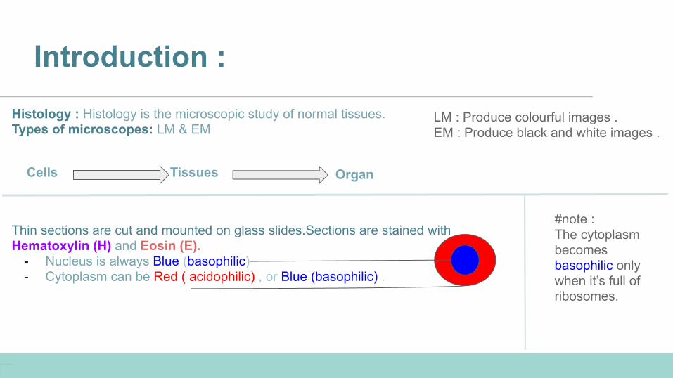
Introduction :Histology : Histology is the microscopic study of normal tissues.Types of microscopes: LM & EM
Cells Tissues Organ
Thin sections are cut and mounted on glass slides.Sections are stained withHematoxylin (H) and Eosin (E).
- Nucleus is always Blue (basophilic)- Cytoplasm can be Red ( acidophilic) , or Blue (basophilic) .
#note : The cytoplasm becomes basophilic only when it’s full of ribosomes.
LM : Produce colourful images .EM : Produce black and white images .
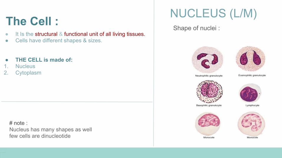
The Cell : ● It Is the structural & functional unit of all living tissues.● Cells have different shapes & sizes.
● THE CELL is made of: 1. Nucleus2. Cytoplasm
NUCLEUS (L/M)Shape of nuclei :
# note : Nucleus has many shapes as wellfew cells are dinucleotide
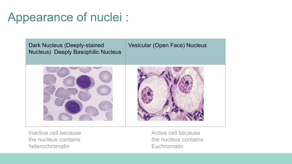
Appearance of nuclei :
Dark Nucleus (Deeply-stained Nucleus) Deeply Basophilic Nucleus
Vesicular (Open Face) Nucleus
Inactive cell because the nucleus contains heterochromatin
Active cell because the nucleus contains Euchromatin
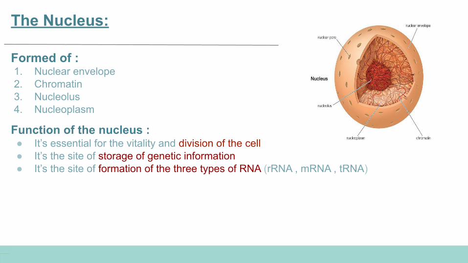
The Nucleus:
Formed of : 1. Nuclear envelope 2. Chromatin3. Nucleolus 4. Nucleoplasm
Function of the nucleus : ● It’s essential for the vitality and division of the cell ● It’s the site of storage of genetic information ● It’s the site of formation of the three types of RNA (rRNA , mRNA , tRNA)

Nuclear envelope :
Definition:A double membrane with many pores surroundings the Nucleus .
Formed of :1- Outer membrane2- Inner membrane3- Nuclear pores ( provide communication between nucleus and cytoplasm)
#note : * the only double membrane surrounded structures in the cell are Nucleus and Mitochondria .
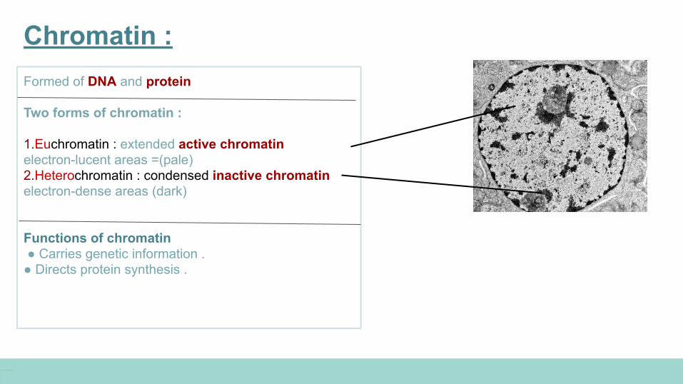
Chromatin : Formed of DNA and protein Two forms of chromatin :
1.Euchromatin : extended active chromatin electron-lucent areas =(pale) 2.Heterochromatin : condensed inactive chromatin electron-dense areas (dark)
Functions of chromatin ● Carries genetic information .● Directs protein synthesis .
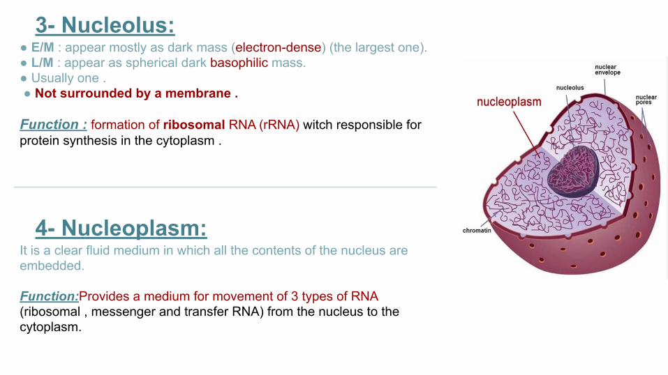
3- Nucleolus:● E/M : appear mostly as dark mass (electron-dense) (the largest one). ● L/M : appear as spherical dark basophilic mass. ● Usually one . ● Not surrounded by a membrane .
Function : formation of ribosomal RNA (rRNA) witch responsible for protein synthesis in the cytoplasm .
4- Nucleoplasm:It is a clear fluid medium in which all the contents of the nucleus are embedded.
Function:Provides a medium for movement of 3 types of RNA(ribosomal , messenger and transfer RNA) from the nucleus to the cytoplasm.
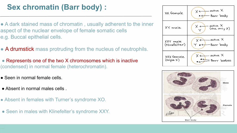
Sex chromatin (Barr body) :
● A dark stained mass of chromatin , usually adherent to the inner aspect of the nuclear envelope of female somatic cells e.g. Buccal epithelial cells.
● A drumstick mass protruding from the nucleus of neutrophils.
● Represents one of the two X chromosomes which is inactive (condensed) in normal female (heterochromatin).
● Seen in normal female cells.
● Absent in normal males cells .
● Absent in females with Turner’s syndrome XO.
● Seen in males with Klinefelter’s syndrome XXY.
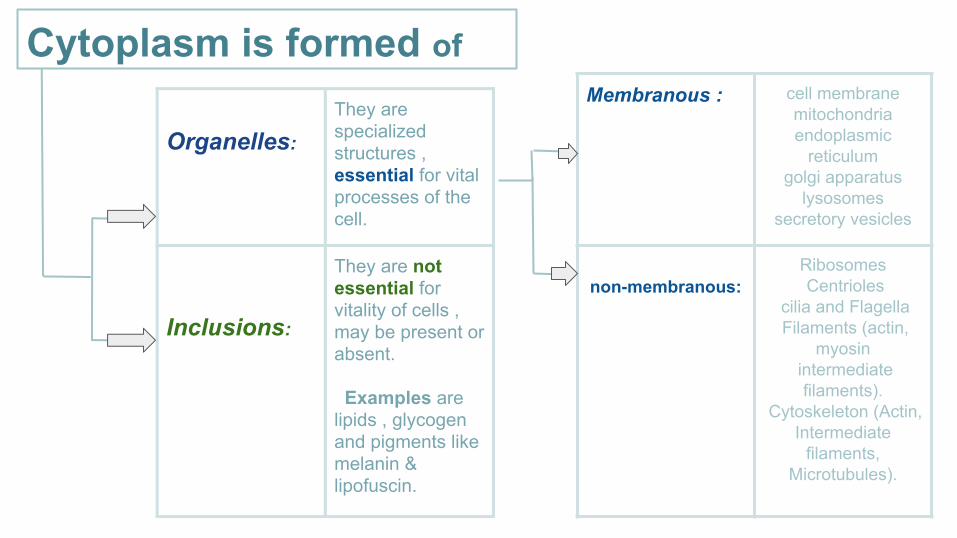
Organelles:
They are specialized structures , essential for vital processes of the cell.
Inclusions:
They are not essential for vitality of cells , may be present or absent.
Examples are lipids , glycogen and pigments like melanin & lipofuscin.
Membranous : cell membrane mitochondriaendoplasmic
reticulumgolgi apparatus
lysosomessecretory vesicles
non-membranous: Ribosomes Centrioles
cilia and Flagella Filaments (actin,
myosin intermediate
filaments). Cytoskeleton (Actin,
Intermediate filaments,
Microtubules).
Cytoplasm is formed of
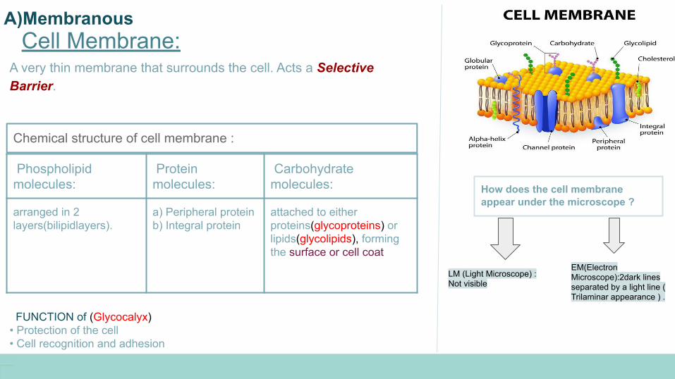
Cell Membrane:
How does the cell membrane appear under the microscope ?
LM (Light Microscope) : Not visible
EM(Electron Microscope):2dark lines separated by a light line ( Trilaminar appearance ) .
A very thin membrane that surrounds the cell. Acts a Selective Barrier.
Phospholipid molecules:
Protein molecules:
Carbohydrate molecules:
arranged in 2 layers(bilipidlayers).
a) Peripheral protein b) Integral protein
attached to either proteins(glycoproteins) or lipids(glycolipids), forming the surface or cell coat
Chemical structure of cell membrane :
FUNCTION of (Glycocalyx) • Protection of the cell• Cell recognition and adhesion
A)Membranous
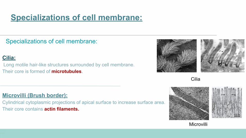
Specializations of cell membrane:
Specializations of cell membrane:
Cilia: Long motile hair-like structures surrounded by cell membrane.Their core is formed of microtubules.
Microvilli (Brush border):Cylindrical cytoplasmic projections of apical surface to increase surface area.Their core contains actin filaments.
Microvilli
Cilia
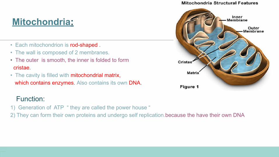
Mitochondria:
• Each mitochondrion is rod-shaped .• The wall is composed of 2 membranes. • The outer is smooth, the inner is folded to form cristae.• The cavity is filled with mitochondrial matrix, which contains enzymes. Also contains its own DNA.
Function:1) Generation of ATP “ they are called the power house “2) They can form their own proteins and undergo self replication.because the have their own DNA
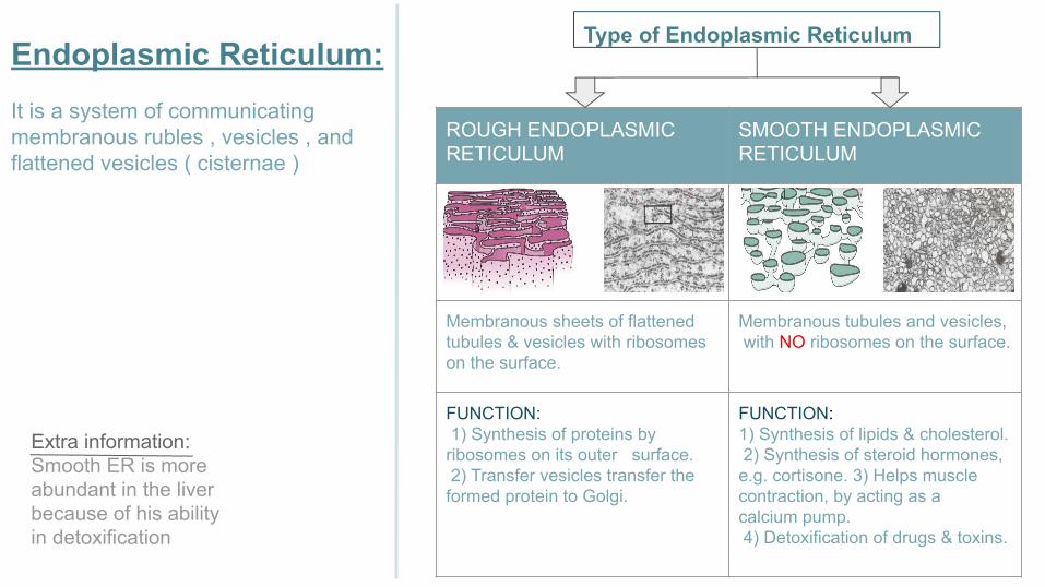
ROUGH ENDOPLASMIC RETICULUM
SMOOTH ENDOPLASMIC RETICULUM
Membranous sheets of flattened tubules & vesicles with ribosomes on the surface.
Membranous tubules and vesicles, with NO ribosomes on the surface.
FUNCTION: 1) Synthesis of proteins by ribosomes on its outer surface. 2) Transfer vesicles transfer the formed protein to Golgi.
FUNCTION: 1) Synthesis of lipids & cholesterol. 2) Synthesis of steroid hormones, e.g. cortisone. 3) Helps muscle contraction, by acting as a calcium pump. 4) Detoxification of drugs & toxins.
Extra information: Smooth ER is more abundant in the liver because of his ability in detoxification
Endoplasmic Reticulum:It is a system of communicating membranous rubles , vesicles , and flattened vesicles ( cisternae )
Type of Endoplasmic Reticulum
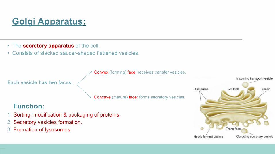
Golgi Apparatus:
• The secretory apparatus of the cell. • Consists of stacked saucer-shaped flattened vesicles.
Each vesicle has two faces:
Function:1. Sorting, modification & packaging of proteins.2. Secretory vesicles formation. 3. Formation of lysosomes
Convex (forming) face: receives transfer vesicles.
Concave (mature) face: forms secretory vesicles.
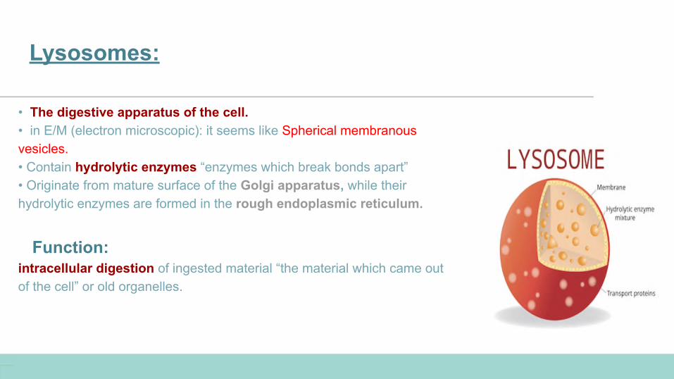
Lysosomes:
• The digestive apparatus of the cell. • in E/M (electron microscopic): it seems like Spherical membranous vesicles. • Contain hydrolytic enzymes “enzymes which break bonds apart”• Originate from mature surface of the Golgi apparatus, while their hydrolytic enzymes are formed in the rough endoplasmic reticulum.
Function:intracellular digestion of ingested material “the material which came out of the cell” or old organelles.
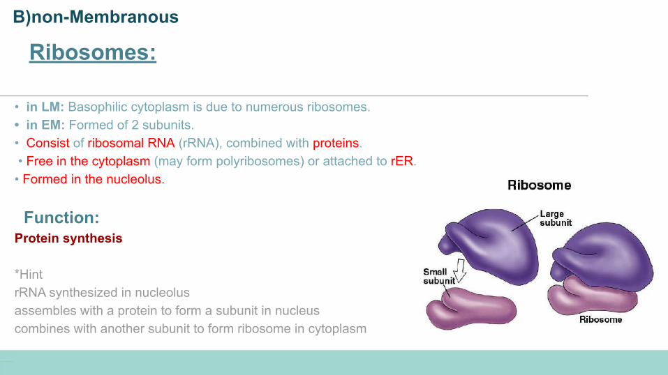
Ribosomes:
• in LM: Basophilic cytoplasm is due to numerous ribosomes.• in EM: Formed of 2 subunits. • Consist of ribosomal RNA (rRNA), combined with proteins. • Free in the cytoplasm (may form polyribosomes) or attached to rER. • Formed in the nucleolus.
Function:Protein synthesis
*HintrRNA synthesized in nucleolusassembles with a protein to form a subunit in nucleus combines with another subunit to form ribosome in cytoplasm
B)non-Membranous
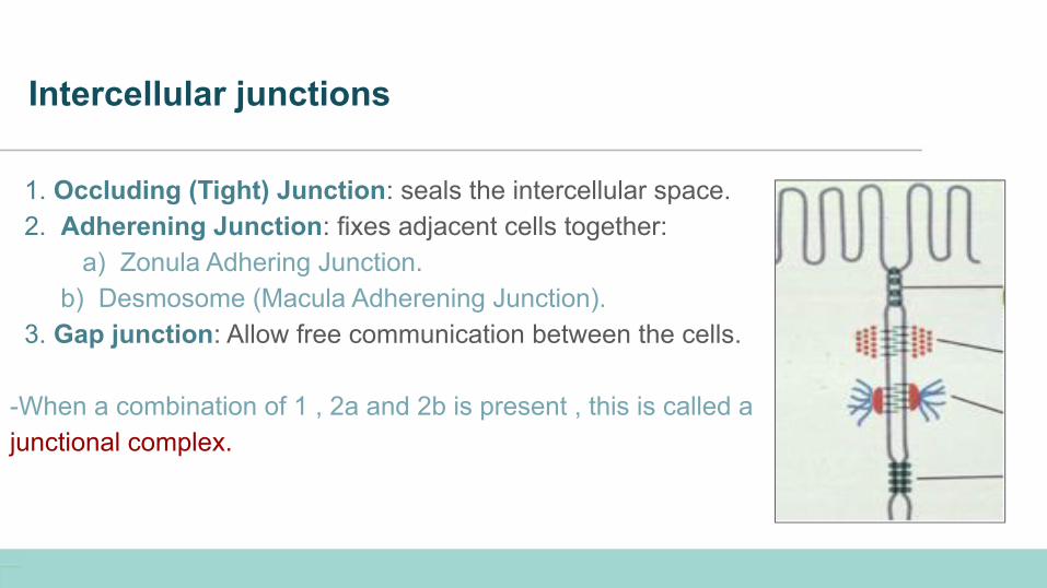
Intercellular junctions
1. Occluding (Tight) Junction: seals the intercellular space. 2. Adherening Junction: fixes adjacent cells together: a) Zonula Adhering Junction. b) Desmosome (Macula Adherening Junction). 3. Gap junction: Allow free communication between the cells.
-When a combination of 1 , 2a and 2b is present , this is called a junctional complex.
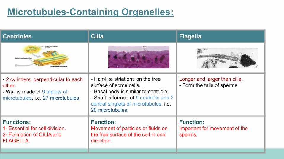
Microtubules-Containing Organelles:
Centrioles Cilia Flagella
- 2 cylinders, perpendicular to each other.- Wall is made of 9 triplets of microtubules, i.e. 27 microtubules
- Hair-like striations on the free surface of some cells.- Basal body is similar to centriole.- Shaft is formed of 9 doublets and 2 central singlets of microtubules, i.e. 20 microtubules.
Longer and larger than cilia.- Form the tails of sperms.
Functions:1- Essential for cell division.2- Formation of CILIA and FLAGELLA.
Function:Movement of particles or fluids on the free surface of the cell in one direction.
Function:Important for movement of the sperms.
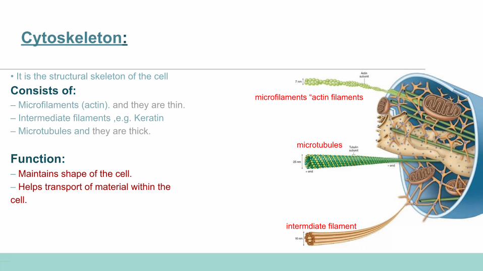
Cytoskeleton:
• It is the structural skeleton of the cellConsists of:– Microfilaments (actin). and they are thin.– Intermediate filaments ,e.g. Keratin– Microtubules and they are thick.
Function:– Maintains shape of the cell.– Helps transport of material within thecell.
intermdiate filament
microtubules
microfilaments “actin filaments
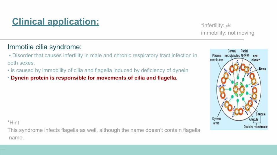
Clinical application:
Immotile cilia syndrome: • Disorder that causes infertility in male and chronic respiratory tract infection in both sexes.• is caused by immobility of cilia and flagella induced by deficiency of dynein• Dynein protein is responsible for movements of cilia and flagella.
*HintThis syndrome infects flagella as well, although the name doesn’t contain flagella name.
*infertility: عقمimmobility: not moving
Q1: it’s the structural and functional unit of all living tissues?
MCQs:
1. The nucleus 2. The cell 3. Mitochondria4. Nuclear envelope
Q2: provides a medium for movement of 3 types of RNA?
1. Nucleoplasm2. Barr body 3. Flagella 4. Cilia
Q3: Composed of 2 membranes ?
1. Mitochondria 2. Nucleus 3. Cell membrane 4. Both 1&2
Q4: which part of the cell compartment is more abundant in the liver ?
1. Rough ER2. Smooth ER3. Golgi apparatus 4. Centrioles
● Answers : ● Q1 : 2● Q2 : 1 ● Q3 : 4 ● Q4 : 2

Team members
Team leaders
● Yazeed Alomar
● Abdulmohsen Albesher
● Abdullah Alburikan
● Mohamed Albabtain
● Mohammed Benhjji
● Mohamed Alquhidan
● Nawaf Alshahrani
● Afnan AlMohsen
● Nourah Alklaib
● Sarah Alobaid
● Sumo Abdulrahman
● Mariam Alruhaimi
● Joud Alarifi
Albara Aldawoud Fatimah AlhelalContact us through :
[email protected] future corrections will be in the editing file :Click Here