geranium robertianum extract
TRANSCRIPT

38
Geranium Robertianum Extract
ICHIMARU PHARCOS CO., LTD. 318-1 Asagi, Motosu-shi, Gifu 501-0475 JAPAN
Phone : (81) 58 320-1032 Fax : (81) 58 320-1039
http://www.ichimaru.co.jp
Requests concerning with intellectual property rights and others The catalogues, technical documents, samples and the like materials of our company’s products that you are provided for this time, are supplied in favor of a confidential relationship between us, and you are strictly requested not to use them in any form as your intellectual property right or like properties. In addition, the contents represented or described in these materials may concern with intellectual property rights owned by others, so that you are respectfully requested to understand and consider that the use and handling of these materials are to be dealt with finally on your own responsibility.
Published on October 6, 2006Revised on May 25, 2007

39
C O N T E N T S
1. Tryptase and Photo aging - 1-
2. Origin - 4-
3. Introduction - 6-
4. Efficacy
Inhibitory Effect on Tryptase - 7-
Inhibitory Effect on Tryptase in epidermal cells - 9-
Inhibitory Effect on Tryptase in human skin 3D model -10-
Inhibitory Effect on Type I Collagen Degradation -11-
Inhibitory Effect on Type IV Collagen Degradation -13-
Improvement of Skin Elasticity on the Human Skin -15-
Recovering of Wrinkle on the Human Skin -17-
Penetration into skin -20-
Moisturizing -22-
Inhibitory Effect on Melanin Production -25-
Inhibitory Effect on Histamine Release -26-
SOD Like Activity -29-
8.Stability -30-
9.Compatibility -33-
10.Specification -36-
11. Reference -37-
1
Tryptase and Photo aging
Mast cell and Tryptase
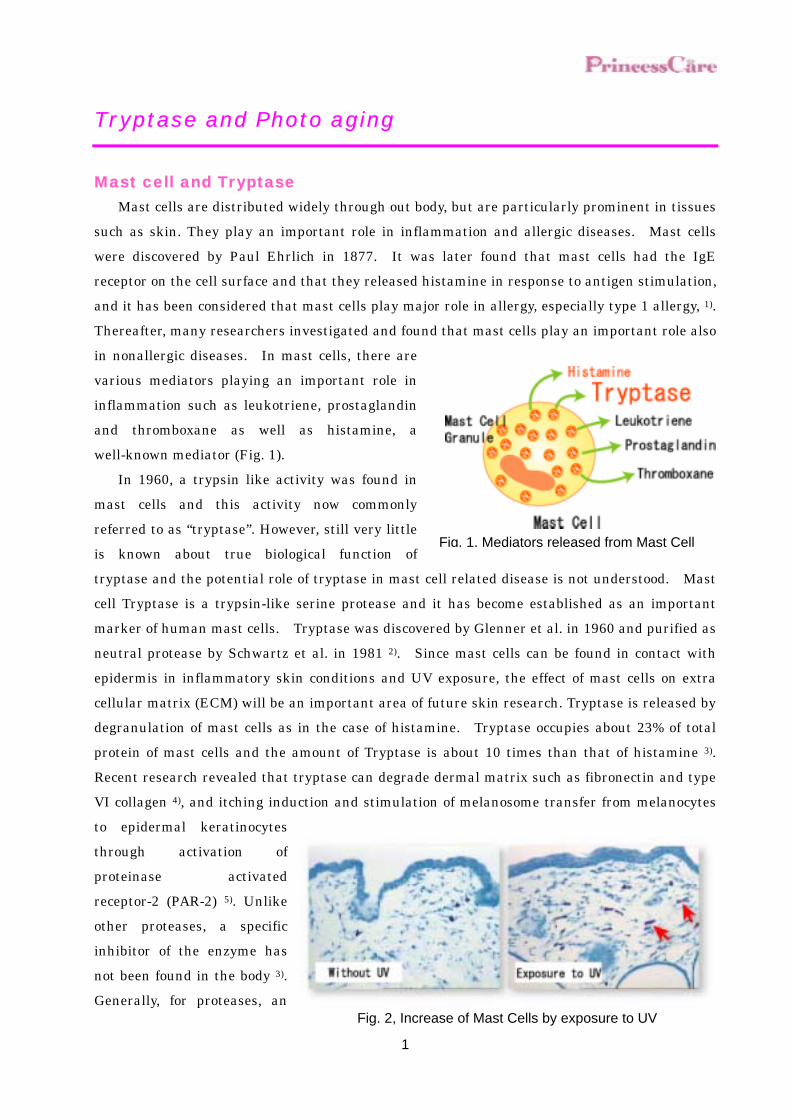
Mast cells are distributed widely through out body, but are particularly prominent in tissues such as skin. They play an important role in inflammation and allergic diseases. Mast cells were discovered by Paul Ehrlich in 1877. It was later found that mast cells had the IgE receptor on the cell surface and that they released histamine in response to antigen stimulation, and it has been considered that mast cells play major role in allergy, especially type 1 allergy, 1). Thereafter, many researchers investigated and found that mast cells play an important role also in nonallergic diseases. In mast cells, there are various mediators playing an important role in inflammation such as leukotriene, prostaglandin and thromboxane as well as histamine, a well-known mediator (Fig. 1).
In 1960, a trypsin like activity was found in mast cells and this activity now commonly referred to as “tryptase”. However, still very little is known about true biological function of tryptase and the potential role of tryptase in mast cell related disease is not understood. Mast cell Tryptase is a trypsin-like serine protease and it has become established as an important marker of human mast cells. Tryptase was discovered by Glenner et al. in 1960 and purified as neutral protease by Schwartz et al. in 1981 2). Since mast cells can be found in contact with epidermis in inflammatory skin conditions and UV exposure, the effect of mast cells on extra cellular matrix (ECM) will be an important area of future skin research. Tryptase is released by degranulation of mast cells as in the case of histamine. Tryptase occupies about 23% of total protein of mast cells and the amount of Tryptase is about 10 times than that of histamine 3). Recent research revealed that tryptase can degrade dermal matrix such as fibronectin and type VI collagen 4), and itching induction and stimulation of melanosome transfer from melanocytes to epidermal keratinocytes through activation of proteinase activated receptor-2 (PAR-2) 5). Unlike other proteases, a specific inhibitor of the enzyme has not been found in the body 3). Generally, for proteases, an
Fig. 2, Increase of Mast Cells by exposure to UV
Fig. 1, Mediators released from Mast Cell
2
inhibitor exists in the body, for example trypsin inhibitor for trypsin, to regulate the enzyme activity. For Tryptase, however, a specific inhibitor has not been found in the body and the Tryptase activity is hardly inhibited by general protease inhibitors. Therefore, the Tryptase activity is not regulated positively in the body and there is a possibility that the effect of Tryptase may be maintained for a long time once secreted.
Tryptase and Photo aging Bosset et al. has recently reported
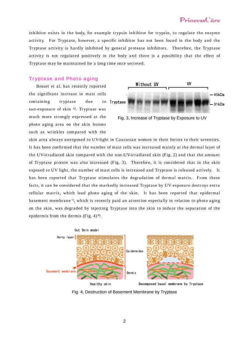
the significant increase in mast cells containing tryptase due to sun-exposure of skin 6). Tryptase was much more strongly expressed at the photo aging area on the skin lesions such as wrinkles compared with the skin area always unexposed to UV-light in Caucasian women in their forties to their seventies.
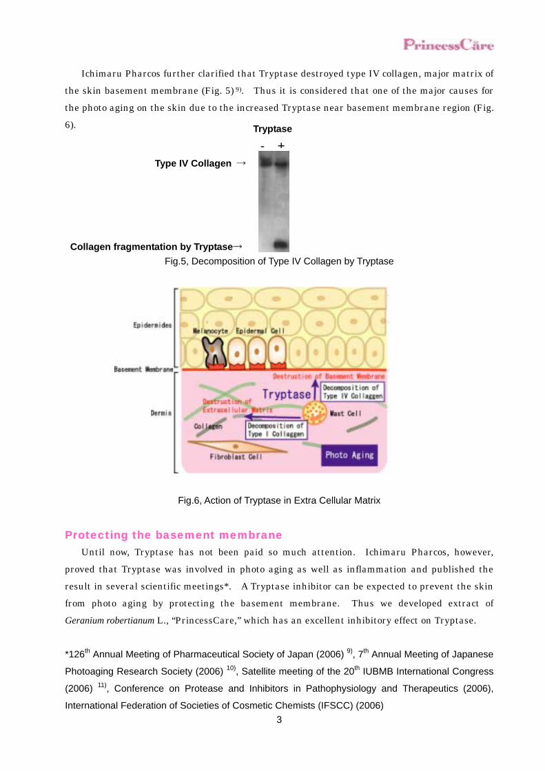
It has been confirmed that the number of mast cells was increased mainly at the dermal layer of the UV-irradiated skin compared with the non-UV-irradiated skin (Fig. 2) and that the amount of Tryptase protein was also increased (Fig. 3). Therefore, it is considered that in the skin exposed to UV light, the number of mast cells is increased and Tryptase is released actively. It has been reported that Tryptase stimulates the degradation of dermal matrix. From these facts, it can be considered that the markedly increased Tryptase by UV exposure destroys extra cellular matrix, which lead photo aging of the skin. It has been reported that epidermal basement membrane 7), which is recently paid an attention especially in relation to photo aging on the skin, was degraded by injecting Tryptase into the skin to induce the separation of the epidermis from the dermis (Fig. 4) 8).
Fig. 4, Destruction of Basement Membrane by Tryptase
Fig. 3, Increase of Tryptase by Exposure to UV
3
Ichimaru Pharcos further clarified that Tryptase destroyed type IV collagen, major matrix of the skin basement membrane (Fig. 5) 9). Thus it is considered that one of the major causes for the photo aging on the skin due to the increased Tryptase near basement membrane region (Fig. 6).
Fig.5, Decomposition of Type IV Collagen by Tryptase
Fig.6, Action of Tryptase in Extra Cellular Matrix
Protecting the basement membrane Until now, Tryptase has not been paid so much attention. Ichimaru Pharcos, however,
proved that Tryptase was involved in photo aging as well as inflammation and published the result in several scientific meetings*. A Tryptase inhibitor can be expected to prevent the skin from photo aging by protecting the basement membrane. Thus we developed extract of Geranium robertianum L., “PrincessCare,” which has an excellent inhibitory effect on Tryptase. *126th Annual Meeting of Pharmaceutical Society of Japan (2006) 9), 7th Annual Meeting of Japanese
Photoaging Research Society (2006) 10), Satellite meeting of the 20th IUBMB International Congress
(2006) 11), Conference on Protease and Inhibitors in Pathophysiology and Therapeutics (2006),
International Federation of Societies of Cosmetic Chemists (IFSCC) (2006)
Type IV Collagen →
Tryptase
- +
Collagen fragmentation by Tryptase→

4
What is Basement membrane? A basement membrane exists between the epidermis and the dermis. The basement
membrane consists of type IV collagen forming membraneous structure as the fundamental skeleton, laminin-5 and type VII collagen (Fig. 22). The basement membrane involves not only in the connection between the epidermis and dermis, but also in the differentiation of the epidermis and in the transfer of the signal transmitters among the cells. Therefore, the basement membrane plays an important role in the maintenance of healthy skin conditions.
In the light-exposed skin, the basement membrane has received the damages such as multiplication and rupture, which have been considered to be the cause for skin aging.
Fig. 22, Construction of basement membrane
Integrin
Iaminin-5
Basement membrane
keratinocyte
Epidermal cells (Fibroblast)
Type VII Collagen
5
Origin
PrincessCare is extract obtained from the whole
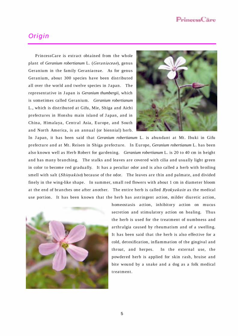
plant of Geranium robertianum L. (Geraniaceae), genus Geranium in the family Geraniaceae. As for genus Geranium, about 300 species have been distributed all over the world and twelve species in Japan. The representative in Japan is Geranium thumbergii, which is sometimes called Geranium. Geranium robertianum L., which is distributed at Gifu, Mie, Shiga and Aichi prefectures in Honshu main island of Japan, and in China, Himalaya, Central Asia, Europe, and South and North America, is an annual (or biennial) herb. In Japan, it has been said that Geranium robertianum L. is abundant at Mt. Ibuki in Gifu prefecture and at Mt. Reisen in Shiga prefecture. In Europe, Geranium robertianum L. has been also known well as Herb Robert for gardening. Geranium robertianum L. is 20 to 40 cm in height and has many branching. The stalks and leaves are covered with cilia and usually light green in color to become red gradually. It has a peculiar odor and is also called a herb with broiling smell with salt (Shioyakiso) because of the odor. The leaves are thin and palmate, and divided finely in the wing-like shape. In summer, small red flowers with about 1 cm in diameter bloom at the end of branches one after another. The entire herb is called Byokyakuin as the medical use portion. It has been known that the herb has astringent action, milder diuretic action,
homeostasis action, inhibitory action on mucus secretion and stimulatory action on healing. Thus the herb is used for the treatment of numbness and arthralgia caused by rheumatism and of a swelling. It has been said that the herb is also effective for a cold, detoxification, inflammation of the gingival and throat, and herpes. In the external use, the powdered herb is applied for skin rash, bruise and bite wound by a snake and a dog as a folk medical treatment.

6
Introduction
This product is an extract obtained by extracting the whole plant of Geranium robertianum L.
(Geraniaceae) with 1,3-butylene glycol solution. Efficacy
PrincessCare has inhibitory effect on Tryptase derived from human mast cell. ! Inhibitory effect on Tryptase ! Inhibitory Effect on Tryptase in epidermal cells ! Inhibitory Effect on Tryptase in human skin 3D model
In vitro (considered to relate to Tryptase inhibition) ! Inhibitory effect on Type I collagen degradation ! Inhibitory effect on Type IV collagen degradation
In vitro (considered to penetrate into human skin model) ! Penetration into skin
In vivo (considered to relate to Tryptase inhibition) ! Improvement of Skin Elasticity on the human skin ! Recovering of wrinkle on the human skin
In vivo (considered not to relate to Tryptase inhibition) ! Moisturizing
In vitro (considered not to relate to Tryptase inhibition) ! Inhibitory effect on melanin production ! Inhibitory effect on histamine release ! SOD like activity
7
Inhibitory Effect on Tryptase
Ichimaru Pharcos confirmed that Tryptase was secreted from mast cells infiltrated into the
skin by inflammation and the UV light exposure, and gave great influence on aging in the skin. Thus it was cleared that the inhibition of Tryptase activity was an important key for prevention of aging in the skin. Thus the effect of Princess Care on Tryptase activity was examined Test sample
PrincessCare was diluted by 50% of 1,3-Butylene Glycol solution to adjust 0.00039 to 0.0125 % as product concentration. As control solution, 50% of 1,3-Butylene Glycol solution was used in this test. Test Method
Human mast cell Tryptase used in the present study was extracted from human tonsil and purified by octyl sepharose chromatography and heparin sepharose chromatography. As a substrate, MCA labeled peptide (Boc-Phe-Ser-Arg-MCA ⋅ Peptide institute INC.) was used. The substrate solution was prepared by dissolving the substrate in 0.1 M Tris-HCl buffer solution (pH 7.2) to make the concentration 5 µmol/L. The substrate solution (160 µL), test sample solution (20 µL) and 9 ng/mL Tryptase solution (20 µL) were mixed and incubated at 37°C for 15 minutes. After cooling to room temperature, the fluorescent intensity of the reaction solution (excitation wavelength: 360 nm, emission wavelength: 450 nm) was measured to obtain the enzyme activity. For each test sample, a blank, in which Tris-HCl buffer solution was added in place of the enzyme solution, was employed. The inhibitory rate of each test sample on Tryptase activity was calculated according to the following equation. Inhibitory rate of Tryptase % = (1 - ) x 100
Control fluorescence intensity-Control Blanc fluorescence intensity Sample fluorescence intensity-Sample Blanc fluorescence intensity

8
Result and Discussion Inhibitory rate of Tryptase on PrincessCare was shown in Fig. 7. PrincessCare inhibited
Tryptase activity concentration dependently; IC50 was 0.00245 %. According to the result, PrincessCare is expected to prevent various negative impacts caused
by Tryptase because PrincessCare inhibits Tryptase activity.
0
20
40
60
80
100
0 0.002 0.004 0.006 0.008 0.01 0.012 0.014
Concentration
Inhi
bito
ry r
ate
of T
rypt
ase
(%)
R2=0.9889
Fig. 7, Inhibitory effect on Tryptase
9
Inhibitory Effect on Tryptase in epidermal cells
Tryptase degrades type IV collagen, the major component of the epidermal basement
membrane, and fibronectin, the adhesive protein, which connect between the basement membrane and epidermal cells. Therefore, tryptase can damage the connection (attachment) between the epidermis and the basement membrane. Thus, the inhibitory effect of Princess Care on tryptase activity was examined in in-vitro model.
Test Method Human keratinocytes (Kurabo) were seeded on 6-well plate coated with type IV collagen and
fibronectin (Becton Dickison) and incubated to make 70% to 80% confluent layer in a growth medium (EpiLife-KG2, Kurabo) with a CO2 incubator at 37°C. Thereafter, human mast cell tryptase and PrincessCare (25 ppm) were added to the medium. After 3 to 6-hr cultivation, the condition of keratinocytes was observed under a light microscope.
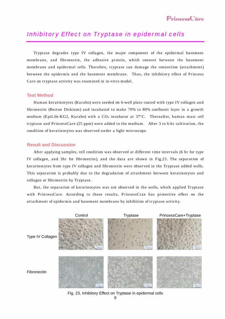
Result and Discussion After applying samples, cell condition was observed at different time intervals (6 hr for type
IV collagen, and 3hr for fibronectin), and the data are shown in Fig.23. The separation of keratinocytes from type IV collagen and fibronectin were observed in the Tryptase added wells. This separation is probably due to the degradation of attachment between keratinocytes and collagen or fibronectin by Tryptase.
But, the separation of keratinocytes was not observed in the wells, which applied Tryptase with PrincessCare. According to these results, PrincessCrae has protective effect on the attachment of epidermis and basement membrane by inhibition of tryptase activity.
Control Tryptase PrincessCare+Tryptase
Type IV Collagen
Fibronectin
Fig. 23, Inhibitory Effect on Tryptase in epidermal cells
10
Inhibitory Effect on Tryptase in human skin 3D model
We have observed degradation of type IV collagen by Tryptase in different in-vitro models.
This experiment is planned to confirm Inhibitory Effect on Tryptase using a human skin 3D model.
Test Method Human mast cell tryptase and Princess Care (50 ppm) were added to the three-dimensional
cultured human skin 3D model (Toyobo) and the culture medium was incubated for 3 hr with a CO2 incubator at 37°C. Thereafter, frozen sections were prepared from the three-dimensional skin model and subjected to immunostaining with a fluorescent antibody specific for type IV collagen (Chemicon). Then the basement membrane (type IV collagen) was observed under a light microscope and a fluorescent microscope.
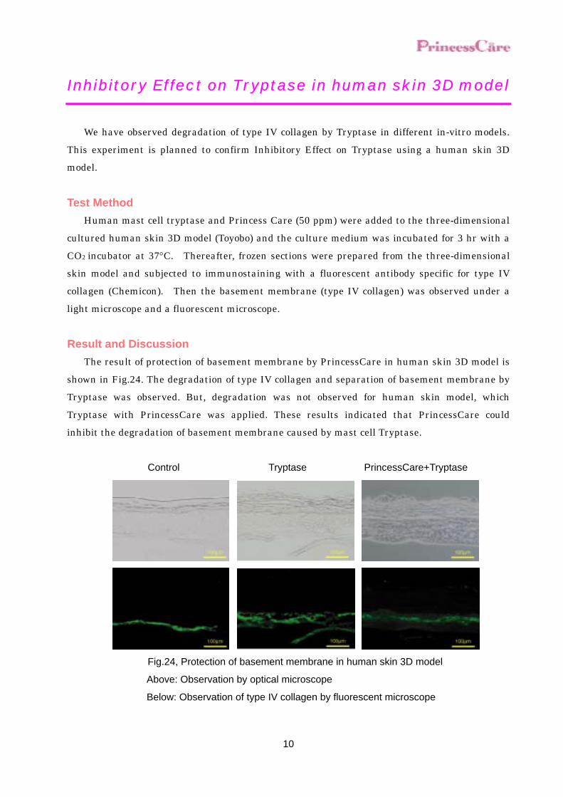
Result and Discussion The result of protection of basement membrane by PrincessCare in human skin 3D model is
shown in Fig.24. The degradation of type IV collagen and separation of basement membrane by Tryptase was observed. But, degradation was not observed for human skin model, which Tryptase with PrincessCare was applied. These results indicated that PrincessCare could inhibit the degradation of basement membrane caused by mast cell Tryptase.
Control Tryptase PrincessCare+Tryptase
Fig.24, Protection of basement membrane in human skin 3D model
Above: Observation by optical microscope
Below: Observation of type IV collagen by fluorescent microscope
11
Inhibitory Effect on Type I Collagen Degradation (Tryptase)
It became clear that as in the case of MMP-1, Tryptase digested type I collagen and degraded dermal matrix to lead to aging in the skin. Thus the inhibitory effect of Princess Care on type I collagen digestion by Tryptase was examined.
Test Sample PrincessCare was adjusted to 0.05 and 0.1 % as product concentration. As control solution,
50% of 1,3-Butylene Glycol solution was used in this test. As positive control, 10 µmol/L of Leupetin, which is a protease inhibitors inhibits Tryptase, was used.
Test Method Ice-cold FITC labeled type I collagen (Sigma-Aldrich) solution (50 µL), 0.1 mol/L Tris-HCl
buffer solution (pH 7.2) (130 µL), 9 ng/mL Tryptase solution (10 µL) and test sample solution (10 µL) were mixed and incubated at 37°C for 3 hours. After the completion of the reaction, ice-cold o-phenanthroline solution (200 µL) was added to the reaction mixture and kept at 4°C for 15 minutes. After centrifuging the mixture at 6000 × g for 10 minutes, the fluorescent intensity of the supernatant (excitation wavelength: 490 nm, emission wavelength: 530 nm) was measured. A blank, in which Tris-HCl buffer solution was added in place of Tryptase and test sample solution, was employed and the fluorescent intensity of the blank was designated as 100. Using the relative values to the blank value, 100, the amount of collagen digested in the presence of each test sample was expressed.
Type I collagen derived from human skin (Sigma-Aldrich) was dissolved in 1 mol/L acetic acid and adjusted the concentration to 1 mg/mL with 50 mmol/L Tris-HCl buffer solution (pH 7.2). The collagen solution thus prepared (10 µL), 16 ng/mL Tryptase solution (10 µL) and test sample solution (10 µL) were mixed and incubated at 37°C for 1 hour. After the completion of the reaction, the reaction mixture was subjected to 8.5% T SDS-PAGE (Bio-Rad) electrophoresis. The gel was stained with coomassie brilliant blue to analyze collagen fragmentation.
12
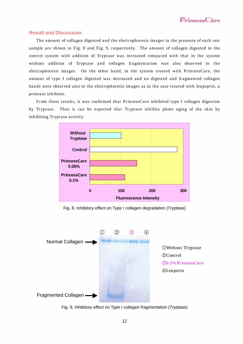
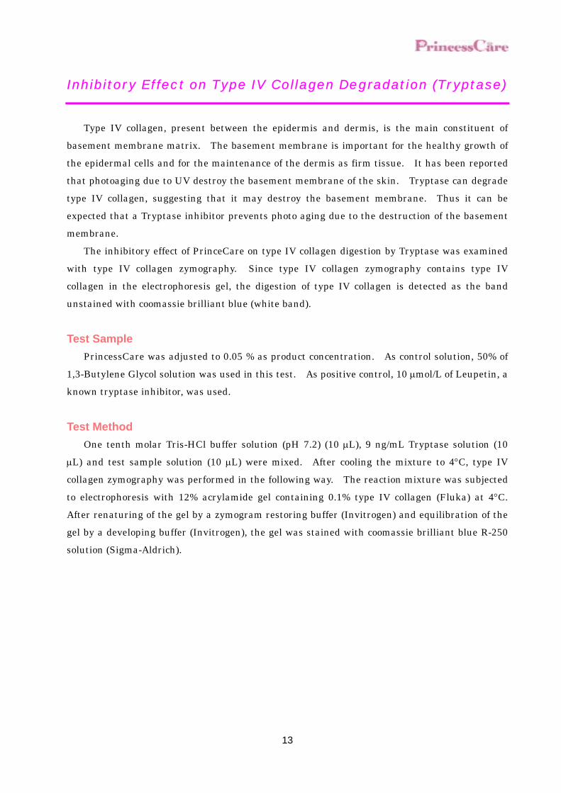
Result and Discussion The amount of collagen digested and the electrophoresis images in the presence of each test
sample are shown in Fig. 8 and Fig. 9, respectively. The amount of collagen digested in the control system with addition of Tryptase was increased compared with that in the system without addition of Tryptase and collagen fragmentation was also observed in the electrophoresis images. On the other hand, in the system treated with PrincessCare, the amount of type I collagen digested was decreased and no digested and fragmented collagen bands were observed also in the electrophoresis images as in the case treated with leupeptin, a protease inhibitor.
From these results, it was confirmed that PrincessCare inhibited type I collagen digestion by Tryptase. Thus it can be expected that Tryptase inhibits photo aging of the skin by inhibiting Tryptase activity.
Fluorescence Intensity
0 100 200 300
PrincessCare0.1%
PrincessCare0.05%
Control
Without Tryptase
Fig. 8, Inhibitory effect on Type I collagen degradation (Tryptase)
Fig. 9, Inhibitory effect on Type I collagen fragmentation (Tryptase)
① ② ③ ④
①Without Tryptase②Control ③0.1% PrincessCare④Leupetin
Normal Collagen
Fragmented Collagen
13
Inhibitory Effect on Type IV Collagen Degradation (Tryptase)
Type IV collagen, present between the epidermis and dermis, is the main constituent of
basement membrane matrix. The basement membrane is important for the healthy growth of the epidermal cells and for the maintenance of the dermis as firm tissue. It has been reported that photoaging due to UV destroy the basement membrane of the skin. Tryptase can degrade type IV collagen, suggesting that it may destroy the basement membrane. Thus it can be expected that a Tryptase inhibitor prevents photo aging due to the destruction of the basement membrane.
The inhibitory effect of PrinceCare on type IV collagen digestion by Tryptase was examined with type IV collagen zymography. Since type IV collagen zymography contains type IV collagen in the electrophoresis gel, the digestion of type IV collagen is detected as the band unstained with coomassie brilliant blue (white band).
Test Sample PrincessCare was adjusted to 0.05 % as product concentration. As control solution, 50% of
1,3-Butylene Glycol solution was used in this test. As positive control, 10 µmol/L of Leupetin, a known tryptase inhibitor, was used.
Test Method One tenth molar Tris-HCl buffer solution (pH 7.2) (10 µL), 9 ng/mL Tryptase solution (10
µL) and test sample solution (10 µL) were mixed. After cooling the mixture to 4°C, type IV collagen zymography was performed in the following way. The reaction mixture was subjected to electrophoresis with 12% acrylamide gel containing 0.1% type IV collagen (Fluka) at 4°C. After renaturing of the gel by a zymogram restoring buffer (Invitrogen) and equilibration of the gel by a developing buffer (Invitrogen), the gel was stained with coomassie brilliant blue R-250 solution (Sigma-Aldrich).
14
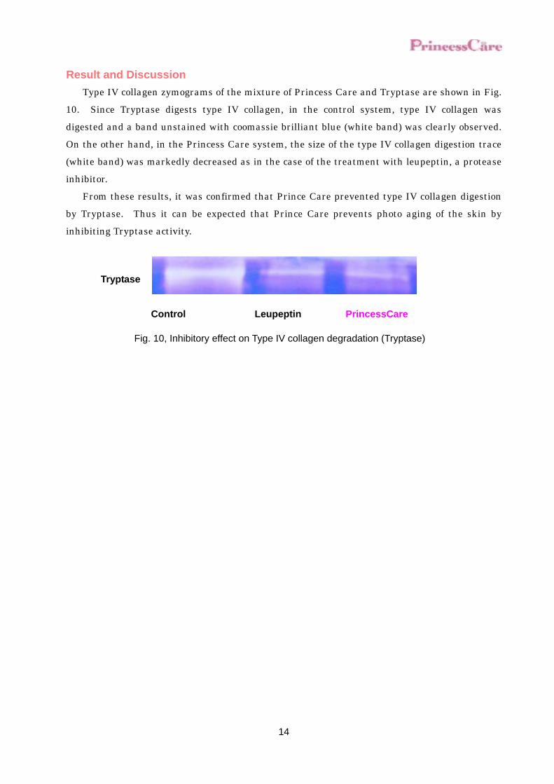
Result and Discussion Type IV collagen zymograms of the mixture of Princess Care and Tryptase are shown in Fig.
10. Since Tryptase digests type IV collagen, in the control system, type IV collagen was digested and a band unstained with coomassie brilliant blue (white band) was clearly observed. On the other hand, in the Princess Care system, the size of the type IV collagen digestion trace (white band) was markedly decreased as in the case of the treatment with leupeptin, a protease inhibitor.
From these results, it was confirmed that Prince Care prevented type IV collagen digestion by Tryptase. Thus it can be expected that Prince Care prevents photo aging of the skin by inhibiting Tryptase activity.
Control Leupeptin PrincessCare
Tryptase
Fig. 10, Inhibitory effect on Type IV collagen degradation (Tryptase)

15
Improvement of Skin Elasticity on the Human Skin
The decreased elasticity of the skin results in the occurrence of externally remarkable aging
symptoms of the skin such as wrinkles and bags under eyes. The decrease in the metabolism of the components in the dermis such as collagen, elastin and hyaluronic acid due to the decreased function of the skin cells has been considered as one of the causes for the decrease in the skin elasticity. PrincessCare has inhibitory effect on Tryptase activity, which destroys basement membrane and dermis collagen and leads to photo aging. Improvement of skin elasticity on PrincessCare was examined.
Test Sample PrincessCare was diluted by purified water to adjust 3 % as product concentration. As
control solution, 50% of 1,3-Butylene Glycol solution was diluted in the same way and used in the test.
Test Method Ten male and female volunteers at age 20s to 50s that gave us written informed consent
were enrolled in this study. Each test sample was applied around the left and right eyes of each subject three times a day for 12 weeks. Before and at 12 weeks after the commencement of the application, the elasticity of the skin was measured by a skin viscosity and elasticity meter ⋅ Cutometer (CUTOMETER SEM474, COURAGE + KHAZAKA Electronic GmbH). The elasticity was calculated by the change of the skin condition when the skin was sucked for 5 sec by instantly reducing the pressure to 500 mb and thereafter the negative pressure was instantly released, which were done twice.
The test was performed several times each in right and left application sites, and the percentage of the change of the mean values between the value before the commencement of the application and that at 28 days after the commencement of the application was calculated to obtain the rate of the change. The measurement was performed 20 min after acclimation in an air conditioning room (room temperature: 20°C and humidity: 50%) after washing the face.

16
Result and Discussion The elasticity before application and 12 weeks later of 10 volunteers is shown in Fig.11.
According to the result, the improvement of elasticity was seen, where PrincessCare was applied; although, the elasticity of aged skin is reported to increase. According to this result, PrincessCare has improvement effect of elasticity.
60
80
100
120
0 12Weeks
Cha
nge
of P
last
icity
PrincessCareControl
Fig. 11, Improvement of Skin Elasticity on the human skin
17
Recovering of Wrinkle on the Human Skin
PrincessCare has inhibitory effect on Tryptase activity, which destroys basement membrane
and dermis collagen and leads to photo aging. The improving effect of PrincessCare on the wrinkles was examined by a replica method.
In this test, the wrinkle area is obtained by calculating by the image analyzer the shadow area due to the unevenness of the wrinkles produced by irradiating light to a replica obtained from the treated site at a certain angle
Test Sample PrincessCare was diluted by purified water to adjust 3 % as product concentration. As
control solution, 50% of 1,3-Butylene Glycol solution was diluted in the same way and used in the test.
Test Method Ten health male and female volunteers at age 20s to 40s that gave us written informed
consent were enrolled in this study. Before the treatment and 12 weeks after the treatment, a test was carried out according to
the procedures for examining dermal shape by replica (SILFLO,AMIC Group).After washing face, volunteers stay in a thermo-hygrostat room (at 20℃, humidity 50%) for 20 minutes. Light
was irradiated to replica from constant angle (tops and bottoms angle 25) and its shade was calculated with the use of an image data processing and picture analyzing software and area of wrinkle was the measured. By calculating ratio of the area of wrinkle per analyzing area, it is compared with control.
18
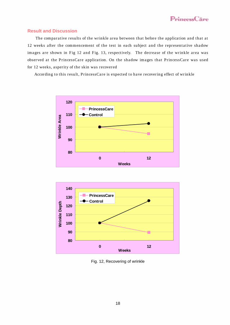
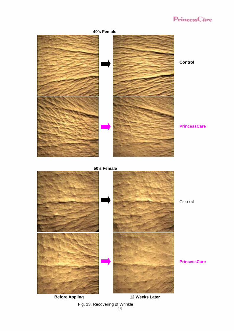
Result and Discussion The comparative results of the wrinkle area between that before the application and that at
12 weeks after the commencement of the test in each subject and the representative shadow images are shown in Fig 12 and Fig. 13, respectively. The decrease of the wrinkle area was observed at the PrincessCare application. On the shadow images that PrincessCare was used for 12 weeks, asperity of the skin was recovered
According to this result, PrincessCare is expected to have recovering effect of wrinkle
80
90
100
110
120
0 12Weeks
Wri
nkle
Are
a
PrincessCareControl
80
90
100
110
120
130
140
0 12Weeks
Wri
nkle
Dep
th
PrincessCareControl
Fig. 12, Recovering of wrinkle
19
Before Appling 12 Weeks Later
Fig. 13, Recovering of Wrinkle
40’s Female
Control
PrincessCare
Control
PrincessCare
50’s Female
20
Penetration into skin
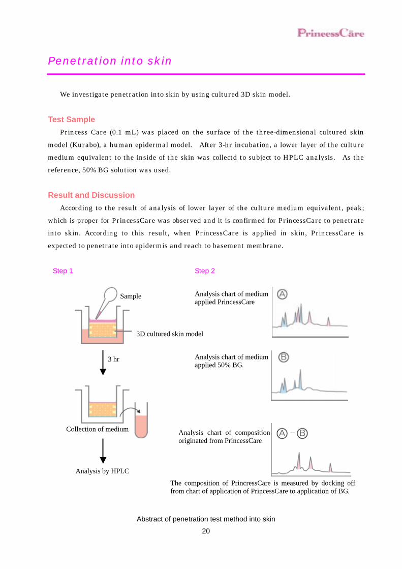
We investigate penetration into skin by using cultured 3D skin model.
Test Sample Princess Care (0.1 mL) was placed on the surface of the three-dimensional cultured skin
model (Kurabo), a human epidermal model. After 3-hr incubation, a lower layer of the culture medium equivalent to the inside of the skin was collectd to subject to HPLC analysis. As the reference, 50% BG solution was used.
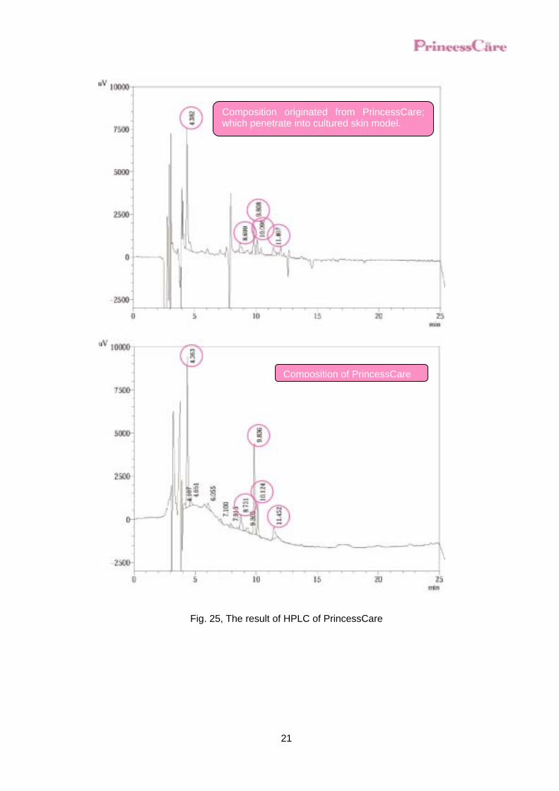
Result and Discussion According to the result of analysis of lower layer of the culture medium equivalent, peak;
which is proper for PrincessCare was observed and it is confirmed for PrincessCare to penetrate into skin. According to this result, when PrincessCare is applied in skin, PrincessCare is expected to penetrate into epidermis and reach to basement membrane.
Abstract of penetration test method into skin
Sample
3D cultured skin model
3 hr
Collection of medium
Analysis by HPLC
Step 1 Step 2
Analysis chart of medium applied PrincessCare
Analysis chart of mediumapplied 50% BG.
Analysis chart of compositionoriginated from PrincessCare
The composition of PrincressCare is measured by docking offfrom chart of application of PrincessCare to application of BG.
21
Fig. 25, The result of HPLC of PrincessCare
Composition originated from PrincessCare;which penetrate into cultured skin model.
Composition of PrincessCare
22
Moisturizing
The protection of the skin from dryness by retaining moisture is the most important for the
prevention of the skin aging. Water content and TEWL (Trans Epidermis Water Loss) were examined to know
moisturizing effect of PrincessCare.
1. Determination of the Water Content in the Horny Layer In this test, water content in the horny layer is determined by measuring the conductance at sending the high frequency current of 3.5 Mhz to the horny layer through the probe.
Test Sample PrincessCare was diluted by purified water to adjust 3 % as product concentration. As
control solution, 50% of 1,3-Butylene Glycol solution was diluted in the same way and used in the test.
Test Method The test detail is explained for volunteers in advance, after confirming, they cooperate in
these tests. Sample is applied on the right and left eye area to 8 volunteers (male and female of 20’s to
50’s years old) three times a day for 12 weeks. Before applying, 4 weeks, 8 weeks, and 12 weeks later, volunteers stay in a thermo-hygrostat room (at 20℃, humidity 50%) for 20 minutes and the subocular water content in the horny layer
is determined 10 times by Impedance Meter (SKICON-200, IBS Co., Ltd.) and the mean value of electric conductivity is calculated.
23
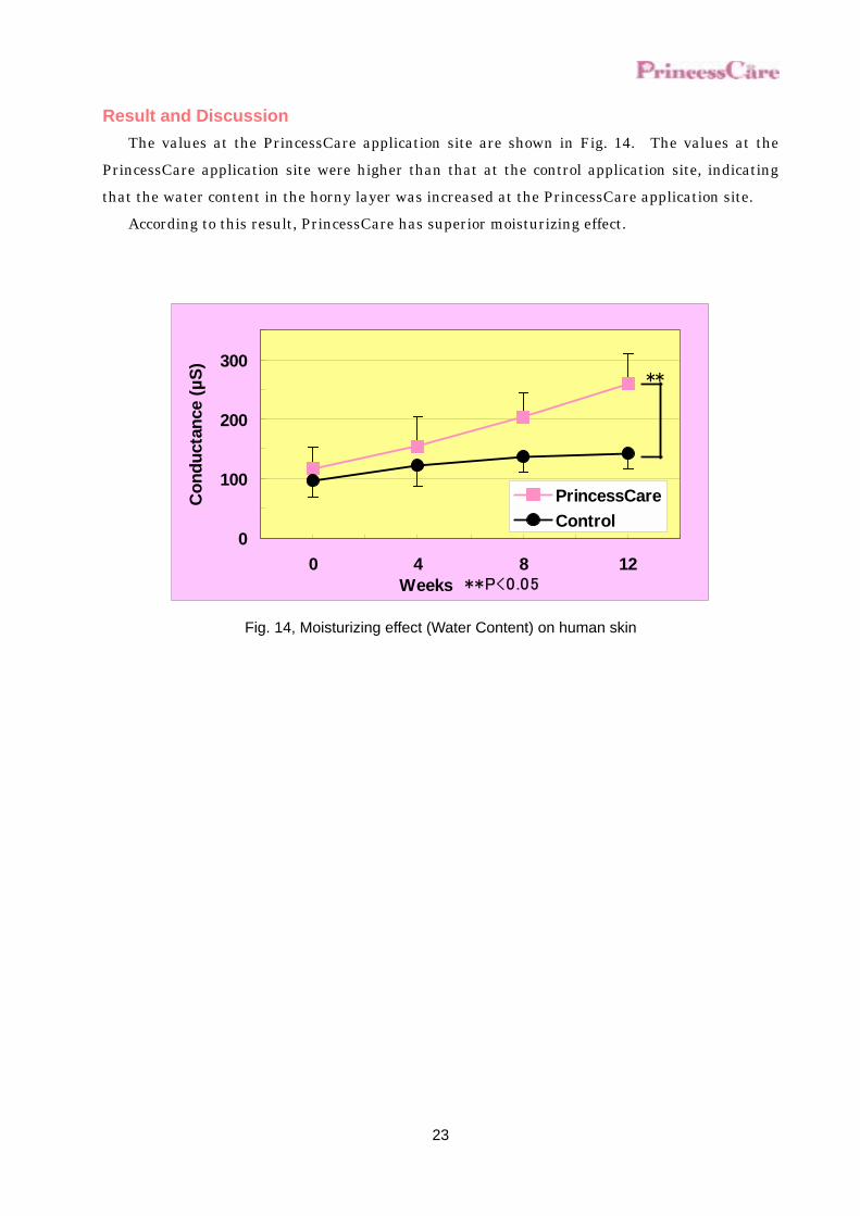
Result and Discussion The values at the PrincessCare application site are shown in Fig. 14. The values at the
PrincessCare application site were higher than that at the control application site, indicating that the water content in the horny layer was increased at the PrincessCare application site.
According to this result, PrincessCare has superior moisturizing effect.
0
100
200
300
0 4 8 12Weeks **P<0.05
Con
duct
ance
(µS)
PrincessCareControl
**
Fig. 14, Moisturizing effect (Water Content) on human skin
24
2. Measurement of Trans Epidermal Water Loss (TEWL) In this test, on the basis that the concentration slope of water is proportional to the water
content evaporated from the skin not higher than about 1 cm from the skin surface, the amount of water evaporated from the skin (the amount of water loss) is obtained by measuring the water concentration slope with putting the probe having two electrodes set at the different heights to the skin. When the barrier function of the skin is decreased, trans epidermal water loss (TEWL) is increased.
Test Sample PrincessCare was diluted by purified water to adjust 3 % as product concentration. As
control solution, 50% of 1,3-Butylene Glycol solution was diluted in the same way and used in the test. Test Method
The test detail is explained for volunteers in advance, and after confirming, they cooperate in these tests.
Sample is applied on the right and left eye area to 8 volunteers (male and female of 20’s to 50’s years old) three times a day for 12 weeks. Before applying, 4 weeks, 8 weeks, and 12 weeks later, volunteers stay in a thermo-hygrostat room (at 20℃, humidity 50%) for 20 minutes and the sub ocular Trans Epidermal Water loss (TEWL) was determined every 2 seconds for 90 seconds by TEWAMETER TM210 (Courage + Khazaka Electronic GmbH) and the mean value from 30 to 90 seconds was calculated.
25
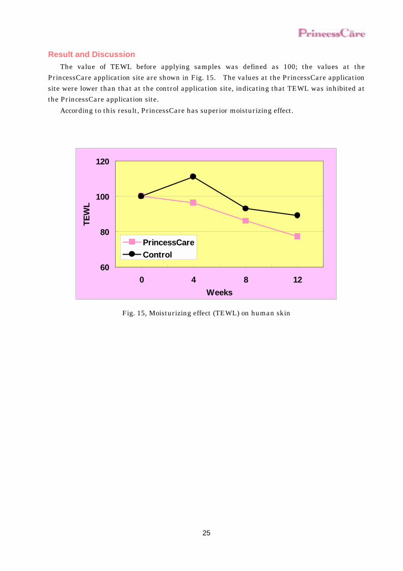
Result and Discussion The value of TEWL before applying samples was defined as 100; the values at the
PrincessCare application site are shown in Fig. 15. The values at the PrincessCare application site were lower than that at the control application site, indicating that TEWL was inhibited at the PrincessCare application site.
According to this result, PrincessCare has superior moisturizing effect.
60
80
100
120
0 4 8 12Weeks
TEW
L
PrincessCareControl
Fig. 15, Moisturizing effect (TEWL) on human skin
26
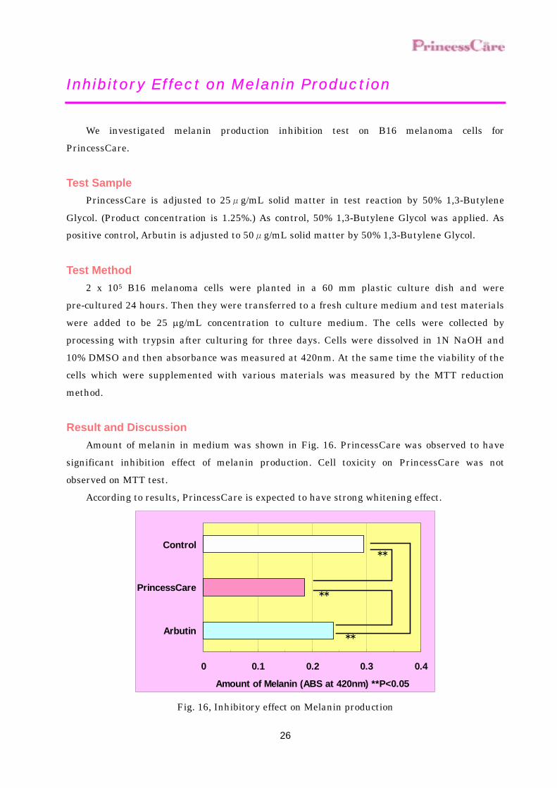
Inhibitory Effect on Melanin Production
We investigated melanin production inhibition test on B16 melanoma cells for PrincessCare.
Test Sample PrincessCare is adjusted to 25μg/mL solid matter in test reaction by 50% 1,3-Butylene
Glycol. (Product concentration is 1.25%.) As control, 50% 1,3-Butylene Glycol was applied. As positive control, Arbutin is adjusted to 50μg/mL solid matter by 50% 1,3-Butylene Glycol.
Test Method 2 x 10⁵ B16 melanoma cells were planted in a 60 mm plastic culture dish and were
pre-cultured 24 hours. Then they were transferred to a fresh culture medium and test materials were added to be 25 µg/mL concentration to culture medium. The cells were collected by processing with trypsin after culturing for three days. Cells were dissolved in 1N NaOH and 10% DMSO and then absorbance was measured at 420nm. At the same time the viability of the cells which were supplemented with various materials was measured by the MTT reduction method.
Result and Discussion Amount of melanin in medium was shown in Fig. 16. PrincessCare was observed to have
significant inhibition effect of melanin production. Cell toxicity on PrincessCare was not observed on MTT test.
According to results, PrincessCare is expected to have strong whitening effect.
0 0.1 0.2 0.3 0.4
Arbutin
PrincessCare
Control
Amount of Melanin (ABS at 420nm) **P<0.05
**
**
**
Fig. 16, Inhibitory effect on Melanin production
27
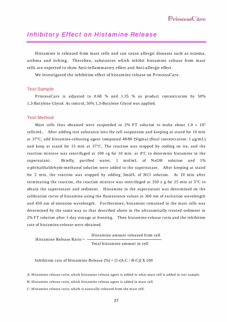
Inhibitory Effect on Histamine Release
Histamine is released from mast cells and can cause allergic diseases such as eczema, asthma and itching. Therefore, substances which inhibit histamine release from mast cells are expected to show Anti-inflammatory effect and Anti-allergic effect.
We investigated the inhibition effect of histamine release on PrincessCare.
Test Sample PrincessCare is adjusted to 0.68 % and 1.35 % as product concentration by 50%
1,3-Butylene Glycol. As control, 50% 1,3-Butylene Glycol was applied.
Test Method Mast cells thus obtained were suspended in 2% FT solution to make about 1.0 × 10⁵
cells/mL. After adding test substance into the cell suspension and keeping at stand for 10 min at 37°C, add histamine-releasing agent compound 48/80 (Sigma) (final concentration: 1 µg/mL) and keep at stand for 15 min at 37°C. The reaction was stopped by cooling on ice, and the reaction mixture was centrifuged at 100 ×g for 10 min. at 4°C to determine histamine in the supernatant. Briefly, purified water, 1 mol/mL of NaOH solution and 1% o-phthaldialdehyde-methanol solution were added to the supernatant. After keeping at stand for 5 min, the reaction was stopped by adding 3mol/L of HCl solution. At 10 min after terminating the reaction, the reaction mixture was centrifuged at 350 x g for 25 min at 5°C to obtain the supernatant and sediment. Histamine in the supernatant was determined on the calibration curve of histamine using the fluorescence values at 360 nm of excitation wavelength and 450 nm of emission wavelength. Furthermore, histamine remained in the mast cells was determined by the same way as that described above in the ultrasonically treated sediment in 2% FT solution after 1-day storage at freezing. Then histamine-release ratio and the inhibition rate of histamine-release were obtained.
Inhibition rate of Histamine Release (%) = [1-(A-C / B-C)] X 100 A: Histamine release ratio; which histamine release agent is added in what mast cell is added in test sample.
B: Histamine release ratio; which histamine release agent is added in mast cell.
C: Histamine release ratio; which is naturally released from the mast cell.
Histamine amount released from cellHistamine Release Ratio =
Total histamine amount in cell

28
Result and Discussion Inhibition rate of histamine release is shown in Fig.17. PrincessCare was observed to have
strong inhibition effect of histamine release by concentration dependence. According to result, PrincessCare is expected to have anti-inflammation effect and
anti-allergic effect.
0 20 40 60 80 100
PrincessCare1.35%
PrincessCare0.68%
Inhibition of Histamine Release (%)
Fig. 17, Inhibitory effect on Histamine release
29
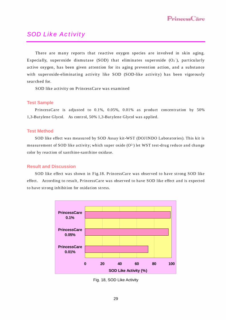
SOD Like Activity
There are many reports that reactive oxygen species are involved in skin aging.
Especially, superoxide dismutase (SOD) that eliminates superoxide (O2−), particularly
active oxygen, has been given attention for its aging prevention action, and a substance with superoxide-eliminating activity like SOD (SOD-like activity) has been vigorously searched for.
SOD like activity on PrincessCare was examined
Test Sample PrincessCare is adjusted to 0.1%, 0.05%, 0.01% as product concentration by 50%
1,3-Butylene Glycol. As control, 50% 1,3-Butylene Glycol was applied.
Test Method SOD like effect was measured by SOD Assay kit-WST (DOJINDO Laboratories). This kit is
measurement of SOD like activity; which super oxide (O2-) let WST test-drug reduce and change color by reaction of xanthine-xanthine oxidase.
Result and Discussion SOD like effect was shown in Fig.18. PrincessCare was observed to have strong SOD like
effect. According to result, PrincessCare was observed to have SOD like effect and is expected to have strong inhibition for oxidation stress.
SOD Like Activity (%)
0 20 40 60 80 100
PrincessCare0.01%
PrincessCare0.05%
PrincessCare0.1%
Fig. 18, SOD Like Activity

30
Stability
Stability of PrincessCare was evaluated.
Long Term stability Store PrincessCare in a cool dark place(4℃), room temperature, window side and at 40℃.
Absorbance values at 470nm were determined.
Result and Discussion Change of Absorbance value is shown in Fig. 19. At any condition, PrincessCare was very
stable. According to the result, the long term stability of PrincessCare is superior.
0
0.2
0.4
0.6
0.8
1
0 1 2 3Months
AB
S(47
0nm
)
Cool dark placeRoom Temp.40℃Window side
Fig. 19, Long term stability
31
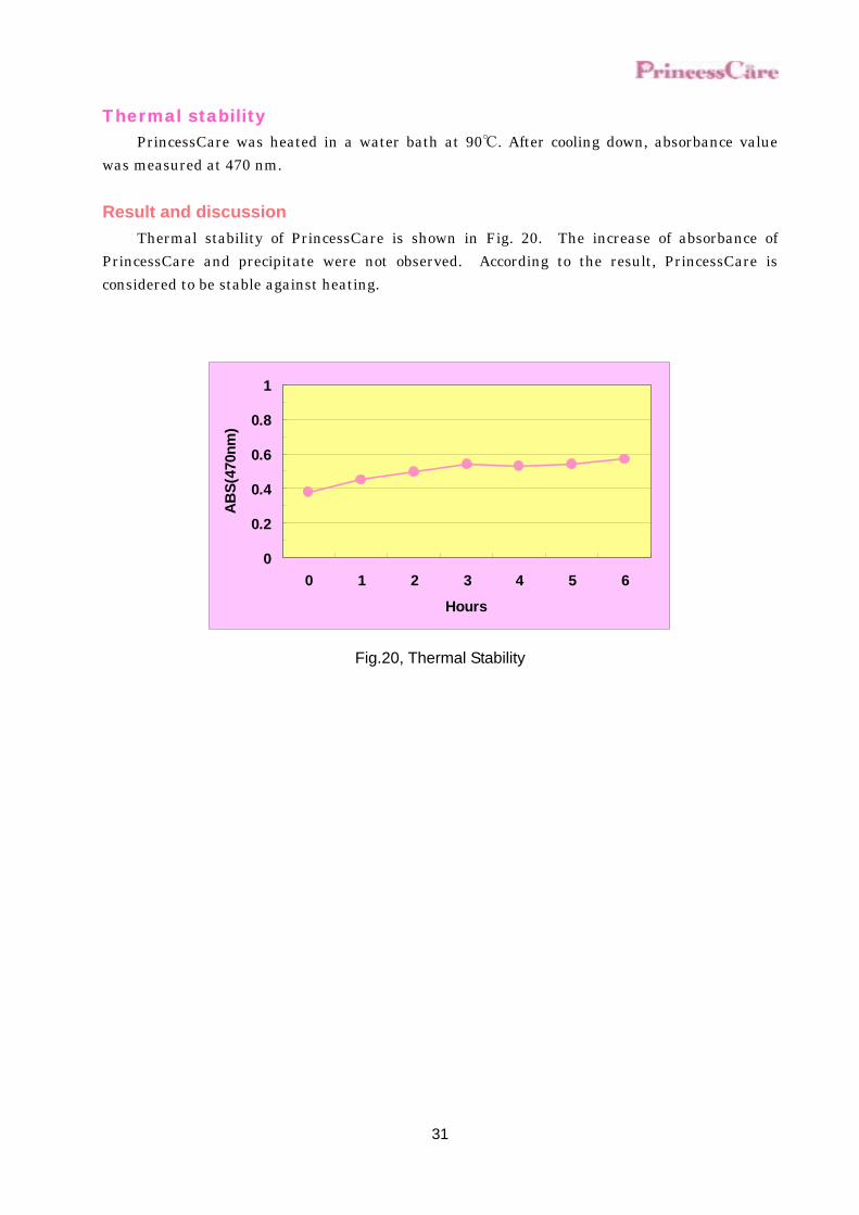
Thermal stability PrincessCare was heated in a water bath at 90℃. After cooling down, absorbance value
was measured at 470 nm.
Result and discussion Thermal stability of PrincessCare is shown in Fig. 20. The increase of absorbance of
PrincessCare and precipitate were not observed. According to the result, PrincessCare is considered to be stable against heating.
0
0.2
0.4
0.6
0.8
1
0 1 2 3 4 5 6
Hours
AB
S(47
0nm
)
Fig.20, Thermal Stability
32
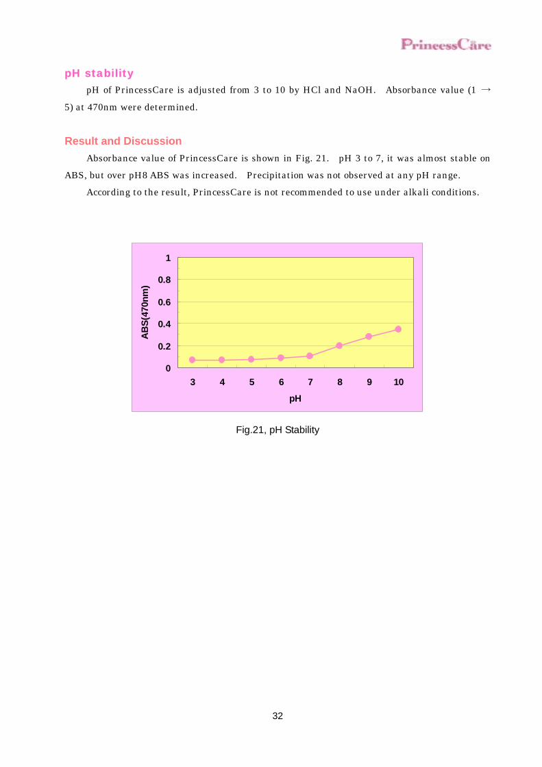
pH stability pH of PrincessCare is adjusted from 3 to 10 by HCl and NaOH. Absorbance value (1 →
5) at 470nm were determined.
Result and Discussion Absorbance value of PrincessCare is shown in Fig. 21. pH 3 to 7, it was almost stable on
ABS, but over pH8 ABS was increased. Precipitation was not observed at any pH range. According to the result, PrincessCare is not recommended to use under alkali conditions.
0
0.2
0.4
0.6
0.8
1
3 4 5 6 7 8 9 10
pH
AB
S(47
0nm
)
Fig.21, pH Stability

33
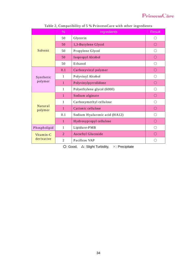
Compatibility
Compatibility of PrincessCare with other materials was evaluated.
Test Method PrincessCare was diluted to 5 %, and the test samples were adjusted by purified water as
shown on the each table. After 24 hours, mixed solution was evaluated.
Result and Discussion Table 1, Compatibility of 5 % PrincessCare with Surfactant
% Ingredients Result
2.8 Stearyl Trimethyl Ammonium Chloride ○
3.0 Cetyltrimehylammonium Chloride ○
Cation
2.7 Lauryltrimethylammonium Chloride ○
10.0 Triethanolamine Lauryl Sulfate ○
25.0 Sodium Laureth Sulfate ○
25.0 Triethanolamine Laureth Sulfate △
6.25 Laureth-6 Carboxylic Acid ○
10.0 Sodium N-Cocoyl-N-methyl Taurate ○
10.0 Potassium N-Cocoyl Glycinate ○
7.5 Sodium Lauroyl Methylaminopropionate ○
Anion
25.0 Sodium Tetradecenesulfonate ○
10.0 Polyethylene Glycol (50) Oleyl Ether ○
10.0 Coconut Tatty Acid Diethanolamide ○
10.0 Sorbeth-60 Tetraoleate ○
10.0 Polyoxyethylene Sorbitan Monooleate (20E.O.) ○
Nonion
10.0 Polyoxyethylene Hydrogenated Castor Oil (60E.O.) ○
Silicone 10.0 Polyoxyethylene・Methylpolysiloxane Copolymer ○
3.5 Lauryl Dimethylaminoacetic Acid Betaine ○
4.0 Sodium N-Cocoyl-N-Carboxymethyl-N-Hydroxyethyl Ethylenediamide ○
Ampholytic
2.9 Lauroyl Amide Propylhydroxysulfobetaine ○
○ : Good, △: Slight Turbidity, ×: Precipitate
34
Table 2, Compatibility of 5 % PrincessCare with other ingredients % Ingredients Result
50 Glycerin ○
50 1,3-Butylene Glycol ○
50 Propylene Glycol ○
50 Isopropyl Alcohol ○
Solvent
50 Ethanol ○
0.1 Carboxyvinyl polymer ○
1 Polyvinyl Alcohol ○
1 Polyvinylpyrrolidone ○ Synthetic polymer
1 Polyethylene glycol (6000) ○
1 Sodium alginate ○
1 Carboxymethyl cellulose ○
1 Cationic cellulose ○
0.1 Sodium Hyaluronic acid (HA12) ○
Natural polymer
1 Hydroxypropyl cellulose ○
Phospholipid 1 Lipidure-PMB ○
2 Ascorbyl Glucoside ○ Vitamin-C derivative 2 Pacificos VAP ○
○: Good, △: Slight Turbidity, ×: Precipitate
35
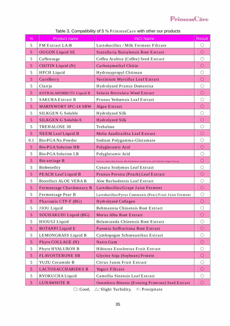
Table 3, Compatibility of 5 % PrincessCare with other our products
% Product name INCI Name Result
5 FM Extract LA-B Lactobacillus / Milk Ferment Filtrate ○ 5 OUGON Liquid SE Scutellaria Baicalensis Root Extract ○ 5 Caffenoage Coffea Arabica (Coffee) Seed Extract ○ 5 CHITIN Liquid (N) Carboxymethyl Chitin ○ 5 HPCH Liquid Hydroxypropyl Chitosan ○ 5 CureBerry Vaccinium Myrtillus Leaf Extract ○ 5 Clairju Hydrolyzed Prunus Domestica ○ 5 KOTHALAHIMBUTU Liquid B Salacia Reticulata Wood Extract ○ 5 SAKURA Extract B Prunus Yedoensis Leaf Extract ○ 5 MARINWORT IPC-14 SBW Algae Extract ○ 5 SILKGEN G Soluble Hydrolyzed Silk ○ 5 SILKGEN G Soluble-S Hydrolyzed Silk ○ 5 TREHALOSE 30 Trehalose ○ 5 NEEM Leaf Liquid B Melia Azadirachta Leaf Extract ○
0.1 Bio-PGA Na Powder Sodium Polygamma-Glutamate ○ 5 Bio-PGA Solution HB Polyglutamic Acid ○ 5 Bio-PGA Solution LB Polyglutamic Acid ○ 5 Bio antiage B Pueraria Lobata Root Extract, Aloe Barbadensis Leaf Extract, and Chlorella Vulgaris Extract ○ 5 Biobenefity Cynara Scolymus Leaf Extract ○ 5 PEACH Leaf Liquid B Prunus Persica (Peach) Leaf Extract ○ 5 Biocellact ALOE VERA B Aloe Barbadensis Leaf Extract ○ 5 Fermentage Chardonnary B Lactobacillus/Grape Juice Ferment ○ 5 Fermentage Pear B Lactobacillus/Pyrus Communis (Pear) Fruit Juice Ferment ○ 5 Pharconix CTP-F (BG) Hydrolyzed Collagen ○ 5 JIOU Liquid Rehmannia Chinensis Root Extract ○ 5 SOUHAKUHI Liquid (BG) Morus Alba Root Extract ○ 5 HIOUGI Liquid Belamcanda Chinensis Root Extract ○ 5 BOTANPI Liquid E Paeonia Suffruticosa Root Extract ○ 5 LEMONGRASS Liquid B Cymbopogon Schoenanthus Extract ○ 5 Phyto COLLAGE (N) Natto Gum ○ 5 Phyto HYALURON B Hibiscus Esculentus Fruit Extract ○ 5 FLAVOSTERONE SB Glycine Soja (Soybean) Protein ○ 5 YUZU Ceramide B Citrus Junos Fruit Extract ○ 5 LACTOSACCHARIDES B Yogurt Filtrate ○ 5 RYOKUCHA Liquid Camellia Sinensis Leaf Extract ○ 5 LUNAWHITE B Oenothera Biennis (Evening Primrose) Seed Extract ○
○: Good, △: Slight Turbidity, ×: Precipitate
36
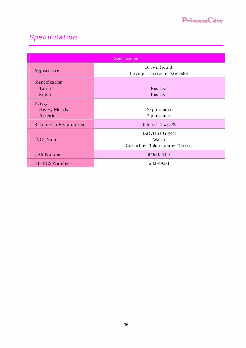
Specification
Specification
Appearance Brown liquid, having a characteristic odor
Identification Tannin Sugar
Positive Positive
Purity Heavy Metals Arsenic
20 ppm max. 2 ppm max.
Residue on Evaporation 0.6 to 1.4 w/v %
INCI Name Butylene Glycol
Water Geranium Robertianum Extract
CAS Number 84650-11-3
EILECS Number 283-492-1
37
Reference
1. Sachiko AKIMOTO et al., Clinic on Mast Cell, Sentan Igaku-sya, Tokyo, 212-217 (2001) 2. Akiko YOSHIMI et al., Clinic on Mast Cell, Sentan Igaku-sya, Tokyo, 112-119 (2001) 3. Andrew F. WALLS, “MAST CELLS AND BASOPHILS”, ACADEMIC PRESS, London, 291 -
309 (2001) 4 . Jenny HALLGREN, et.al., FEBS Journal. , 273, 1871 - 1895 (2006) 5. Anke RATTENHOLL, et.al., Drug Development Research, 59, 408 – 416 (2003) 6. S. BOSSET, et.al., Br. J. Dermatol., 149, 826 - 835 (2003) 7. Shinji INOMATA, et.al., J. Invest. Dermatol. , 120, 1 - 7 (2003) 8. Renata KAMINSKA, et.al., J. Invest. Dermatol. , 113, 31 – 39 (2005) 9. Arunasiri IDDAMALGODA , et al., 126th Annual Meeting of Pharmaceutical Society of Japan,
Proceedings, 149 (2006) 10. Arunasiri IDDAMALGODA, et al., 7th Annual Meeting of Japanese Photoaging Research
Society, Proceedings, 14 (2006) 11. Arunasiri IDDAMALGODA, et al., Satellite meeting of the 20th IUBMB International
Congress, Proceedings, 48 (2006) 12. Minoru OKADA et al., Newly Illustrated Medicinal Plants of the world, Hokuryu Co.,Ltd.,
239 (2002) 13. Deni Bown, Herb Encyclopedia, Seibundo Shinkosha Inc., Tokyo, 554 (1997) 14. Tatemi SHIMIZU, ASAHI Encyclopedia The World of Plants, Asahi Shinbun Company,
Tokyo, 164-165 (1990) 15. Shoji HAYASHI et al., J. Soc. Cosmet. Chem. Japan., 27 (3), 335-373, (1993) 16. Motoji TAKAHASHI, FRAGRANCE JOURNAL, 18(12, 34 - 40 (1990)
17. Tomoko SUGAWARA et al., Ningen Dock, 20 (3) 483-486 (2005) 18. Toshihiko TSUJI et al., J. Soc. Cosmet. Chem. Japan., 25 (4), 246-253, (1992) 19. Michinori KUBO, YAKUGAKU ZASSHI, 110 (1), 59-67 (1990)