from a to z: a potential role for grid cells in spatial navigation
TRANSCRIPT

Barry and Bush Neural Systems & Circuits 2012, 2:6http://www.neuralsystemsandcircuits.com/content/2/1/6
REVIEW Open Access
From A to Z: a potential role for grid cells inspatial navigationCaswell Barry1,2,3* and Daniel Bush1,2
Abstract
Since their discovery, the strikingly regular and spatially stable firing of entorhinal grid cells has attracted theattention of experimentalists and theoreticians alike. The bulk of this work has focused either on the assumptionthat the principal role of grid cells is to support path integration or the extent to which their multiple firinglocations can drive the sparse activity of hippocampal place cells. Here, we propose that grid cells are bestunderstood as part of a network that combines self-motion and environmental cues to accurately track an animal’slocation in space. Furthermore, that grid cells - more so than place cells - efficiently encode self-location inallocentric coordinates. Finally, that the regular structure of grid firing fields represents information about therelative structure of space and, as such, may be used to guide goal directed navigation.
IntroductionHalf a century’s worth of research has established, beyonddoubt, the role of the hippocampal formation in memory.Damage to the human hippocampus and surrounding cor-tex results in profound amnesia for events occurring afterthe insult and also for those occurring beforehand in atemporally graded fashion [1,2]. Such patients exhibitimpaired spatial cognition, have difficulty navigating [3],remembering the relative location of objects [4], and evenvisualizing imagined scenes [5]. In model organisms, suchas the rat, lesions of the hippocampal formation producesimilar deficits, including an impaired ability to navigate [6]and failure to recognize novel spatial arrangements [7].Single unit recordings made in the early 1970s first inti-mated a neural basis for these functions in the form ofplace cells (Figure 1a), hippocampal pyramidal neuronswith spatially localized firing fields (place fields) [8].Prompted by this discovery, O’Keefe and Nadel proposedthat place cells constitute part of a hippocampal networkwhich functions as a cognitive map; representing an ani-mal’s location within its environment relative to otherobjects and, hence, enabling flexible navigation strategies,including novel short-cuts and detours [9]. Furthermore,they suggested that this cognitive map is the basis of
* Correspondence: [email protected] Inst of Cognitive Neuroscience, London, UK2UCL Inst of Neurology, London, UKFull list of author information is available at the end of the article
© 2012 Barry and Bush; licensee BioMed CentCommons Attribution License (http://creativecreproduction in any medium, provided the or
human episodic memory, the spatial framework beingembellished to encode the content of specific events.While place cells provide an undeniably spatial signal,
attempts to derive models that would support flexiblenavigation proved difficult for several reasons. First, it wasinitially believed that place cells simply encoded an animal’scurrent location, with no capacity to represent the route to,or site of, a navigational goal. Second, the sparsely distribu-ted, irregular place fields of an ensemble of place cells donot obviously convey information about the relativeproximity of those fields in a given environment. Directconnectivity between place cells, such as that found inCA3, could encode the distance between place fields [10].However, the synaptic weight matrix would have to belearned for each new environment and could not supportaccurate navigation across unvisited areas, a feat that manyanimals, including rodents, are capable of [11]. Both thesebarriers now seem to be falling away. The increasingly wellunderstood phenomena of preplay and replay in ensemblesof hippocampal place cells [12,13] together with the dis-covery of entorhinal grid cells with periodic spatial firingfields [14,15], indicate that the representation of space inthe hippocampal formation is both non-local and spatiallystructured. Here we argue that grid cells, and not placecells, principally encode self-location in allocentric coordi-nates and, furthermore, that they also represent the relativeproximity of spaces in an animal’s environment. Finally, be-cause grid cells encode spatial information in this way, theyare likely to be a key part of a network supporting vector
ral Ltd. This is an Open Access article distributed under the terms of the Creativeommons.org/licenses/by/2.0), which permits unrestricted use, distribution, andiginal work is properly cited.
5.6Hz 3.2Hz
0.760.79
1.01
0.78
1.01 0.81
a
d
b
c
100cm
70cm
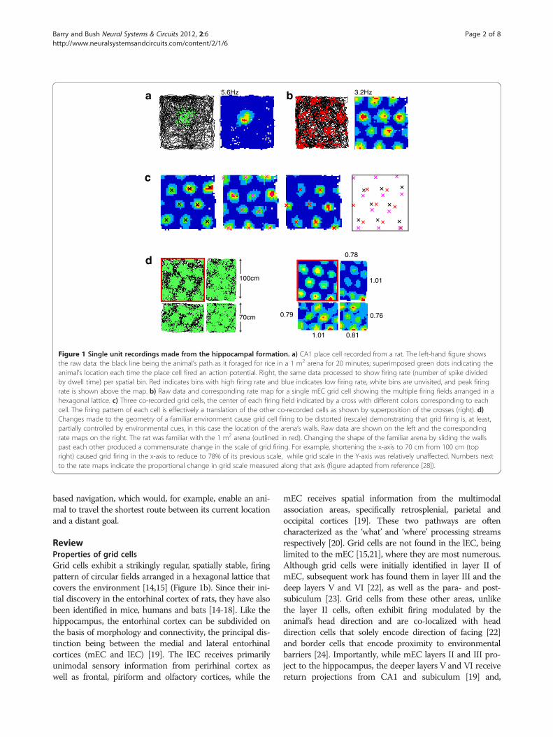
Figure 1 Single unit recordings made from the hippocampal formation. a) CA1 place cell recorded from a rat. The left-hand figure showsthe raw data: the black line being the animal’s path as it foraged for rice in a 1 m2 arena for 20 minutes; superimposed green dots indicating theanimal’s location each time the place cell fired an action potential. Right, the same data processed to show firing rate (number of spike dividedby dwell time) per spatial bin. Red indicates bins with high firing rate and blue indicates low firing rate, white bins are unvisited, and peak firingrate is shown above the map. b) Raw data and corresponding rate map for a single mEC grid cell showing the multiple firing fields arranged in ahexagonal lattice. c) Three co-recorded grid cells, the center of each firing field indicated by a cross with different colors corresponding to eachcell. The firing pattern of each cell is effectively a translation of the other co-recorded cells as shown by superposition of the crosses (right). d)Changes made to the geometry of a familiar environment cause grid cell firing to be distorted (rescale) demonstrating that grid firing is, at least,partially controlled by environmental cues, in this case the location of the arena’s walls. Raw data are shown on the left and the correspondingrate maps on the right. The rat was familiar with the 1 m2 arena (outlined in red). Changing the shape of the familiar arena by sliding the wallspast each other produced a commensurate change in the scale of grid firing. For example, shortening the x-axis to 70 cm from 100 cm (topright) caused grid firing in the x-axis to reduce to 78% of its previous scale, while grid scale in the Y-axis was relatively unaffected. Numbers nextto the rate maps indicate the proportional change in grid scale measured along that axis (figure adapted from reference [28]).
Barry and Bush Neural Systems & Circuits 2012, 2:6 Page 2 of 8http://www.neuralsystemsandcircuits.com/content/2/1/6
based navigation, which would, for example, enable an ani-mal to travel the shortest route between its current locationand a distant goal.
ReviewProperties of grid cellsGrid cells exhibit a strikingly regular, spatially stable, firingpattern of circular fields arranged in a hexagonal lattice thatcovers the environment [14,15] (Figure 1b). Since their ini-tial discovery in the entorhinal cortex of rats, they have alsobeen identified in mice, humans and bats [14-18]. Like thehippocampus, the entorhinal cortex can be subdivided onthe basis of morphology and connectivity, the principal dis-tinction being between the medial and lateral entorhinalcortices (mEC and lEC) [19]. The lEC receives primarilyunimodal sensory information from perirhinal cortex aswell as frontal, piriform and olfactory cortices, while the
mEC receives spatial information from the multimodalassociation areas, specifically retrosplenial, parietal andoccipital cortices [19]. These two pathways are oftencharacterized as the ‘what’ and ‘where’ processing streamsrespectively [20]. Grid cells are not found in the lEC, beinglimited to the mEC [15,21], where they are most numerous.Although grid cells were initially identified in layer II ofmEC, subsequent work has found them in layer III and thedeep layers V and VI [22], as well as the para- and post-subiculum [23]. Grid cells from these other areas, unlikethe layer II cells, often exhibit firing modulated by theanimal’s head direction and are co-localized with headdirection cells that solely encode direction of facing [22]and border cells that encode proximity to environmentalbarriers [24]. Importantly, while mEC layers II and III pro-ject to the hippocampus, the deeper layers V and VI receivereturn projections from CA1 and subiculum [19] and,

Barry and Bush Neural Systems & Circuits 2012, 2:6 Page 3 of 8http://www.neuralsystemsandcircuits.com/content/2/1/6
subsequently, project back to the shallow layers, placinggrid cells within a processing loop that encompasses mostof the hippocampal formation [19].The scale of the grid pattern, measured as the distance be-
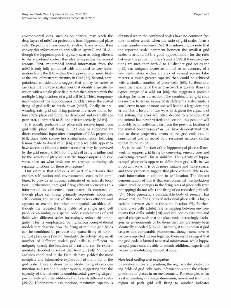
tween neighboring peaks, increases along the dorso-ventralmEC gradient, mirroring a similar trend in hippocampalplace fields [15,25]. The smallest, most dorsal, scale is typic-ally 20 to 25 cm in the rat, reaching in excess of severalmeters in the intermediate region of the gradient [15,26](Figure 2). This may explain how this remarkable patternwas missed by early electrophysiology studies, which tar-geted ventral mEC and found only broadly tuned spatial fir-ing (for example, [27]). Interestingly, grid scale increases indiscontinuous increments and the increment ratio, at leastbetween the smaller scales, is constant [28]. Grid cellsrecorded from the same electrode, which are, therefore,proximate in the brain, typically have a common scale andorientation but a random offset relative to each other andthe environment [15]. As such, their firing patterns are ef-fectively identical translations of one another and a smallnumber of cells will ‘tile’ the complete environment(Figure 1c). It also appears that grids of different scalerecorded ipsilaterally have a common orientation, such thatthe hexagonal arrangement of their firing fields share thesame three axes, albeit with some localized distortions[15,28,29].
Hippocampus(dentate gyrus,
CA1 & CA3)
Posterior
Mediaentorhcortex
Figure 2 Grid scale increases along a dorso-ventral gradient in the mtimes are shown, both cells were recorded in a familiar 1 m2 arena. Approxcell exhibits a considerably larger size of firing fields and distance between
Mechanisms of grid field formationDue to their invariant spatial metric, theoretical models ofgrid cell activity have almost exclusively described their fir-ing in terms of a system that integrates idiothetic cues inorder to update self-location. In fact, it is hard to see howsuch a regular pattern that is coherent between neighboringcells could be otherwise produced. However, these modelsdiffer significantly in the manner by which they account forthe formation of the grid field; either by continuousattractor dynamics or oscillatory interference. Attractormodels hypothesize that grid cell activity reflects a ‘packet’or ‘packets’ of localized excitation on a flat energylandscape provided by recurrent connections, and that thisactivation can be smoothly shifted by translational inputfrom speed modulated head direction or conjunctive cells[30-32]. Conversely, oscillatory interference models positthat grid firing is generated by interference between velocitycontrolled oscillators (VCOs) – individual cells or smallnetworks that increase their firing frequency according tothe speed of movement in a preferred direction – and abaseline oscillation [33-36]. The phase difference betweenthese oscillations then reflects displacement in the preferreddirection of the VCOs. Hence, interference between thebaseline oscillation and VCOs with preferred directionsthat differ by multiples of 60° produces periodic spatialtuning with six fold rotational symmetry.
100cm
linal
EC. Two grid cells recorded from the same animal but at differentimate recording locations in the mEC are indicated. The more ventralfiring fields than the dorsal cell.

Barry and Bush Neural Systems & Circuits 2012, 2:6 Page 4 of 8http://www.neuralsystemsandcircuits.com/content/2/1/6
Evidence in favor of the attractor model comes from theexistence of the requisite speed modulated head directionand conjunctive cells in mEC, post- and para- subiculum,which also appear to have the necessary anatomicalconnections to grid cells in those regions [22,37]. Theobserved topographical clustering of grid scales and orien-tation is also a necessary component of attractor models[28]. Criticism of this class of model is generally focused onthe apparent lack of recurrent excitatory connectionsbetween principal cells in layer II mEC, although contra-dictory reports do exist and attractor dynamics might bemaintained in the deeper layers by recurrent inhibition, belimited to the dense entorhinal cell islands, or be located ineither the pre- or para- subiculum [23,31,37-40]. Moreover,the stable operation of any attractor network requiresprecisely tuned synaptic weights to prevent drift or disrup-tion of the activity packet, and it is not clear how suchconnectivity would be developed or maintained in vivo.Evidence in support of the oscillatory interference models
comes from recordings of putative velocity controlled oscil-lator cells in the anterior thalamus, medial septum andhippocampus that exhibit cosine directional tuning of firingfrequency [41]. Similarly, inactivation of the medial septumin rodents, which generates the theta frequency oscillationsthat dominate the hippocampus during translational move-ment, quickly eliminates the grid field firing pattern [42,43].Most compelling, though, is evidence that links the theta-band frequency of membrane potential oscillations in mECgrid cells with their scale, as predicted by the model [44,45].Manipulations that show a change in one of these proper-ties is accompanied by the expected change in the other arealso encouraging [46,47]. The principal criticism of thisclass of model relates to the difficulty of maintaining pre-cisely timed oscillations in neural circuits – as any erroraccumulated in the VCOs will quickly disrupt the resultantgrid field [39,48]. Simulations indicate that this is not neces-sarily the case, however. While independent realisticallynoisy oscillators would quickly decohere, a population ofcoupled oscillators would not [36,41]. Similarly, the obser-vation of phase precession in grid cell firing is not onlyexplained by this class of model but also indicates that thetiming of neural oscillations can be maintained with a highdegree of fidelity in vivo [49]. Recently, recordings madefrom crawling fruit bats appear to present a serious chal-lenge for the oscillatory interference models: the bats ex-hibit no continuous theta in the entorhinal or hippocampalLFP and have grid cells with no theta-band modulation ofthe spike train [18]. However, the very low movementspeed and firing rates make these results difficult to inter-pret as they render theta-band modulation of the spiketrain hard to detect [50]. The same group has presentedplace cell recordings from flying bats but it is unclear if thisdata exhibit theta-band modulation because of artefactscreated by the animals’ 7-8Hz wing beats [51]. Interestingly,
several models of grid cell firing have recently been pub-lished which incorporate both recurrent connectivity andtemporal dynamics (for example, [41,52]). Interaction be-tween these two, possibly redundant, mechanisms may ac-count for the disparity in results so far reported fromdifferent species.
Path integrationSo what is the function of grid cells? Or, more meaning-fully, what information do grid cells encode? Again, mostmodels and theoretical studies have focused either on theassumption that their principal function is to support pathintegration [30,31,34,36,53,54] or the extent to which theirmultiple firing locations can drive the unitary firing of placecells [55-57]. It is to the first of these points that we turn.Path integration is a basic navigational strategy observed
across a wide range of species in which an animal’s currentposition relative to some reference point is maintained bycontinually integrating the direction and distance movedaccording to idiothetic cues [11,58]. The experimentalstudies described above delineate several properties of gridcells that provide indirect evidence for an involvement inpath integration. First, they are co-localized with headdirection and conjunctive cells that exhibit coherent spatialtuning across different environments, such that all the in-formation required to perform path integration is presentin the local circuit [22,59]. Second, the grid field isgenerated rapidly in a novel environment, updated in theabsence of visual input, and stable to the removal of localcues [15,59]. Finally, the spatial scale of the grid field isfixed across familiar environments and striking in its regu-larity, thereby providing a coherent, consistent and reliableestimate of allocentric distance travelled in a context inde-pendent fashion [15,59].That said, grid firing does not simply track accumu-
lated idiothetic cues, it encodes an animal’s locationin allocentric space and, once established, is clearlystabilized and controlled by environmental (allothetic)cues. For example, grid firing is stable between visitsto an environment, over distances and durations thatare unlikely to be accurately judged on the basis ofidiothetic information alone [15]. More obviously, theorientation of grid firing is controlled by movementof a single polarizing cue in an otherwise symmetricalcircular environment [15]. Similarly, the spacing andregularity of the grid pattern is influenced by manipu-lations of the shape and position of boundaries withinan animal’s environment (Figure 1d). In fact, whenself-motion and environmental cues are placed incontradiction, grid firing is initially more stronglyshaped by the latter [28,60].How might environmental cues become associated with
grid firing? An interesting suggestion raised by O’Keefe andBurgess [61] is that information about the location of
Barry and Bush Neural Systems & Circuits 2012, 2:6 Page 5 of 8http://www.neuralsystemsandcircuits.com/content/2/1/6
environmental cues, such as boundaries, may reach thedeep layers of mEC via projections from hippocampal placecells. Projections from deep to shallow layers would thenconvey this information to grid cells in layers II and III. Al-though the hippocampus is typically seen as being efferentto the entorhinal cortex, this idea is appealing for severalreasons. First, multimodal spatial information from themEC is only fully combined with unimodal sensory infor-mation from the lEC within the hippocampus, most likelyat the level of recurrent circuitry in CA3 [21]. Second, com-putational considerations suggest that it may be easier toassociate the multiple spatial cues that identify a specific lo-cation with a single place field rather than directly with themultiple firing locations of a grid cell [61]. Third, temporaryinactivation of the hippocampus quickly causes the spatialfiring of grid cells to break down [49,62]. Finally, in pre-weanling rats, grid cell firing patterns are never found be-fore stable place cell firing has developed and normally ap-pear later, at days p20 to 21 and p16 respectively [63,64].It is equally probable that place cells receive input from
grid cells: place cell firing in CA1 can be supported bydirect entorhinal input after disruption of CA3 projections[64]; place fields convey less spatial information followinglesions made to dorsal mEC [66]; and place fields appear tohave access to idiothetic information that may be conveyedby the grid network [67]. So, if grid cell firing is influencedby the activity of place cells in the hippocampus and viceversa, then on what basis can we attempt to distinguishseparate functions for these two regions?Our claim is that grid cells are part of a network that
enables self-motion and environmental cues to be com-bined to provide an optimal estimate of an animal’s loca-tion. Furthermore, that grid firing efficiently encodes thisinformation in allocentric coordinates. In contrast, al-though place cell firing primarily represents allocentricself-location, the nature of that code is less efficient andappears to encode for other, non-spatial, variables. Al-though the repeated firing fields of a single grid cellproduce an ambiguous spatial code, combinations of gridfields with different scales increasingly reduce this ambi-guity. This is emphasized by numerous computationalmodels that describe how the firing of multiple grid fieldscan be combined to produce the sparse firing of hippo-campal place cells [55-57]. Similarly, the activity of a smallnumber of different scaled grid cells is sufficient touniquely specify the location of a rat and can be experi-mentally decoded to reveal that location [14]. Numericalanalyses conducted in the Fiete lab have yielded the mostcomplete and informative exploration of the limits of thegrid code. These analyses demonstrate that grid cells canfunction as a residue number system, suggesting that thecapacity of the network is combinatorial, growing dispro-portionately with the addition of units with different scales[39,68]. Under certain assumptions, maximum capacity is
obtained when the combined scales have no common fac-tors, in other words when the ratio of grid scales form aprime number sequence [68]. It is interesting to note thatthe reported scale increment between the smallest gridscales is around 1.65, a good approximation for the ratiobetween the prime numbers 5 and 3 [28]. If these assump-tions are met, then with 8 to 10 distinct grid scales themEC can uniquely locate an animal to an accuracy of afew centimeters within an area of several square kilo-meters; a much greater capacity than could be achievedwith a similar number of place cells [68]. Furthermore,since the capacity of the grid network is greater than thetypical range of a wild rat [68], this suggests a possiblestrategy for error correction. The combinatorial grid codeis sensitive to errors in any of its differently scaled units; asmall error in one or more unit will lead to a large decodingerror. This is helpful in two ways: first, given the capacity ofthe system, the error will often decode to a position thatthe animal has never visited; and second, this position willprobably be unrealistically far from the previous location ofthe animal. Sreenivasan et al [32] have demonstrated that,due to these properties, errors in the grid code can beconstrained and corrected by a recurrent network similarto that found in CA3.So, is the sole function of the hippocampal place cell net-
work to support grid firing by conveying sensory cues andcorrecting errors? This is unlikely. The activity of hippo-campal place cells appear to differ from grid cells in twoimportant ways: it is both more variable and more sparse,and these properties suggest that place cells are able to en-code information in addition to self-location. The clearestdemonstration of this is that environmental manipulationswhich produce changes in the firing rates of place cells (rateremapping) do not affect the firing of co-recorded grid cells[59]. More generally, a considerable body of research hasshown that the firing rates of individual place cells is highlyvariable between visits to the same location [69]. Further-more, place cells exhibit rate remapping between environ-ments that differ subtly [70], and can accumulate rate andspatial changes such that the place code increasingly distin-guishes environments or locations that had previously beenidentically encoded [70-72]. Currently, it is unknown if gridcells exhibit comparable phenomena, though none have sofar been reported. Taken together, these results suggest thatthe grid code is limited to spatial information, while hippo-campal place cells are able to encode additional experientialfactors by modulating the spatial code.
Non-local coding and navigationIn addition to current position, the regularly distributed fir-ing fields of grid cells carry information about the relativeproximity of places in an environment. For example, whena rat is traveling in a single dimension, movement from oneregion of peak grid cell firing to another indicates
Barry and Bush Neural Systems & Circuits 2012, 2:6 Page 6 of 8http://www.neuralsystemsandcircuits.com/content/2/1/6
displacement by some integer multiple of the grid scale. Inprincipal, the calculations that update grid firing by inte-grating idiothetic cues can be reversed in order to extractthe translational vector between two allocentric locations.Such a process could provide the basis for a navigation sys-tem that would enable an animal to travel directly from itscurrent location to a non-visible goal, a task which rodentsand other animals can ably perform [11]. Might a grid cellnetwork support these navigational abilities?Preliminary theoretical results indicate that it is possible to
create a network that will extract both the distance and dir-ection of a goal from the activity of a population of grid cells[73,74]. However, as yet, there are no published models thatactually direct navigation on the basis of grid firing (see noteadded in proof). Entorhinal lesion studies indicate that dam-age to the grid network does impair an animal’s ability toreach a hidden goal but, because the entorhinal cortex is re-ciprocally connected with the hippocampus, it can be diffi-cult to interpret these results. That said, lesions focused onthe shallow layers of dorsal mEC eliminated spatial prefer-ence in rats trained on the Morris water maze [75]. Import-antly, the animals were subsequently able to relearn the task,indicating that some degree of spatial processing was pre-served. Less specific entorhinal lesions also produce deficitsin the water maze and particularly impact an animal’s abilityto navigate directly to the escape platform. Interestingly, therats change strategy as a result, searching for the goal closeto cues placed within the maze [76]. Lesions made to theentorhinal and parietal cortex also produce path integra-tive deficits in a homing task [77]. Similarly, bilateral dis-connection of the entorhinal-hippocampal circuit wasfound to impair detection of a spatial change when famil-iar objects were moved relative to one another, possiblyindicating a deficit in the ability to judge relative position[78]. However, contradictory results do exist, for exampleBurwell et al. [79] did not detect navigational deficits aftermaking entorhinal lesions in rats. A likely source of thereported variability is that several of these studies wereconducted before the discovery of grid cells, and lesionswere made without knowledge of the precise topograph-ical arrangement of those cells within entorhinal cortex.Finally, accumulating electrophysiological results from the
last 15 years have increasingly shown that place cells can firenon-locally, effectively encoding trajectories removed fromthe animal’s current location [12,13,80]. These events typic-ally occur during hippocampal sharp waves, brief periods ofactivity characterized by a reduction in inhibition and transi-ent high frequency oscillations (100 to 200 Hz ‘ripples’ [9])in the local field potential, as well as during REM sleep. Ithas been known for some time that hippocampal ripplesreach the entorhinal cortex [81] and preliminary results in-dicate that grid cells also participate in these preplay events[81]. What remains unproven is whether these events arerelated to task demands and might, therefore, indicate the
route that an animal will subsequently follow to reach a goal.Though not directly related to preplay, recordings madefrom mEC while rats performed a T-maze alternation taskshowed that cells in this region (though not explicitly gridcells) modulated their firing according to the route that theanimal was following [83]. Similar results have been notedfor place cells [84]. However, in this case the authors com-pared the mEC modulation with co-recorded place cells andfound that the entorhinal effect was larger and more inform-ative about the animal’s future actions. fMRI studies also im-plicate the entorhinal cortex in navigational planning. Forexample, a study of London taxi drivers navigating in a vir-tual reality rendition of central London demonstrated thatentorhinal activity positively correlated with Euclidian dis-tance to a goal [85].
ConclusionsIt seems clear that the regular firing pattern of grid cellsrepresent an efficient strategy for encoding self-location inallocentric coordinates. Furthermore, that grid cells andplace cells form part of a network that combines idiotheticand allothetic cues to accurately track an animal’s move-ment through space. It is also clear that the activity of apopulation of grid cells encodes information about the rela-tive structure of space. What is currently unknown iswhether this metric is accessible to other structures in thebrain and, if so, whether it is employed during navigation.Existing results and theoretical models suggest this may bethe case, but it will require more precisely targeted investi-gations using new techniques, such as optogenetics, to con-firm or deny this hypothesis.
Note added in proof: Since submission of this manu-script two computational models that incorporate gridcells and perform goal directed spatial navigation have,in fact, been published [86,87].
Competing interestsThe authors declare that they have no competing interests.
AcknowledgementsThis work was supported by the Wellcome Trust U.K. The authors would liketo thank Neil Burgess for assistance in preparing this manuscript.
Author details1UCL Inst of Cognitive Neuroscience, London, UK. 2UCL Inst of Neurology,London, UK. 3UCL Inst of Behavioural Neuroscience, London, UK.
Author contributionsCB and DB made equal academic contributions to this work and bothparticipated in drafting the manuscript. Both authors read and approved thefinal manuscript.
Received: 28 February 2012 Accepted: 18 April 2012Published: 30 May 2012
References1. Scoville WB, Milner B: Loss of recent memory after bilateral hippocampal
lesions. J Neurol Neurosurg Psychiatry 1959, 20:11–21.

Barry and Bush Neural Systems & Circuits 2012, 2:6 Page 7 of 8http://www.neuralsystemsandcircuits.com/content/2/1/6
2. Squire LR: Memory systems of the brain: a brief history and currentperspective. Neurobiol Learn Mem 2004, 82:171–177.
3. Burgess N, Maguire EA, O’Keefe J: The human hippocampus: spatial andepisodic memory. Neuron 2002, 35:625–641.
4. Hartley T, Bird CM, Chan D, Cipolotti L, Husain M, Vargha-Khadem F, BurgessN: The hippocampus is required for short-term topographical memory inhumans. Hippocampus 2007, 17:34–48.
5. Hassabis D, Kumaran D, Vann SD, Maguire EA: Patients with hippocampalamnesia cannot imagine new experiences. Proc Natl Acad Sci USA 2007,104:1726–1731.
6. Morris RGM, Garrud P, Rawlins JNP, O’Keefe J: Place navigation impaired inrats with hippocampal lesions. Nature 1982, 297:681–683.
7. Brown MW, Aggleton JP: Recognition memory: what are the roles of theperirhinal cortex and hippocampus? Nature reviews. Neuroscience 2001, 2:51–61.
8. O’Keefe J, Dostrovsky J: The hippocampus as a spatial map. Preliminaryevidence from unit activity in the freely-moving rat. Brain Res 1971, 34:171–175.
9. O’Keefe J, Nadel L: The hippocampus as a cognitive map. 1st edition. Oxford:Oxford University Press; 1978.
10. Muller RU, Stead M, Pach J: The hippocampus as a cognitive graph. J GenPhysiol 1996, 107:663–694.
11. Etienne AS, Jeffery KJ: Path integration in mammals. Hippocampus 2004,14:180–192.
12. Wilson MA, McNaughton BL: Reactivation of hippocampal ensemblememories during sleep. Science 1994, 265:676–679.
13. Louie K, Wilson MA: Temporally structured replay of awake Hippocampalensemble activity during rapid eye movement sleep. Neuron 2001, 29:145–156.
14. Fyhn M, Molden S, Witter MP, Moser EI, Moser M: Spatial representation inthe entorhinal cortex. Science 2004, 305:1258–1264.
15. Hafting T, Fyhn M, Moser M, Moser EI: Microstructure of a spatial map inthe entorhinal cortex. Nature 2005, 436:801–806.
16. Fyhn M, Hafting T, Witter MP, Moser EI, Moser M-B: Grid cells in mice.Hippocampus 2008, 18:1230–1238.
17. Doeller CF, Barry C, Burgess N: Evidence for grid cells in a human memorynetwork. Nature 2010, 463:657–661.
18. Yartsev MM, Witter MP, Ulanovsky N: Grid cells without theta oscillationsin the entorhinal cortex of bats. Nature 2011, 479:103–107.
19. Amaral DG, Witter MP: Hippocampal Formation. In The rat nervous system.1st edition. Edited by Paxinos G. Academic Press, San Diego. 1995:443–486.
20. Mishkin M, Ungerleider LG: Contribution of striate inputs to thevisuospatial functions of parieto- preoccipital cortex in monkeys.Behav Brain Res 1982, 6:57–77.
21. Hargreaves EL, Roa G, Lee I, Knierim JJ: Major dissociation between medial andlateral entorhinal input to dorsal hippocampus. Science 2005, 308:1792–1794.
22. Sargolini F, Fyhn M, Hafting T, McNaughton BL, Witter MP, Moser MB, MoserEI: Conjunctive representation of position, direction, and velocity inentorhinal cortex. Science 2006, 312:758–762.
23. Boccara CN, Sargolini F, Thoresen VH, Solstad T, Witter MP, Moser EI, MoserMB: Grid cells in pre- and parasubiculum. Nat Neurosci 2010, 13:987–994.
24. Solstad T, Boccara CN, Kropff E, Moser M-B, Moser EI: Representation ofgeometric borders in the entorhinal cortex. Science 2008, 322:1865–1868.
25. Kjelstrup KB, Solstad T, Brun VH, Hafting T, Leutgeb S, Witter MP, Moser EI,Moser MB: Finite scale of spatial representation in the hippocampus.Science 2008, 321:140–143.
26. Brun VH, Solstad T, Kjelstrup KB, Fyhn M, Witter MP, Moser EI, Moser MB:Progressive increase in grid scale from dorsal to ventral medialentorhinal cortex. Hippocampus 2008, 18:1200–1212.
27. Quirk GJ, Muller RU, Kubie JL, Ranck JB: The positional firing properties ofmedial entorhinal neurons: description and comparison withhippocampal place cells. J Neurosci 1992, 12:1945–1963.
28. Barry C, Hayman R, Burgess N, Jeffery KJ: Experience-dependent rescalingof entorhinal grids. Nat Neurosci 2007, 10:682–684.
29. Stensland H, Kirkesola T, Moser MB, Moser E: Orientational geometry ofEntorhinal Grid Cells. Society for Neuroscience 2010, 101:14.
30. Fuhs MC, Touretzky DS: A spin glass model of path integration in ratmedial entorhinal cortex. J Neurosci 2006, 26:4266–4276.
31. McNaughton BL, Battaglia FP, Jensen O, Moser EI, Moser MB: Pathintegration and the neural basis of the “cognitive map”. Nat Rev Neurosci2006, 7:663–678.
32. Sreenivasan S, Fiete I: Grid cells generate an analog error-correcting codefor singularly precise neural computation. Nat Neurosci 2011,14:1330–1337.
33. Barry C, Burgess N, O’Keefe J: An oscillatory interference model of grid cellfiring. Hippocampus 2007, 17:801–812.
34. Blair HT, Gupta K, Zhang K: Phase coding and central pattern generationby ring attractors: a model of theta cells, grid cells, and place cells.Hippocampus 2008, 18:1239–1255.
35. Burgess N: The oscillatory interference model of grid cell firing: theoryand predictions. Hippocampus 2008, 18:1157–1174.
36. Zilli EA, Hasselmo ME: Coupled noisy spiking neurons as velocity-controlledoscillators in a model of grid cell spatial firing. J Neurosci 2010,30:13850–13860.
37. Witter MP, Moser EI: Spatial representation and the architecture of theentorhinal cortex. Trends Neurosci 2006, 29:671–678.
38. Dhillon A, Jones R: Laminar differences in recurrent excitatorytransmission in the rat entorhinal cortex in vitro. Neuroscience 2000,99:413–422.
39. Burak Y, Fiete IR: Accurate path integration in continuous attractornetwork models of grid cells. PLoS Comput Biol 2009, 5:e1000291.
40. Burgalossi A, et al: Microcircuits of functionally identified neurons in therat medial entorhinal cortex. Neuron 2011, 70:773–786.
41. Welday aC, Shlifer IG, Bloom ML, Zhang K, Blair HT: Cosine directionaltuning of theta cell burst frequencies: evidence for spatial coding byoscillatory interference. J Neurosci 2011, 31:16157–16176.
42. Brandon MP, Bogaard AR, Libby CP, Connerney MA, Gupta K, Hasselmo ME:Reduction of theta rhythm dissociates grid cell spatial periodicity fromdirectional tuning. Science 2011, 332:595–599.
43. Koenig J, Linder AN, Leutgeb JK, Leutgeb S: The spatial periodicity of gridcells is not sustained during reduced theta oscillations. Science 2011,332:592–595.
44. Jeewajee A, Barry C, O’Keefe J, Burgess N: Grid cells and theta asoscillatory interference: electrophysiological data from freely movingrats. Hippocampus 2008, 18:1175–1185.
45. Giocomo LM, Zilli EA, Fransen E, Hasselmo ME: Temporal frequency ofsubthreshold oscillations scales with entorhinal grid field spacing.Science 2007, 315:1719–1722.
46. Giocomo LM, Hussaini SA, Zheng F, Kandel ER, Moser MB, Moser EI: Gridcells use HCN1 channels for spatial scaling. Cell 2011, 147:1159–1170.
47. Barry C, O’Keefe J, Burgess N: Effect of novelty on grid cell firing.Society for Neuroscience 2009, 101:24.
48. Zilli EA, Yoshida M, Tahvildari B, Giocomo LM, Hasselmo ME: Evaluation ofthe oscillatory interference model of grid cell firing through analysis andmeasured period variance of some biological oscillators. PLoS ComputBiol 2009, 5:e1000573.
49. Hafting T, Fyhn M, Bonnevie T, Moser M-B, Moser EI: Hippocampus-independent phase precession in entorhinal grid cells. Nature 2008,453:1248–1252.
50. Barry C, Bush D, O’Keefe J, Burgess B: Models of grid cells and thetaoscillations. Nature 2012, 488.
51. Ulanovsky N, Yartsev MM: Evidence for theta oscillations in thehippocampus of flying bats. Society for Neuroscience Abstract 2011, 937:27.
52. Navratilova Z, Giocomo LM, Fellous J-M, Hasselmo ME, McNaughton BL:Phase precession and variable spatial scaling in a periodic attractor mapmodel of medial entorhinal grid cells with realistic after-spike dynamics.Hippocampus 2011, doi:10.1002/hipo.20939.
53. Burgess N, Barry C, O’Keefe J: An oscillatory interference model of grid cellfiring. Hippocampus 2007, 17:801–812.
54. Blair HT, Welday AC, Zhang K: Scale-invariant memory representationsemerge from Moire interference between grid fields that produce thetaoscillations: a computational model. J Neurosci 2007, 27:3211–3229.
55. Solstad T, Moser EI, Einevoll GT: From grid cells to place cells: amathematical model. Hippocampus 2006, 16:1026–1031.
56. de Almeida L, Idiart M, Lisman JE: The input-output transformation of thehippocampal granule cells: from grid cells to place fields. J Neurosci 2009,29:7504–7512.
57. Monaco JD, Abbott LF: Modular realignment of entorhinal grid cellactivity as a basis for hippocampal remapping. J Neurosci 2011,31:9414–9425.
58. Mittelstaedt ML, Mittelstaedt H: Homing by path integration in a mammal.Naturwissenschaften 1980, 67:566–567.
59. Fyhn M, Hafting T, Treves A, Moser M-B, Moser EI: Hippocampalremapping and grid realignment in the entorhinal cortex. Nature 2007,446:190–194.

Barry and Bush Neural Systems & Circuits 2012, 2:6 Page 8 of 8http://www.neuralsystemsandcircuits.com/content/2/1/6
60. Derdikman D, Whitlock JR, Tsao A, Fyhn M, Hafting T, Moser MB, Moser EI:Fragmentation of grid cell maps in a multicompartment environment.Nat Neurosci 2009, 12:1325–1332.
61. O’Keefe J, Burgess N: Dual phase and rate coding in hippocampal placecells: theoretical significance and relationship to entorhinal grid cells.Hippocampus 2005, 15:853–866.
62. Bonnevie T, Fyhn M, Hafting T, Moser EI, Moser M-B: Misalignment ofentorhinal grid fields after hippocampal inactivation. Society forNeuroscience 2006, 68:8.
63. Wills TJ, Cacucci F, Burgess N, O’Keefe J: Development of the hippocampalcognitive map in preweanling rats. Science 2010, 328:1573–1576.
64. Langston RF, Ainge JA, Couey JJ, Canto CB, Bjerknes TL, Witter MP, Moser EI,Moser MB: Development of the spatial representation system in the rat.Science 2010, 328:1576–1580.
65. Brun VH, Otnaess MK, Molden S, Steffenach HA, Witter MP, Moser MB:Moser EIPlace cells and place recognition maintained by directentorhinal-hippocampal circuitry. J Neurosci 2002, 296:2243–2246.
66. Brun VH, Leutgeb S, Wu HQ, Schwarcz R, Witter MP, Moser EI, Moser MB:Impaired spatial representation in CA1 after lesion of direct input fromentorhinal cortex. Neuron 2008, 57:290–302.
67. Gothard KM, Hoffman KL, Battaglia FP, McNaughton BL: Dentate gyrus andCA1 ensemble activity during spatial reference frame shifts in thepresence and absence of visual input. J Neurosci 2001, 21:7284–7292.
68. Fiete IR, Burak Y, Brookings T: What grid cells convey about rat location. JNeurosci 2008, 28:6858–6871.
69. Fenton AA, Muller RU: Place cell discharge is extremely variable duringindividual passes of the rat through the firing field. PNAS 1998,95:3182–3187.
70. Hayman RA, Chakraborty S, Anderson MI, Jeffery KJ: Context-specificacquisition of location discrimination by hippocampal place cells.Eur J Neurosci 2003, 18:2825–2834.
71. Lever C, Wills TJ, Cacucci F, Burgess N, O’Keefe J: Long-term plasticity inhippocampal place-cell representation of environmental geometry.Nature 2002, 416:90–94.
72. Komorowski RW, Manns JR, Eichenbaum H: Robust conjunctive item-placecoding by hippocampal neurons parallels learning what happens where.J Neurosci 2009, 29:9918–9929.
73. Huhn Z, Somogyvari Z, Kiss T, Erdi P: Extraction of distance informationfrom the activity of entorhinal grid cells: a model study. 2009International Joint Conference on Neural Networks 2009, :1298–1303.
74. Kubie JL, Fenton AA, Lytton WW, Burgess N: Grid-cell models fornavigation and context discrimination. Soc Neurosci 2009, 782:18.
75. Steffenach H-A, Witter M, Moser M-B, Moser EI: Spatial memory in the ratrequires the dorsolateral band of the entorhinal cortex. Neuron 2005,45:301–313.
76. Parron C, Poucet B, Save E: Entorhinal cortex lesions impair the use ofdistal but not proximal landmarks during place navigation in the rat.Behav Brain Res 2004, 154:345–352.
77. Parron C, Save E: Evidence for entorhinal and parietal corticesinvolvement in path integration in the rat. Exp Brain Res 2004,159:349–359.
78. Parron C, Poucet B, Save E: Cooperation between the hippocampus andthe entorhinal cortex in spatial memory: a disconnection study.Behav Brain Res 2006, 170:99–109.
79. Burwell RD, Saddoris MP, Bucci DJ, Wiig KA: Corticohippocampalcontributions to spatial and contextual learning. J Neurosci 2004,24:3826–3836.
80. O’Neill J, Senior T, Csicsvari J: Place-selective firing of CA1 pyramidal cellsduring sharp wave/ripple network patterns in exploratory behavior.Neuron 2006, 49:143–155.
81. Chrobak JJ, Buzsáki G: Selective activation of deep layer (V-VI)retrohippocampal cortical neurons during hippocampal sharp waves inthe behaving rat. J Neurosci 1994, 14:6160–6170.
82. Gupta K, Keller L, Hasselmo ME: Entorhinal neurons rotate responses tolocal cues on T-Maze during spatial alternation and appetitive traceconditioning. Soc Neurosci 2011, 730:05.
83. Lipton PA, White JA, Eichenbaum H: Disambiguation of overlappingexperiences by neurons in the medial entorhinal cortex. J Neurosci 2007,27:5787–5795.
84. Wood ER, Dudchenko PA, Robitsek RJ, Eichenbaum H: Hippocampalneurons encode information about different types of memory episodesoccurring in the same location. Neuron 2000, 27:623–633.
85. Spiers HJ, Maguire EA: A navigational guidance system in the humanbrain. Hippocampus 2007, 17:618–626.
86. Erdem UM, Hasselmo M: A goal-directed spatial navigation model usingforward trajectory planning based on grid cells. The European journal ofneuroscience 2012, 35:916–931.
87. Kubie JL, Fenton AA: Linear Look-ahead in Conjunctive Cells: AnEntorhinal Mechanism for Vector-Based Navigation. Frontiers in NeuralCircuits 2012, doi: 10.33.
doi:10.1186/2042-1001-2-6Cite this article as: Barry and Bush: From A to Z: a potential role for gridcells in spatial navigation. Neural Systems & Circuits 2012 2:6.
Submit your next manuscript to BioMed Centraland take full advantage of:
• Convenient online submission
• Thorough peer review
• No space constraints or color figure charges
• Immediate publication on acceptance
• Inclusion in PubMed, CAS, Scopus and Google Scholar
• Research which is freely available for redistribution
Submit your manuscript at www.biomedcentral.com/submit