exercise combined with low-level gaba(a) receptor
TRANSCRIPT
Instructions for use
Title Exercise combined with low-level GABA(A) receptor inhibition up-regulates the expression of neurotrophins in themotor cortex
Author(s) Takahashi, Kazurna; Maejima, Hiroshi; Ikuta, Gaku; Mani, Hiroki; Asaka, Tadayoshi
Citation Neuroscience letters, 636, 101-107https://doi.org/10.1016/j.neulet.2016.10.052
Issue Date 2017-01-01
Doc URL http://hdl.handle.net/2115/68033
Rights ©2017. This manuscript version is made available under the CC-BY-NC-ND 4.0 licensehttp://creativecommons.org/licenses/by-nc-nd/4.0/
Rights(URL) http://creativecommons.org/licenses/by-nc-nd/4.0/
Type article (author version)
File Information Neurosci. Lett.636_101-107.pdf
Hokkaido University Collection of Scholarly and Academic Papers : HUSCAP
Exercise combined with low-level GABAA receptor inhibition up-regulates the expression of neurotrophins in
the motor cortex.
Kazuma Takahashi1, Hiroshi Maejima2, Gaku Ikuta1, Hiroki Mani1, Tadayoshi Asaka1,
1. Department of Health Sciences, School of Medicine, Hokkaido University, Kita 12 Nishi 5, Kita-ku,
Sapporo, 060-0812, Japan
2. Department of Rehabilitation Science, Faculty of Health Sciences, Hokkaido University, Kita 12 Nishi 5,
Kita-ku, Sapporo, 060-0812, Japan
Corresponding Author
Hiroshi Maejima, PhD
Department of Rehabilitation Science, Faculty of Health Sciences, Hokkaido University
Kita 12 Nishi 5, Kita-ku Sapporo 060-0812, Japan
Phone & Fax: +81-11-706-3328
E-mail: [email protected]
Key words: neurotrophin, exercise, GABA, motor cortex
Abstract
Neurotrophins play a crucial role in neuroplasticity, neurogenesis, and neuroprotection in the central
nervous system. Aerobic exercise is known to increase the expression of BDNF in the cerebral cortex. Several
animal studies have evaluated the tonic inhibition of GABAergic synapses to enhance hippocampal plasticity
as well as learning and memory, whereas the effects of GABAergic inhibition on plasticity in the cerebral
cortex related to motor learning are not well characterized. The objective of the present study was to examine
the interactive effect of low-level GABAA receptor inhibition and exercise on the expression of neurotrophins
including BDNF in the murine motor cortex. ICR mice were randomly distributed among 4 groups based on
two factors of GABAA receptor inhibition and exercise, i.e. control group, an exercise group, a bicuculline
group, and an exercise plus bicuculline group. We administered GABAA receptor antagonist, bicuculline
intraperitoneally to the mice (bicuculline and exercise plus bicuculline group) at a non-epileptic dose of 0.25
mg/kg, whereas the mice (exercise and exercise plus bicuculline group) were exercised on a treadmill for 1
hour every day. After two week intervention, the expression of mRNA and protein abundance of neurotrophins
in the motor cortex was assayed using Real time PCR and ELISA. BDNF gene expression was significantly
increased by approximately 3-fold in the bicuculline group relative to the control, exercise, and bicuculline
plus exercise groups. Protein abundance of BDNF expression was significantly increased by approximately
3-fold in the bicuculline plus exercise group relative to other groups. Therefore, the present study revealed that
combined GABAA receptor inhibition and moderate aerobic exercise up-regulated BDNF protein expression
in the motor cortex without producing side effects on motor or cognitive functions. Alterations in BDNF
expression could positively contribute to plasticity by regulating the balance between EPSPs and IPSPs in the
motor cortex and thus providing a more appropriate neuronal condition for motor learning and recovery.
1. Introduction
Kinesiotherapy is a primary intervention designed to improve and rehabilitate the motor function of
patients with central nervous system disorders. Motor recovery in patients with central nervous system
disorders such as cerebrovascular accident (CVA) is regulated by cortical neuroplasticity, which is a product
of broad spectrum synaptic modification (i.e., synaptic facilitation or inhibition). An important therapeutic
goal of motor recovery is therefore to maximize neuronal plasticity and the facilitation of motor learning
during the kinesiotherapy in patients.
Neuronal activity involves excitatory postsynaptic potentials (EPSPs) and inhibitory postsynaptic
potentials (IPSPs). Glutamatergic synapses expressing N-methyl-D-aspartate (NMDA) receptors and
aminomethylphosphonic acid (AMPA) receptors mainly regulate EPSPs, whereas GABAergic inputs from
interneurons produce IPSPs. In a clinical trial designed to control the balance between motor-related EPSPs
and IPSPs, transcranial direct current stimulation (tDCS) to the cortex was used to enhance the motor recovery
of CVA patients receiving kinesiotherapy [1]. The application of tDCS to the motor-related cortex enhances
motor learning [1]. Stagg et al. reported that tDCS inhibited GABAergic neurons and reduced IPSPs to
enhance the excitability of the target neuronal network [2, 3]. In addition to the acute neuronal effects of tDCS,
several animal studies have evaluated the tonic inhibition of GABAergic synapses using GABAA or GABAB
receptor antagonists to enhance hippocampal plasticity as well as learning and memory [4-9]. While these
strategies should take care to avoid excitotoxicity induced by the over-activation of NMDA receptors, the
non-epileptic inhibition of GABAergic synapses could positively affect neuronal plasticity in motor learning
during kinesiotherapy.
Neurotrophins play a crucial role in neuroplasticity, neurogenesis, and neuroprotection (neuronal
maintenance and survival) in the central nervous system. The neurotrophin family includes nerve growth
factor (NGF), brain-derived neurotrophic factor (BDNF), neurotrophin 3 (NT-3), and neurotrophin 4/5 (NT-4).
Of these, BDNF has a high affinity for the tyrosine receptor kinase B (TrkB) receptor, which transduces
intracellular signals for neuroplasticity in the brain [10, 11]. Therefore, it is reasonable to hypothesize that the
up-regulation of BDNF could enhance motor learning and neuroplasticity. Aerobic exercise is known to
increase the expression of BDNF in the cerebral cortex, hippocampus, and cerebellar cortex [12-16].
Moreover, previous literature indicates that aerobic exercise enhances cognitive function by increasing BDNF
expression in the hippocampus [14, 15]. We thus hypothesized that aerobic exercise would enhance motor
learning through the up-regulation of BDNF in the motor-related cortex. Furthermore, if we can find a novel
therapeutic intervention which efficiently enhances the exercise-induced up-regulation of BDNF expression in
the motor cortex, we can expect an effective pre-conditioning for kinesiotherapy to rehabilitate the motor
function of patients with central nervous system disorders such as CVA. Thus, we focused on exercise
combined with drug-based low-level GABAA receptor inhibition in this study. In particular, , we can expect
more chronic drug-based inhibition of GABAergic synapses compared with the transient inhibition by tDCS.
The objective of the present study was to examine the interactive effect of GABAA receptor
inhibition and aerobic exercise on the expression of neurotrophins including BDNF and NT-4 in the murine
motor cortex. We hypothesized that aerobic exercise in the presence of low-level GABAergic synaptic
inhibition would enhance the expression of BDNF and NT-4 in the motor cortex in a manner potentially
related to the neuroplasticity of cortical neurons.
2. Materials and methods
2.1. Animals
Twenty-nine 30-week-old female ICR mice were randomly distributed among 4 groups: a control
group (Con, n = 7), an exercise group (Ex, n = 7), a bicuculline group (Bic, n = 8), and an exercise plus
bicuculline group (Bic&Ex, n = 7). Mice were housed in a temperature- and humidity-controlled room
on a 12-hour light/dark cycle with food and water available ad libitum. All study procedures were
approved by the ethics committee for animal research of Hokkaido University in Japan and conducted
according to the guidelines of the committee.
2.2. Aerobic exercise
According to previous literature reporting the significant up-regulation of BDNF mRNA after
moderate treadmill exercise [13, 15], mice (Ex and Bic&Ex) were familiarized with treadmill exercise at a
slow speed using mild electric shocks and subsequently exercised on a treadmill every day for 2 weeks. In
each session, the treadmill speed was gradually increased to 15 m/min in order to avoid stress (i.e., 3 min at
6m/min, 3 min at 9 m/min, 5 min at 12 m/min, and 50 min at 15m/min in the absence of electric shocks). Mice
were exercised at the same time each day (between the hours of 14:00 and 16:00). Sedentary mice (Con and
Bic) were kept in their home cages during exercise sessions.
2.3. Drug administration
Castellano et al. [4, 5] reported that intraperitoneal administration of the GABAA receptor antagonist
bicuculline produced dose-dependent improvements in learning and memory retention, with sufficient effects
at a non-epileptic dose of 0.5 mg/kg. Accordingly, we administered bicuculline intraperitoneally to mice (Bic
and Bic&Ex) at a subeffective dose of 0.25 mg/kg every day for 2 weeks; this dose was selected to avoid
possible neurotoxicity during the 2-week administration period. Bicuculline was administered to Bic&Ex mice
30 min before treadmill exercise and no seizures were noted after drug administration.
2.4. Behavioral tests
Motor and cognitive functions were evaluated to examine the possible negative effects of bicuculline
administration after exercise intervention. To evaluate motor control, we used the rotarod test, inclined plane
test, and wire hang test. In the rotarod test, mice were placed on a rod with a diameter of 30 mm (MK-630B,
Muromachi Kikai, Japan) and the rod was rotated at 30 rpm. The latency to falling off of the rod was
measured. If a mouse remained on the rod for 180 s, the test was stopped and the latency was recorded as 180s.
In the wire hang test, a 50-cm-wide plastic-coated metal wire with a diameter of 2 mm was placed at a height
of 1 m above the safe floor. Mice were made to hang from the wire and the latency to falling was measured. In
the inclined plane test, a 50-cm square acrylic platform with an angular gauge was fixed to a laboratory bench.
Mice were placed on the platform and the platform was inclined at 10 °/s (raised on the cranial side). The
incline angle at which mice slipped was measured. The mean value from three trials was recorded for each
test.
To evaluate recognition memory, the novel-object recognition test was used according to the
methods of Wang et al. [17]. The test included two sessions. After habituation to the test arena (40 cm × 40
cm), 2 identical cylinder blocks were placed at opposite corners of the arena. Each mouse was allowed to
explore for 10 min during the first session. In the second session (24 h after the first session), one of the
cylinder blocks was exchanged for a novel cube block. Mice were placed into the test arena and allowed to
explore for 5 min during the second session. Object investigation was scored as the number of nose touches to
each block during the second session. The ratio of nose touches to the novel block versus that of both blocks
was calculated to evaluate recognition memory.
In addition, to assess the effects of interventions on the wakefulness (sleeping) during night-time
when mice are regularly active, we monitored sleeping mice with eyes closed at 12 time points during
night-time.
2.5. Tissue collection
Mice were sacrificed and decapitated 30 h after the final exercise or at a corresponding time in order
to avoid the acute effects of interventions. The motor cortex of each hemisphere was surgically excised under
sterile RNase-free conditions on ice. The right motor cortex was prepared for real-time PCR while the left
motor cortex was prepared for analysis by enzyme-linked immunosorbent assay (ELISA). Tissue or tissue
suspensions were immediately frozen in liquid nitrogen and stored at –80°C until use.
2.6. RNA purification and reverse transcriptase-polymerase chain reaction (RT-PCR)
The right motor cortex was roughly dissociated in 300 µL of a RNA stabilization reagent (RNAlater;
QIAGEN, Venlo, Netherlands) in a microfuge tube. Purification of total RNA was performed using the
RNeasy Lipid Tissue Mini Kit (QIAGEN, USA). Tissue was transferred to a new microtube for dissociation
with a disposable homogenizer (BioMasherII, Nippi, Japan), 1 mL of kit Qiasol regent was added to the tube,
and finally the suspension was homogenized three times with a Powermasher homogenizer (PowermasherII,
Nippi, Japan) set at 450 rpm. The remainder of the procedure was performed according to kit manufacturer
specifications. The absorbance of purified total RNA was measured at 260 nm (OD260) and 280 nm (OD280)
with a UV mini-1240 spectrophotometer (NanoDrop ND-2000, Thermoscientific, USA) to assess the level of
purity and RNA concentration of each sample. An aliquot containing 100 ng of RNA was used for reverse
transcription polymerase chain reaction (RT-PCR).
RT-PCR was performed using a High Capacity cDNA Reverse Transcription Kit (QIAGEN)
according to manufacturer specifications. Briefly, 2.5 ng of RNA per sample was used for each reaction. The
thermal profile was a single cycle of 25°C for 10 min, 37°C for 120 min, and 85°C for 5 s followed by
incubation at 4°C using a Dice thermal cycler (TP600, TakaraBio, Japan).
2.7. Quantitative PCR based on real-time PCR
Real-time PCR was performed using a Step One Plus kit (Applied Biosystems, Foster City, CA,
USA). Primers and TaqMan probes specific for the target genes (TaqMan Gene Expression Assay) were
obtained from Applied Biosystems (Foster City, CA, USA). The expression levels of target neurotrophins
(BDNF and NT-4/5), the neurotrophin receptor (TrkB), and a marker of neuronal activity (c-fos) were assayed
using β-actin as an internal control. Individual codes of TaqMan Gene Expression Assays were as follows:
BDNF (Mm334042), NT-4 (Mm01701591), TrkB (Mm00435422), c-fos (Mm01302932), and β-actin
(4352341E). Individual reactions contained 0.5 µL of target primer, 0.5 µL of β-actin primer (TaqMan Gene
Expression Assay, Applied Biosystems), 3 µL of RNase/DNase-free H2O, 5 µL of TaqMan Gene Expression
Master Mix (Applied Biosystems), and 1 µL of sample cDNA in a total volume of 10 µL. The thermal profile
was a single cycle of 50°C for 2 min and 95°C for 10 min, followed by 40 cycles of 95°C for 15 s and 60°C
for 60 s.
The relative expression level of each target gene was determined relative to the transcript of β-actin
using the comparative (ΔΔCt) method. Averages of triplicate Ct values were used for the analysis. Transcript
abundance was normalized to the average of the adult control group for each target gene.
2.8. ELISA
Cortical neurotrophins were assayed using the ELISA method. Total protein from the left motor
cortex was extracted using a Minute Total Protein Extraction Kit (Invent Biotechnologies, Inc. USA)
according to manufacturer specifications. Additionally, we added 1 mL protease inhibitor cocktail PIC-2
(ITSI-Biosciences, PA, USA) including 100 mM AEBSF, 80 μM aprotinin, 5 mM bestatin, 1.5 mM E-64, 2
mM leupeptin, and 1 mM pepstatin to 20 mL of Native Cell Lysis Buffer (SN-002) supplied in the protein
extraction kit. Each frozen tissue was placed on a filter cartridge and homogenized by grinding with a plastic
rod 50–60 times. A volume of 200 μL lysis buffer was added to the tube and the tissue was additionally
homogenized with the plastic rod 30–60 times. The filter cartridge was centrifuged at 13,000 rpm to collect
supernatants containing native total protein extract. Following total protein extraction, the density of extracted
total protein was assayed using a spectrophotometer (NanoDrop ND-2000, Thermoscientific, USA).
Cortical BDNF or NT-4 was assayed using an ELISA BDNF E-maxR kit (Promega, USA) or a
biosenseisR Neurtorophin 4/5 (NT4/5) RapidTM ELISA Kit (Biosensis, USA) according to manufacturer
specifications. Aliquots of 100 μL sample protein including approximately 1.0 mg total protein were
transferred to a 96-well ELISA plate. Final absorbances were read at 450 nm using a plate reader (Model 680
Microplate Reader, Bio-Rad Laboratories, Inc., CA, USA).
2.9. Statistics
A one-way analysis of variance (ANOVA) was performed using SPSS ver.11.6 (SPSS Inc., Chicago,
IL, USA) for each measurement. When a significant effect of group was detected in ANOVA, Boferroni
correction method was used for post hoc multiple comparison. Statistical significance was established at P <
0.05. All data were shown in mean ± S.E.M.
3. Results
3.1 Behavioral tests
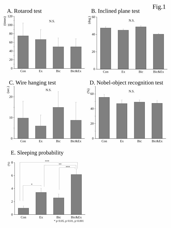
There were no significant group effects observed for the rotarod test, wire hang test, or novel-object
recognition test (Fig.1A-D), indicating that neither exercise nor bicuculline administration significantly
affected motor function or cognitive function. The probability of mice’s sleeping (%) was shown Fig.1E. An
ANOVA showed significant group effect and post hoc analyses demonstrated that sleeping probability was
significantly increased in the Bic&Ex group relative to the Con, Ex, and Bic groups. The sleeping probability
was also increased in the Ex group than in the Con group.
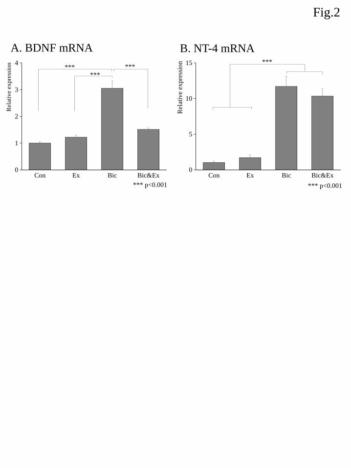
3.2. Expression of mRNA
The relative mRNA expression of BDNF among different groups is shown in Fig. 2A. An ANOVA
showed a significant group effect and post hoc analyses demonstrated that BDNF gene expression was
significantly increased by approximately 3-fold in the Bic group relative to the Con, Ex, and Bic&Ex groups.
The relative mRNA expression of NT-4 among different groups is shown in Fig. 2B. An ANOVA
showed a significant group effect and post hoc analyses demonstrated that NT-4 gene expression was
significantly increased by approximately 10-fold in the Bic and Bic&Ex groups relative to the Con and Ex
groups.
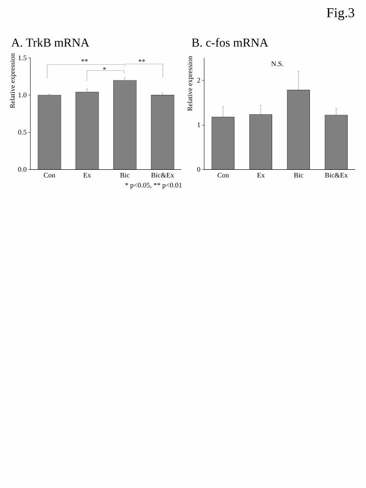
The relative mRNA expression of TrkB among different groups is shown in Fig. 3A. An ANOVA
showed a significant group effect and post hoc analyses demonstrated that the TrkB gene expression was
slightly but significantly elevated in the Bic group relative to the Con, Ex, and Bic&Ex groups.
The relative mRNA expression of c-fos among different groups is shown in Fig. 3B. An ANOVA did
not indicate a significant group effect.
3.3 Protein abundance
The relative protein abundances of BDNF and NT-4 are shown in Fig. 4. An ANOVA showed a
significant group effect on BDNF expression and post hoc analyses demonstrated that BDNF expression was
significantly increased by approximately 3-fold in the Bic&Ex group relative to the Con, Ex and Bic groups.
Alternatively, the ANOVA for NT-4 expression did not show a significant group effect (Fig. 4B).
4. Discussion
The present study revealed that low-level inhibition of the GABAA receptor for 2 weeks increased
the expression of BDNF mRNA in the motor cortex, indicating enhanced transcription of BDNF. In addition,
low-level inhibition of GABAA up-regulated the transcriptional expression of the BDNF receptor, TrkB, as
well as that of NT-4. However, neither exercise nor GABAA receptor inhibition alone modified the protein
expression of BDNF, whereas exercise in combination with low-level GABAA receptor inhibition significantly
increased the protein expression of BDNF in the motor cortex. Since BDNF is a potent neurotrophic factor
that enhances neuronal differentiation, survival, and LTP, it is possible that increased BDNF protein
expression induced by aerobic exercise in the presence of low-level GABAA receptor inhibition could provide
a more effective neural condition for motor learning during kinesiotherapy. As described in previous literature,
low-level GABAA receptor inhibition did not cause the decline of motor or cognitive functions [4, 5], as
evidenced by a lack of effect of any intervention in the motor and cognitive behavioral tests evaluated in this
study. Thus, this study provides evidence that moderate exercise in the presence of low-level GABAA receptor
inhibition is likely safe, increases the expression of BDNF in the motor cortex, and could potentially enhance
motor learning or motor recovery in patients receiving kinesiotherapy.
Several animal studies have shown that the inhibition of GABAergic signaling improves learning
and memory, whereas the enhancement of GABAergic neurotransmission in the hippocampus or prefrontal
cortex impairs learning and memory. Jong Hoon Ryu et al. reported that administration of bicuculline or
bicuculline methiodide directly into the hippocampus enhanced memory acquisition in a passive avoidance
task in a manner related to hippocampal extracellular signal-regulated kinase signaling [7, 8]. Castellano et al.
[4, 5] reported that bicuculline or picrotoxin produced dose-dependent enhancements in memory retention,
whereas the GABAA receptor agonist muscimol produced dose-dependent impairments in memory acquisition
and retention. Similar results were reported by Yousefi et al.[9]. Kim et al. reported that bicuculline
methiodide administration into the hippocampus or prefrontal cortex enhanced memory consolidation by
increasing hippocampal BDNF expression [6]. In addition, GABAA receptor antagonists are reported to
induce long term potentiation (LTP) in hippocampal neurons [18] and up-regulate BDNF mRNA expression in
hypothalamic neurons [19] in vitro. Taken together, these reports suggest that adequate inhibition of
GABAergic synapses could positively contribute to plasticity in the central nervous system. Unfortunately, the
effects of GABAergic inhibition on plasticity in the cerebral cortex related to motor learning or motor skill
acquisition are not well characterized in vivo or in vitro. Recently, Stagg et al. reported that the inhibition of
GABAergic synapses with tDCS prior to motor training enhanced the acquisition of trained motor skills [2, 3].
Lee et al. reported that axonal remodeling for motor recovery after traumatic brain injury required the
down-regulation of GABAA receptors [20]. Thus, the inhibition of GABAergic synapses appears to provide a
more appropriate condition for motor learning. In this study, low-level GABAA receptor inhibition enhanced
the gene expression of BDNF in the motor cortex, which may indicate an improved capacity for
neuroplasticity and thus motor learning. Moreover, when exercise, which is known to potentiate brain BDNF
expression, was co-applied with bicuculline, dramatic increases in protein BDNF expression were observed in
the motor cortex, indicating that aerobic exercise in the presence of low-level GABAA receptor inhibition
might facilitate motor cortical plasticity.
Since previous reports demonstrated that 60 min of moderate treadmill exercise [13, 15] or voluntary
wheel exercise over a period longer than 7 days [12] significantly increased the expression of BDNF in the
brain, we used moderate treadmill exercise for a period longer than 7 days in the present study. However, we
were unable to detect a positive effect of exercise on BDNF expression in the motor cortex. To this end, we
believe that the acute and chronic effects of exercise on BDNF expression should be considered. Huang et al.
[12] reported the time-dependent up-regulation of BDNF expression by treadmill exercise, indicating that
BDNF up-regulation over a 4-week period of treadmill exercise was confirmed 2-3 h after the last exercise
session, but not 2 days later; therefore, the effects of exercise on BDNF expression may be acute. In addition,
Ramussen et al. [16] reported that moderate treadmill exercise for 2 h up-regulated BDNF expression in the
hippocampus 0–6 h after the last exercise session, while this effect disappeared after 24 h. Therefore, it is
reasonable that the 2-week forced treadmill exercise intervention used in this study had no chronic effect on
BDNF expression as assayed 30 h after the final exercise session.
Interestingly, combined exercise and bicuculline administration increased BDNF protein expression.
Recent studies show that aerobic exercise reduces short-interval intracortical inhibition (SICI) induced by
paired-pulse transcranial magnetic stimulation (TMS) used to assess GABAergic inhibition in human motor
cortex [21, 22]. Given that the neuronal expression of BDNF is enhanced in an activity-dependent manner,
additional reduction of intracotical ingibition by exercise to GABAA receptor inhibition with bicuculline could
enhance neural activity and generate an elevation of BDNF.
Of note, combined exercise and bicuculline administration did not have an effect on BDNF gene
expression, whereas bicuculline alone increased BDNF gene expression. These data contradicted our protein
expression findings, wherein combined exercise and bicuculline administration up-regulated BDNF protein
expression. Discrepancies between exercise-induced modifications of BDNF mRNA and protein levels have
been previously reported [12, 23]. There are several possible reasons underlying this discrepancy.
First, protein expression reflects mRNA translation after transcription. Exercise can affect intricate
intracellular signals that enhance the translation of proteins in the central nervous system, for example by
modulating effector proteins that interact with the 5’ or 3’ untranslated regions of dendritic mRNAs [24, 25].
One possibility is that the translation (rather than transcription) of BDNF was enhanced by combined exercise
and bicuculline administration. Therefore, low-level GABAA receptor inhibition might have potentiated
exercise-induced increases in BDNF translation in the motor cortex. This can also potentially explain our
observations regarding NT-4: despite the transcriptional up-regulation of NT-4 by bicuculline administration,
there was no effect of bicuculline on NT-4 protein expression.
Second, the discrepancy could be explained by the time lag between transcriptional up-regulation,
translation, and subsequent behavioral modification. To assess the chronic effects of exercise and bicuculline
administration in this study, mice were sacrificed 30 h after the last intervention. As abovementioned, several
studies have indicated that exercise acutely increases the expression of BDNF in a manner that peaks 2–3 h
after exercise and gradually decreases to baseline 24 h after exercise [12, 16]. On the contrary, previous
literatures show that protein expression starts to increase after 6 h after exercise and 9 h after GABAA receptor
inhibition by bicuculline methioide administration [6, 26]. In addition, mice sleep longer and more deeply
after exercise [27]; acute over-excitation induced by bicuculline administration might potentiate this
exercise-induced behavioral modification, i.e. mice might sleep longer and more deeply, reducing their regular
nighttime activities just prior to sacrifice. In fact, the probability of sleeping during night time was extremely
higher in the Bic&Ex group relative to the Con, Ex, Bic groups (Fig.1E). Given that the neuronal expression
of BDNF is enhanced in an activity-dependent manner, decreased nighttime activity immediately prior to
sacrifice may have limited the extent of BNDF up-regulation in the motor cortex. In agreement with this
hypothesis, combined exercise and bicuculline administration tended to decrease the expression of c-fos, a
marker for neuronal activity, relative to bicuculline administration alone, although a significant difference was
not detected due to the limited sample size of each group.
This study had some limitations. First, we did not directly assay the inhibition of GABAergic
synapses using real-time electrophysiological analyses. Second, we did not assay plasticity in the motor cortex
nor did we directly evaluate motor learning or skill acquisition. Future studies should address these issues in
order to validate our hypotheses and findings. Third, the discrepancy between modifications of BDNF mRNA
and protein expression is not clearly explained as in most previous studies [12, 23]. Additional time-course
detections of BDNF mRNA and protein are expected to elucidate the mechanism underlying the discrepancy
as the next stage following this study.
In conclusion, the present study revealed that combined GABAA receptor inhibition and moderate
aerobic exercise up-regulated BDNF protein expression in the motor cortex without producing side effects on
motor or cognitive functions. Alterations in BDNF expression could positively contribute to plasticity by
regulating the balance between EPSPs and IPSPs in the motor cortex. Thus the advantage of the interactive
effect of aerobic exercise combined with low-level GABAA receptor inhibition could be providing a more
appropriate neuronal condition for motor learning and recovery. Therefore, it is expected that this advantage
of the interactive effects without neural toxicity could contribute to pre-conditioning for kinesiotherapy to
rehabilitate the motor function of patients with central nervous system disorders such as CVA in future.
Acknowledgements
We would like to thank Dr. Kaori Tsutsumi in Hokkaido University for her kind support and assistance with
the use of her laboratory equipment. This study was supported by JSPS KAKENHI Grant 24500619.
References
[1] N. Kubis, Non-Invasive Brain Stimulation to Enhance Post-Stroke Recovery, Frontiers in neural
circuits 10 (2016) 56.
[2] U. Amadi, C. Allman, H. Johansen-Berg, C.J. Stagg, The Homeostatic Interaction Between Anodal
Transcranial Direct Current Stimulation and Motor Learning in Humans is Related to GABAA Activity,
Brain stimulation 8(5) (2015) 898-905.
[3] C.J. Stagg, V. Bachtiar, H. Johansen-Berg, The role of GABA in human motor learning, Current
biology : CB 21(6) (2011) 480-4.
[4] C. Castellano, I.B. Introini-Collison, J.L. McGaugh, Interaction of beta-endorphin and GABAergic
drugs in the regulation of memory storage, Behavioral and neural biology 60(2) (1993) 123-8.
[5] C. Castellano, J.L. McGaugh, Effects of post-training bicuculline and muscimol on retention: lack of
state dependency, Behavioral and neural biology 54(2) (1990) 156-64.
[6] D.H. Kim, J.M. Kim, S.J. Park, M. Cai, X. Liu, S. Lee, C.Y. Shin, J.H. Ryu, GABA(A) receptor blockade
enhances memory consolidation by increasing hippocampal BDNF levels, Neuropsychopharmacology :
official publication of the American College of Neuropsychopharmacology 37(2) (2012) 422-33.
[7] D.H. Kim, J.M. Kim, S.J. Park, S. Lee, C.Y. Shin, J.H. Cheong, J.H. Ryu, Hippocampal extracellular
signal-regulated kinase signaling has a role in passive avoidance memory retrieval induced by GABAA
Receptor modulation in mice, Neuropsychopharmacology : official publication of the American College of
Neuropsychopharmacology 37(5) (2012) 1234-44.
[8] J.M. Kim, D.H. Kim, Y. Lee, S.J. Park, J.H. Ryu, Distinct roles of the hippocampus and perirhinal
cortex in GABAA receptor blockade-induced enhancement of object recognition memory, Brain research
1552 (2014) 17-25.
[9] B. Yousefi, M. Nasehi, F. Khakpai, M.R. Zarrindast, Possible interaction of cholinergic and GABAergic
systems between MS and CA1 upon memory acquisition in rats, Behavioural brain research 235(2) (2012)
231-43.
[10] L.F. Reichardt, Neurotrophin-regulated signalling pathways, Philosophical transactions of the Royal
Society of London. Series B, Biological sciences 361(1473) (2006) 1545-64.
[11] V.K. Sandhya, R. Raju, R. Verma, J. Advani, R. Sharma, A. Radhakrishnan, V. Nanjappa, J.
Narayana, B.L. Somani, K.K. Mukherjee, A. Pandey, R. Christopher, T.S. Prasad, A network map of
BDNF/TRKB and BDNF/p75NTR signaling system, Journal of cell communication and signaling 7(4)
(2013) 301-7.
[12] A.M. Huang, C.J. Jen, H.F. Chen, L. Yu, Y.M. Kuo, H.I. Chen, Compulsive exercise acutely
upregulates rat hippocampal brain-derived neurotrophic factor, J Neural Transm (Vienna) 113(7) (2006)
803-11.
[13] S. Mojtahedi, M.R. Kordi, S.E. Hosseini, S.F. Omran, M. Soleimani, Effect of treadmill running on the
expression of genes that are involved in neuronal differentiation in the hippocampus of adult male rats,
Cell biology international 37(4) (2013) 276-83.
[14] S.A. Neeper, F. Gomez-Pinilla, J. Choi, C.W. Cotman, Physical activity increases mRNA for
brain-derived neurotrophic factor and nerve growth factor in rat brain, Brain research 726(1-2) (1996)
49-56.
[15] R.M. O'Callaghan, R. Ohle, A.M. Kelly, The effects of forced exercise on hippocampal plasticity in the
rat: A comparison of LTP, spatial- and non-spatial learning, Behavioural brain research 176(2) (2007)
362-6.
[16] P. Rasmussen, P. Brassard, H. Adser, M.V. Pedersen, L. Leick, E. Hart, N.H. Secher, B.K. Pedersen, H.
Pilegaard, Evidence for a release of brain-derived neurotrophic factor from the brain during exercise,
Experimental physiology 94(10) (2009) 1062-9.
[17] H. Wang, G.D. Ferguson, V.V. Pineda, P.E. Cundiff, D.R. Storm, Overexpression of type-1 adenylyl
cyclase in mouse forebrain enhances recognition memory and LTP, Nature neuroscience 7(6) (2004)
635-42.
[18] T. Saleewong, A. Srikiatkhachorn, M. Maneepark, A. Chonwerayuth, S. Bongsebandhu-phubhakdi,
Quantifying altered long-term potentiation in the CA1 hippocampus, Journal of integrative neuroscience
11(3) (2012) 243-64.
[19] F. Marmigere, F. Rage, L. Tapia-Arancibia, GABA-glutamate interaction in the control of BDNF
expression in hypothalamic neurons, Neurochemistry international 42(4) (2003) 353-8.
[20] S. Lee, M. Ueno, T. Yamashita, Axonal remodeling for motor recovery after traumatic brain injury
requires downregulation of gamma-aminobutyric acid signaling, Cell death & disease 2 (2011) e133.
[21] A.E. Smith, M.R. Goldsworthy, T. Garside, F.M. Wood, M.C. Ridding, The influence of a single bout of
aerobic exercise on short-interval intracortical excitability, Experimental brain research 232(6) (2014)
1875-82.
[22] T. Yamaguchi, T. Fujiwara, W. Liu, M. Liu, Effects of pedaling exercise on the intracortical inhibition
of cortical leg area, Experimental brain research 218(3) (2012) 401-6.
[23] M. Borsoi, C.B. Antonio, L.G. Muller, A.F. Viana, V. Hertzfeldt, P.S. Lunardi, C. Zanotto, P. Nardin,
A.P. Ravazzolo, S.M. Rates, C.A. Goncalves, Repeated forced swimming impairs prepulse inhibition and
alters brain-derived neurotrophic factor and astroglial parameters in rats, Pharmacology, biochemistry,
and behavior 128 (2015) 50-61.
[24] G. Baj, V. Pinhero, V. Vaghi, E. Tongiorgi, Signaling pathways controlling activity-dependent local
translation of BDNF and their localization in dendritic arbors, Journal of cell science 129(14) (2016)
2852-64.
[25] C.R. Bramham, D.G. Wells, Dendritic mRNA: transport, translation and function, Nature reviews.
Neuroscience 8(10) (2007) 776-89.
[26] H. Soya, T. Nakamura, C.C. Deocaris, A. Kimpara, M. Iimura, T. Fujikawa, H. Chang, B.S. McEwen,
T. Nishijima, BDNF induction with mild exercise in the rat hippocampus, Biochemical and biophysical
research communications 358(4) (2007) 961-7.
[27] M. Lancel, S.K. Droste, S. Sommer, J.M. Reul, Influence of regular voluntary exercise on spontaneous
and social stress-affected sleep in mice, The European journal of neuroscience 17(10) (2003) 2171-9.
Figure legends
Figure 1. Effects of exercise and bicuculline administration on motor control in the rotarod test (A), inclined
plane test (B), and wire hang test (C); on recognition memory as evaluated by the novel object recognition test
(D); and on the probability of sleeping during night-time (E). Data are shown as the mean ± S.E.M. (n = 7–8).
Figure 2. Effects of exercise and bicuculline administration on the mRNA expression of BDNF (A) and NT-4
(B). The expression level of each target gene was determined relative to the transcript of β-actin using the
comparative (ΔΔCt) method. Transcript abundance was normalized to the average of the control group for
each target gene. Data are shown as the mean ± S.E.M. (n = 7–8). *** indicates p < 0.001 (Boferroni
correction).
Figure 3. Effects of exercise and bicuculline administration on the mRNA expression of TrkB (A) and c-fos
(B). The expression level of each target was determined relative to the transcript of β-actin using the
comparative (ΔΔCt) method. Transcript abundance was normalized to the average of the control group for
each target gene. Data are shown as the mean ± S.E.M. (n = 7–8). * indicates p < 0.05 and ** indicates p <
0.01 (Boferroni correction ).
Figure 4. Effects of exercise and bicuculline administration on protein expression of BDNF (A) and NT-4 (B).
Protein levels were determined by enzyme-linked immunosorbent assay and standardized to total protein in
each well. Data are shown as the mean ± S.E.M. (n = 7–8). *** indicates p < 0.01 ( Boferroni correction).
Con Ex Bic Bic&Ex0
20
40
60
80
100
120
(tim
es)
N.S.
Con Ex Bic Bic&Ex0
20
40
60
(deg
.)
N.S.
Con Ex Bic Bic&Ex0
10
20
(sec
.)
N.S.
Con Ex Bic Bic&Ex0
20
40
60(%)
N.S.
Fig.1A. Rotarod test B. Inclined plane test
C. Wire hanging test D. Nobel-object recognition test
E. Sleeping probability
Con Ex Bic Bic&Ex0
2
4
6
8
* p<0.05, p<0.01, p<0.001
*****
***
(%
)
*
Con Ex Bic Bic&Ex0
1
2
3
4
***
*** ***
*** p<0.001
Rel
ativ
e ex
pre
ssio
n
Con Ex Bic Bic&Ex0
5
10
15
*** p<0.001R
elat
ive
expre
ssio
n ***
A. BDNF mRNA B. NT-4 mRNA
Fig.2
Con Ex Bic Bic&Ex0.0
0.5
1.0
1.5
* p<0.05, ** p<0.01
**
Rel
ativ
e ex
pre
ssio
n
*
**
Con Ex Bic Bic&Ex0
1
2
Rel
ativ
e ex
pre
ssio
n
N.S.
A. TrkB mRNA B. c-fos mRNA
Fig.3
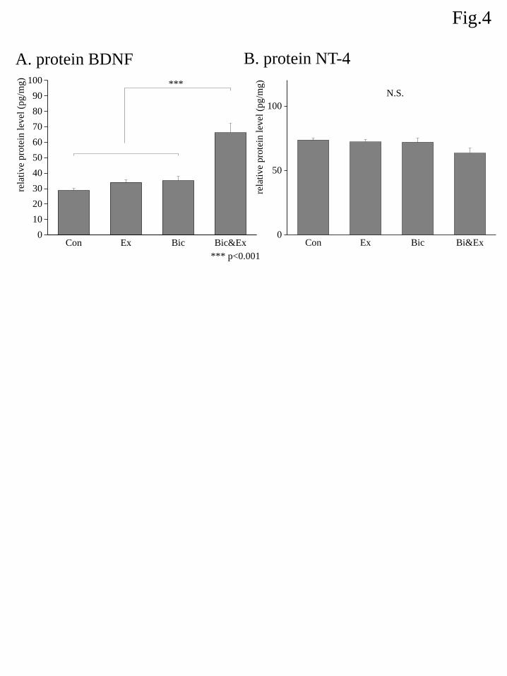
Fig.4
A. protein BDNF B. protein NT-4
Con Ex Bic Bi&Ex0
50
100
rela
tive
pro
tein
lev
el (
pg
/mg)
N.S.
Con Ex Bic Bic&Ex0
10
20
30
40
50
60
70
80
90
100
*** p<0.001
rela
tive
pro
tein
lev
el (
pg
/mg)
***