a unified model of the gaba(a) receptor comprising agonist and
TRANSCRIPT
General rights Copyright and moral rights for the publications made accessible in the public portal are retained by the authors and/or other copyright owners and it is a condition of accessing publications that users recognise and abide by the legal requirements associated with these rights.
• Users may download and print one copy of any publication from the public portal for the purpose of private study or research. • You may not further distribute the material or use it for any profit-making activity or commercial gain • You may freely distribute the URL identifying the publication in the public portal
If you believe that this document breaches copyright please contact us providing details, and we will remove access to the work immediately and investigate your claim.
Downloaded from orbit.dtu.dk on: Apr 01, 2018
A Unified Model of the GABA(A) Receptor Comprising Agonist and BenzodiazepineBinding Sites
Kongsbak, Kristine Grønning; Bergmann, Rikke; Sørensen, Pernille Louise; Sander, Tommy; Balle,ThomasPublished in:P L o S One
Link to article, DOI:10.1371/journal.pone.0052323
Publication date:2013
Document VersionPublisher's PDF, also known as Version of record
Link back to DTU Orbit
Citation (APA):Kongsbak, K. G., Bergmann, R., Sørensen, P. L., Sander, T., & Balle, T. (2013). A Unified Model of theGABA(A) Receptor Comprising Agonist and Benzodiazepine Binding Sites. P L o S One, 8(1), e52323. DOI:10.1371/journal.pone.0052323
A Unified Model of the GABAA Receptor ComprisingAgonist and Benzodiazepine Binding SitesRikke Bergmann1., Kristine Kongsbak1,2., Pernille Louise Sørensen1, Tommy Sander3, Thomas Balle4*
1 Department of Drug Design and Pharmacology, Faculty of Health and Medical Sciences, University of Copenhagen, Copenhagen, Denmark, 2 National Food Institute,
Technical University of Denmark, Søborg, Denmark, 3 Novo Nordisk A/S, Bagsværd, Denmark, 4 Faculty of Pharmacy, The University of Sydney, Sydney, New South Wales,
Australia
Abstract
We present a full-length a1b2c2 GABA receptor model optimized for agonists and benzodiazepine (BZD) allostericmodulators. We propose binding hypotheses for the agonists GABA, muscimol and THIP and for the allosteric modulatordiazepam (DZP). The receptor model is primarily based on the glutamate-gated chloride channel (GluCl) from C. elegans andincludes additional structural information from the prokaryotic ligand-gated ion channel ELIC in a few regions. Availablemutational data of the binding sites are well explained by the model and the proposed ligand binding poses. We suggest aGABA binding mode similar to the binding mode of glutamate in the GluCl X-ray structure. Key interactions are predictedwith residues a1R66, b2T202, a1T129, b2E155, b2Y205 and the backbone of b2S156. Muscimol is predicted to bind similarly,however, with minor differences rationalized with quantum mechanical energy calculations. Muscimol key interactions arepredicted to be a1R66, b2T202, a1T129, b2E155, b2Y205 and b2F200. Furthermore, we argue that a water molecule couldmediate further interactions between muscimol and the backbone of b2S156 and b2Y157. DZP is predicted to bind withinteractions comparable to those of the agonists in the orthosteric site. The carbonyl group of DZP is predicted to interactwith two threonines a1T206 and c2T142, similar to the acidic moiety of GABA. The chlorine atom of DZP is placed near theimportant a1H101 and the N-methyl group near a1Y159, a1T206, and a1Y209. We present a binding mode of DZP in whichthe pending phenyl moiety of DZP is buried in the binding pocket and thus shielded from solvent exposure. Our full lengthGABAA receptor is made available as Model S1.
Citation: Bergmann R, Kongsbak K, Sørensen PL, Sander T, Balle T (2013) A Unified Model of the GABAA Receptor Comprising Agonist and BenzodiazepineBinding Sites. PLoS ONE 8(1): e52323. doi:10.1371/journal.pone.0052323
Editor: Eugene A. Permyakov, Russian Academy of Sciences, Institute for Biological Instrumentation, Russian Federation
Received August 15, 2012; Accepted November 16, 2012; Published January 7, 2013
Copyright: ! 2013 Bergmann et al. This is an open-access article distributed under the terms of the Creative Commons Attribution License, which permitsunrestricted use, distribution, and reproduction in any medium, provided the original author and source are credited.
Funding: Financial support was obtained from The Velux Foundation (post doctoral grant to RB) and NeuroSearch A/S (funding ogf research assistant KK). Thefunders had no role in study design, data collection and analysis, decision to publish, or preparation of the manuscript.
Competing Interests: TS is employed at Novo Nordisk A/S. Post Doc RB was funded by The Velux Foundation and Reseach Assistant KK was funded byNeurosearch A/S. This does not alter the authors’ adherence to all the PLoS ONE policies on sharing data and materials.
* E-mail: [email protected]
. These authors contributed equally to this work.
Introduction
c-aminobutyric acid (GABA) is the major inhibitory neuro-transmitter in the central nervous system (CNS) as opposed toglutamic acid, which is the primary excitatory CNS-neurotrans-mitter (Figure 1). Structurally, the two compounds are similar, andin fact GABA is formed in vivo by decarboxylation of glutamate.GABAA receptors (GABAARs) are involved in a number ofimportant functions such as cognition, learning, and memory andin disorders such as epilepsy, anxiety, schizophrenia, sleepdisorders, and depression [1]. The GABAARs belong to the Cys-Loop receptor family that also includes nicotinic acetylcholinereceptors (nAChRs), serotonine type 3 receptors (5-HT3Rs) andglycine receptors (GlyRs). All Cys-Loop receptors are homomericor heteromeric assemblies of five subunits forming a central ion-conducting pore (Figure 2). The GABAARs and GlyRs conductanions whereas nAChRs and 5-HT3Rs are cation selective. Eachsubunit is made up of an extracellular domain (ECD) consisting ofmainly b-sheets, and a trans-membrane domain (TMD) consistingof four membrane spanning a-helices. GABAAR subunits includea1–6, b1–3, c1–3, d, e, p, h, r1–3 and the most abundant GABAARsubunit combination in the human CNS is the a1b2c2 subtype
where the endogenous neurotransmitter GABA binds in each ofthe interfaces between b2 and a1 subunits (Figure 2). A modulatorysite for benzodiazepine (BZD) like compounds is found in ahomologous position between a1 and c2 subunits.
Despite decades of research and a wealth of experimental andtheoretical studies, the exact binding mode of key agonistsincluding GABA is still unknown. The same is the case for theBZDs. Key agonists for the GABA binding site include the highaffinity agonist muscimol [2,3] and the partial agonist THIP [4,5],which is a structurally restrained muscimol analog (Figure 1).THIP was long in clinical trials for treatment of insomnia, but wasdiscontinued. Still, the GABAAR agonist binding site is regarded apromising drug target and represents an intriguing alternative tothe BZD binding site, which has long been the target for allostericmodulators including BZDs such as diazepam (Figure 1). BZDsare still one of the most prescribed classes of drugs for thetreatment of insomnia, anxiety, and convulsions [6,7].
So far, drug discovery efforts have relied mainly on indirectstructural insight from focused [8–12] or unified pharmacophoremodels recapitulating the structure-activity relationships (SAR) ofcompounds synthesized during more than fifty years of activemedicinal chemistry research in the field [13,14]. Homology
PLOS ONE | www.plosone.org 1 January 2013 | Volume 8 | Issue 1 | e52323
models, on the other hand, have had little practical impact on thedesign process despite a number of models reported in theliterature [15–25]. The models were mainly built using thehomologous acetylcholine binding proteins (AChBPs) as templates.The AChBPs have supplied insight into a number of structural
features of Cys-Loop receptors. The position of loops A–F(Figure 2) known from mutational studies to participate in ligandbinding were established with the first AChBP structure [26]. Ahigh degree of flexibility has later on been observed for the C-loop,which is a hair-pin shaped loop that embraces the orthostericbinding sites and shields from the solvent [27]. It was observed thatdepending on the type of ligand in the binding site, the C-loopeither exists in a closed (agonist) conformation or an open(antagonist) conformation allowing large inhibitors to enter thebinding site. This C-loop movement has also been speculated to belinked to the activation mechanism of Cys-Loop receptors [28].
Although, the AChBPs have proven valuable templates formodeling of nAChRs [26,29–32] they suffer from a lack ofconservation of binding site residues with respect to GABAARs,which makes them unsuitable as stand-alone templates forGABAAR homology modeling. To compensate for the lack ofconservation of binding site residues, we have recently reported anovel strategy for GABAAR modeling utilizing experimentalrestraints and multiple templates including AChBPs from differentspecies [27,33], a mouse a1 nAChR subunit [34], and the bacterialorthologs from Gloeobacter violaceus (GLIC) [35] and Erwiniachrysanthemii (ELIC) [36] in the alignment generation and modelbuilding steps [37]. In particular, inclusion of the ELIC structureadds important conserved binding site residues to the pool oftemplate structures. Using this strategy, a reliable model of theGABAAR ECD with focus on the orthosteric ligand bindinginterface of the a1b2c2 GABAAR in its non-activated (antagonized)state was obtained. The model was consistent with experimentaldata and capable of rationalizing the structure activity relation-ships (SAR) of a series of GABAAR orthosteric antagonists [37].
With the recent release of atomic resolution structures of aeukaryotic glutamate gated chloride channel (GluCl) [38] from thenematode C. elegans, the molecular basis for modeling ofpentameric ligand gated anion channels has improved consider-ably. The GluCl structure has an unprecedented high sequenceidentity compared to the GABAAR; 30%, 36%, and 31% relatingto a1, b2 and c2 subunits, respectively, and even higher identities
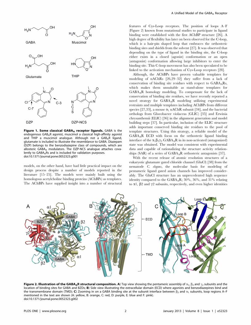
Figure 1. Some classical GABAA receptor ligands. GABA is theendogenous GABAR agonist, muscimol a classical high-affinity agonistand THIP a muscimol analogue. Although not a GABAR ligand,glutamate is included to illustrate the resemblance to GABA. Diazepam(DZP) belongs to the benzodiazepine class of compounds, which areallosteric GABAA modulators. The DZP-NCS analogue attaches cova-lently to GABAARs and is included for validation purposes.doi:10.1371/journal.pone.0052323.g001
Figure 2. Illustration of the GABAAR structural composition. A) Top view showing the pentameric assembly of a1, b2 and c2 subunits and thelocation of binding sites for GABA and BZDs; B) Side view illustrating the extracellular domain (ECD) where agonists and benzodiazepines bind andthe transmembrane domain (TMD); C) Zooming in on a GABA binding site at the subunit interface between b2 and a1 subunits, loop regions A–Fmentioned in the text are shown (A: yellow, B: orange, C: red, D: purple, E: blue and F: pink).doi:10.1371/journal.pone.0052323.g002
A Unified Model of the GABAA Receptor
PLOS ONE | www.plosone.org 2 January 2013 | Volume 8 | Issue 1 | e52323
with respect to the ligand binding cores (,48% in an 8 A radiusfrom glutamate in GluCl). The GluCl structure was crystallized inpresence of its agonist glutamate and was captured in its presumedopen state.
In this report we demonstrate the use of the GluCl structure astemplate for construction of a GABAA receptor homology modelcomprising both the ECD and TMD portions of the receptor. Weshow that when combined with the structure of the bacterial ELICchannel, a reliable GABAAR model based entirely on full lengthreceptor X-ray structures can be obtained. The model is built inthe open state with GABA in the two orthosteric binding sitesbetween b2 and a1 subunits. The BZD binding site between the a1
and c2 subunit is adapted to the positive allosteric modulatordiazepam (DZP). The model is capable of explaining SARs,mutational data, and data from studies of covalent linking of aDZP-derivative to cysteine mutants of the receptor. Therefore, thevalidated model might also serve as a tool for structure guideddesign of new agonists and allosteric modulators and may form thelink to interpretation of previously reported pharmacophoremodels [11,14,39] in a structural context.
Methods
Homology modelingTemplates, sequences and sequence alignment. The X-
ray structure of GluCl co-crystallised with glutamate (PDB code3RIF) [38] was used as primary template for homology modelingof the most abundant subtype of the GABAAR, a1b2c2. In a fewimportant regions with low sequence identity to GluCl thebacterial homologue ELIC (PDB code 2VL0) [40], which has a20% sequence identity to GluCl, was included as template as well.The extent to which each template structure was used is specifiedin Figure 3, in which the definitions for the general secondarystructural elements of Cys-Loop receptors referred to throughoutthis paper are also indicated. The rationale for including ELIC astemplate in the areas highlighted in Figure 3 were the following: 1)In the b1 and b2 sheets the ELIC structure contains aromaticresidues in positions 19 and 38 resembling those in the GABAAR;2) In the b6–b7-loop (Cys-Loop) and in the b7 and b10 strandsELIC was included as template for the b2 subunit to captureinformation about the conformations of and interactions betweenGABAAR b2E155 and b2R207; 3) In the M2–M3-loop ELIC wasincluded as template due to the presence of Pro residues inhomologous positions.
The sequence alignment was obtained following the procedurereported by Sander et al. [37] First, a structural alignment of the twotemplate structures was generated using Pymol 1.3 [41]. Subse-quently, all human GlyR a-subunits and all human GABAARsubunits were aligned to the GluCl sequence as profile alignmentswith iteration on the last alignment using ClustalX v. 2.0.12 [42].The GlyR a-subunits represent the closest human homologs toGluCl and were included to aid identification of semi-conservedmotifs. In three regions, namely, 1) in and after the N-terminal a-helix, 2) in loop F, and 3) in loop C, manual adjustments of thegenerated alignment were performed to ensure proper alignmentof conserved motifs. 1) In the N-terminal a-helix the motifrepresented by the GABAA a1 sequence ILDRLLDGYDNRLRPwas misaligned by ClustalX due to the presence of insertions in theGlyR a-subunit sequences and the GABAAR r-subunit sequences.Therefore, this motif was reestablished as described by Sander etal. [37] 2) In loop F varying sequence lengths and low sequenceidentity resulted in a poor alignment and many gaps. We identifieda hydrophobic-X-hydrophobic motif (corresponding to VVV inthe GABAAR a1-subunit), forming a short b-strand of three
residues in GluCl, ELIC, the bacterial ion channel GLIC (PDBID: 3EAM) the mouse nAChR a1-subunit (PDB ID: 2QC1) and inAChBPs from Aplysia californica (PDB ID: 2BYQ) and Bulinustruncatus (PDB ID: 2BJ0) [27,34,35,43]. The generated alignmentwas manually adjusted to re-establish this motif in the GABAARsequences. 3) In loop C, the automatically generated alignmentfrom ClustalX had gaps in the GABAAR sequences in the b-sheetregions. These were manually moved to the tip of the loop as it isgenerally accepted that the length of loop C varies betweenfamilies and subtypes of Cys-Loop receptors. 4) Finally, wetruncated the M3–M4 intracellular loop and inserted an AGTtripeptide according to the GluCl structure. The manuallyadjusted alignment is reported in Figure 3.
Prior to model building the GluCl X-ray structure was preparedas follows. The FAB fragments (chains F–O) as well as allheteroatoms were removed except glutamates in the orthostericbinding sites between chains A, B and C, D. Then the a-carboxylicacid moiety was deleted from the glutamate ligands, resulting in aGluCl template structure with GABA in the two orthostericbinding sites between chains A, B and C, D.
Model building, evaluation and selection. The programMODELLER 9v7 [44] was used for homology modeling using the‘‘automodel class’’, which includes no other restraints than spatialrestraints gathered from the sequence alignment. 100 models weregenerated, and the refinement level ‘‘refine.slow’’ was applied.GABA molecules were modeled into the two b2-a1 subunitinterfaces in the GABAA receptor model as rigid bodies.
The final model selection was performed according to theconsensus scoring approach described by Sander et al. [37] usingthe ProSA z-score [45], the energy according to the OPLS 2001force field [46,47] as implemented in Maestro [48], and theMODELLER built in scoring functions, molpdf and DOPE score[44]. The consensus 10 best scoring models were assessed visuallyfor physico-chemical requirements such as packing of hydrophobicresidues in hydrophobic environments and solvent exposure ofcharged residues. Also, the interactions between the modeledGABA molecule and the receptor model were assessed as part ofthe selection criteria.
Model refinementThe selected model was subjected to the protein preparation
wizard in Maestro [48], which adds hydrogen atoms, assigns bondorders, creates disulphide bonds and samples hydrogen bondnetworks. Furthermore, the protein preparation wizard assessesthe protonation state of His, Lys, Arg, Glu, and Asp. As is seen inthe GluCl structure E293 interacts with R245 and D316,indicating that this residue exists in its protonated form. Therefore,the corresponding five Glu residues (two a1E302, two b2E298, andone c2E313) in the homology model, which also coordinates toArg and Asp were protonated. The protein preparation wizardand the PROPKA web server [49–51] further supported thisassessment. All other residues were kept at their standardprotonation states (neutral His, protonated Lys and Arg anddeprotonated Glu and Asp). Finally, an energy minimization witha flat-bottomed Cartesian constraint and a convergence thresholdset to an RMSD of 0.3 A was performed.
The model was further refined as follows: 1) The rotameric stateof a1R66 (in chain D) was optimized for optimal bidentateinteractions with GABA using the side chain refinement tool inPrime [52]; 2) Hydrogen bond networks between the GABAmolecules and the receptor model were manually optimized byselecting appropriate rotamers of a1T129 similar to the homol-ogous S121 in the GluCl structure; 3) Loop A (residues 99–102) inthe BZD site carrying a1H101 was sampled using the loop
A Unified Model of the GABAA Receptor
PLOS ONE | www.plosone.org 3 January 2013 | Volume 8 | Issue 1 | e52323
sampling protocol in Prime [52] in order to obtain an orientationof a1Asn102 in agreement with the template structure; 4) Arotamer of b2K196 able to make a salt bridge with b2E153 wasselected (Table 1).
Ligand docking and binding site characterizationGABA, muscimol, and THIP were created in their ionized
states in Maestro 9.2 [53] followed by conformational searcheswith MacroModel 9.9 (default settings) [54]. The global energyminimum conformations were identified and used as inputconformations for docking. The agonists were docked into theorthosteric binding site between chains A and B using the GlideInduced Fit Docking (IFD) protocol [55,56] and the ExtraPrecision (XP) scoring function [57]. By default the IFD procedureallows amino acid side chains to adapt to the docked ligand in a
5 A sphere. Docking poses were selected based on compliancewith mutational data (Table 1) and common interaction patternsin the binding site. Finally, selected ligand poses including residuesin an 8 A sphere were energy minimized to convergence usingMacroModel 9.9.
The program GRID [58,59] was used to characterize the non-bonded water interaction properties of the vacant binding pocketbetween chains A and B (GABA site) of the refined model usingthe water probe (OH2). A grid spacing of 0.33 A was used and allother settings were kept at their default values.
Quantum mechanical (QM) calculations was performed usingJaguar 7.8 [60]. For muscimol, a relaxed coordinate scan wasperformed to determine conformational energies when varying theamino-methyl side chain dihedral angle in a step size of 10ubetween 0–180u. The Poisson-Bolzmann aqueous solvation model
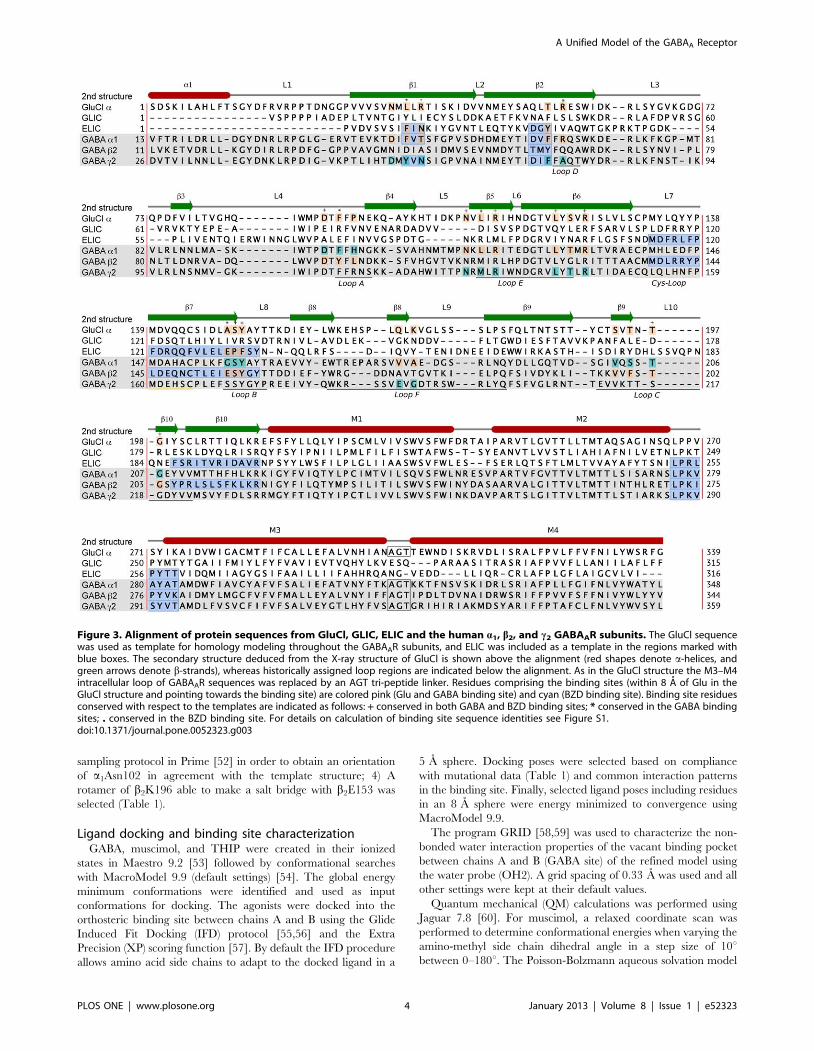
Figure 3. Alignment of protein sequences from GluCl, GLIC, ELIC and the human a1, b2, and c2 GABAAR subunits. The GluCl sequencewas used as template for homology modeling throughout the GABAAR subunits, and ELIC was included as a template in the regions marked withblue boxes. The secondary structure deduced from the X-ray structure of GluCl is shown above the alignment (red shapes denote a-helices, andgreen arrows denote b-strands), whereas historically assigned loop regions are indicated below the alignment. As in the GluCl structure the M3–M4intracellular loop of GABAAR sequences was replaced by an AGT tri-peptide linker. Residues comprising the binding sites (within 8 A of Glu in theGluCl structure and pointing towards the binding site) are colored pink (Glu and GABA binding site) and cyan (BZD binding site). Binding site residuesconserved with respect to the templates are indicated as follows: + conserved in both GABA and BZD binding sites; * conserved in the GABA bindingsites; . conserved in the BZD binding site. For details on calculation of binding site sequence identities see Figure S1.doi:10.1371/journal.pone.0052323.g003
A Unified Model of the GABAA Receptor
PLOS ONE | www.plosone.org 4 January 2013 | Volume 8 | Issue 1 | e52323
[61] and otherwise default settings were selected (B3LYP/6-31G**). Gas-phase energies were extracted from the results inorder to consider only the steric energies.
The docking procedure described for the agonists was alsoattempted for DZP at the BZD site, but none of the obtained posescould be rationalized by experimental data. DZP was docked usingits assumed bioactive conformation as input for docking [62–66].QM partial charges were calculated using Jaguar 7.6 defaultsettings. Subsequently, DZP was manually docked to the BZDbinding site according to experimental evidence: Docked DZPshould 1) have the Cl-substituent positioned in the vicinity of orpointing towards a1H101, a1N102 [67], a1G157, a1V202, anda1V211 [68], 2) have the C-3 atom positioned in the vicinity of ordirected towards a1S205 and a1T206 [69], 3) have the N-methylsubstituent directed towards an exit from the binding cavity [70],and 4) have the pending phenyl ring positioned in a lipophiliccavity [71,72]. Following manual positioning of DZP, the a1-c2
interface was allowed to adapt to DZP using the side chainprediction tool in Prime in which backbone and residue samplingwithin 4 A of DZP was performed. As a final step, Prime performsa minimization of the complex, the docked ligand and thesurrounding residues in question (backbone and side chains). Theprotein and the ligand were thus allowed to adapt to each other.The final model was further validated using 1) mutational studiesfrom the literature (Table 2), 2) in silico covalent docking of a Cys-reactive DZP derivative, and 3) assessment of SAR data from theliterature in a structural context.
As a validation of the DZP binding mode, the Cys-reactive BZDderivative DZP-NCS [67,68,73,74] (Figure 1) was covalently
docked to an a1H101C variant of the homology model using the‘‘covalent docking’’ module in Prime. The covalent dockingmodule works by eliminating two atoms in order to form a newbond between the reacting molecules/species. Since Prime cannothandle simultaneous reduction of a double bond and formation ofa new bond, the isothiocyanato group of DZP-NCS was reducedto a methanethioamide group prior to submission of the job. Thethiol-hydrogen was defined as the leaving receptor atom. Theconformation of the attachment residue was sampled and all otherresidues were kept fixed.
Finally, a 48 ns molecular dynamics simulation was performedto assess the stability of the final GABAAR model. Details aresupplied as Model S1.
Results and Discussion
With the improved structural templates available from efforts instructural biology it is now possible to build a1b2c2 GABAARmodels based entirely on full length receptor templates. We havecreated a full-length a1b2c2 GABA model mainly based on theglutamate bound GluCl X-ray structure and partly using thebacterial Cys-Loop homolog ELIC as an additional template. Themodel has been optimized in the GABA and BZD binding sites forthe agonists GABA, muscimol and THIP and the modulatordiazepam. The GluCl X-ray structure with glutamate bound wascrystallized in an open state with a negatively charged ion in thelower part of the TMD [38]. The ELIC structure, on the otherhand, was crystallized in a putatively closed state in absence of abound ligand [36]. Since only a few residue positions in our model
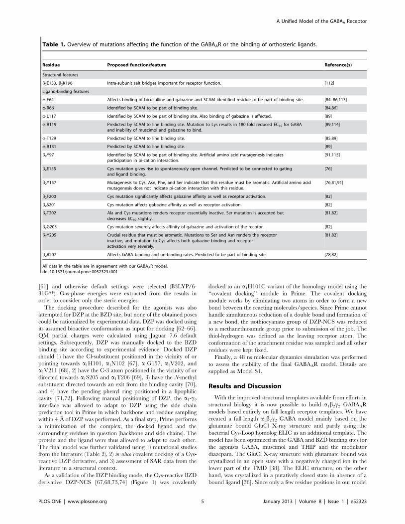
Table 1. Overview of mutations affecting the function of the GABAAR or the binding of orthosteric ligands.
Residue Proposed function/feature Reference(s)
Structural features
b2E153, b2K196 Intra-subunit salt bridges important for receptor function. [112]
Ligand-binding features
a1F64 Affects binding of bicuculline and gabazine and SCAM identified residue to be part of binding site. [84–86,113]
a1R66 Identified by SCAM to be part of binding site. [84,86]
a1L117 Identified by SCAM to be part of binding site. Also binding of gabazine is affected. [89]
a1R119 Predicted by SCAM to line binding site. Mutation to Lys results in 180 fold reduced EC50 for GABAand inability of muscimol and gabazine to bind.
[89,114]
a1T129 Predicted by SCAM to line binding site. [85,89]
a1R131 Predicted by SCAM to line binding site. [89]
b2Y97 Identified by SCAM to be part of binding site. Artificial amino acid mutagenesis indicatesparticipation in pi-cation interaction.
[91,115]
b2E155 Cys mutation gives rise to spontaneously open channel. Predicted to be connected to gatingand ligand binding.
[76]
b2Y157 Mutagenesis to Cys, Asn, Phe, and Ser indicate that this residue must be aromatic. Artificial amino acidmutagenesis does not indicate pi-cation interaction with this residue.
[76,81,91]
b2F200 Cys mutation significantly affects gabazine affinity as well as receptor activation. [82]
b2S201 Cys mutation affects gabazine affinity as well as receptor activation. [82]
b2T202 Ala and Cys mutations renders receptor essentially inactive. Ser mutation is accepted butdecreases EC50 slightly.
[81,82]
b2G203 Cys mutation severely affects affinity of gabazine and activation of the recptor. [82]
b2Y205 Crucial residue that must be aromatic. Mutations to Ser and Asn renders the receptorinactive, and mutation to Cys affects both gabazine binding and receptoractivation very severely.
[81,82]
b2R207 Affects GABA binding and un-binding rates. Predicted to be part of binding site. [78,82]
All data in the table are in agreement with our GABAAR model.doi:10.1371/journal.pone.0052323.t001
A Unified Model of the GABAA Receptor
PLOS ONE | www.plosone.org 5 January 2013 | Volume 8 | Issue 1 | e52323
have been modeled based on the ELIC structure, the overallarchitecture of the a1b2c2 GABA model is obtained from theGluCl structure. Therefore, we regard our model as being in theopen state.
Model assessmentThe pentameric GABAAR a1b2c2 ECD-TMD homology model
comprised 1676 residues distributed with 335, 334, and 333residues in a1, b2, and c2 GABAAR subunits respectively. Theselected model had good backbone geometry with 98.8% of theresidues in favorable or additionally allowed regions in theProcheck v. 3.5.4 Ramachandran plot [75]. All residues indisallowed regions in the Ramachandran plot were situated insolvent exposed loop regions distant to the binding site. TheProSA z-scores for the selected homology model were within theaccepted area for X-ray structures from the PDB. Furthermore,stability of the model was assessed by a 48 ns molecular dynamicscalculation which showed essentially no drift after termination ofthe equilibration protocol (see Figure S2).
Validation by mutational data. Both the GABA and BZDbinding sites have been heavily investigated by site-directedmutagenesis (Tables 1 and 2). Among these mutations, some havebeen used to suggest which residues line the binding sites, whichresidues interact directly with different ligands, and which are
regarded as important structural features of the receptor. Theseexperimental data, including those listed in Tables 1 and 2 thatwere not directly imposed during model refinement, are explain-able by our homology model with the suggested poses of GABAand DZP as described below.
Agonist binding modelGABA. The X-ray structure of GluCl with glutamate bound
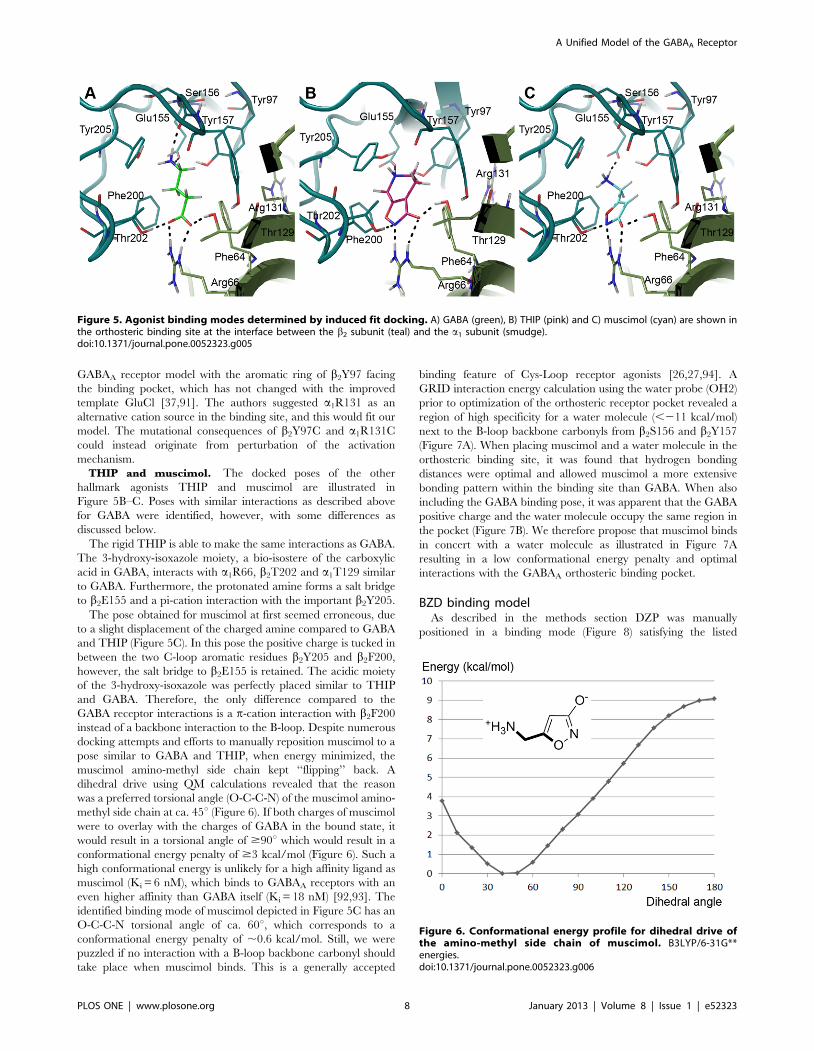
presents a good indication of how GABA would bind to the GABAreceptors. Our a1b2c2 GABA model confirms that a similarGABA binding mode interacts well with the receptor and is inagreement with the experimental mutational data presented inTable 1. In this binding mode GABA forms salt bridges witha1R66 and b2E155 and hydrogen bonds with a1T129, b2T202,and the backbone of b2S156. Finally, there is a p-cationinteraction with b2Y205. The GABA binding mode from ourdocking study is illustrated in Figure 5A. As described in themethods section GABA was modeled into the binding pockets ofour receptor model as rigid bodies, which allowed space for GABAin the binding site. However, hydrogen bonding network was notoptimized in the modeling process, hence, a few side chainsneeded adjustments as described in the methods section.
The GABAA orthosteric binding site apparently resembles theGluCl glutamate binding site to a large extent. However, there are
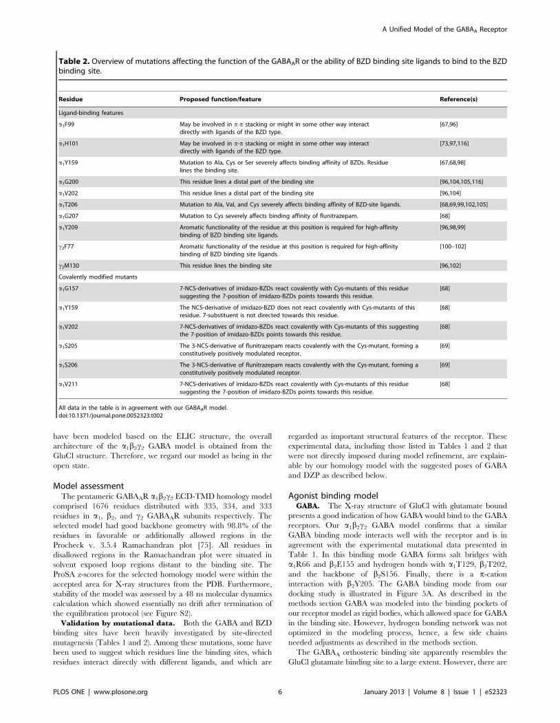
Table 2. Overview of mutations affecting the function of the GABAAR or the ability of BZD binding site ligands to bind to the BZDbinding site.
Residue Proposed function/feature Reference(s)
Ligand-binding features
a1F99 May be involved in p-p stacking or might in some other way interactdirectly with ligands of the BZD type.
[67,96]
a1H101 May be involved in p-p stacking or might in some other way interactdirectly with ligands of the BZD type.
[73,97,116]
a1Y159 Mutation to Ala, Cys or Ser severely affects binding affinity of BZDs. Residuelines the binding site.
[67,68,98]
a1G200 This residue lines a distal part of the binding site [96,104,105,116]
a1V202 This residue lines a distal part of the binding site [96,104]
a1T206 Mutation to Ala, Val, and Cys severely affects binding affinity of BZD-site ligands. [68,69,99,102,105]
a1G207 Mutation to Cys severely affects binding affinity of flunitrazepam. [68]
a1Y209 Aromatic functionality of the residue at this position is required for high-affinitybinding of BZD binding site ligands.
[96,98,99]
c2F77 Aromatic functionality of the residue at this position is required for high-affinitybinding of BZD binding site ligands.
[100–102]
c2M130 This residue lines the binding site [96,102]
Covalently modified mutants
a1G157 7-NCS-derivatives of imidazo-BZDs react covalently with Cys-mutants of this residuesuggesting the 7-position of imidazo-BZDs points towards this residue.
[68]
a1Y159 The NCS-derivative of imidazo-BZD does not react covalently with Cys-mutants of thisresidue. 7-substituent is not directed towards this residue.
[68]
a1V202 7-NCS-derivatives of imidazo-BZDs react covalently with Cys-mutants of this suggestingthe 7-position of imidazo-BZDs points towards this residue.
[68]
a1S205 The 3-NCS-derivative of flunitrazepam reacts covalently with the Cys-mutant, forming aconstitutively positively modulated receptor.
[69]
a1S206 The 3-NCS-derivative of flunitrazepam reacts covalently with the Cys-mutant, forming aconstitutively positively modulated receptor.
[69]
a1V211 7-NCS-derivatives of imidazo-BZDs react covalently with Cys-mutants of this residuesuggesting the 7-position of imidazo-BZDs points towards this residue.
[68]
All data in the table is in agreement with our GABAAR model.doi:10.1371/journal.pone.0052323.t002
A Unified Model of the GABAA Receptor
PLOS ONE | www.plosone.org 6 January 2013 | Volume 8 | Issue 1 | e52323
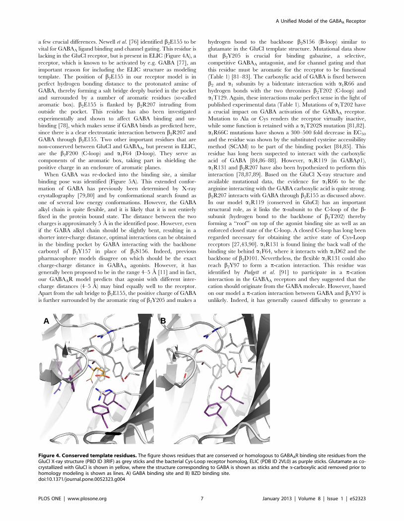
a few crucial differences. Newell et al. [76] identified b2E155 to bevital for GABAA ligand binding and channel gating. This residue islacking in the GluCl receptor, but is present in ELIC (Figure 4A), areceptor, which is known to be activated by e.g. GABA [77], animportant reason for including the ELIC structure as modelingtemplate. The position of b2E155 in our receptor model is inperfect hydrogen bonding distance to the protonated amine ofGABA, thereby forming a salt bridge deeply buried in the pocketand surrounded by a number of aromatic residues (so-calledaromatic box). b2E155 is flanked by b2R207 intruding fromoutside the pocket. This residue has also been investigatedexperimentally and shown to affect GABA binding and un-binding [78], which makes sense if GABA binds as predicted here,since there is a clear electrostatic interaction between b2R207 andGABA through b2E155. Two other important residues that arenon-conserved between GluCl and GABAA, but present in ELIC,are the b2F200 (C-loop) and a1F64 (D-loop). They serve ascomponents of the aromatic box, taking part in shielding thepositive charge in an enclosure of aromatic planes.
When GABA was re-docked into the binding site, a similarbinding pose was identified (Figure 5A). This extended confor-mation of GABA has previously been determined by X-raycrystallography [79,80] and by conformational search found asone of several low energy conformations. However, the GABAalkyl chain is quite flexible, and it is likely that it is not entirelyfixed in the protein bound state. The distance between the twocharges is approximately 5 A in the identified pose. However, evenif the GABA alkyl chain should be slightly bent, resulting in ashorter inter-charge distance, optimal interactions can be obtainedin the binding pocket by GABA interacting with the backbonecarbonyl of b2Y157 in place of b2S156. Indeed, previouspharmacophore models disagree on which should be the exactcharge-charge distance in GABAA agonists. However, it hasgenerally been proposed to be in the range 4–5 A [11] and in fact,our GABAAR model predicts that agonist with different inter-charge distances (4–5 A) may bind equally well to the receptor.Apart from the salt bridge to b2E155, the positive charge of GABAis further surrounded by the aromatic ring of b2Y205 and makes a
hydrogen bond to the backbone b2S156 (B-loop) similar toglutamate in the GluCl template structure. Mutational data showthat b2Y205 is crucial for binding gabazine, a selective,competitive GABAA antagonist, and for channel gating and thatthis residue must be aromatic for the receptor to be functional(Table 1) [81–83]. The carboxylic acid of GABA is fixed betweenb2 and a1 subunits by a bidentate interaction with a1R66 andhydrogen bonds with the two threonines b2T202 (C-loop) anda1T129. Again, these interactions make perfect sense in the light ofpublished experimental data (Table 1). Mutations of a1T202 havea crucial impact on GABA activation of the GABAA receptor.Mutation to Ala or Cys renders the receptor virtually inactive,while some function is retained with a a1T202S mutation [81,82].a1R66C mutations have shown a 300–500 fold decrease in EC50
and the residue was shown by the substituted cysteine accessibilitymethod (SCAM) to be part of the binding pocket [84,85]. Thisresidue has long been suspected to interact with the carboxylicacid of GABA [84,86–88]. However, a1R119 (in GABAr1),a1R131 and b2R207 have also been hypothesized to perform thisinteraction [78,87,89]. Based on the GluCl X-ray structure andavailable mutational data, the evidence for a1R66 to be thearginine interacting with the GABA carboxylic acid is quite strong.b2R207 interacts with GABA through b2E155 as discussed above.In our model a1R119 (conserved in GluCl) has an importantstructural role, as it links the a-subunit to the C-loop of the b-subunit (hydrogen bond to the backbone of b2T202) therebyforming a ‘‘roof’’ on top of the agonist binding site as well as anenforced closed state of the C-loop. A closed C-loop has long beenregarded necessary for obtaining the active state of Cys-Loopreceptors [27,43,90]. a1R131 is found lining the back wall of thebinding site behind a1F64, where it interacts with a1D62 and thebackbone of b2D101. Nevertheless, the flexible a1R131 could alsoreach b2Y97 to form a p-cation interaction. This residue wasidentified by Padgett et al. [91] to participate in a p-cationinteraction in the GABAA receptors and they suggested that thecation should originate from the GABA molecule. However, basedon our model a p-cation interaction between GABA and b2Y97 isunlikely. Indeed, it has generally caused difficulty to generate a
Figure 4. Conserved template residues. The figure shows residues that are conserved or homologous to GABAAR binding site residues from theGluCl X-ray structure (PBD ID 3RIF) as grey sticks and the bacterial Cys-Loop receptor homolog, ELIC (PDB ID 2VL0) as purple sticks. Glutamate as co-crystallized with GluCl is shown in yellow, where the structure corresponding to GABA is shown as sticks and the a-carboxylic acid removed prior tohomology modeling is shown as lines. A) GABA binding site and B) BZD binding site.doi:10.1371/journal.pone.0052323.g004
A Unified Model of the GABAA Receptor
PLOS ONE | www.plosone.org 7 January 2013 | Volume 8 | Issue 1 | e52323
GABAA receptor model with the aromatic ring of b2Y97 facingthe binding pocket, which has not changed with the improvedtemplate GluCl [37,91]. The authors suggested a1R131 as analternative cation source in the binding site, and this would fit ourmodel. The mutational consequences of b2Y97C and a1R131Ccould instead originate from perturbation of the activationmechanism.
THIP and muscimol. The docked poses of the otherhallmark agonists THIP and muscimol are illustrated inFigure 5B–C. Poses with similar interactions as described abovefor GABA were identified, however, with some differences asdiscussed below.
The rigid THIP is able to make the same interactions as GABA.The 3-hydroxy-isoxazole moiety, a bio-isostere of the carboxylicacid in GABA, interacts with a1R66, b2T202 and a1T129 similarto GABA. Furthermore, the protonated amine forms a salt bridgeto b2E155 and a pi-cation interaction with the important b2Y205.
The pose obtained for muscimol at first seemed erroneous, dueto a slight displacement of the charged amine compared to GABAand THIP (Figure 5C). In this pose the positive charge is tucked inbetween the two C-loop aromatic residues b2Y205 and b2F200,however, the salt bridge to b2E155 is retained. The acidic moietyof the 3-hydroxy-isoxazole was perfectly placed similar to THIPand GABA. Therefore, the only difference compared to theGABA receptor interactions is a p-cation interaction with b2F200instead of a backbone interaction to the B-loop. Despite numerousdocking attempts and efforts to manually reposition muscimol to apose similar to GABA and THIP, when energy minimized, themuscimol amino-methyl side chain kept ‘‘flipping’’ back. Adihedral drive using QM calculations revealed that the reasonwas a preferred torsional angle (O-C-C-N) of the muscimol amino-methyl side chain at ca. 45u (Figure 6). If both charges of muscimolwere to overlay with the charges of GABA in the bound state, itwould result in a torsional angle of $90u which would result in aconformational energy penalty of $3 kcal/mol (Figure 6). Such ahigh conformational energy is unlikely for a high affinity ligand asmuscimol (Ki = 6 nM), which binds to GABAA receptors with aneven higher affinity than GABA itself (Ki = 18 nM) [92,93]. Theidentified binding mode of muscimol depicted in Figure 5C has anO-C-C-N torsional angle of ca. 60u, which corresponds to aconformational energy penalty of ,0.6 kcal/mol. Still, we werepuzzled if no interaction with a B-loop backbone carbonyl shouldtake place when muscimol binds. This is a generally accepted
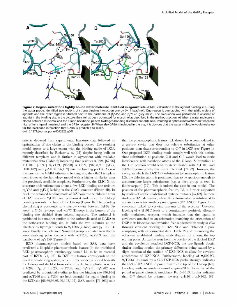
binding feature of Cys-Loop receptor agonists [26,27,94]. AGRID interaction energy calculation using the water probe (OH2)prior to optimization of the orthosteric receptor pocket revealed aregion of high specificity for a water molecule (,211 kcal/mol)next to the B-loop backbone carbonyls from b2S156 and b2Y157(Figure 7A). When placing muscimol and a water molecule in theorthosteric binding site, it was found that hydrogen bondingdistances were optimal and allowed muscimol a more extensivebonding pattern within the binding site than GABA. When alsoincluding the GABA binding pose, it was apparent that the GABApositive charge and the water molecule occupy the same region inthe pocket (Figure 7B). We therefore propose that muscimol bindsin concert with a water molecule as illustrated in Figure 7Aresulting in a low conformational energy penalty and optimalinteractions with the GABAA orthosteric binding pocket.
BZD binding modelAs described in the methods section DZP was manually
positioned in a binding mode (Figure 8) satisfying the listed
Figure 5. Agonist binding modes determined by induced fit docking. A) GABA (green), B) THIP (pink) and C) muscimol (cyan) are shown inthe orthosteric binding site at the interface between the b2 subunit (teal) and the a1 subunit (smudge).doi:10.1371/journal.pone.0052323.g005
Figure 6. Conformational energy profile for dihedral drive ofthe amino-methyl side chain of muscimol. B3LYP/6-31G**energies.doi:10.1371/journal.pone.0052323.g006
A Unified Model of the GABAA Receptor
PLOS ONE | www.plosone.org 8 January 2013 | Volume 8 | Issue 1 | e52323
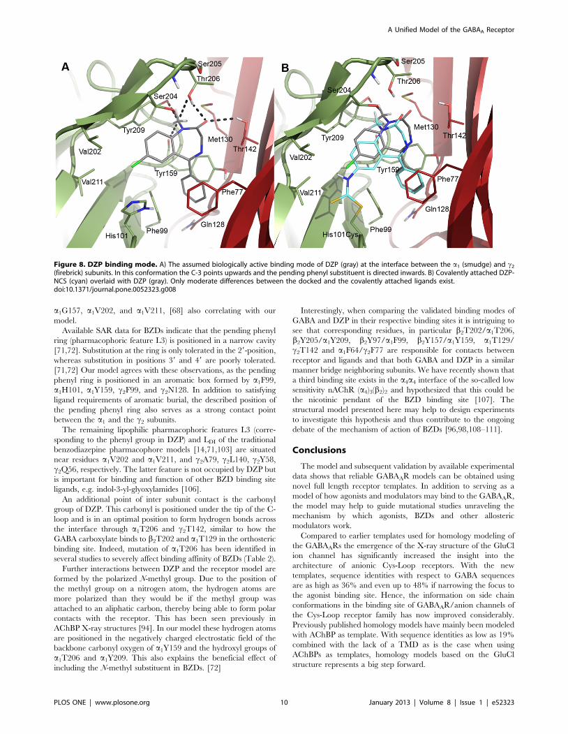
criteria deduced from experimental literature data followed byoptimization of side chains in the binding pocket. The resultingmodel agrees to a large extent with the binding mode of DZP,recently described by Richter et al. [95] despite being built ondifferent templates and is further in agreement with availablemutational data (Table 2) indicating that residues a1F99, [67,96]a1H101, [73,97] a1Y159, [96,98] a1Y209, [96,98,99] c2F77,[100–102] and c2M130 [96,102] line the binding pocket. As wasthe case for the GABA othosteric binding site, the GluCl templatecontributes to the homology model with a higher similarity thanthe previously available templates. Furthermore, the ELIC X-raystructure adds information about a few BZD binding site residues(c2Y58 and c2F77) lacking in the GluCl structure (Figure 4B). Inbrief, the obtained binding mode of DZP orients the chlorine atomof DZP towards a1H101 and positions it underneath the C-looppointing towards the base of the C-loop (Figure 8). The pendingphenyl ring is positioned in a narrow cavity between a1F99 (A-loop), a1Y159 (B-loop), and c2F77 (D-loop) in the bottom of thebinding site shielded from solvent exposure. The carbonyl ispositioned in a manner similar to the carboxylic acid of GABA inthe orthosteric binding site. It links the two subunits at theinterface by hydrogen bonds to a1T206 (C-loop) and c2T142 (D-loop). Finally, the polarized N-methyl group is situated near the C-loop enabling polar contacts with a1T206, a1Y209 and thebackbone of a1Y159 (Figure 8).
BZD pharmacophore models based on SAR data havepredicted a lipophilic pharmacophoric feature (in the traditionalBZD pharmacophore terminology named L1) to be an essentialpart of BZDs [71,103]. In DZP this feature corresponds to thefused aromatic ring system, which in the model is buried beneaththe C-loop and shielded from solvent by the hydrophobic residuesa1V202, Cb of a1T206, a1Y209, and a1V211. a1V202 waspredicted by mutational studies to line the binding site [96,104]and a1T206 and a1Y209 are both essential for ligand binding tothe BZD site [68,69,96,98,99,102,105]. SAR studies [71,103] state
that the pharmacophoric feature, L1, should be accommodated ina narrow cavity that does not tolerate substitution at otherpositions than that corresponding to C-7 in DZP (see Figure 1).Our proposed DZP binding mode comply well with this notion,since substitution at positions C-8 and C-9 would lead to stericinterference with backbone atoms of the C-loop. Substitution atthe C-6 position would lead to steric clashes with a1H101 andc2F99 explaining why this is not tolerated. [71,72] However, thecavity, in which the DZP C-7 substituent (pharmacophoric featureL2), the chlorine atom, is positioned, has to be spacious enough toaccommodate larger substituents (e.g. a nitro group as seen influnitrazepam) [72]. This is indeed the case in our model. Theposition of the pharmacophoric feature, L2, is further supportedby studies of covalent labeling of cysteine mutants [67,69]. In thesestudies, a DZP derivative, where the chlorine atom is substituted toa cysteine-reactive isothiocyanate group (DZP-NCS, Figure 1), iscovalently linked to cysteine mutants of the receptor. Covalentlabeling of a1H101C leads to a constitutively positively allosteri-cally modulated receptor, which indicates that the ligand iscovalently attached in an orientation matching the orientation ofDZP in its bioactive conformation. We replicated this study in silicothrough covalent docking of DZP-NCS and obtained a posecomplying with experimental data (Table 2) and resembling theotherwise established binding mode (Figure 8B) among the topscoring poses. As can be seen from the overlay of the docked DZPand the covalently attached DZP-NCS, the two ligands obtainsimilar binding modes, the primary difference being caused by aslight rotation of the scaffold of DZP-NCS to allow for covalentattachment of DZP-NCS. Furthermore, labeling of a1S205C,a1T206C mutants by a C-3 DZP-NCS probe strongly indicatesthe C-3 of DZP-NCS to point towards the tip of the C-loop [69].Labeling with an imidazobenzodiazepine-NCS derivative of thepartial negative allosteric modulator Ro15-4513, further indicatesthat C-7 should be oriented towards a1H101, a1N102, [67]
Figure 7. Region suited for a tightly bound water molecule identified in agonist site. A GRID calculation at the agonist binding site, usingthe water probe, identified two regions of strong binding interaction energy (211 kcal/mol). One region is overlapping with the acidic moiety ofagonists and the other region is situated next to the backbone of b2S156 and b2Y157 (grey mesh). The calculation was performed in absence ofagonist in the binding site. In the picture, the site has been optimized for muscimol as described in the methods section. A) When a water molecule isplaced between muscimol and the B-loop backbone, perfect hydrogen bonding distances are obtained, resulting in optimal interactions between thehigh affinity ligand muscimol and the GABA receptor. B) When also GABA is included in the site, it is obvious that the water molecule would make upfor the backbone interaction that GABA is predicted to make.doi:10.1371/journal.pone.0052323.g007
A Unified Model of the GABAA Receptor
PLOS ONE | www.plosone.org 9 January 2013 | Volume 8 | Issue 1 | e52323
a1G157, a1V202, and a1V211, [68] also correlating with ourmodel.
Available SAR data for BZDs indicate that the pending phenylring (pharmacophoric feature L3) is positioned in a narrow cavity[71,72]. Substitution at the ring is only tolerated in the 29-position,whereas substitution in positions 39 and 49 are poorly tolerated.[71,72] Our model agrees with these observations, as the pendingphenyl ring is positioned in an aromatic box formed by a1F99,a1H101, a1Y159, c2F99, and c2N128. In addition to satisfyingligand requirements of aromatic burial, the described position ofthe pending phenyl ring also serves as a strong contact pointbetween the a1 and the c2 subunits.
The remaining lipophilic pharmacophoric features L3 (corre-sponding to the phenyl group in DZP) and LDI of the traditionalbenzodiazepine pharmacophore models [14,71,103] are situatednear residues a1V202 and a1V211, and c2A79, c2L140, c2Y58,c2Q56, respectively. The latter feature is not occupied by DZP butis important for binding and function of other BZD binding siteligands, e.g. indol-3-yl-glyoxylamides [106].
An additional point of inter subunit contact is the carbonylgroup of DZP. This carbonyl is positioned under the tip of the C-loop and is in an optimal position to form hydrogen bonds acrossthe interface through a1T206 and c2T142, similar to how theGABA carboxylate binds to b2T202 and a1T129 in the orthostericbinding site. Indeed, mutation of a1T206 has been identified inseveral studies to severely affect binding affinity of BZDs (Table 2).
Further interactions between DZP and the receptor model areformed by the polarized N-methyl group. Due to the position ofthe methyl group on a nitrogen atom, the hydrogen atoms aremore polarized than they would be if the methyl group wasattached to an aliphatic carbon, thereby being able to form polarcontacts with the receptor. This has been seen previously inAChBP X-ray structures [94]. In our model these hydrogen atomsare positioned in the negatively charged electrostatic field of thebackbone carbonyl oxygen of a1Y159 and the hydroxyl groups ofa1T206 and a1Y209. This also explains the beneficial effect ofincluding the N-methyl substituent in BZDs. [72]
Interestingly, when comparing the validated binding modes ofGABA and DZP in their respective binding sites it is intriguing tosee that corresponding residues, in particular b2T202/a1T206,b2Y205/a1Y209, b2Y97/a1F99, b2Y157/a1Y159, a1T129/c2T142 and a1F64/c2F77 are responsible for contacts betweenreceptor and ligands and that both GABA and DZP in a similarmanner bridge neighboring subunits. We have recently shown thata third binding site exists in the a4a4 interface of the so-called lowsensitivity nAChR (a4)3(b2)2 and hypothesized that this could bethe nicotinic pendant of the BZD binding site [107]. Thestructural model presented here may help to design experimentsto investigate this hypothesis and thus contribute to the ongoingdebate of the mechanism of action of BZDs [96,98,108–111].
Conclusions
The model and subsequent validation by available experimentaldata shows that reliable GABAAR models can be obtained usingnovel full length receptor templates. In addition to serving as amodel of how agonists and modulators may bind to the GABAAR,the model may help to guide mutational studies unraveling themechanism by which agonists, BZDs and other allostericmodulators work.
Compared to earlier templates used for homology modeling ofthe GABAARs the emergence of the X-ray structure of the GluClion channel has significantly increased the insight into thearchitecture of anionic Cys-Loop receptors. With the newtemplates, sequence identities with respect to GABA sequencesare as high as 36% and even up to 48% if narrowing the focus tothe agonist binding site. Hence, the information on side chainconformations in the binding site of GABAAR/anion channels ofthe Cys-Loop receptor family has now improved considerably.Previously published homology models have mainly been modeledwith AChBP as template. With sequence identities as low as 19%combined with the lack of a TMD as is the case when usingAChBPs as templates, homology models based on the GluClstructure represents a big step forward.
Figure 8. DZP binding mode. A) The assumed biologically active binding mode of DZP (gray) at the interface between the a1 (smudge) and c2
(firebrick) subunits. In this conformation the C-3 points upwards and the pending phenyl substituent is directed inwards. B) Covalently attached DZP-NCS (cyan) overlaid with DZP (gray). Only moderate differences between the docked and the covalently attached ligands exist.doi:10.1371/journal.pone.0052323.g008
A Unified Model of the GABAA Receptor
PLOS ONE | www.plosone.org 10 January 2013 | Volume 8 | Issue 1 | e52323
Based on homology modeling, advanced docking methods, QMcalculations and a vast amount of collected experimental data, wehave identified binding hypotheses for GABA, muscimol, THIPand diazepam and optimized the binding sites accordingly. OurGABAAR model is modeled in the open state according to theGluCl glutamate bound structure and is intended for creatingbinding hypotheses of agonists or BZD site modulators. The modelis made available in Model S1.
Supporting Information
Model S1 The GABAAR model described in this paper.(ZIP)
Figure S1 Calculations of binding site sequence identi-ties.(PDF)
Figure S2 RMSD plot and details of a 48 ns moleculardynamics simulation.(PDF)
Author Contributions
Conceived and designed the experiments: RB KK PS TS TB. Performedthe experiments: RB KK PS. Analyzed the data: RB KK PS TS TB. Wrotethe paper: RB KK TS TB.
References
1. Johnston GA (2005) GABA(A) receptor channel pharmacology. Curr PharmDes 11: 1867–1885.
2. Johnston GA, Curtis DR, De Groat WC, Duggan AW (1968) Central actions ofibotenic acid and muscimol. Biochem Pharmacol 17: 2488–2489.
3. Ebert B, Frolund B, Diemer NH, Krogsgaard-Larsen P (1999) Equilibriumbinding characteristics of [3H]thiomuscimol. Neurochem Int 34: 427–434.
4. Krogsgaard-Larsen P, Johnston GA, Lodge D, Curtis DR (1977) A new class ofGABA agonist. Nature 268: 53–55.
5. Frolund B, Kristiansen U, Brehm L, Hansen AB, Krogsgaard-Larsen P, et al.(1995) Partial GABAA receptor agonists. Synthesis and in vitro pharmacologyof a series of nonannulated analogs of 4,5,6,7-tetrahydroisoxazolo[5,4-c]pyridin-3-ol. J Med Chem 38: 3287–3296.
6. McKernan RM, Rosahl TW, Reynolds DS, Sur C, Wafford KA, et al. (2000)Sedative but not anxiolytic properties of benzodiazepines are mediated by theGABAA receptor a1 subtype. Nat Neurosci 3: 587–592.
7. Rosahl TW, Sur C, Reynolds DS, Collinson N, Macauley A, et al. (2000)Towards an understanding of the role or the GABAergic system in anxiety,learning and memory. European Journal of Neuroscience 12: 514–514.
8. Grant JA, Bonnick T, Gossell-Williams M, Clayton T, Cook JM, et al. (2010)Synthesis, pharmacological studies and molecular modeling of some tetracyclic1,3-diazepinium chlorides. Bioorg Med Chem 18: 909–921.
9. He X, Huang Q, Ma C, Yu S, McKernan R, et al. (2000) Pharmacophore/receptor models for GABAA/BzR a2b3c2, a3b3c2 and a4b3c2 recombinantsubtypes. Included volume analysis and comparison to a1b3c2, a5b3c2, anda6b3c2 subtypes. Drug Des Discov 17: 131–171.
10. Huang Q, Cox ED, Gan T, Ma C, Bennett DW, et al. (1999) Studies ofmolecular pharmacophore/receptor models for GABAA/benzodiazepinereceptor subtypes: binding affinities of substituted beta-carbolines at recombi-nant alpha x beta 3 gamma 2 subtypes and quantitative structure-activityrelationship studies via a comparative molecular field analysis. Drug DesDiscov 16: 55–76.
11. Ghoshal N, Vijayan RSK (2010) Pharmacophore models for GABAA
modulators: implications in CNS drug discovery. Exp Opin Drug Discov 5:441–460.
12. Frolund B, Tagmose L, Liljefors T, Stensbol TB, Engblom C, et al. (2000) Anovel class of potent 3-isoxazolol GABA(A) antagonists: design, synthesis, andpharmacology. J Med Chem 43: 4930–4933.
13. Krogsgaard-Larsen P, Frølund B, Liljefors T (2002) Specific GABA(A) agonistsand partial agonists. Chem Rec 2: 419–430.
14. Clayton T, Chen JL, Ernst M, Richter L, Cromer BA, et al. (2007) An updatedunified pharmacophore model of the benzodiazepine binding site on gamma-aminobutyric acid(a) receptors: correlation with comparative models. CurrMed Chem 14: 2755–2775.
15. Berezhnoy D, Gibbs TT, Farb DH (2009) Docking of 1,4-benzodiazepines inthe a1/c2 GABAA receptor modulator site. Mol Pharmacol 76: 440–450.
16. Ci SQ, Ren TR, Ma CX, Su ZG (2007) Modeling of ak/c2 (k = 1, 2, 3 and 5)interface of GABAA receptor and docking studies with zolpidem: implicationsfor selectivity. J Mol Graph Model 26: 537–545.
17. Cromer BA, Morton CJ, Parker MW (2002) Anxiety over GABAA receptorstructure relieved by AChBP. Trends Biochem Sci 27: 280–287.
18. Sancar F, Ericksen SS, Kucken AM, Teissere JA, Czajkowski C (2007)Structural determinants for high-affinity zolpidem binding to GABA-Areceptors. Mol Pharmacol 71: 38–46.
19. Gharaghani S, Khayamian T, Keshavarz F (2011) A structure-based QSARand docking study on imidazo[1,5-a][1,2,4]-triazolo[1,5-d][1,4,]benzodiaze-pines as Selective GABA(A) alpha5 inverse agonists. Chemical biology & drugdesign 78: 612–621.
20. Ernst M, Brauchart D, Boresch S, Sieghart W (2003) Comparative modeling ofGABAA receptors: limits, insights, future developments. Neuroscience 119:933–943.
21. Sawyer GW, Chiara DC, Olsen RW, Cohen JB (2002) Identification of thebovine c-aminobutyric acid type A receptor a subunit residues photolabeled bythe imidazobenzodiazepine [3H]Ro15-4513. J Biol Chem 277: 50036–50045.
22. Ci S, Ren T, Su Z (2008) Investigating the putative binding-mode of GABAand diazepam within GABA A receptor using molecular modeling. Protein J27: 71–78.
23. Law RJ, Lightstone FC (2009) Modeling neuronal nicotinic and GABAreceptors: important interface salt-links and protein dynamics. Biophys J 97:1586–1594.
24. Cheng J, Ju XL (2010) Homology modeling and atomic level binding study ofGABA(A) receptor with novel enaminone amides. Eur J Med Chem 45: 3595–3600.
25. O’Mara M, Cromer B, Parker M, Chung SH (2005) Homology model of theGABAA receptor examined using Brownian dynamics. Biophys J 88: 3286–3299.
26. Brejc K, van Dijk WJ, Klaassen RV, Schuurmans M, van Der Oost J, et al.(2001) Crystal structure of an ACh-binding protein reveals the ligand-bindingdomain of nicotinic receptors. Nature 411: 269–276.
27. Hansen SB, Sulzenbacher G, Huxford T, Marchot P, Taylor P, et al. (2005)Structures of Aplysia AChBP complexes with nicotinic agonists and antagonistsreveal distinctive binding interfaces and conformations. Embo J 24: 3635–3646.
28. Chang YC, Wu W, Zhang JL, Huang Y (2009) Allosteric activation mechanismof the cys-loop receptors. Acta Pharmacol Sin 30: 663–672.
29. Dutertre S, Ulens C, Buttner R, Fish A, van Elk R, et al. (2007) AChBP-targeted alpha-conotoxin correlates distinct binding orientations with nAChRsubtype selectivity. The EMBO journal 26: 3858–3867.
30. Unwin N (2005) Refined structure of the nicotinic acetylcholine receptor at 4 Aresolution. J Mol Biol 346: 967–989.
31. Bisson WH, Westera G, Schubiger PA, Scapozza L (2008) Homology modelingand dynamics of the extracellular domain of rat and human neuronal nicotinicacetylcholine receptor subtypes alpha4beta2 and alpha7. Journal of MolecularModeling 14: 891–899.
32. Harpsoe K, Ahring PK, Christensen JK, Jensen ML, Peters D, et al. (2011)Unraveling the high- and low-sensitivity agonist responses of nicotinicacetylcholine receptors. The Journal of neuroscience: the official journal ofthe Society for Neuroscience 31: 10759–10766.
33. Celie PH, van Rossum-Fikkert SE, van Dijk WJ, Brejc K, Smit AB, et al. (2004)Nicotine and carbamylcholine binding to nicotinic acetylcholine receptors asstudied in AChBP crystal structures. Neuron 41: 907–914.
34. Dellisanti CD, Yao Y, Stroud JC, Wang ZZ, Chen L (2007) Crystal structure ofthe extracellular domain of nAChR alpha1 bound to alpha-bungarotoxin at1.94 A resolution. Nat Neurosci 10: 953–962.
35. Bocquet N, Nury H, Baaden M, Le Poupon C, Changeux JP, et al. (2009) X-ray structure of a pentameric ligand-gated ion channel in an apparently openconformation. Nature 457: 111–114.
36. Hilf RJC, Dutzler R (2008) X-ray structure of a prokaryotic pentameric ligand-gated ion channel. Nature 452: 375–379.
37. Sander T, Frolund B, Bruun AT, Ivanov I, McCammon JA, et al. (2011) Newinsights into the GABA(A) receptor structure and orthosteric ligand binding:receptor modeling guided by experimental data. Proteins 79: 1458–1477.
38. Hibbs RE, Gouaux E (2011) Principles of activation and permeation in ananion-selective Cys-loop receptor. Nature 474: 54–60.
39. Frolund B, Jorgensen AT, Tagmose L, Stensbol TB, Vestergaard HT, et al.(2002) Novel class of potent 4-arylalkyl substituted 3-isoxazolol GABA(A)antagonists: synthesis, pharmacology, and molecular modeling. J Med Chem45: 2454–2468.
40. Hilf RJ, Dutzler R (2008) X-ray structure of a prokaryotic pentameric ligand-gated ion channel. Nature 452: 375–379.
41. Berman HM, Westbrook J, Feng Z, Gilliland G, Bhat TN, et al. (2000) TheProtein Data Bank. Nucleic Acids Res 28: 235–242.
42. Larkin MA, Blackshields G, Brown NP, Chenna R, McGettigan PA, et al.(2007) Clustal W and Clustal X version 2.0. Bioinformatics 23: 2947–2948.
43. Celie PH, Klaassen RV, van Rossum-Fikkert SE, van Elk R, van Nierop P, etal. (2005) Crystal structure of acetylcholine-binding protein from Bulinustruncatus reveals the conserved structural scaffold and sites of variation innicotinic acetylcholine receptors. J Biol Chem 280: 26457–26466.
A Unified Model of the GABAA Receptor
PLOS ONE | www.plosone.org 11 January 2013 | Volume 8 | Issue 1 | e52323
44. Sali A, Blundell TL (1993) Comparative protein modelling by satisfaction ofspatial restraints. J Mol Biol 234: 779–815.
45. Sippl MJ (1993) Recognition of errors in three-dimensional structures ofproteins. Proteins 17: 355–362.
46. Kaminski GA, Friesner RA, Tirado-Rives J, Jorgensen WL (2001) Evaluationand reparametrization of the OPLS-AA force field for proteins via comparisonwith accurate quantum chemical calculations on peptides. Journal of PhysicalChemistry B 105: 6474–6487.
47. Jorgensen WL, Maxwell DS, TiradoRives J (1996) Development and testing ofthe OPLS all-atom force field on conformational energetics and properties oforganic liquids. Journal of the American Chemical Society 118: 11225–11236.
48. Schrodinger L (2010) Maestro. 9.1 ed. New York, NY, USA.49. Olsson MHM, Sondergaard CR, Rostkowski M, Jensen JH (2011) PROPKA3:
Consistent Treatment of Internal and Surface Residues in Empirical pK(a)Predictions. Journal of Chemical Theory and Computation 7: 525–537.
50. Jensen JH, Bas DC, Rogers DM (2008) Very fast prediction and rationalizationof pK(a) values for protein-ligand complexes. Proteins-Structure Function andBioinformatics 73: 765–783.
51. Li H, Robertson AD, Jensen JH (2005) Very fast empirical prediction andrationalization of protein pK(a) values. Proteins-Structure Function andBioinformatics 61: 704–721.
52. Schrodinger L (2010) Prime. 2.2 ed. New York, NY, USA.53. Maestro v. 9.2, Schrodinger, LLC, New York, NY, 2011.54. MacroModel v. 9.9, Schrodinger, LLC, New York, NY, 2011.55. Sherman W, Beard HS, Farid R (2006) Use of an induced fit receptor structure
in virtual screening. Chem Biol Drug Des 67: 83–84.56. Sherman W, Day T, Jacobson MP, Friesner RA, Farid R (2006) Novel
procedure for modeling ligand/receptor induced fit effects. J Med Chem 49:534–553.
57. Friesner RA, Murphy RB, Repasky MP, Frye LL, Greenwood JR, et al. (2006)Extra precision glide: docking and scoring incorporating a model ofhydrophobic enclosure for protein-ligand complexes. J Med Chem 49: 6177–6196.
58. Goodford PJ (1985) A computational procedure for determining energeticallyfavorable binding sites on biologically important macromolecules. J Med Chem28: 849–857.
59. Molecular Discovery L (2005) GRID 22. Pinner, Middlesex, UK.60. Jaguar v. 7.8, Schrodinger, LLC, New York, NY, 2011.61. Tannor DJ, Marten B, Murphy R, Friesner RA, Sitkoff D, et al. (1994)
Accurate First Principles Calculation of Molecular Charge Distributions andSolvation Energies from Ab Initio Quantum Mechanics and ContinuumDielectic Theory. J Am Chem Soc 116: 11875–11882.
62. Young R, Glennon RA, Dewey WL (1984) Stereoselective stimulus effects of 3-methylflunitrazepam and pentobarbital. Life Sci 34: 1977–1983.
63. Fellegvari I, Visy J, Valko K, Lang T, Simonyi M (1989) Investigation ofConformational Diastereomers of 2,3-Benzodiazepines by High-PerformanceLiquid-Chromatography. J Liq Chromatogr 12: 2719–2732.
64. Simonyi M (1990) Chiral recognition by central benzodiazepine receptors. ActaPharm Nord 2: 145–154.
65. Maksay G, Tegyey Z, Simonyi M (1991) Central benzodiazepine receptors: invitro efficacies and potencies of 3-substituted 1,4-benzodiazepine stereoisomers.Mol Pharmacol 39: 725–732.
66. Simonyi M, Maksay G (1990) Conformational Recognition by CentralBenzodiazepine Receptors. Bioorg Chem 18: 1–12.
67. Tan KR, Baur R, Gonthier A, Goeldner M, Sigel E (2007) Two neighboringresidues of loop A of the alpha1 subunit point towards the benzodiazepinebinding site of GABAA receptors. FEBS Lett 581: 4718–4722.
68. Tan KR, Gonthier A, Baur R, Ernst M, Goeldner M, et al. (2007) Proximity-accelerated chemical coupling reaction in the benzodiazepine-binding site ofgamma-aminobutyric acid type A receptors: superposition of different allostericmodulators. J Biol Chem 282: 26316–26325.
69. Tan KR, Baur R, Charon S, Goeldner M, Sigel E (2009) Relative positioningof diazepam in the benzodiazepine-binding-pocket of GABA receptors.J Neurochem 111: 1264–1273.
70. Sigel E, Stephenson FA, Mamalaki C, Barnard EA (1983) A gamma-aminobutyric acid/benzodiazepine receptor complex of bovine cerebral cortex.J Biol Chem 258: 6965–6971.
71. Zhang W, Koehler KF, Zhang P, Cook JM (1995) Development of acomprehensive pharmacophore model for the benzodiazepine receptor. DrugDes Discov 12: 193–248.
72. Sternbach LH (1971) 1,4-benzodiazepines. Chemistry and some aspects of thestructure-activity relationship. Angew Chem Int Ed Engl 10: 34–43.
73. Berezhnoy D, Nyfeler Y, Gonthier A, Schwob H, Goeldner M, et al. (2004) Onthe benzodiazepine binding pocket in GABAA receptors. J Biol Chem 279:3160–3168.
74. Berezhnoy D, Baur R, Gonthier A, Foucaud B, Goeldner M, et al. (2005)Conformational changes at benzodiazepine binding sites of GABA(A) receptorsdetected with a novel technique. J Neurochem 92: 859–866.
75. Laskowski RA, MacArthur MW, Moss DS, Thornton JM (1993) PROCHECK- a program to check the stereochemical quality of protein structures. J AppCryst 26: 283–291.
76. Newell JG, McDevitt RA, Czajkowski C (2004) Mutation of glutamate 155 ofthe GABAA receptor beta2 subunit produces a spontaneously open channel: atrigger for channel activation. J Neurosci 24: 11226–11235.
77. Thompson AJ, Alqazzaz M, Ulens C, Lummis SC (2012) The pharmacologicalprofile of ELIC, a prokaryotic GABA-gated receptor. Neuropharmacology 63:761–767.
78. Wagner DA, Czajkowski C, Jones MV (2004) An arginine involved in GABAbinding and unbinding but not gating of the GABA(A) receptor. J Neurosci 24:2733–2741.
79. Steward EG, Player RB, Warner D (1973) the Crystal Structure of gamma-Aminobutyric Acid Hydrochloride: A refinement. Acta Cryst B 29: 2825–2826.
80. Tomita K (1965) Jap J Brain Physiol 61: 1–4.81. Amin J, Weiss DS (1993) GABAA receptor needs two homologous domains of
the beta-subunit for activation by GABA but not by pentobarbital. Nature 366:565–569.
82. Wagner DA, Czajkowski C (2001) Structure and dynamics of the GABAbinding pocket: A narrowing cleft that constricts during activation. J Neurosci21: 67–74.
83. Gynther BD, Curtis DR (1986) Pyridazinyl-GABA derivatives as GABA andglycine antagonists in the spinal cord of the cat. Neurosci Lett 68: 211–215.
84. Boileau AJ, Evers AR, Davis AF, Czajkowski C (1999) Mapping the agonistbinding site of the GABAA receptor: evidence for a beta-strand. J Neurosci 19:4847–4854.
85. Jansen M, Rabe H, Strehle A, Dieler S, Debus F, et al. (2008) Synthesis ofGABAA receptor agonists and evaluation of their alpha-subunit selectivity andorientation in the GABA binding site. J Med Chem 51: 4430–4448.
86. Holden JH, Czajkowski C (2002) Different residues in the GABA(A) receptoralpha 1T60-alpha 1K70 region mediate GABA and SR-95531 actions. J BiolChem 277: 18785–18792.
87. Harrison NJ, Lummis SC (2006) Molecular modeling of the GABA(C) receptorligand-binding domain. J Mol Model 12: 317–324.
88. Cromer BA, Morton CJ, Parker MW (2002) Anxiety over GABA(A) receptorstructure relieved by AChBP. Trends Biochem Sci 27: 280–287.
89. Kloda JH, Czajkowski C (2007) Agonist-, antagonist-, and benzodiazepine-induced structural changes in the alpha1 Met113-Leu132 region of theGABAA receptor. Mol Pharmacol 71: 483–493.
90. Hibbs RE, Sulzenbacher G, Shi J, Talley TT, Conrod S, et al. (2009) Structuraldeterminants for interaction of partial agonists with acetylcholine bindingprotein and neuronal alpha7 nicotinic acetylcholine receptor. Embo J 28:3040–3051.
91. Padgett CL, Hanek AP, Lester HA, Dougherty DA, Lummis SC (2007)Unnatural amino acid mutagenesis of the GABA(A) receptor binding siteresidues reveals a novel cation-pi interaction between GABA and beta 2Tyr97.J Neurosci 27: 886–892.
92. Bostrom J, Norrby PO, Liljefors T (1998) Conformational energy penalties ofprotein-bound ligands. J Comput Aided Mol Des 12: 383–396.
93. Madsen C, Jensen AA, Liljefors T, Kristiansen U, Nielsen B, et al. (2007) 5-Substituted imidazole-4-acetic acid analogues: synthesis, modeling, andpharmacological characterization of a series of novel gamma-aminobutyricacid(C) receptor agonists. J Med Chem 50: 4147–4161.
94. Rohde LA, Ahring PK, Jensen ML, Nielsen EO, Peters D, et al. (2012)Intersubunit bridge formation governs agonist efficacy at nicotinic acetylcholinealpha4beta2 receptors: unique role of halogen bonding revealed. J Biol Chem287: 4248–4259.
95. Richter L, de Graaf C, Sieghart W, Varagic Z, Morzinger M, et al. (2012)Diazepam-bound GABA(A) receptor models identify new benzodiazepinebinding-site ligands. Nat Chem Biol 8: 455–464.
96. Hanson SM, Morlock EV, Satyshur KA, Czajkowski C (2008) Structuralrequirements for eszopiclone and zolpidem binding to the gamma-aminobu-tyric acid type-A (GABAA) receptor are different. J Med Chem 51: 7243–7252.
97. Davies M, Bateson AN, Dunn SM (1998) Structural requirements for ligandinteractions at the benzodiazepine recognition site of the GABA(A) receptor.J Neurochem 70: 2188–2194.
98. Amin J, Brooks-Kayal A, Weiss DS (1997) Two tyrosine residues on the alphasubunit are crucial for benzodiazepine binding and allosteric modulation ofgamma-aminobutyric acidA receptors. Mol Pharmacol 51: 833–841.
99. Buhr A, Schaerer MT, Baur R, Sigel E (1997) Residues at positions 206 and209 of the alpha1 subunit of gamma-aminobutyric AcidA receptors influenceaffinities for benzodiazepine binding site ligands. Mol Pharmacol 52: 676–682.
100. Buhr A, Baur R, Sigel E (1997) Subtle changes in residue 77 of the gammasubunit of alpha1beta2gamma2 GABAA receptors drastically alter the affinityfor ligands of the benzodiazepine binding site. J Biol Chem 272: 11799–11804.
101. Wingrove PB, Thompson SA, Wafford KA, Whiting PJ (1997) Key aminoacids in the gamma subunit of the gamma-aminobutyric acidA receptor thatdetermine ligand binding and modulation at the benzodiazepine site. MolPharmacol 52: 874–881.
102. Sigel E, Schaerer MT, Buhr A, Baur R (1998) The benzodiazepine bindingpocket of recombinant alpha1beta2gamma2 gamma-aminobutyric acidAreceptors: relative orientation of ligands and amino acid side chains. MolPharmacol 54: 1097–1105.
103. Diaz-Arauzo H, Koehler KF, Hagen TJ, Cook JM (1991) Synthetic andcomputer assisted analysis of the pharmacophore for agonists at benzodiaze-pine receptors. Life Sci 49: 207–216.
104. Buhr A, Baur R, Malherbe P, Sigel E (1996) Point mutations of the alpha 1beta 2 gamma 2 gamma-aminobutyric acid(A) receptor affecting modulation ofthe channel by ligands of the benzodiazepine binding site. Mol Pharmacol 49:1080–1084.
A Unified Model of the GABAA Receptor
PLOS ONE | www.plosone.org 12 January 2013 | Volume 8 | Issue 1 | e52323
105. Schaerer MT, Buhr A, Baur R, Sigel E (1998) Amino acid residue 200 on thealpha1 subunit of GABA(A) receptors affects the interaction with selectedbenzodiazepine binding site ligands. Eur J Pharmacol 354: 283–287.
106. Taliani S, Cosimelli B, Da Settimo F, Marini AM, La Motta C, et al. (2009)Identification of anxiolytic/nonsedative agents among indol-3-ylglyoxylamidesacting as functionally selective agonists at the gamma-aminobutyric acid-A(GABAA) alpha2 benzodiazepine receptor. J Med Chem 52: 3723–3734.
107. Harpsoe K, Ahring PK, Christensen JK, Jensen ML, Peters D, et al. (2011)Unraveling the high- and low-sensitivity agonist responses of nicotinicacetylcholine receptors. J Neurosci 31: 10759–10766.
108. Campo-Soria C, Chang Y, Weiss DS (2006) Mechanism of action ofbenzodiazepines on GABAA receptors. Br J Pharmacol 148: 984–990.
109. Walters RJ, Hadley SH, Morris KD, Amin J (2000) Benzodiazepines act onGABAA receptors via two distinct and separable mechanisms. Nat Neurosci 3:1274–1281.
110. Sharkey LM, Czajkowski C (2008) Individually monitoring ligand-inducedchanges in the structure of the GABAA receptor at benzodiazepine binding siteand non-binding-site interfaces. Mol Pharmacol 74: 203–312.
111. Morlock EV, Czajkowski C (2011) Different residues in the GABAA receptorbenzodiazepine binding pocket mediate benzodiazepine efficacy and binding.Mol Pharmacol 80: 14–22.
112. Venkatachalan SP, Czajkowski C (2008) A conserved salt bridge critical forGABA(A) receptor function and loop C dynamics. Proc Natl Acad Sci U S A105: 13604–13609.
113. Sigel E, Baur R, Kellenberger S, Malherbe P (1992) Point mutations affectingantagonist affinity and agonist dependent gating of GABAA receptor channels.Embo J 11: 2017–2023.
114. Westh-Hansen SE, Witt MR, Dekermendjian K, Liljefors T, Rasmussen PB, etal. (1999) Arginine residue 120 of the human GABAA receptor alpha 1, subunitis essential for GABA binding and chloride ion current gating. Neuroreport 10:2417–2421.
115. Boileau AJ, Newell JG, Czajkowski C (2002) GABA(A) receptor beta 2 Tyr97and Leu99 line the GABA-binding site. Insights into mechanisms of agonistand antagonist actions. J Biol Chem 277: 2931–2937.
116. Wingrove PB, Safo P, Wheat L, Thompson SA, Wafford KA, et al. (2002)Mechanism of alpha-subunit selectivity of benzodiazepine pharmacology atgamma-aminobutyric acid type A receptors. Eur J Pharmacol 437: 31–39.
A Unified Model of the GABAA Receptor
PLOS ONE | www.plosone.org 13 January 2013 | Volume 8 | Issue 1 | e52323