distinctive patterns of gaba, receptor subunit mrnas in 13
TRANSCRIPT

The Journal of Neuroscience, September 1994, M(9): 54175428
Distinctive Patterns of GABA, Receptor Subunit mRNAs in 13 Cell Lines
Rachel F. Tyndale,Q Tim G. Hales,3s4 Richard W. Olsen,1,4,5,6 and Allan J. Tobin2.4,6
‘Department of Pharmacology, School of Medicine, *Department of Biology, 3Department of Anesthesiology, School of Medicine, 4Brain Research Institute, 5Mental Retardation Research Center, and 6Molecular Biology Institute, University of California, Los Angeles, California 90024
We have investigated the GABA, receptor mRNA compo- sition in 13 cell lines, using 13 subunit-specific oligo-primers (al-6,81-$71-3, and 6) and reverse transcriptase PCR am- plification. Cell lines (B35, B65, 8103, 8104, RINm5F, Ratl, PC1 2, C6, Cl 7, C27, @TC3, NB41 A3, AtT-20), derived from diverse tissue origins, were investigated in order to identify homogeneous cellular sources with distinctive GABA, re- ceptor subunits. Fifteen GABA, receptor subunits have been cloned from mammalian tissue (those listed above plus the retinal subunits pl and ~2). This multiplicity of GABA, re- ceptor subunits underlies the diverse pharmacology of the GABA, receptor. Attempts to understand the regulation and pharmacology of individual subunits and of the heterooli- gomeric receptor combinations have been impeded by a lack of pure populations of cells expressing GABA, receptor sub- units. Permanent cell lines provide such a resource. Each GABA, receptor subunit mRNA, al -5,/31-3, ~1-3, and 6, was detected in at least one cell line. All cell lines examined contained detectable levels of at least one GABA, receptor subunit mRNA. Each cell line contained distinctive combi- nations of subunit mRNAs. None of the cell lines examined contained detectable amounts of ar6 mRNA. These cell lines, which transcribe GABA, receptor subunit mRNAs, provide useful cellular sources for transcriptional and pharmacolog- ical studies. Our data also suggest that endogenous GABA, receptor subunit mRNAs may be present in cells that are routinely used for transfection studies, and that this ex- pression might confound interpretation of the studies. In the following companion article, we have looked for functional GABA, receptor Cl- ion channels in these cell lines, using the patch-clamp technique (Hales and Tyndale, 1994).
[Key words: GABA, receptors, PCR, cell lines, neuroblas- tomas, NB4 lA3, C6, AtT-20, PC 12, RINmSF, BTC3, fibro- blasts]
Received Jan. 3, 1994, revised Mar. 2, 1994; accepted Mar. 15, 1994.
This work was supported by the Medical Research Council of Canada (R.F.T.); the Department of Anesthesiology, UCLA (T.G.H.); and NIH Grants NS22256 (A.J.T.) and NS28772 (R.W.O.). We acknowledge the following people for their kind donation of GABA, receptor cDNA clones: Drs. Robert Macdonald, Dolan Pritchett, Larry Mahan, Paul Whiting, David Weiss, and Jim Sikela. We thank Drs. Jose Segovia, Tony Sandrasegra, and George Lawless for their help.
Correspondence should be addressed to Rachel F. Tyndale Ph.D., Department of Biology, 405 Hilgard Avenue, University of California, Los Angeles, California 90024. Copyright 0 1994 Society for Neuroscience 0270-6474/94/145417-12$05.00/O
Cell lines allow the study of a homogeneous population of cells, thus permitting the examination of such issues as the influence of drug and growth factor treatments on protein and mRNA levels, as well as providing a tissue source for studies of tran- scriptional regulation (von Blankenfeld et al., 1990). While cell lines may or may not accurately represent in viva cell types, they offer many advantages for the study of protein and mRNA regulation, subunit assembly, and pharmacology. Many inves- tigators have used permanent cell lines to study the function and regulation of neurotransmitter receptors (e.g., Lukas, 1989; Hales et al., 1992).
GABA type A (GABA,) receptors are ligand-gated Cl- chan- nels modulated by a variety of drugs (Burt and Kamatchi, 199 1; Hales and Olsen, 1994). They are found in the CNS and the periphery, both in neuronal and non-neuronal cell types (Bor- mann and Clapham, 1985; Bormann and Kettenmann, 1988; Olsen and Tobin, 1990).
Studies of the purified nicotinic ACh receptor and GABA, receptor proteins suggest that the GABA, receptor is a penta- merit membrane-spanning channel of assorted subunit com- position (Anand et al., 199 1; Olsen and Tobin, 1990). To date, 15 mammalian GABA, receptor subunits have been cloned and expressed (arl-6, @l-3,71-3,6, andpl-2; forreview, see Tyndale et al., 1994). Evidence for heterogeneity of the GABA, receptor complex arises from immunohistochemical, in situ hybridiza- tion and ligand binding techniques, as well as autoradiography, immunochemical and immunoprecipitation studies (Olsen et al., 1990; Houser, 1991; McKernan et al., 1991; Wisden et al., 1992; Bureau and Olsen, 1993; Endo and Olsen, 1993; Mertens et al., 1993). The identities of the individual subunits that com- bine in vivo to provide this heterogeneity, and their respective GABA, receptor pharmacology are unknown. These combi- nations have not been identified, partly because of the difficulty in identifying pure populations of homogeneous cells in which to assess subunit composition.
Functional GABA, receptors have been reported in only two cell lines, the murine hypothalamic GTl-7 cell line and the human IMR-32 neuroblastoma cell line (Hales et al., 1992; Anderson et a!., 1993; Noble et al., 1993). In addition, the p3 subunit mRNA has been detected in several cell lines of diverse tissue origin (Kirkness and Fraser, 1993).
The cell lines were chosen on the basis that their disparate tissue origins might provide distinct subunit patterns. We in- vestigated eight cell lines derived from the CNS. The NB41A3 are a murine neuroblastoma cell line, while the B35, B65, B103, and B 104 cells, derived from nitrosoethylurea-induced rat brain
5418 Tyndale et al. - GABA, Receptor Subunit mRNAs in 13 Cell Lines
kb
2.0- 1.6- l.O-
0.5-
2.0- 1.6- 1.0-
0.5-
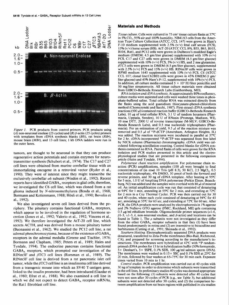
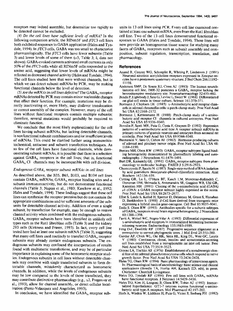
Figure (A) non-neuronal enolase (25 cycles) and (B) @actin (25 cycles) primers with templates from cDNA synthesis blanks (B/k), rat brain (RBr), mouse brain (MEr), and 13 cell lines; 1 kb DNA ladders were run in the outer lanes.
tumors, are thought to be neuronal in that they can produce regenerative action potentials and contain enzymes for neuro- transmitter synthesis (Schubert et al., 1974). The C 17 and C27 cell lines were obtained from murine cerebellar tissue with an immortalizing oncogene in a retroviral vector (Ryder et al., 1990). They were of interest since they might transcribe the exclusively cerebellar ot6 subunit (Wisden et al., 1992). Recent reports have identified GABA, receptors in glial cells; therefore, we investigated the C6 cell line, which was cloned from a rat glioma induced by N-nitrosomethylurea (Benda et al., 1968; Bormann and Kettenmann, 1988; HGsli et al., 1990; Bovolin et al., 1992).
We also investigated seven cell lines derived from the pe- riphery. The pituitary contains functional GABA, receptors, which appear to be involved in the regulation of hormone se- cretion (Jones et al., 1992; Valerio et al., 1992; Vincens et al., 1992). We therefore investigated the AtT-20 cell line, which secrets ACTH, and was derived from a mouse pituitary tumor (Buonassisi et al., 1962). We studied the PC12 cell line, a rat adrenal pheochromocytoma, because of the existence of GABA, receptors in the adrenal medulla (Greene and Tischler, 1976; Bormann and Clapham, 1985; Peters et al., 1989; Hales and Tyndale, 1994). The endocrine pancreas contains functional GABA, receptors, which prompted our investigation of the RINmSF and PTC3 cell lines (Rorsman et al., 1989). The RINmSF cell line is derived from a rat pancreatic islet cell tumor, while the PTC3 cell line is an insulinoma cell line derived from a transgenic mouse into which an SV40 T-antigen gene, linked to the insulin promotor, had been introduced (Gazdar et al., 1980; Efrat et al., 1988). We also examined a cell line in which we did not expect to detect GABA, receptor mRNAs, the Rat 1 fibroblast cell line.
Materials and Methods
Tissue culture. Cells were cultured in 75 cm2 tissue culture flasks at 37°C in 5% CO,, 95% air and 100% humidity. NB41A3 cells from the Amer- ican Type Culture Collection (ATCC, CCL 147) were grown in Ham’s F-10 medium supplemented with 2.5% (v/v) fetal calf serum (FCS), 15% (v/v) horse serum (HS). AtT-20 (ATCC CCL 89), B35, B65, B103, B 104, Rat 1, and PC 12 cells were grown in Dulbecco’s modified Eagle’s medium (DMEM; 4.5 gm/liter glucose) supplemented with lOoh (v/v) FCS. Cl7 and C27 cells were grown in DMEM (4.5 gm/liter glucose) supplemented with 10% (v/v) FCS, 5% (v/v) HS, and 2 mM glutamine. PTC3 cells were grow-n in DMEM (4.5 gm/liter glucose), supplemented with 2.5% (v/v) FCS and 15% (v/v) HS. RINmSF cells were grown in RPM1 medium 1640 supplemented with 10% (v/v) FCS. C6 (ATCC CCL 107; clonal line:C62BD) cells were grown in 45% DMEM (1 gm/ liter glucose) and 45% Ham’s F- 12, supplemented with 10% (v/v) FCS. In addition, all culture media contained 5 x lo4 IU/liter penicillin and 50 mg/liter streptomycin. All tissue culture materials were obtained from GIBCO-Bethesda Research Labs (Gaithersburg, MD).
RNA isolation andcDNA synthesis. At approximately 80% confluency, culture media were aspirated and cells were washed three times in phos- phate-buffered saline. Total cellular RNA was extracted directly from the flasks using the acid guanidium thiocyanate-phenol-chloroform method (Chomczynski and Sacchi, 1987). First-strand cDNA synthesis reactions (500 pl) contained reaction buffer (GIBCO-Bethesda Research Labs), 10 rg of total cellular RNA, 3.1 U of random hexamers (Phar- macia, Uppsala, Sweden), 10 U of RNasin (Promega, Madison, WI), 10 mM DTT, 2000 U of reverse transcriptase (M-MLV, GIBCO-Be- thesda Research Labs), and 0.5 mM nucleotide triphosphates (Phar- macia). For an estimation of yield, 9.5 ~1 of the reaction mixture was removed and 0.5 ~1 of 32P-dCTP (Amersham, Arlington Heights, IL) was added. The reaction mixtures were incubated in parallel-at 37°C for 2 hr. cDNA-incoraorated ‘*P-dCTP was seuarated from free label by NICK column (Pharmacia) chromatograph;. cDNA yield was cal- culated following scintillation counting. coni& blanks for cDNA syn- thesis contained no RNA. Paired flasks of cells were grown for the RNA isolation and PCR studies presented in this article and the electro- physiological studies that are presented in the following companion article (Hales and Tyndale, 1994).
Polymerase chain reaction amplification. For polymerase chain re- action (PCR) amplifications, samples (100 ~1) were brought to final concentrations of (in mM) 10 Tris (pH 8.3), 50 KCl, 2.5 MgCl,, 0.5 nucleotide triphosphate, 4% DMSO, 50 pmol of both the forward and reverse primers, and 30 ng of cDNA template. After heating at 95°C for 5 min, 1 .O U of Amplitaq DNA polymerase (Perkin Elmer, Branch- burg, NJ) was added and the samples were overlaid with 80 ul of mineral oil.& initial amplification cycle was run that consisted df denaturing at 94°C for 1 min, annealing at 55°C for 2 min, and extending at 72°C for 2 min in a Coy Thermal Cycler. PCR was then carried out for 30 and 40 cycles, where each cycle consisted of denaturing at 94°C for 45 set, annealing at 55°C for 60 set, and extending at 72°C for 60 sec. After PCR, the DNA products were analyzed by electrophoresis in 1% agarose and 2% NuSieve GTG agarose (FMC, Rockland, ME) gels containing 0.5 &/ml ethidium bromide. Oligonucleotide primer sequences ((~1-6, 81-3, 71-3, 6, non-neuronal enolase, and @actin) and locations can be found in Table 1. The P subunits were not investigated as thev differ from the other GABA, receptor subunits in that ihey (I) have been found predominantly in retina and (2) are insensitive to bicuculline and barbiturates (Cutting et al., 199 1; Shimada et al., 1992).
Southern blotting. Electrophoretically separated DNA products were denatured, transferred to Zeta-Probe membranes (Bio-Rad, Richmond, CA), and prepared for screening according to the manufacturer’s in- structions. The membranes were hybridized at 42°C with ‘IP random- primed cDNA probes for 15 hr in hybridization buffer (50% formamide, 5 x Denhart’s, 5 x SSPE, 0.1% SDS, 100 &ml salmon sperm DNA). The membranes were washed in 0.15 x SSC and 0.1% SDS at 25°C for 20 min, followed by four washes at 65-75°C for 30 min each. Exposure times varied from 10 min to 1 hr.
Control studies. PCR amplification was carried out at 40 cycles with 30 ng of template in order to determine which subunits were transcribed in the cell lines. In preliminary studies 40 cycles was deemed appropriate based on the following: (1) subunits were detected after 40 cycles that were not seen after 30 cycles of PCR (e.g., see Table 3), but additional subunits were not detected after 50 cycles; and (2) the comparison be- tween amplification from rat brain regions with published in situ studies
The Journal of Neuroscience, September 1994, 14(9) 5419
Table 1. Primer sequences for PCR
Starting Size Subunit Direction Primer sequence at base (W
LYl
ot2
a3(i)
a3(ii)
&4(i)
a4(ii)
or5
a6
81
P2
83
Yl
YLzLs
r3
8
NNE
p-Actin
Forward Reverse Forward Reverse Forward Reverse Forward Reverse Forward Reverse Forward Reverse Forward Reverse Forward Reverse Forward Reverse Forward Reverse Forward Reverse Forward
Forward Reverse Forward Reverse Forward Reverse Forward Reverse Forward Reverse
S-ATCTTTGGGCCTGGACCCTCATTCT-3’ Y-CGGGCTGGCTCCCTTGTCCACTC-3’ S-GAGGACAAAATTGAGCACTTGCA-3’ 5’-GAGTTGTTAAGTCGAAGGATATT-3’ 5’-GAATCAAGACGACAAGAACCT-3’ 5’-CAGATTTGTTCTTCCCAAGAG-3’ 5’-TTTATTCTTGGACTCTTGGGAAGAACA-3’ 5’-CTTCATCTCCAGGGCCTCTGGTACCT-3’ 5’-CTGGACCAAAGGCCCTGAGA-3’ 5’-TTTTCCTTCAGTACTGGGGCAGCTG-3’ 5’-TTTAAACGAATCCCCAGGACAGAA-3’ 5’-TGCCATTTCTCATAATTCTAA-3’ 5’-ACTTTGGCTTTTCACAAATGCCAA-3’ 5’-AGAAGGTTGAGAGGGAGACGTT-3’ 5’-AGGGTGACCTGACCTGGCATTTCAGTGAACCATAGG-3’ 5’-TCATGGTGTACAGGATCGTTCCA-3’ 5’-ACAGTACAAAATCGAGAGAGTTTG-3’ 5’-TCCACCTTCTTGGACACCATCTTG-3’ 5’-ATAAACTCATCACCAAGAAAGTTG-3’ 5’-AAGTCCCATTACTGCTTCTGATGT-3’ 5’-TGGAGCACCGTCTGGTCTCCAGGA-3’ 5’-TCGATCATTCTTGGCCTTGGCTGT-3’ 5’-AGTACAAGTGGAAAAAGCCC-3’ 5’-TCAGCTTCCTGTCTCTGGTGGTT-3’ 5’-GTGGAGTATGGTACCCTGCACTATTTTGTG-3’ 5’-CAGAAGGCGGTAGGGAAGAAGATCCGAGCA-3’ 5’-TGCTCGGTCCAGGAGGGTAGA-3’ 5’-CTGATCAGCTGCCTCAACTGAATTTTT-3’ 5’-GACTACGTGGGCTCCAACCTGGA-3’ 5’-ACTGTGGAGGTGATGCGGATGCT-3’ 5’-ACTCCGAGACAATGATAAGACCC-3’ 5’-AGGTGCGAATCCACCCTCATCA-3’ 5’-CACCACAGCTGAGAGGGAAATCGTGCGTGA-3’ 5’-ATTTGCGGTGCACGATGGAGGGGCCGGACT-5’
29 608
3 347
91 691 659
1143 609
1112 81
475 80
371 -31 445
671 644
1158 641
1059 650
1114 1048 1358
48 639
88 485 134 637 603
1120
580
345
601
484
503
387
292
476
664
515
419
465
311
591
398
504
517
Numbcring is from the ATG. Published sequences used to design primers and putative exons (E) that they span: oil (Khrestchatisky et al., 1989; Lolait et al., 1989; El-6), a2 (Kbrestchatisky et al., 1991; El-4), (~3 [Malherbe et al., 1990; a3(i):El-6; a3(ii):E6-81, a4 [Wisden et al., 1991; a4(i):E6-8, a4(ii):El-41, a5 (originally described as u4 by Khrestchatisky et al., 1989; Malherbe et al., 1990; Pritchett and Seeburg, 1990; El-4), a6 (Ltiddens et al., 1990; El-4), 81 (Ymer et al., 1989; El-6), 82 (Ymer et al., 1989; E6-9), j33 (Lolait et al., 1989; Ymer et al., 1989; E6-S), yl (Ymer et al., 1990; E6-S), -r2 (Shivers et al., 1989; E&9), 73 (Knoflach et al., 1991; Herb et al., 1992; El-6), d (Shivers et al., 1989; Zhao and Joho, 1990; El-F& NNE (non-neuronal enolase; Sakimura et al., 1985), and @-actin (Nude1 et al., 1983).
(data not shown) suggested that 40 cycles would detect mRNAs of high and low abundance.
The oligo-primer pairs may amplify with different efficiencies, sug- gesting that comparisons of mRNA levels between different subunits, based& the amount of PCR product produced, may be inappropriate. However, PCR products for individual oligo-primer pairs between cell lines can be compared as the problem of varying efficiencies has been eliminated. Thus, amplification was also carried out at 30 cycles in order to identify cell lines with higher or lower levels of any one subunit.
No differences were observed between cells harvested at 50-1001 confluency with respect to the levels of GABA, receptor subunit PCR products/30 ng total cDNA (data not shown). All experiments were subsequently performed on cells harvested at approximately 80% con- fluency. All of the cell lines described maintained the same level and composition of GABA, receptor mRNAs, as detected by PCR, over at least three different passage numbers. The figures demonstrate results from representative experiments.
Due to the possible detection, by PCR, of contaminant cDNA/RNA, we used multiple controls. (1) RNA blanks that were taken through the cDNA synthesis steps were used as controls for PCR. These blanks were used in every PCR reaction, for every set ofprimers. (2) Samples without primers were run for every cDNA template for each PCR experiment.
(3) Primers for ubiquitous mRNAs were also used to ensure that the RNA isolation and cDNA synthesis had been successful. (4) Rat and/ or mouse brain (as appropriate) positive controls were run for each experiment as the cell lines are derived from one of these two species. For many of the GABA, receptor subunits only sequence data from the rat was available for design of the primer sets. While there is high sequence identity between the cloned mouse and rat GABA, receptors, mouse brain cDNA as well as rat brain cDNA was used as a positive control to ensure that all of the primer sets hybridized to cDNA of both species.
Specificity of the GABA, receptor primers. Specificity of the GABA, receptor primers was tested as follows. (1) For each set of primers, PCR DNA was cut with restriction endonucleases and the appropriate size products were produced (data not shown). (2) Southern blotting was performed on PCR derived DNA products. ‘The primers designeh for individual GABA, receotor subunit mRNAs amnlified nroducts of the expected size (see ‘Table 1; Figs. 2, 3, 5-10) and hybridized specifically with the appropriate labeled cDNA. In addition, the cDNA probes did not hybridize to bands amplified from inappropriate primer sets. (3) Primers were able to amplify products of the correct size from the appropriate GABA, receptor cDNA clones and not from others (data not shown).
5420 Tyndale et al. l GABA, Receptor Subunit mRNAs in 13 Cell tines
kb
2.0- 1.6- l.O-
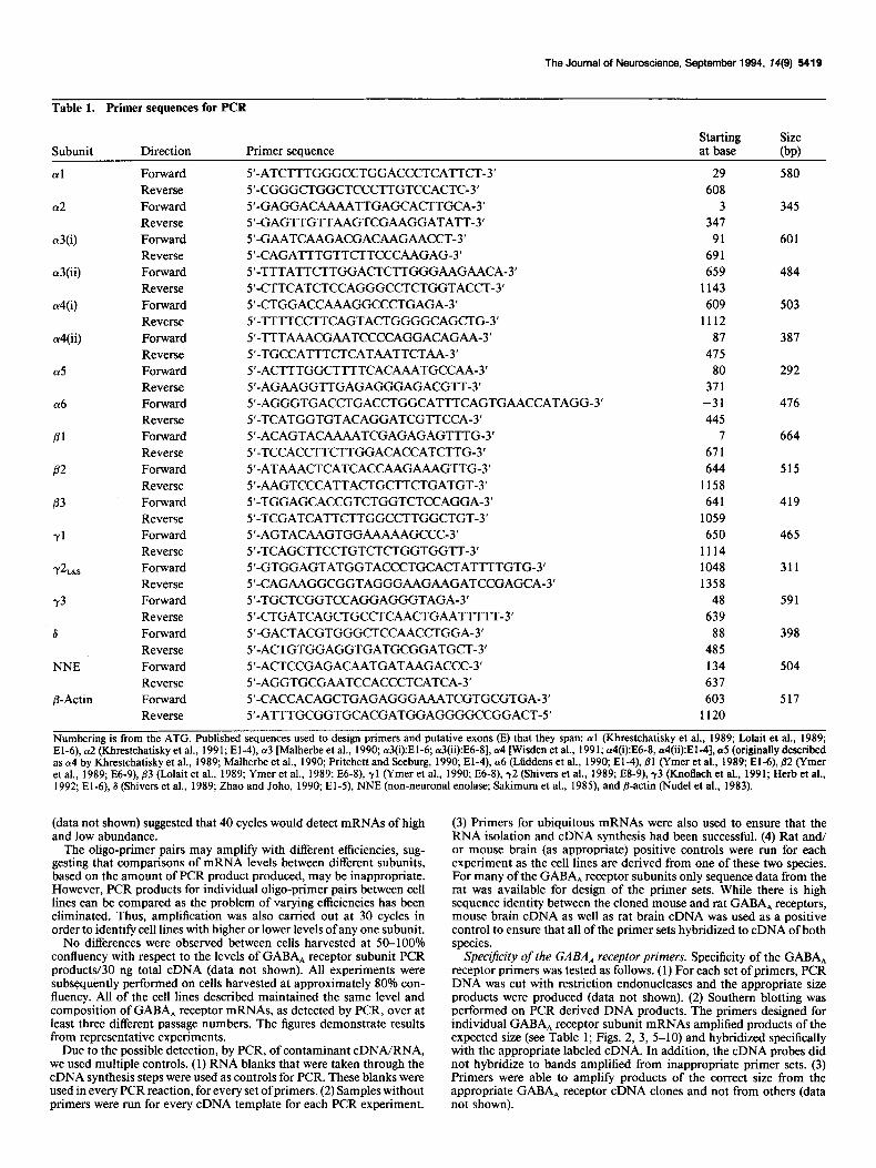
Figure 2. PCR products using GA- BA, receptor subunit 83 primers and cell line cDNA templates for 30 cycles of amplification. Positive controls in- clude the j33 primers against rat brain (RB) and mouse brain (MB) cDNA templates while the negative controls include an RNA blank (Blk). A 1 kb DNA ladder was run in lane 3. Southern blots with @l (B), 82 (C), and 83 cDNA (0) probes were performed. @ 1 (lane I) and ,82 (lane 2) primer sets were used with rat brain cDNA template to gen- erate DNA products as negative and positive controls for the Southern blots.
Since cDNA clones for GABA, receptor 013 and a4 subunits were unavailable for testing the specificity of the (~3 and a4 primers, two sets of primers for each of these subunits were designed. Both primer sets amplified the correctly sized DNA products, which, upon digestion with restriction endonucleases, produced the expected products. (~3 PCR product was found in RINmSF, BTC3, B104, Cl7 cells using either a3(i) or a3(ii) primer sets, while (~4 PCR product was found in RINmSF and j3TC3 cells with either Ly4(i) or Ly4(ii) primer sets (Table 2). In addition, low-stringency Southern blotting with GABA, receptor 32P- labeled (Y-cDNA clones (al, (~2, (~5) detected the appropriately sized DNA products from PCR using the a3 and cu4 primers (data not shown).
All of the primer sets produced PCR DNA products of the same size from rat and mouse brain cDNA (e.g., Figs. l-3). In addition, rat cDNA probes hybridized to the DNA products produced from mouse brain cDNA templates (e.g., Figs. 2, 3).
Genomic DNA contamination of cellular RNA. The possibility of ge- nomic DNA contamination of total cellular RNA was determined by DNase treatment of at least one RNA sample for each cell line. No differences were observed between DNAase treated or nontreated RNA samples in the level or composition of GABA, receptor mRNAs de- tected by PCR in any cell line. Therefore, DNase treatment was not routinely performed. In addition, GABA, receptor intron-exon struc- ture has been characterized for the mouse 6 subunit, the human @l subunit, part of the human 83, and the chicken @4 (Sommer et al., 1990; Kirkness et al., 199 1; Lasham et al., 199 1; Kirkness and Fraser, 1993). These subunits display highly conserved exon boundaries. By aligning
PI probe
82 probe
P3 probe
subunits of known gene structure with those of unknown structure, the location of the introns in the unstudied subunit genes can be estimated (Feng and Doolittle, 1987). The primer sets were therefore designed to amplify across multiple introns (the postulated introns that each set crosses can be found in the Table 1 notes). I f PCR amplification of genomic DNA contamination had been present, a product of altered size would have been expected as the primers span multiple introns.
Results PCR DNA products were detected from all of the cell lines with non-neuronal enolase and @-actin primers (Fig. 1). Negative controls lacking RNA in the cDNA synthesis do not demon- strate any detectable DNA bands (Fig. l), while positive controls containing total mouse or rat brain cDNA demonstrate detect- able DNA bands.
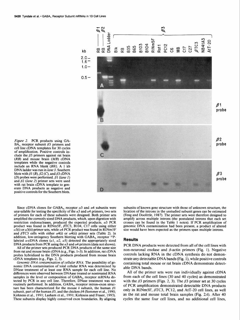
All of the primer sets were run individually against cDNA from each of the cell lines (30 and 40 cycles) as demonstrated with the 83 primers (Figs. 2, 3). The p3 primer set at 30 cycles of PCR amplification demonstrated detectable DNA products only in RINmSF, PTC3, PC12, and AtT-20 cell lines, as well as the rat and mouse total brain samples (Fig. 2~9. After 40 cycles the same four cell lines, and no additional cell lines,
The Journal of Neuroscience, September 1994, 74(9) 5421
kb
PI probe
P2 probe
Figure 3. PCR products using GA- BA, receptor subunit ,83 primers and
.- cell line cDNA templates for 40 cycles of amplification. Positive controls in-
. elude the 83 primers against rat brain (RB) and mouse brain (MB) cDNA templates while the negative controls include an RNA blank (BZQ. A 1 kb DNA ladder was run in lane 3. Southern blots with 81 (B), /32 (C), and B.3 (0) cDNA probes were performed. 81 (lane I) and j32 (lane 2) primer sets were used with rat brain cDNA template to gen- erate DNA products as negative and positive controls for the Southern blots.
demonstrated 83 DNA products (Fig. 3.4). Primer sets, 01 and The Pl and ,82 cDNA probes only hybridized to the lanes con- /32, were used in PCR with rat brain cDNA template as controls taining the corresponding primer sets and not to the lanes with for the Southern blotting (Figs. 24 34. The p3 cDNA probe ,83 primer (Figs. 2B,C, 3B,C). These data suggest that the p-pri- hybridized to the /I3 primer generated PCR products in the mers amplify products only from their corresponding mRNAs. Southern blots but not to Pl or p2 PCR products (Figs. 20,3D). These results are typical of the experiments that were run with
Table 2. Cell lines in which individual GABA, receptor subunit mRNAs are found
Subunit Cell line
Cul RINmSF, B35, B65, B103, B104 (Y2 RINmSF, B35, B65, B103, Cl7 a3 RINmSF, BTC3, B104, Cl7 cz4 RINmSF, BTC3 ff5 RINmSF, AtT20
81 RINm5F
82 RINmSF, B35, B65, B103, B104, C6 P3 RINmSF, PTC3, PC12, AtT20
71 RINmSF, @TC3, B35, B65, B103, B104, Cl7
7% RINmSF, B35, B65, B103, PC12, C17, C6, C27
7% RINmSF, B35, B65, B103, PC12, C17,C6, C27
r3 RINm5F 6 RINmSF, flTC3, B35, B65, B014, PC12, AtT20, NB41A3, C17, C6, C27, Rat1
GABA, receptor subunit mRNAs found by PCR after 40 cycles of amplification.
5422 Tyndale et al. * GABA, Receptor Subunit mRNAs in 13 Cell Lines
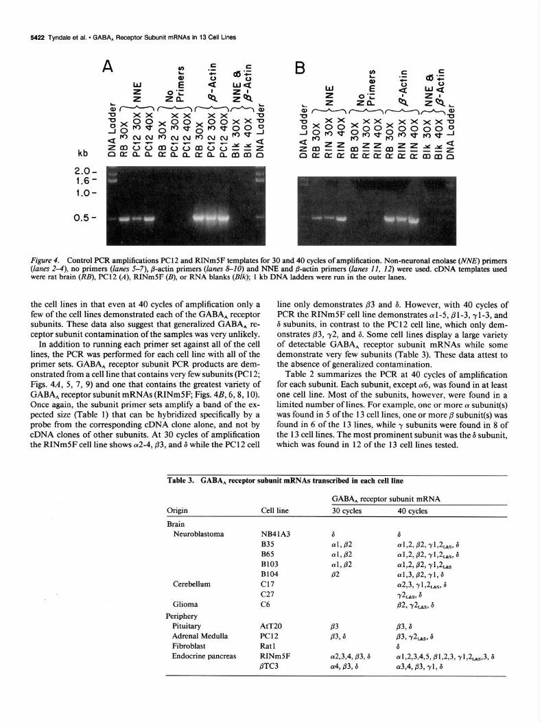
Figure 4. Control F’CR amplifications PC1 2 and RINm5F templates for 30 and 40 cycles of amplification. Non-neuronal enolase (WV,!?) primers (lanes 2-4), no primers (lanes 5-7), @-actin primers (lanes 8-10) and NNE and @-actin primers ([anes II, 12) were used. cDNA templates used were rat brain (RB), PC12 (A), RINmSF (B), or RNA blanks @NC); 1 kb DNA ladders were run in the outer lanes.
the cell lines in that even at 40 cycles of amplification only a few of the cell lines demonstrated each of the GABA, receptor subunits. These data also suggest that generalized GABA, re- ceptor subunit contamination of the samples was very unlikely.
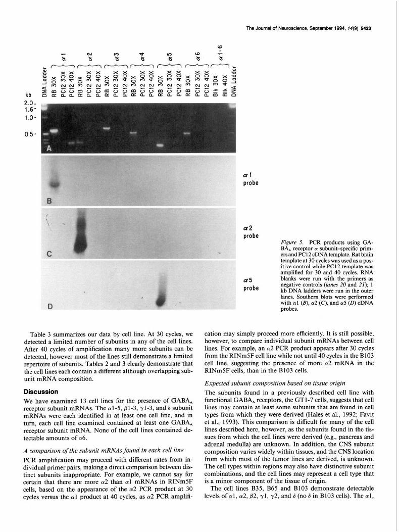
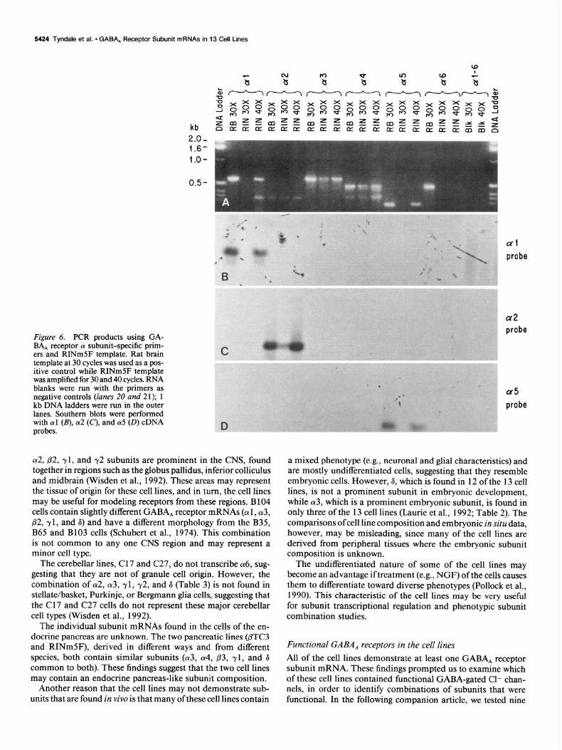
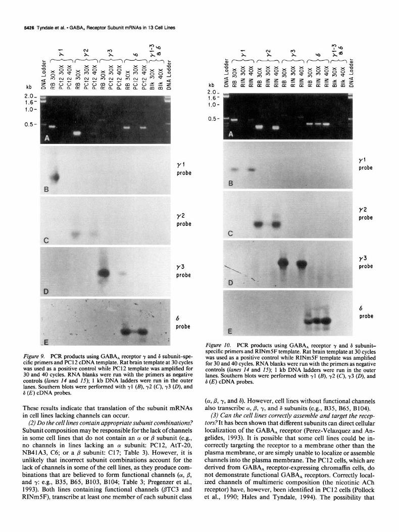
In addition to running each primer set against all of the cell lines, the PCR was performed for each cell line with all of the primer sets. GABA, receptor subunit PCR products are dem- onstrated from a cell line that contains very few subunits (PC1 2; Figs. 4A, 5, 7, 9) and one that contains the greatest variety of GABA, receptor subunit mRNAs (RINmSF; Figs. 4B, 6,8, 10). Once again, the subunit primer sets amplify a band of the ex- pected size (Table 1) that can be hybridized specifically by a probe from the corresponding cDNA clone alone, and not by cDNA clones of other subunits. At 30 cycles of amplification the RINm5F cell line shows a2-4,,83, and 6 while the PC1 2 cell
line only demonstrates 03 and 6. However, with 40 cycles of PCR the RINmSF cell line demonstrates al-5,81-3,rl-3, and 6 subunits, in contrast to the PC 12 cell line, which only dem- onstrates p3, 72, and 6. Some cell lines display a large variety of detectable GABA, receptor subunit mRNAs while some demonstrate very few subunits (Table 3). These data attest to the absence of generalized contamination.
Table 2 summarizes the PCR at 40 cycles of amplification for each subunit. Each subunit, except (~6, was found in at least one cell line. Most of the subunits, however, were found in a limited number of lines. For example, one or more a subunit(s) was found in 5 of the 13 cell lines, one or more p subunit(s) was found in 6 of the 13 lines, while y subunits were found in 8 of the 13 cell lines. The most prominent subunit was the 6 subunit, which was found in 12 of the 13 cell lines tested.
Table 3. GABA, receptor subunit mRNAs transcribed in each cell line
Origin
Brain Neuroblastoma
Cerebellum
Glioma Periphery
Pituitary Adrenal Medulla Fibroblast Endocrine pancreas
Cell line
NB41A3 B35 B65 B103 B104 Cl7 C27 C6
AtT20 PC12 Rat1 RINm5F /3TC3
GABA, receptor subunit mRNA 30 cycles 40 cycles
6 6 al, P2 (u1,2,82, YLL, 6 al, P2 ~1,2,82, Yl,L,, 6 (Yl,P2 al,& P2, Y1,Ls P2 ch3,82, Yl, 6
ff2,3, ~1,2,,, 6 Y%&,, 6 m, Y&&s, 6
P3 83,s P3,6 83, Y2,,, 6
6 a2,3,4,83,6 a1,2,3,4,5, ~1~2~3, ~1,2,,,3,6 a49 P3,6 a3949 83, Yl, 6
The Journal of Neuroscience, September 1994, 74(9) 5423
al probe
a2 probe
a5 probe
Figure 5. PCR products using GA- BA, receptor CY subunit-specific prim- ers and PC 12 cDNA template. Rat brain template at 30 cycles was used as a pos- itive control while PC1 2 template was amplified for 30 and 40 cycles. RNA blanks were run with the-primers as negative controls (lanes 20 and 21); 1 kb DNA ladders were run in the outer lanes. Southern blots were performed with cul (B), ~y2 (C), and 015 (0) cDNA probes.
Table 3 summarizes our data by cell line. At 30 cycles, we detected a limited number of subunits in any of the cell lines. After 40 cycles of amplification many more subunits can be detected, however most of the lines still demonstrate a limited repertoire of subunits. Tables 2 and 3 clearly demonstrate that the cell lines each contain a different although overlapping sub- unit mRNA composition.
Discussion We have examined 13 cell lines for the presence of GABA, receptor subunit mRNAs. The (~1-5, pl-3, ~1-3, and 6 subunit mRNAs were each identified in at least one cell line, and in turn, each cell line examined contained at least one GABA, receptor subunit mRNA. None of the cell lines contained de- tectable amounts of (~6.
A comparison of the subunit mRNAs found in each cell line PCR amplification may proceed with different rates from in- dividual primer pairs, making a direct comparison between dis- tinct subunits inappropriate. For example, we cannot say for certain that there are more 012 than cul mRNAs in RINmSF cells, based on the appearance of the (~2 PCR product at 30 cycles versus the al product at 40 cycles, as a2 PCR amplifi-
cation may simply proceed more efficiently. It is still possible, however, to compare individual subunit mRNAs between cell lines. For example, an cu2 PCR product appears after 30 cycles from the RINmSF cell line while not until 40 cycles in the B 103 cell line, suggesting the presence of more (~2 mRNA in the RINmSF cells, than in the B103 cells.
Expected subunit composition based on tissue origin The subunits found in a previously described cell line with functional GABA, receptors, the GT l-7 cells, suggests that cell lines may contain at least some subunits that are found in cell types from which they were derived (Hales et al., 1992; Favit et al., 1993). This comparison is difficult for many of the cell lines described here, however, as the subunits found in the tis- sues from which the cell lines were derived (e.g., pancreas and adrenal medulla) are unknown. In addition, the CNS subunit composition varies widely within tissues, and the CNS location from which most of the tumor lines are derived, is unknown. The cell types within regions may also have distinctive subunit combinations, and the cell lines may represent a cell type that is a minor component of the tissue of origin.
The cell lines B35, B65 and B103 demonstrate detectable levels of al, a2, @2, yl, 72, and 6 (no 6 in B103 cells). The al,
5424 Tyndale et al. - GABA, Receptor Subunit mRNAs in 13 Cell Lines
kb 2.0- 1.6- l.O-
Figure 6. PCR products using GA- BA, receptor cx subunit-specific prim- ers and RINmSF template. Rat brain template at 30 cycles was used as a pos- itive control while RINmSF template was amplified for 30 and 40 cycles. RNA blanks were run with the primers as negative controls (lanes 20 and 21); 1 kb DNA ladders were run in the outer lanes. Southern blots were performed with al (B), cz2 (C), and (~5 (0) cDNA probes.
~y2, p2, yl, and y2 subunits are prominent in the CNS, found together in regions such as the globus pallidus, inferior colliculus and midbrain (Wisden et al., 1992). These areas may represent the tissue of origin for these cell lines, and in turn, the cell lines may be useful for modeling receptors from these regions. B 104 cells contain slightly different GABA, receptor mRNAs (cul , (~3, p2, yl, and 6) and have a different morphology from the B35, B65 and B103 cells (Schubert et al., 1974). This combination is not common to any one CNS region and may represent a minor cell type.
The cerebellar lines, C 17 and C27, do not transcribe a6, sug- gesting that they are not of granule cell origin. However, the combination of (r2, (~3, yl, 72, and 6 (Table 3) is not found in stellate/basket, Purkinje, or Bergmann glia cells, suggesting that the Cl7 and C27 cells do not represent these major cerebellar cell types (Wisden et al., 1992).
The individual subunit mRNAs found in the cells of the en- docrine pancreas are unknown. The two pancreatic lines @TC3 and RINmSF), derived in different ways and from different species, both contain similar subunits (cy3, (~4, ,83, yl, and 6 common to both). These findings suggest that the two cell lines may contain an endocrine pancreas-like subunit composition.
Another reason that the cell lines may not demonstrate sub- units that are found in vivo is that many of these cell lines contain
ul probe
a2 probe
a5 probe
a mixed phenotype (e.g., neuronal and glial characteristics) and are mostly undifferentiated cells, suggesting that they resemble embryonic cells. However, 6, which is found in 12 of the 13 cell lines, is not a prominent subunit in embryonic development, while (~3, which is a prominent embryonic subunit, is found in only three of the 13 cell lines (Laurie et al., 1992; Table 2). The comparisons ofcell line composition and embryonic in situ data, however, may be misleading, since many of the cell lines are derived from peripheral tissues where the embryonic subunit composition is unknown.
The undifferentiated nature of some of the cell lines may become an advantage if treatment (e.g., NGF) of the cells causes them to differentiate toward diverse phenotypes (Pollock et al., 1990). This characteristic of the cell lines may be very useful for subunit transcriptional regulation and phenotypic subunit combination studies.
Functional GABA, receptors in the cell lines All of the cell lines demonstrate at least one GABA, receptor subunit mRNA. These findings prompted us to examine which of these cell lines contained functional GABA-gated Cl- chan- nels, in order to identify combinations of subunits that were functional. In the following companion article, we tested nine

The Journal of Neuroscience, September 1994, 14(9) 5425
kb
2.0- 1.6-
l.O-
Pi probe
: P2 : / . probe I
kb
2.0- 1.6-
l.O-
B1 probe
P2 probe
P3 probe
Figure 7. PCR products using GABA, receptor B subunit-specific primers and PC 12 cDNA template. Rat brain template at 30 cycles was used as a positive control while PC12 template was amplified for 30 and 40 cycles. RNA blanks were run with the primers as negative con- trols (lanes I1 and 12); 1 kb DNA ladders were run in the outer lanes. Southern blots were performed with pl (B), ,82 (C), and 63 (0) cDNA probes.
of the cell lines for GABA-gated Cl- channels, using the whole- cell configuration of the patch-clamp technique. Only two of the cell lines, the RINmSF and PTC3 cells, responded to GABA (Hales and Tyndale, 1994). However, none of the other cell lines tested, even though they contained GABA, receptor subunit mRNAs (Table 3) expressed functional GABA, receptor Cl- channels. These included the B65, B104, PC12, AtT-20, NB41A3, C17, and C6 cells (Hales and Tyndale, 1994). The Rat1 cells were not tested as the NB4 lA3 cells also contain only the 6 subunit mRNA (and in greater quantity than the Rat1 cells; Table 3), and did not respond to GABA. Likewise, the C27 cells were not tested because they show an even more limited subunit mRNA composition than their sibling line, the C 17 cells (Table 3), which did not demonstrate functional chan- nels. In addition, Kasckow et al. (1992) did not find functional GABA, Cl- channels in the B35, B65, or B103 cells.
Figure 8. PCR products using GABA, receptor p subunit-specific primers and RINm5F template. Rat brain template at 30 cycles was used as a positive control while RINmSF template was amplified for 30 and 40 cycles. RNA blanks were run with the primers as negative controls (lanes I1 and 12); 1 kb DNA ladders were run in the outer lanes. Southern blots were performed with @l (B), @2 (C), and p3 (0) cDNA probes.
What makes a functional GABA-gated chloride channel?
Many factors might contribute to why cell lines with subunit mRNAs are not producing detectable channels. These include (1) lack of subunit translation, (2) incorrect subunit combina- tions, (3) inability to assemble or localize channels correctly, (4) insufficient amounts of mRNAs, and/or (5), defective mRNAs. These alternative explanations are discussed below.
(1) Do the cell lines translate receptor mRNA? While the B35, B65, B103, and B104 cell lines contained GABA, mRNAs, GABA, receptor binding sites and (Y subunit immunoreactivity, they do not demonstrate functional channels (Table 3; Napias et al., 1980; Kasckow et al., 1992; Hales and Tyndale, 1994). Likewise, the Cl7 cells have mRNAs (Table 3) bind 3H-mus- cimol and 3H-flunitrazepam (Sapp, Tyndale, and Olsen, un- published observations), but do not have functional channels.
5426 Tyndale et al. l GABA, Receptor Subunit mRNAs in 13 Cell Lines
Y’ probe
Y2 probe
Y3 probe
D
“ .-i.% 6
probe I ’
Figure 9. PCR products using GABA, receptor y and 6 subunit-spe- cific primers and PC 12 cDNA template. Rat brain template at 30 cycles was used as a positive control while PC12 template was amplified for 30 and 40 cycles. RNA blanks were run with the primers as negative controls (lanes 14 and 15); 1 kb DNA ladders were run in the outer lanes. Southern blots were performed with yl (B), 72 (C), 73 (D), and d (E) cDNA probes.
These results indicate that translation of the subunit mRNAs in cell lines lacking channels can occur.
(2) Do the cell lines contain appropriate subunit combinations? Subunit composition may be responsible for the lack ofchannels in some cell lines that do not contain an (Y or /? subunit (e.g., no channels in lines lacking an (Y subunit: PC12, AtT-20, NB4 lA3, C6; or a p subunit: Cl 7; Table 3). However, it is unlikely that incorrect subunit combinations account for the lack of channels in some of the cell lines, as they produce com- binations that are believed to form functional channels (cy, p, and y: e.g., B35, B65, B103, B104; Table 3; Pregenzer et al., 1993). Both lines containing functional channels @TC3 and RINmSF), transcribe at least one member of each subunit class
Y’ probe
Y2 probe
Y3 probe
6 probe
Figure IO. PCR products using GABA, receptor y and 6 subunit- specific primers and RINm5F template. Rat brain template at 30 cycles was used as a positive control while RINmSF template was amplified for 30 and 40 cycles. RNA blanks were run with the primers as negative controls (lanes 14 and 15); 1 kb DNA ladders were run in the outer lanes. Southern blots were performed with yl (B), ~2 (C), 73 (O), and 6 (E) cDNA probes.
(a, p, y, and 6). However, cell lines without functional channels also transcribe (Y, P, y, and 6 subunits (e.g., B35, B65, B104).
(3) Can the cell lines correctly assemble and target the recep- tors?It has been shown that different subunits can direct cellular localization of the GABA, receptor (Perez-Velazquez and An- gelides, 1993). It is possible that some cell lines could be in- correctly targeting the receptor to a membrane other than the plasma membrane, or are simply unable to localize or assemble channels into the plasma membrane. The PC 12 cells, which are derived from GABA, receptor-expressing chromaffin cells, do not demonstrate functional GABA, receptors. Correctly local- ized channels of multimeric composition (the nicotinic ACh receptor) have, however, been identified in PC1 2 cells (Pollock et al., 1990; Hales and Tyndale, 1994). The possibility that
The Journal of Neuroscience, September 1994, 14(9) 5427
receptors may indeed assemble, but desensitize too rapidly to be detected cannot be excluded.
(4) Do the cell lines have suficient levels of mRNA? In the following companion article, the RINmSF and /3TC3 cell lines both exhibited responses to GABA application (Hales and Tyn- dale, 1994). In PTC3 cells, GABA was too small to characterize pharmacologically. The PTC3 cells have fewer subunits (Table 3) and lower levels of some of them ((~3, Table 3; 6, data not shown). GABA-evoked currents activated small currents in only 50% of the PTC3 cells while all RINmSF cells responded to the amino acid, suggesting that lower levels of mRNAs might be reflected as decreased channel activity (Hales and Tyndale, 1994). The cell lines studied here that were without channels, but in which we can detect subunit mRNAs by PCR, may be making functional channels below the level of detection.
(5) Are the mRNAs in cell lines defective?The GABA, receptor mRNAs detected by PCR may contain minor nucleotide changes that effect their function. For example, mutations may be di- rectly inactivating or, more likely, may disfavor translocation or correct assembly of the complex. However, many of the cell lines without functional receptors contain multiple subunits; therefore, several mutations would probably be required to eliminate function.
We conclude that the most likely explanation(s) for the cell lines having subunit mRNAs, but lacking detectable channels, is nonfunctional subunit combinations and/or insufficient levels of mRNAs. This could be clarified further using immunohis- tochemical, antisense and subunit transfection techniques. As so few of the cell lines have functional channels, while dem- onstrating subunit mRNAs, it is possible that there is a selection against GABA, receptors in the cell lines; that is, functional GABA, Cl- channels may be incompatible with cell division.
Endogenous GABA, receptor subunit mRNAs in cell lines As described above, the B35, B65, B103, and B104 cell lines contain GABA, mRNAs, GABA, receptor binding sites and LY subunit immunoreactivity, but do not demonstrate functional channels (Table 3; Napias et al., 1980; Kasckow et al., 1992; Hales and Tyndale, 1994). These findings suggest that these cell lines can synthesize the receptor proteins but do not contain the appropriate combinations and/or sufficient amounts of the sub- units for detectable channel activity. Addition of even a single subunit, by transfection for example, may be enough to restore channel activity when combined with the endogenous subunits. GABA, receptor subunits have been identified in unlikely cell types such as the Rat 1 fibroblast cells (Table 3) and the kidney 293 cells (Kirkness and Fraser, 1993). In fact, every cell line tested here had at least one subunit mRNA (Table 3), suggesting that many cell lines used routinely to transfect GABA, receptor subunits may already contain endogenous subunits. The en- dogenous subunits may confound the interpretation of results found with multimeric transfections, and may be of particular importance in explaining some of the homomeric receptor stud- ies. Endogenous subunits in cell lines without detectable chan- nels may combine with single transfected subunits to form de- tectable channels, mistakenly characterized as homomeric channels. In addition, while the levels of endogenous subunits may be low compared to the levels of those transfected, they may contribute distinctive pharmacology (e.g., ~2, Pregenzer et al., 1993), allow for channel assembly, or direct cellular local- ization (Perez-Velazquez and Angelides, 1993).
In conclusion, we have identified the GABA, receptor sub-
units in 13 cell lines using PCR. Every cell line examined con- tained at least one subunit mRNA, even from the Rat 1 fibroblast cell line. Two of the 13 cell lines demonstrated functional re- sponses to GABA (Hales and Tyndale, 1994). These lines will now provide an homogeneous tissue source for studying many facets of GABA, receptors such as subunit assembly and com- position, subunit regulation, transcription, translation, and pharmacology.
References Anand R, Conroy WG, Schoepfer R, Whiting P, Lindstrom J (1991)
Neuronal nicotinic acetylcholine receptors expressed in Xenopus oo- cytes have a pentameric quatemary structure. J Biol Chem 266: 11192- 11198.
Anderson SMP, De Souza RJ, Cross AJ (1993) The human neurob- lastoma cell line, IMR-32 possesses a GABA, receptor lacking the benzodiazepine modulatory site. Neuropharmacology 32:455-460.
Benda P, Lightbody J, Sato G, Levine L, Sweet W (1968) Differentiated rat glial cell strain in tissue culture. Science 16 1:370-37 1.
Bormann J, Clapham DE (1985) y-Aminobutyric acid receptor chan- nels in adrenal chromaffin cells: a patch-clamp study. Proc Nat1 Acad Sci USA 82:2168-2172.
Bormann J, Kettenmann H (1988) Patch-clamp study of r-amino- butyric acid receptor Cl- channels in cultured astrocytes. Proc Nat1 Acad Sci USA 85:9336-9340.
Bovolin P, Santi MR, Puia G, Costa E, Grayson D (1992) Expression patterns of y-aminobutyric acid type A receptor subunit mRNAs in primary cultures of granule neurons and astrocytes from neonatal rat cerebella. Proc Nat1 Acad Sci USA 89:9344-9348.
Buonassisi V, Sato G, Cohen AI (1962) Hormone-producing cultures of adrenal and pituitary tumor origin. Proc Nat1 Acad Sci USA 48: 1184-1190.
Bureau MH, Olsen RW (1993) GABA, receptor subtypes: ligand bind- ing heterogeneity demonstrated by photoaffinity labeling and auto- radiography. J Neurochem 6 1: 1479-l 49 1.
Burt DR, Kamatchi GL ( 199 1) GABA, receptor subtypes: from phar- macology to molecularbiology. FASEB J 529 16-2923. -
Chomczvnski P. Sacchi N f 1987) Sinale-sten method of RNA isolation by acid guanidium thiocyanate-ph&ol-chloroform extraction. Anal Biochem 162:156-159.
Cutting GR, Lu L, CYHara BF, Kasch LM, Montrose-Rafizadeh C, Donovan DM, Shimada S, Antonarakis SE, Guggino WB, Uhl GR, Kazazian HH (199 1) Cloning of the r-aminobutyric acid (GABA) pl cDNA: a GABA receptor subunit highly expressed in the retina. Proc Nat1 Acad Sci USA 8812673-2677.
Efrat S, Linde S, Kofod H, Spector D, Delannoy M, Grant S, Hanahan D, Baekkeskov S (1988) &Cell lines derived from transgenic mice expressing a hybrid insulin gene-oncogene. Cell Biol 85:9037-904 1.
Endo S, Olsen RW (1993) Antibodies specific for a-subunit subtypes of GABA, receptors reveal brain regional heterogeneity. J Neurochem 60:1388-1398.
Favit A, Wetsel WC, Negro-Vilar A (1993) Differential expression of r-aminobutyric acid receptors in immortalized luteinizing hormone- releasing neurons. Endocrinology 133: 1983-l 989.
Feng D-F, Doolittle RF (1987) Progressive sequence alignment as a prerequisite to correct phylogenetic trees. J Mol Evol 25:351-360.
Gazdar AF, Chick WL, Oie HK, Sims HL, King DL, Weir GC, Lauris V (1980) Continuous, clonal, insulin- and somatostatin-secreting cell lines established from a transplantable rat islet cell tumor. Proc Nat1 Acad Sci USA 77:3519-3523.
Greene LA, Tischler AS (1976) Establishment of a noradrenergic clon- al line of rat adrenal pheochromocytoma cells which respond to nerve growth factor. Proc Nat1 Acad Sci USA 73~2424-2428.
Hales TG, Olsen RW (1994) Basic pharmacology of intravenous agents. In: Pharmacological basis of anesthesiology: basic science and clinical applications (Bowdle TA, Horita AH, Karasch ED, eds), in press. Chichester: Churchill Livingstone.
Hales TG, Tyndale RF (1994) Few cell lines with GABA, mRNAs have functional receptors. J Neurosci 14:5429-5436.
Hales TG, Kim H, Longoni B, Olsen RW, Tobin AJ (1992) Immor- talized hypothalamic GT l-7 neurons express functional y-amino- butyric acid type-A receptors. Mol Pharmacol 42: 197-202.
Herb A, Wisden W, Ltiddens H, Puia G, Vicini S, Seeburg PH (1992)
5428 Tynclale et al. l GABA, Receptor Subunit mRNAs in 13 Cell Lines
The third y subunit of the y-aminobutyric acid type A receptor family. Proc Nat1 Acad Sci USA 89:1433-1437.
Hijsli L, Hi% E, Redle S, Rojas J, Schramek H (1990) Action of baclofen, GABA and antagonists on the membrane potential of cul- tured astrocytes of rat spinal cord. Neurosci Lett 117:307-3 12.
Houser CR (1991) GABA neurons in seizure disorders: a review of immunocytochemical studies. Neurochem Res 16:295-308.
Jones TH, Brown BL, Cullen DR, Dobson PRM (1992) Effect of the GABA, agonist muscimol on prolactin secretion from human pro- lactin-secreting adenomas and GH3 rat pituitary tumour cells. Horm Res 37:113-118.
Kasckow JW, Tillakaratne NJK, Kim H, Strecker GJ, Tobin AJ, Olsen RW (1992) Expression of GABA, receptor polypeptides in clonal rat ceil lines. Brain Res 581:143-147. - - --
Khrestchatiskv M. MacLennan AJ. Chiana MY. Xu W. Jackson M. Brecha N, Sfernhi C, Olsen RW, Tobin G (1989) A n&l a-subunii in rat brain GABA, receptors. Neuron 3~745-753.
Khrestchatisky M, MacLennan AJ, Tillakaratne NJK, Chiang MY, To- bin AJ (1991) Sequence and regional distribution of the mRNA encoding the a2 polypeptide of rat r-aminobutyric acid, receptors. J Neurochem 56: 17 17-l 722.
Kirkness EF, Fraser CM (1993) A strong promoter element is located between alternative exons of a gene encoding the human y-amino- butyric acid-type A receptor 83 subunit (GABRB3). J Biol Chem 268: 4420-4428.
Kirkness EF, Kusiak JW, Fleming JT, Menninger J, Gocayne JD, Ward DC, Venter JC (199 1) Isolation, characterization, and localization of human genomic DNA encoding the p-1 subunit of the GABA, receptor (GABRBI). Genomics 10:985-995.
Knoflach F, Rhyner T, Villa M, Kellenberger S, Drescher U, Malherbe P, Sigel E, M&hler H (199 1) The r-3-subunit of the GABA, receptor confers sensitivitv to benzodiazeuine recentor liaands. FEBS Lett 293: 191-194. -
_ -
Lasham A, Vreugdenhil E, Bateson AN, Barnard EA, Darlison MG (199 1) Conserved organization of r-aminobutyric acid-A receptor genes: cloning and analysis of the chicken @4-subunit gene. J Neu- rochem 57:352-355.
Laurie DJ, Wisden W, Seeburg PH (1992) The distribution of 13 GABA, receptor subunit mRNAs in the rat brain. III. Embryonic and postnatal development. J Neurosci 12:4 152-4 172.
Lolait SJ, O’Carroll A-M, Kusano K, Muller J-M, Brownstein MJ, Mahan LC (1989) Cloning and expression of a novel rat GABA, receptor. FEBS Lett 246: 145-148.
Liiddens H, Pritchett D, Khler M, Killisch I, Keinanen K, Monyer H, Sprengel R, Seeburg PH (1990) Cerebellar GABA, receptor selective for a behavioural alcohol antagonist. Nature 346:648-65 1.
Lukas RJ (1989) Pharmacological distinctions between functional nic- otinic acetylcholine receptors on the PC 12 rat pheochromocytoma and the TE67 1 human medulloblastoma. J Pharmacol Exp Ther 25 1: 175-182.
Malherbe P, Sigel E, Baur R, Persohn E, Richards JG, Miihler H (1990) Functional expression and sites of gene transcription of a novel (Y subunit of the GABA, receptor in rat brain. FEBS Lett 260:26 l-265.
McKeman RM, Quirk K, Prince R, Cox PA, Gillard NP, Ragan CI, Whiting P (199 1) GABA, receptor subtypes immunopurified from rat brain with (Y subunit-specific antibodies have unique pharmaco- logical properties. Neuron 7:667-676.
Mertens S, Benke D, MGhler H (1993) GABA, receptor populations with novel subunit combinations and drug binding profiles identified in brain by a5- and b-subunit-specific immunopurification. J Biol Chem 268:5965-5973.
Napias C, Olsen RW, Schubert D (1980) GABA and picrotoxinin receptors in clonal nerve cells. Nature 283:298-299.
Noble PJ, Anderson SMP, De Souza RJ, Cross AJ, Stephenson FA (1993) Identification of the GABA, receptor (~3 subunit in the IMR- 32 neuroblastoma cell line. J Neurochem 61:752-755.
Nude1 U. Zakut R. Shani M. Neuman S. Levv Z. Yaffe D (1983) The nucleoiide sequence of the rat cytoplasmic @-a&n gene. Nbcleic’Acids Res 11:1759-1771.
Olsen RW, Tobin AJ (1990) Molecular biology of GABA, receptors. FASEB J 4:1469-1480.
Olsen RW, McCabe RT, Wamsley JK (1990) GABA, receptor sub- types: autoradiographic comparison of GABA, benzodiazepine, and convulsant binding sites in the rat central nervous system. J Chem Neuroanat 3:59-76.
Perez-Velazquez J, Angelides K (1993) Assembly of GABA, receptor subunits determines sorting and localization in polarized cells. Nature 361:457460.
Peters JA, Lambert JJ, Cottrell GA (1989) An electrophysiological investigation of the characteristics and function of GABA, receptors on bovine adrenomedullary chromaffin cells. Pfluegers Arch 4 15:95- 103.
Pollock JD, Krempin M, Rudy B (1990) Differential effects of NGF, FGF, EGF, CAMP, and dexamethasone on neurite outgrowth and sodium channel expression in PC12 cells. J Neurosci 10:2626-2637.
Pregenzer JF, Im WB, Carter DB, Thomsen DR (1993) Comparison of interactions of [3H]muscimol, t-butylbicyclophosphoro [%]thionate, and [3H]flunitrazepam with cloned r-aminobutvric acid, recenters of the Alp3 and oll&?r2 subtypes. Mdl Pharmacdl43:80i~806:
Pritchett DB, Seeburg PH (1990) y-Aminobutyric acid, receptor (~5- subunit creates novel type II benzodiazepine receptor pharmacology. J Neurochem 54: 1802-l 804.
Rorsman P, Berggren P-O, Bokvist K, Ericson H, MZjhler H, Ostenson C-G, Smith PA (1989) Glucose-inhibition of elucaeon secretion ~ I 1 w
involves activation of GABA,-receptor chloride channels. Nature 341:233-236.
Ryder EF, Snyder EY, Cepko CL (1990) Establishment and charac- terization of multipotent neural cell lines using retrovirus vector me- diated oncogene transfer. J Neurobiol 21:356-375.
Sakimura K. Kushiva E. Obinata M. Takahasi Y (1985) Molecular \ , cloning and the iuclebtide sequence of cDNA to mRNA for non- neuronal enolase (alpha alpha enolase) of rat brain and liver. Nucleic Acids Res 13:43654378.
Schubert D, Heineman S, Carlisle W, Tarikas H, Kimes B, Patrick J, Steinbach JH, Culp W, Brandt BL (1974) Clonal cell lines from the rat central nervoui system. Nature 249:224-227.
Shimada S. Cutting G. Uhl GR (1992) r-Aminobutvric acid A or C receptor? r-Aminob&yric acid p 1 receptor RNA induces bicuculline-, barbiturate-, and benzodiazepine-insensitive y-aminobutyric acid re- sponses in Xenopus oocytes. Mol Pharmacol 41:683-687.
Shivers BD, Killisch I, Sprengel R, Sontheimer H, Kohler M, Schofield PR, Seeburg PH (1989) Two novel GABA, receptor subunits exist in distinct neuronal subpopulations. Neuron 31327-337.
Sommer B, Poustka A, Spurr NK, Seeburg PH (1990) The murine GABA, receptor 6 gene: structure and assignment to human chro- mosome 1. DNA Cell Biol 9:561-568.
Tyndale RF, Olsen RW, Tobin AJ (1994) GABA, Receptors. In: CRC handbook of receptors and channels (North JA, eds), in press. Boca Raton: CRC.
Valerio A, Tinti C, Spano PF, Memo M (1992) Rat pituitary cells selectively express mRNA encoding the short isoform of the 72 GA- BA, receptor subunit. Mol Brain Res 13: 145-l 50.
Vincens M, Shu C, Fortin M, Philibert D, Gaillard-Moguilewsky M (1992) Comparison between the interaction of steroids with [?j]TBPS binding to cerebral cortical and to pituitary membranes: correlation with inhibition of prolactin release. Naunyn Schmiedebergs Arch Pharmacol346:523-526.
von Blankenfeld G, Ymer S, Pritchett DB, Sontheimer H, Ewert M, Seeburg PH, Kettenmann H (1990) Differential benzodiazepine pharmacology of mammalian recombinant GABA, receptors. Neu- rosci Lett 115:269-273.
Wisden W, Herb A, Weiland H, Keinanen K, Lliddens H, Seeburg PH (199 1) Cloning, pharmacological characteristics and expression pat- tern of the rat GABA, receptor (~4 subunit. FEBS Lett 289:227-230.
Wisden W, Laurie DJ, Monyer H, Seeburg PH (1992) The distribution of 13 GABA, receutor subunit mRNAs in the rat brain. I. Telen- cephalon, die&ephalon, mesencephalon. J Neurosci 12: 1040-1062.
Ymer S, Schofield PR, Draguhn A, Werner P, Kohler M, Seeburg PH (1989) GABA, receptor @-subunit heterogeneity: functional expres- sion of cloned cDNAs. EMBO J 8: 1665-l 670.
Ymer S, Draguhn A, Wisden W, Werner P, Keinanen K, Schofield PR, Snrengel R. Pritchett DB. Seebura PH (1990) Structural and func- tibnalchar&terization oithe yl subunit‘ of G’ABA,/benzodiazepine receptors. EMBO J 9:326 l-3267.
Zhao ZY, Joho RH (1990) Isolation of distantly related members in a multigene family using the polymerase chain reaction technique. Biochem Biophys Res Commun 167: 174-l 82.