eco impacto
TRANSCRIPT

7/30/2019 eco impacto
http://slidepdf.com/reader/full/eco-impacto 1/9
CRITICAL CARE MEDICINE
Deep Impact of Ultrasound in the Intensive Care Unit
The “ICU-sound” Protocol
Emilpaolo Manno, M.D.,* Mauro Navarra, M.D.,† Luciana Faccio, M.D.,† Mohsen Motevallian, M.D.,†
Luca Bertolaccini, M.D., Ph.D.,‡ Abdou Mfochive, M.D.,† Marco Pesce, M.D.,†
Andrea Evangelista, M.S.§
ABSTRACT
Background: Ultrasound can influence the diagnosis andimpact the treatment plan in critical patients. The aim of thisstudy was to determine whether, without encountering ma- jor environment- or patient-related limitations, ultrasoundexamination under a critical care ultrasonography protocolcan be performed to detect occult anomalies, to prompt ur-gent changes in therapy or induce further testing or interven-tions, and to confirm or modify diagnosis.Methods: One hundred and twenty-five consecutive pa-tients admitted to a general intensive care unit were assessedunder a critical care ultrasonography protocol, and the data were analyzed prospectively. Systematic ultrasound examina-tion of the optic nerve, thorax, heart, abdomen, and venoussystem was performed at the bedside.Results: Environmental conditions hampered the examina-tion slightly in 101/125 patients (80.8%), moderately in20/125 patients (16%), and strongly in 4/125 patients(3.2%). Ultrasonographic findings modified the admitting diagnosis in 32/125 patients (25.6%), confirmed it in 73/125 patients (58.4%), were not effective in confirming ormodifying it in 17/125 patients (13.6%), and missed it in3/125 patients (2.4%). Ultrasonographic findings prompted
further testing in 23/125 patients (18.4%), led to changes in
medical therapy in 22/125 patients (17.6%), and to invasiveprocedures in 27/125 patients (21.6%).Conclusions: In this series of patients consecutively admittedto an intensive careunit, ultrasound examinationrevealed a highprevalence of unsuspected clinical abnormalities, with the high-estnumber of newultrasound abnormalities detected in patients with septic shock. As part of rapid global assessment of thepatient on admission, our ultrasound protocol holds potentialfor improving healthcare quality.
R APID and accurate diagnosis and treatment are crucialand problematic for patients admitted to an intensive
care unit (ICU). The inaccuracy of physical examination atadmission to the ICU has been extensively reported. Differ-ent diagnostic imaging modalities have been developed, butmost lack sensitivity, availability, and portability. Diagnosticaccuracy can be increased when a brief echocardiographicstudy is added to extend the physical examination.1
Ultrasonography has grown rapidly and gained wide-spread acceptance. In a recent study, up to 36% of patients
* Head of Emergency Department and Intensive Care Unit,† Staff Anesthesiologist, Intensive Care Unit, Maria Vittoria Hospital,Torino, Italy. ‡ Thoracic Surgeon, Thoracic Surgery Unit, S. Croce eCarle Hospital, Cuneo, Italy. § Researcher, Unit of Cancer Epidemi-ology, San Giovanni Battista Hospital, Torino, Italy.
Received from the Emergency Department and Intensive CareUnit, Maria Vittoria Hospital, Torino, Italy. Submitted for publication June 9, 2011. Accepted for publication June 6, 2012. Support wasprovided solely from institutional and/or departmental sources.
Address correspondence to Dr. Manno: Via Ruatta, 34, 10040Rubiana, Torino, Italy. [email protected]. Information on purchasingreprints may be found at www.anesthesiology.org or on the mast-head page at the beginning of this issue. ANESTHESIOLOGY ’s articlesare made freely accessible to all readers, for personal use only, 6months from the cover date of the issue.
Copyright © 2012, the American Society of Anesthesiologists, Inc. Lippincott Williams & Wilkins. Anesthesiology 2012; 117:801–9
What We Already Know about This Topic
• Ultrasound examination is now being utilized in the intensivecare unit to detect lung abnormalities and recruitment
What This Article Tells Us That Is New
• Transthoracic ultrasound examination can be used to diag-nose a multitude of abnormalities, helped modify admittingdiagnoses in 26% of patients, led to changes in medical ther-apy in 18% of patients, and prompted invasive procedures in22% of patients
᭜ This article is accompanied by an Editorial View. Please see:
Pelosi P, Corradi F: Ultrasonography in the intensive care unit:
“Looking at the world through colored glasses.” A NESTHESIOLOGY
2012; 117:696–8.
Anesthesi ology, V 117 • No 4 October 2012801

7/30/2019 eco impacto
http://slidepdf.com/reader/full/eco-impacto 2/9
admitted to an ICU with a noncardiac diagnosis had one ormore occult cardiac abnormalities.2 ICU patients oftenpresent with thoracic and abdominal pathologies, making ultrasound examination essential for prompt diagnosis andtreatment and to prevent the patient’s conditions from dete-
riorating or resulting in death.
3–5
Safe, accurate, rapid, andrepeatable at the bedside, the use of ultrasound in the ICUhas been extensively validated as it provides data that may notbe obtained with other routine methods.6 Furthermore, be-cause of the patient’s critical condition, diagnostic accuracy isessential at ICU admittance.
The “ICU-sound” protocol was developed for a rapidglobal assessment, which linked real-time ultrasonographicevaluation with clinical data. This study was designed to testthe hypothesis that a head-to-toe ultrasound evaluation atICU admittance could increase diagnostic accuracy. A pro-spective study was undertaken to investigate whether theprotocol can be performed without encountering major en-vironment- or patient-related limitations, detect occultanomalies, prompt urgent changes in therapy or further test-ing, and confirm or modify the admitting diagnosis.
Materials and Methods
Between March 2009 and January 2010, a total of 262 con-secutive patients were admitted to the general ICU of Maria Vittoria Hospital, Torino, Italy. Excluded from the analysis were 121 patients who left the ICU alive within 48 h (usually postoperative or drug-poisoning patients); seven others be-
cause of lack of consent; and nine others because of patient-related conditions that strongly hampered ultrasound exam-ination. The final data analysis was performed on 125patients. The study design was approved by theethical review board of our institution (Ethical Committee ASL TO2, Tu-rin, Italy) and written informed consent was obtained. TheICU has six beds and the ICU staff consists of six physiciansskilled in ultrasound.
The admitting diagnosis was made by the attending phy-sician under the supervision of the senior physician and es-tablished on the basis of the diagnosis received from thedepartment of referral, history taking, clinical examination,and laboratory and imaging findings. The admitting diagno-sis was made without beside ultrasonographic evaluation.
Within 12 h of ICU admission, all patients underwenthead-to-toe ultrasound examination performed by a differ-ent attending physician (not the same one as at admittance)under the “ICU-sound” protocol developed at our institu-tion. Table 1 reports the criteria for defining ultrasound-induced changes, confirmation, wrong evaluation, and lack of confirmation of the admitting diagnosis.
The decision to change the admitting diagnosis or medi-cal therapy or to perform invasive procedures was taken by the senior physician jointly with the ICU director, consider-ing or not a gold standard. Not included in the analysis
were the data from a second and third examination per-
formed by the attending physician according to changes inclinical conditions. At the time of ICU admission, the severity of the patient’s
condition was graded according to the new Simplified AcutePhysiology Score (SAPS II).7 A report form completed foreach examination at the patient’s bedside immediately aftersonography included items on environment- or patient-re-lated limitations, ultrasound examination, and epidemio-logic and clinical data on paper-based case report forms. Thetwo ICU ultrasound systems were a CX50 (Philips Health-care, Andover, MA) equipped with a 3–12 MHz linearprobe, a 1–5 MHz sector probe, and a 1–5 MHz convex probe, and a Philips EnVisor C HD (Philips Healthcare)equipped a 5– 8 MHz micro-convex probe, a 2– 4 MHz sec-tor probe, and a 2–5 MHz convex probe.
The ICU-sound protocol comprises the following ultra-sound examinations:
Optic nerve. The optic nerve sheath diameter is measured3 mm posterior to the papilla. This is done only in comatoseor deeply sedated patients.
Chest. Six ultrasound areas are examined: anterior, lateral,and posterolateral views in the upper and lower thoracic wallson each side. The probe is moved ventral-to-dorsal on lon-gitudinal and axial scans.
Heart. Basic views necessary to perform a goal-directed
transthoracic echocardiography are used: parasternal long-
Table 1. Criteria to Define Ultrasound-induced
Modification, Confirmation, Wrong Evaluation, and Lack
of Confirmation of Admitting Diagnosis
Definitions Criteria
Ultrasound-induced
modification of admitting
diagnosis
a) Ultrasound evidence of
an aetiological
diagnosis (unknown)
upon a generic organ
failureb) Ultrasound allows a
different aetiological
diagnosis in
comparison
with the aetiological
admitting diagnosisUltrasound-induced
confirmation of
admitting diagnosis
Ultrasound confirms the
aetiological admitting
diagnosis
Ultrasound-induced wrongevaluation of diagnosis
a) Ultrasound-basedaetiological diagnosis is
not confirmed by gold
standardb) Ultrasound missed
aetiological diagnosis
evidenced by gold
standardLack of confirmation of
diagnosis by ultrasound
Ultrasound is not effective
in confirming or
modifying aetiological
diagnosis
Effectiveness of a Critical Care Ultrasound Protocol
Anesthesi ology 2012; 117:801–9 Manno et al.802
7/30/2019 eco impacto
http://slidepdf.com/reader/full/eco-impacto 3/9
axis and short-axis views, apical five-chamber, four-chamberand two-chamber views, and subcostal views. Transesopha-geal echocardiography is not used.
Abdomen. The probe is moved over six abdominal areas.The areas are epigastrium: longitudinal view; right hypoco-ndrium: axial, longitudinal and coronal views; mesogastrial:axial and longitudinal views; left hypocondrium: coronal;hypogastrium: longitudinal; and right iliac fossa: axial view.
Venous system. Lower limb (right and left femoral andpopliteal veins), upper limb (right and left basilica, cephalicand axillary veins), neck vessels (right and left jugular veins),mild compression maneuver.
On completion of each examination, the transducer iscleaned with a germicidal detergent. For the purposes of thisstudy, specific diagnostic challenges and critical abnormali-ties were prospectively defined (table 2). Findings without a direct impact on diagnosis and/or therapy (renal or hepaticcysts, gallstones, prostatic hypertrophy, hepatic steatosis)
were not taken into account.
Statistical Analysis
The association between ICU mortality and number of pathologic findings was evaluated using logistic regressionmodels. Patients were classified into three groups according to the number of pathologic findings: none, only 1, 2, ormore. Crude and SAPS-adjusted odds ratios (ORs) were re-ported along with its 95% CI. In the estimation of SAPS-adjusted ORs, the SAPS score was included as continuousvariable in the logistic model. Cohen’s was applied to mea-sure agreement between pairs of readers and averaged over allpairs to evaluate overall agreement.8 Statistical analysis wascarried out with R 2.15.0 (R Foundation for Statistical Com-puting, Vienna, Austria).
Results
The study population was 125 patients. Table 3 reports pa-tient demographics; table 4 reports the distribution of admit-
ting diagnosis and SAPS II scores. In nine cases, patient-
Table 2. Specific Diagnostic Challenge and Critical Ultrasound Findings Prospectively Defined in ICU-sound Protocol
Clinical Diagnosis Ultrasound Finding
Neurologic examination —Intracranial hypertension Optic nerve sheath diameter more than 5 mmThoracic examination —
Pneumothorax Absence of “lung sliding,” absence of B-lines, detection ofthe “lung point”
Lung consolidation Hypoechoic area with an air bronchogram: static or
dinamicCardiogenic pulmonary edema More than 3 B-lines/examinated area; extended from the
lung bases to the medium and superior fields, bilaterally,
symmetrically, without pleural line abnormalities ARDS/ALI Nonhomogeneous B-line distribution (more than 3 B-lines/
examinated area); presence of spared areas and pleural
line abnormalities; subpleural consolidationsPleural effusion Echo-poor or echo-free space between the pleura
visceralis and parietal pleura Asthma/COPD/Normal lung aeration Bilateral A lines with lung slidingHeart examination —
Valvular disease Moderate/severe valvular insufficiency/stenosisEF Ͻ35% EF less than 35%LV, LA dilatation LA more than 5 cm, LV more than 6 cmDilated RV, RA with overload pattern —Pericardial effusion Moderate/severe pericardial effusion more than 2 cm Valve vegetation Valve vegetationLVH — Abnormal abdomen examination —Peritoneal effusion Anechogenic or moderately echogenic patternCholecystitis Gallbladder distension, pericholecystic fluid, gallbladder
wall more than 3.5 mm, Echo–Murphy signHydronephrosis Dilatated pelvis and collecting system, hypoechoic area in
the kidney hilumParenchymal abnormalities (pancreas, spleen liver,
kidney, bladder)
Parenchimal abnormalities, bladder assessment for
retention Abnormal venous system examination —DVT positive vein compression test Positive vein compression test
ALI ϭ acute lung injury; ARDS ϭ adult respiratory distress syndrome; COPD ϭ chronic obstructive pulmonary disease; DVT ϭ deepvein thrombosis; EF ϭ ejection fraction; ICU ϭ intensive care unit; LA ϭ left atrium; LV ϭ left ventricle; LVH ϭ left ventricularhypertrophy; RA ϭ right atrium; RV ϭ right ventricle.
CRITICAL CARE MEDICINE
Anesthesi ology 2012; 117:801–9 Manno et al.803

7/30/2019 eco impacto
http://slidepdf.com/reader/full/eco-impacto 4/9
related conditions (obesity, subcutaneous emphysema,bandages, digestive gas) strongly hampered the examinationin some areas, so that the protocol could not be performedentirely; these cases were excluded from the study. Environ-ment-related limitations were defined as brightness of roomillumination, space around the patient, noise, and isolation.Examinations were carried out under 0–1 limiting condi-tions resulted in “good or slightly impaired quality of theexamination” in 101/125 (80.8%) cases, under 2 limiting conditions resulting in “moderately impaired quality” in 20/125 (16%) cases, and under 3–4 limiting conditions result-ing in “impaired quality” but still possible in 4/125 (3.2%)cases. The time needed to complete scanning ranged from 17to 54 min (median, 19.5 min).
To simplify data collection, the ultrasound findings weresubdivided in three categories: new pathologic findings (pre-viously unknown abnormalities); pathologic findings (al-ready known abnormalities); and normal findings. All new pathologic findings were downloaded and stored on a server.This was done to allow us to later retrieve the findings anddiscuss difficult cases and images with colleagues.
Examinations showed “normal” findings in 18/125 patients(14.4%) and ultrasound abnormalities in 107/125 patients
(85.6%). Table 5 reports the distribution of the abnormal find-ings. In the group of 107 patients with ultrasound abnormali-ties, examination revealed 254 pathologic finding(s), 136 of which were classified as “new pathologic finding(s).” It is clearthat the same patient could have had more than one pathologic
finding. Table 6 illustrates the number of pathologic findingsobserved and the related ICU mortality.Ultrasound examination confirmed the admitting diag-
nosis in 73/125 cases (58.4%), modified it in 32/125 cases(25.6%), was ineffective in confirming or modifying it in17/125 cases (13.6%), and missed it in 3/125 cases (2.4%).
The ultrasonographic findings prompted: 1) further test-ing in 23/125 patients (18.4%, 95% CI: 12.0–26.3%); 2)changes in medical therapy in 22/125 patients (17.6%, 95%CI: 11.4 –25.4%; and 3) invasive procedures in 27/125 pa-tients (21.6, 95% CI: 14.7–29.8%). The changes in medicaltherapy in the 22 patients were subdivided into: administra-tion of a thrombolytic drug (n ϭ 1); increase in dosage of low-molecular-weight heparin (n ϭ 2); administration of nitric oxide (dilated right ventricle, right atrial with overloadpattern, severe tricuspid regurgitation in patients with theadult respiratory distress syndrome) (nϭ 3); administrationof an inotropic drug (n ϭ 6); administration of a vasoactivedrug (nϭ 4); diuretic therapy (n ϭ 3); and antibiotic ther-apy (n ϭ 3). Subsequent to ultrasound examination, 27invasive procedures were performed: thoracic drainage (n ϭ23); pericardiocentesis (n ϭ 1); paracentesis (n ϭ 1); andemergency bronchoscopy (nϭ 2). The procedures were car-ried out under ultrasound guidance, and no procedure-re-lated complications occurred.
In 61/125 cases (48.8%), the senior physician ordered a gold standard procedure (computed tomography, magneticresonance imaging, transesophageal echocardiography) thatconfirmed the ultrasonographic findings. Ultrasound exam-ination missed abnormalities in three cases: Pancreatitis (nϭ1) and pneumonia (nϭ 1) were demonstrated by computed
Table 3. Patient Demographics
Characteristics
Total
Patients
(n ϭ 125)
Ultrasound
Abnormalities
With
(n ϭ 107)
Without
(n ϭ 18)
Age* 64 — — Age range (yr) 27–88 27–88 30–84Gender — — —
Male 75 62 13Female 50 45 5
* Value given as mean.
Table 4. Study Population Stratified According to Admitting Diagnosis, SAPS II Score at Admission, and Number of
New Ultrasound Abnormalities
Diagnostic Group
No. of
Patients %
SAPS II Score*
(95% CI)
No. of New
Ultrasonographic
Abnormalities
Cardiac arrest 14 11.2 52 (43–60) 19COPD-Asthma 16 12.8 39 (32–46) 13Trauma 9 7.2 29 (19–38) 3 Acute Cardiac Decompensation 27 21.6 40 (36–45) 34 ARDS/Pneumonia 11 8.8 42 (32–55) 13Postoperative complications 14 11.2 40 (33–47) 14Meningo-encephalitis 5 4 42 (35–49) 2Neurologic disease 5 4 36 (18–55) 2Septic shock 18 14.4 47 (39–55) 28Other 6 4.8 42 (34–50) 8Total/average 125 — 41 (39–44) 136
* Score at admission; values given as mean.
ARDSϭ adult respiratory distress syndrome; COPD ϭ chronic obstructive pulmonary disease; SAPS II ϭ Simplified Acute PhysiologyScore.
Effectiveness of a Critical Care Ultrasound Protocol
Anesthesi ology 2012; 117:801–9 Manno et al.804

7/30/2019 eco impacto
http://slidepdf.com/reader/full/eco-impacto 5/9
tomography, and transesophageal echocardiography re-vealed a severe aortic insufficiency (n ϭ 1).
ICU mortality was higher, but not statistically significant,in patients with two or more pathologic findings respect topatients without any findings (SAPS-adjusted OR 2.49,95% CI: 0.79–7.85) (table 6). The level of agreement be-tween two sets of dichotomous findings was compared using Cohen’s . We created a crosstabs table to compare the ul-trasonographic findings recorded by the two physicians. Theaverage coefficient of agreement in interpretation was 0.69for all readers; the values ranged from 0.52 to 0.86, con-
firming good agreement among all observers.
Discussion
Our study is unique in that it is a surveillance study of 125consecutive patients whose evaluation entailed a global as-
sessment encompassing clinical and ultrasound examination.Unlike previous works,2–6 it was performed to evaluate a protocol for head-to-toe ultrasound examination performedby a team of ICU physicians experienced in ultrasonography.Real-time ultrasound evaluation (within 12 h of admission) was done by the attending physician. The approach is basedon the assumption that intensive care physicians withenough expertise can interpret sonographic images.
Ultrasound training is not mandatory for ICU physiciansin Italy, although proposals for graded competence have beenadvanced.9–11 Since 2000, the Society for Academic
Emergency Medicine guidelines for physician training inemergency ultrasonography 12 have informed the continu-ing medical education program of our ICU physicians. Dur-ing their time in the course, physicians are required to per-form 150 practice sections, including 60 heart, 10 chest, 60
Table 5. Distribution of Pathological Findings on Ultrasound Examination and Distribution of Changes in Medical
Therapy and Invasive Procedures
Findings
Findings,
No. (%)
New
Findings,
No. (%)
Changes in
Medical Therapy,
No. of Cases (%)
Invasive
Procedures, No.
of Cases (%)
Neurologic Abnormalities — — —Intracranial hypertension 8 (3.1) 6 (4.4) — —
Thoracic Abnormalities — — — —Pneumothorax 7 (2.7) 3 (2.2) — 3 (2.4)Lung consolidation 38 (14.9) 23 (16.9) 3 (2.4) 2 (1.6)Cardiogenic pulmonary edema 10 (3.9) 4 (2.9) 3 (2.4) — ARDS/ALI 4 (1.5) — — —Pleural effusion 35 (13.7) 25 (18.3) — 20 (16)
Cardiac Abnormalities — — — — Valvular disease 30 (11.8) 17 (12.5) — —EF 35% or less 35 (13.7) 15 (11.0) 6 (4.8) —LV, LA dilatation 16 (6.3) 3 (2.2) — —Pericardial effusion 10 (3.9) 6 (4.4) — 1 (0.8)RA thrombus 1 (0.3) 1 (0.7) 1 (0.8) —
LVH 24 (9.4) 6 (4.4) — —Dilated RV, RA with overload pattern 6 (2.36) 4 (2.94) 3 (2.4) — Valve vegetations 1 (0.3) 1 (0.7) 1 (0.8) —
Abdominal Abnormalities — — — —Peritoneal effusion 11 (4.3) 9 (6.6) 1 (0.8) 1 (0.8)Cholecystitis 3 (1.1) 2 (1.4) 2 (1.6) —Hydronephrosis 3 (1.1) 3 (2.2) — —Parenchymal abnormalities hepatosplenomegaly,
end stage kidney, pancreatitis
8 (3.1) 6 (4.4) — —
Venous System Abnormalities — — — —DVT 4 (1.5) 2 (1.4) 2 (1.6) —
Total 254 136 22 (17.6) 27 (21.6)
ALI ϭ acute lung injury; ARDS ϭ adult respiratory distress syndrome; DVT ϭ deep vein thrombosis; EF ϭ ejection fraction; LA ϭ leftatrium; LV ϭ left ventricle; LVH ϭ left ventricular hypertrophy; RA ϭ right atrium; RV ϭ right ventricle.
Table 6. ICU Mortality According to Number of Pathological Findings
Pathological Findings No. (%) ICU Mortality, No. (%) OR (95% CI) adjOR* (95% CI)
None 45 (36.0%) 9 (25.7%) 1 (Reference) 1 (Reference)Only 1 41 (32.8%) 9 (25.7%) 1.13 (0.4–3.18) 0.98 (0.28–3.38)2 or more 39 (31.2%) 17 (48.6%) 3.09 (1.18–8.13) 2.49 (0.79–7.85)
* SAPS II-adjusted odds ratios.
ICU ϭ intensive care unit; OR ϭ odds ratio; SAPS II ϭ Simplified Acute Physiology Score.
CRITICAL CARE MEDICINE
Anesthesi ology 2012; 117:801–9 Manno et al.805

7/30/2019 eco impacto
http://slidepdf.com/reader/full/eco-impacto 6/9
abdomen, and 20 peripheral vein ultrasound examinations. After the course, the physicians receive 1 yr of tutoredinstruction.
Drawn up in 2008, the ICU-sound protocol was testedfor 6 months before entering daily clinical practice. Our data
suggest that the protocol is not excessively time-consuming:a mean duration of 19.5 min to complete the scan isacceptable considering the protocol’s complexity. Patient-and environment-related ultrasonography limitations hadlittle influence on the majority of the ultrasound examina-tions in this study.
We used optic nerve sonography as an additional nonin-vasive diagnostic tool to detect increased intracranial pressurepreceding emergency computed tomography or the decisionto start invasive monitoring of intracranial pressure.13–15 Ul-trasound examination revealed new findings of optic nervesheath enlargement in six cases, in all of which computedtomography showed unknown intracranial hypertension,demonstrating the importance of this easy-to-perform ultra-sound evaluation at admittance to a general ICU.
Two cases of unknown deep venous thrombosis wereidentified, one an internal jugular vein thrombosis (fig. 1).Upper extremity venous thrombosis is thought to be quiterare, but a recent report found that 18% of all cases involveupper extremity thrombi.16 Patients in the ICU setting areespecially vulnerable to developing upper extremity throm-bosis, and the clinical symptoms associated with an upper-extremity clot are frequently absent because of the extensivecollateral network.17,18 A combined strategy using echocar-diography and venous ultrasonography is a reliable method
for diagnosing pulmonary embolism at the bedside.19,20
Pleural effusion is often encountered in ICU patients; thediagnosis relies mostly on an anteroposterior chest radio-graph obtained at the bedside. Pleural ultrasonography, analternative imaging modality, has been validated againstchest computed tomography, the accepted reference for di-
agnostic methods to identify pleural disease.21,22 The preva-lence of significant pleural effusions in a medical ICU varies widely from 8.4 to 62%.23,24
In our study, effusions (small effusions less than 200 ml were not considered) were found in 35/125 patients (28%),
20 (57.1%) of whom underwent drainage and thoracenthe-sis. The estimated drainage volume was obtained using theformula proposed by Balik et al.
25 It has been recently shown, however, that this formula can underestimate thevolume,26 whereas a multiplane approach can increase theaccuracy of quantifying small and moderate pleural effusion. Although more time-consuming (10 min), an accurate eval-uation of effusion volume is a critical element in deciding whether to perform thoracenthesis. And it becomes evenmore relevant when weaning is considered, because drainagemay reduce the work of breathing and increase respiratory muscle efficiency.27
The decision to drain was based on our clinical practice(reduced chest wall compliance, difficult weaning, refractory hypodynamic circulatory states) or when ultrasound sug-gested an infectious effusion (homogeneous echogenicity,septation, fibrin strands, nodular pleural changes). Pleuraleffusion can cause dissociation between effective preloadand cardiac filling pressures. Drainage may be effective inpatients with refractory hypodynamic circulatory states,particularly when there is evidence for diastolic chambercollapse.28
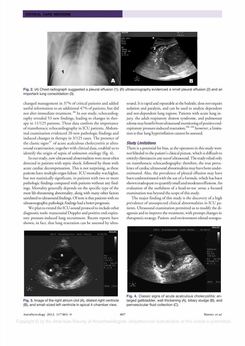
Ultrasound examination was effective in evidencing thepresence of anterior pneumothorax not detected by supineanteroposterior chest radiography (three cases). This finding holds clinical relevance, because during positive pressureventilation a small pneumothorax may progress and causehemodynamic instability. Ultrasound has proved to be moresensitive than anteroposterior chest radiography in the diag-nosis of pneumothorax 29,30 and can decrease the need forcomputed tomography for the diagnosis of occult pneumo-thorax.31 Lower lobe parenchymal consolidation without airbronchogram visualization can be difficult to distinguishfrom pleural effusion on an anteroposterior chest radio-graph.32 Ultrasonography showing consolidation (with or without pleural effusion) can help in avoiding a possiblemistake because of a misread chest radiograph (fig. 2).
Lung sonography is a useful aid in differentiating cardio-genic respiratory failure from acute airflow limitation, con-solidation, pleural effusion, or pulmonary embolism.33–35 Inour study, the diagnostic efficacy of lung ultrasound to dif-ferentiate dyspneic patients is well represented. Lung ultra-sound pointed out 55 new findings, enabling us to differen-tiate the etiologic diagnosis in patients with a genericadmitting diagnosis of acute respiratory insufficiency. Com-bining the data from lung sonography and echocardiography can enhance the diagnostic accuracy in differentiating respi-ratory insufficiency (fig. 3).
In a previous study, transthoracic echocardiography per-
formed by intensivists provided new information and
Fig. 1. Internal jugular vein thrombosis (A) in a young patient
with varicella pneumoniae and adult respiratory distress syn-
drome. Thrombophilia workup revealed anti-2 glycoprotein
I positivity.
Effectiveness of a Critical Care Ultrasound Protocol
Anesthesi ology 2012; 117:801–9 Manno et al.806
7/30/2019 eco impacto
http://slidepdf.com/reader/full/eco-impacto 7/9
changed management in 37% of critical patients and addeduseful information to an additional 47% of patients, but didnot alter immediate treatment.36 In our study, echocardiog-raphy revealed 53 new findings, leading to changes in ther-apy in 11/125 patients. These data confirm the importanceof transthoracic echocardiography in ICU patients. Abdom-inal examination evidenced 20 new pathologic findings andinduced changes in therapy in 3/125 cases. The presence of the classic signs37 of acute acalculous cholecystitis at ultra-sound examination, together with clinical data, enabled us toidentify the origin of sepsis of unknown etiology (fig. 4).
In our study, new ultrasound abnormalities were most oftendetected in patients with septic shock, followed by those withacute cardiac decompensation. This is not surprising, as thesepatients have multiple organ failure. ICU mortality was higher,but not statistically significant, in patients with two or more
pathologic findings compared with patients without any find-ings. Mortality generally depends on the specific type of themost life-threatening abnormality, along with many other factorsunrelated to ultrasound findings. Of note is that patients with noultrasonographic pathologic findings had a better prognosis.
We plan to extend the ICU-sound protocol to include otherdiagnostic tools: transcranial Doppler and positive end-expira-tory pressure-induced lung recruitment. Recent reports haveshown, in fact, that lung reaeration can be assessed by ultra-
sound. It is rapid and repeatable at the bedside, does not requiresedation and paralysis, and can be used to analyze dependentand not-dependent lung regions. Patients with acute lung in- jury, the adult respiratory distress syndrome, and pulmonary edema maybenefitfromultrasound monitoring of positiveend-expiratory pressure-induced reaeration;38–40 however, a limita-tion is that lung hyperinflation cannot be assessed.
Study Limitations
There is a potential for bias, as the operators in this study werenot blinded to the patient’s clinical picture, which is difficult toentirely eliminatein any useof ultrasound. The study relied only on transthoracic echocardiography; therefore, the true preva-lence of cardiac ultrasound abnormalities may havebeen under-estimated. Also, the prevalence of pleural effusion may havebeen underestimated with the use of a formula, which has been
showninadequate to quantifysmall andmoderateeffusions. Anevaluation of the usefulness of a head-to-toe versus a focusedexamination was beyond the scope of this study.
The major finding of this study is the discovery of a highprevalence of unsuspected clinical abnormalities in ICU pa-tients. Ultrasound examination permitted us to modify the di-agnosis and to improve the treatment, with prompt changes intherapeuticstrategy. Patient- and environment-related sonogra-
Fig. 2. ( A ) Chest radiograph suggested a pleural effusion (1); ( B ) ultrasonography evidenced a small pleural effusion (2) and an
important lung consolidation (3).
Fig. 3. Image of the right atrium clot (A), dilated right ventricle
(B), and small-sized left ventricle in apical 4-chamber view.
Fig. 4. Classic signs of acute acalculous cholecystitis: en-
larged gallbladder, wall thickening (A), biliary sludge (B), and
perivescicular fluid collection (C).
CRITICAL CARE MEDICINE
Anesthesi ology 2012; 117:801–9 Manno et al.807

7/30/2019 eco impacto
http://slidepdf.com/reader/full/eco-impacto 8/9
phy limitations had little influence on themajority of the exam-inations. The test can be performed by the attending ICU phy-sician, with minimal risk of overuse or misinterpretation.Moreover, ultrasound use in the ICU could be optimized by making ultrasonography a routine part of intensive care training
during residency.
41
In experienced hands, rapid global assessmentof critical patients at ICU admittance under this ultrasound proto-col holds potential for improving healthcare quality.
References1. Fedson S, Neithardt G, Thomas P, Lickerman A, Radzienda M,
DeCara JM, Lang RM, Spencer KT: Unsuspected clinically important findings detected with a small portable ultrasounddevice in patients admitted to a general medicine service.
J Am Soc Echocar diogr 2003; 16:901–5
2. Bossone E, DiGiovine B, Watts S, Marcovitz PA, Carey L, Watts C, Armstron g WF: Range and prevalen ce of cardiacabnormalities in patients hospitalized in a medical ICU.Chest 2002; 122:1370–6
3. Crone-Munzebrock W, Wegener C, Nicolas V, Pothmann W:The relevance of sonography in the diagnosis of acute abdo-men in the intensive care unit. Rofo 1990; 153:379–84
4. Schacherer D, Klebl F, Goetz D, Buettner R, Zierhut S,Schoelmerich J, Langgartner J: Abdominal ultrasound in theintensive care unit: A 3-year survey on 400 patients. Inten-sive Care Med 2007; 33:841–4
5. Rozycki GS, Pennington SD, Feliciano DV: Surgeon-per-formed ultrasound in the critical care setting: Its use as anextension of the physical examination to detect pleural ef-fusion. J Trauma 2001; 50:636–42
6. Lichtenstein D, Axler O: Intensive use of general ultrasoundin the intensive care unit. Prospective study of 150 consec-utive patients. Intensive Care Med 1993; 19:353–5
7. Le Gall JR, Lemeshow S, Saulnier F: A new Simplified Acute
Physiology Score (SAPS II) based on a European/North Amer-ican multicenter study. JAMA 1993; 270:2957–63
8. Cohen J: A coefficient of agreement for nominal scales. EducPsychol Meas 1960; 20:37–46
9. Mayo PH, Beaulieu Y, Doelken P, Feller-Kopman D, HarrodC, Kaplan A, Oropello J, Vieillard-Baron A, Axler O, Lichten-stein D, Maury E, Slama M, Vignon P: American College of Chest Physicians/La Societ e de Reanimation de Langue Fran-caise statement on competence in critical care ultrasonogra-phy. Chest 2009; 135:1050–60
10. Neri L, Storti E, Lichtenstein D: Toward an ultrasound cur-riculum for critical care medicine. Crit Care Med 2007;35:S290–304
11. Marik PE, Mayo P: Certification and training in critical careultrasound. Intensive Care Med 2008; 34:215–7
12. Mateer J, Plummer D, Heller M, Olson D, Jehle D, Overton D,Gussow L: Model curriculum for physician training in emer-gency ultrasonography. Ann Emerg Med 1994; 23:95–102
13. Geeraerts T, Launey Y, Martin L, Pottecher J, Vigue B, Duranteau J, Benhamou D: Ultrasonography of the optic nerve sheath may beuseful for detecting raised intracranial pressure after severe braininjury. Intensive Care Med 2007; 33:1704–11
14. Kimberly HH, Shah S, Marill K, Noble V: Correlation of opticnerve sheath diameter with direct measurement of intracra-nial pressure. Acad Emerg Med 2008; 15:201–4
15. Blaivas M, Theodoro D, Sierzenski PR: Elevated intracra-nial pressure detected by bedside emergency ultrasonog-raphy of the optic nerve sheath. Acad Emerg Med 2003;10:376–81
16. Mustafa S, Stein PD, Patel KC, Otten TR, Holmes R, Silbergleit A: Upper extremity deep venous thrombosis. Chest 2003;123:1953–6
17. Cronan JJ: Venous thromboembolic disease: The role of US.Radiology 1993; 186:619–30
18. Monreal M, Lafoz E, Ruiz J, Valls R, Alastrue A: Upper-extremity deep venous thrombosis and pulmonary embo-lism. A prospective study. Chest 1991; 99:280–3
19. Mathis G, Blank W, Reissig A, Lechleitner P, Reuss J, Schuler A, Beckh S: Thoracic ultrasound for diagn osing pulmonary
embolism: A prospective multicenter study of 352 patients.Chest 2005; 128:1531–8
20. Mansencal N, Vieillard-Baron A, Beauchet A, Farcot JC, ElHajjam M, Dufaitre G, Brun-Ney D, Lacombe P, Jardin F,Dubourg O: Triage patients with suspected pulmonary em-bolism in the emergency department using a portable ultra-sound device. Echocardiography 2008; 25:451–6
21. McLoud TC, Flower CD: Imaging the pleura: Sonography, CT, andMR imaging. AJR Am J Roentgenol 1991; 156:1145–53
22. Lichtenstein D, Goldstein I, Mourgeon E, Cluzel P, Grenier P,Rouby JJ: Comparative diagnostic performances of auscultation,chest radiography, and lung ultrasonography in acute respiratory distress syndrome. A NESTHESIOLOGY 2004; 100:9–15
23. Fartoukh M, Azoulay E, Galliot R, Le Gall JR, Baud F, ChevretS, Schlemmer B: Clinically documented pleural effusions in
medical ICU patients: How useful is routine thoracentesis?Chest 2002; 121:178–84
24. Mattison LE, Coppage L, Alderman DF, Herlong JO, Sahn SA:Pleural effusions in the medical ICU: Prevalence, causes, andclinical implications. Chest 1997; 111:1018–23
25. Balik M, Plasil P, Waldauf P, Pazout J, Fric M, Otahal M, Pachl J: Ultras ound estimation of volume of pleura l fluid in me-chanically ventilated patients. Intensive Care Med 2006; 32:318–2
26. Remerand F, Dellamonica J, Mao Z, Ferrari F, Bouhemad B, Jianxi n Y, Arbelot C, Lu Q, Ichaï C, Rouby JJ: Multiplaneultrasound approach to quantify pleural effusion at the bed-side. Intensive Care Med 2010; 36:656–4
27. Doelken P, Abreu R, Sahn SA, Mayo PH: Effect of thoracen-tesis on respiratory mechanics and gas exchange in the
patient receiving mechanical ventilation. Chest 2006; 130:1354–61
28. Kopterides P, Lignos M, Papanikolaou S, Papadomichelakis E,Mentzelopoulos S, Armaganidis A, Panou F: Pleural effusioncausing cardiac tamponade: Report of two cases and review of literature. Heart Lung 2006; 35:66–7
29. Blaivas M, Lyon M, Duggal S: A prospective comparison of supinechest radiography and bedside ultrasound for the diagnosis of traumatic pneumothorax. Acad Emerg Med 2005; 12:844–9
30. Zhang M, Liu ZH, Yang JX, Gan JX, Xu SW, You XD, JiangGY: Rapid detection of pneumothorax by ultrasonogra-phy in patients with multiple trauma. Crit Care 2006;10:R112
31. Lichtenstein DA, Meziere G, Lascols N, Biderman P, Courret JP,Gepner A, Goldstein I, Tenoudji-Cohen M: Ultrasound diagnosis of occult pneumothorax. Crit Care Med 2005; 33:1231–8
32. Woodring JH: Recognition of pleural effusion on supineradiographs: How much fluid is required? AJR Am J Roent-genol 1984; 142:59–64
33. Lichtenstein DA, Meziere G: Relevance of lung ultrasound i nthe diagnosis of acute respiratory failure: The BLUE protocol.Chest 2008; 134:117–25
34. Volpicelli G, Cardinale L, Garofalo G, Veltri A: Usefulness of lungultrasound in the bedside distinction between pulmonary edemaand exacerbation of COPD. Emerg Radiol 2008; 15:145–51
35. Reissig A, Copetti R, Kroegel C: Current role of emergency ultrasound of the chest. Crit Care Med 2011; 39:839–45
36. Manasia AR, Nagaraj HM, Kodali RB, Croft LB, Oropello JM,Kohli-Seth R, Leibowitz AB, DelGiudice R, Hufanda JF, Ben-
jamin E, Goldman ME: Feasibi lity and potential clinical utili ty of goal-directed transthoracic echocardiography performedby noncardiologist intensivists using a small hand-carried
Effectiveness of a Critical Care Ultrasound Protocol
Anesthesi ology 2012; 117:801–9 Manno et al.808

7/30/2019 eco impacto
http://slidepdf.com/reader/full/eco-impacto 9/9
device (SonoHeart) in critically ill patients. J Cardiothorac Vasc Anesth 2005; 19:155–9
37. Molenat F, Boussuges A, Valantin V, Sainty JM: Gallbladder abnormalities in medical ICU patients: An ultrasonographicstudy. Intensive Care Med 1996; 22:356–8
38. Liteplo AS, Murray AF, Kimberly HH, Noble VE: Real-timeresolution of sonographic B-lines in a patient with pulmo-
nary edema on continuous positive airway pressure. Am JEmerg Med 2010; 28:541.e5–8
39. Via G, Lichtenstein D, Mojoli F, Rodi G, Neri L, Storti E,
Klersy C, Iotti G, Braschi A: Whole lung lavage: A uniquemodel for ultrasound assessment of lung aeration changes.Intensive Care Med 2010; 36:999 –1007
40. Bouhemad B, Brisson H, Le-Guen M, Arbelot C, Lu Q, Rouby JJ: Bedside ultras ound assessment of positi ve end-expi ratory pressure-induced lung recruitment. Am J Respir Crit CareMed 2011; 183:341–7
41. Eisen LA, Leung S, Gallagher AE, Kvetan V: Barriers to ultrasoundtraining in critical care medicine fellowships: A survey of programdirectors. Crit Care Med 2010; 38:1978–83
ANESTHESIOLOGY REFLECTIONS FROM THE WOOD LIBRARY-MUSEUM
Cocaine/Eucaine Sales Register: Deja Vu or Tete-beche?
If this image of a Cocaine and Eucaine Register of Sales seems like deja vu, that is because it is the
“reverse” or “upside-down” book built into the opposite pagings of the Cocaine and Eucaine Register of
Purchases, which was featured as one of our September 2012 Anesthesiology Reflections. Such an
unusual binding is called a tete-beche (French for “head-to-toe”). In other words, if you start at the front
cover of the Purchases, read right-hand pages to the last page, close the book to reveal the back cover,
and then rotate the book 180 degrees, you will see that Purchase’s back cover is actually the upside-
down front cover of Sales. Now if you were a librarian at the Wood Library-Museum, how would you
shelve Sales—upside up or upside down? (Copyright © the American Society of Anesthesiologists, Inc.)
George S. Bause, M.D., M.P.H., Honorary Curator, ASA’s Wood Library-Museum of Anesthesiology,
Park Ridge, Illinois, and Clinical Associate Professor, Case Western Reserve University, Cleveland, Ohio.
CRITICAL CARE MEDICINE
Anesthesi ology 2012; 117:801–9 Manno et al.809