disruption of semantic network in mild alzheimer’s...
TRANSCRIPT

Disruption of semantic metwork in mild Alzheimer's disease revealed by restingstate fMRI
Article (Published Version)
http://sro.sussex.ac.uk
Mascali, Daniele, DiNuzzo, Mauro, Serra, Laura, Mangia, Silvia, Maraviglia, Bruno, Bozzali, Marco and Giove, Federico (2018) Disruption of semantic metwork in mild Alzheimer's disease revealed by resting-state fMRI. Neuroscience, 371. pp. 38-48. ISSN 1873-7544
This version is available from Sussex Research Online: http://sro.sussex.ac.uk/id/eprint/73749/
This document is made available in accordance with publisher policies and may differ from the published version or from the version of record. If you wish to cite this item you are advised to consult the publisher’s version. Please see the URL above for details on accessing the published version.
Copyright and reuse: Sussex Research Online is a digital repository of the research output of the University.
Copyright and all moral rights to the version of the paper presented here belong to the individual author(s) and/or other copyright owners. To the extent reasonable and practicable, the material made available in SRO has been checked for eligibility before being made available.
Copies of full text items generally can be reproduced, displayed or performed and given to third parties in any format or medium for personal research or study, educational, or not-for-profit purposes without prior permission or charge, provided that the authors, title and full bibliographic details are credited, a hyperlink and/or URL is given for the original metadata page and the content is not changed in any way.

NEUROSCIENCE
RESEARCH ARTICLED. Mascali et al. / Neuroscience 371 (2018) 38–48
Disruption of Semantic Network in Mild Alzheimer’s Disease Revealedby Resting-State fMRI
Daniele Mascali, a* Mauro DiNuzzo, b,a Laura Serra, c Silvia Mangia, d Bruno Maraviglia, a,c Marco Bozzali c andFederico Giove a,c
aCentro Fermi – Museo Storico della Fisica e Centro Studi e Ricerche ‘‘Enrico Fermi”, Piazza del Viminale 1, 00184 Rome, Italy
bCenter for Basic and Translational Neuroscience, Division of Glial Disease and Therapeutics, Faculty of Health and Medical Sciences,
University of Copenhagen, Blegdamsvej 3B, 2200 Copenhagen, Denmark
cFondazione Santa Lucia IRCCS, Via Ardeatina 306, 00142 Rome, Italy
dCenter for Magnetic Resonance Research, Department of Radiology, University of Minnesota, 2021 6th ST SE, Minneapolis, MN 55455, United
States
Abstract—Subtle semantic deficits can be observed in Alzheimer’s disease (AD) patients even in the early stagesof the illness. In this work, we tested the hypothesis that the semantic control network is deregulated in mild ADpatients. We assessed the integrity of the semantic control system using resting-state functional magnetic reso-nance imaging in a cohort of patients with mild AD (n = 38; mean mini-mental state examination = 20.5) and in agroup of age-matched healthy controls (n= 19). Voxel-wise analysis spatially constrained in the left fronto-temporal semantic control network identified two regions with altered functional connectivity (FC) in AD patients,specifically in the pars opercularis (POp, BA44) and in the posterior middle temporal gyrus (pMTG, BA21). Usingwhole-brain seed-based analysis, we demonstrated that these two regions have altered FC even beyond thesemantic control network. In particular, the pMTG displayed a wide-distributed pattern of lower connectivity toseveral brain regions involved in language-semantic processing, along with a possibly compensatory higher con-nectivity to the Wernicke’s area. We conclude that in mild AD brain regions belonging to the semantic control net-work are abnormally connected not only within the network, but also to other areas known to be critical forlanguage processing. � 2017 The Author(s). Published by Elsevier Ltd on behalf of IBRO. This is an open access article under
the CC BY-NC-ND license (http://creativecommons.org/licenses/by-nc-nd/4.0/).
Key words: Alzheimer’s disease, semantic control network, posterior middle temporal gyrus, inferior frontal gyrus, resting-state
fMRI, voxel-wise functional connectivity.
INTRODUCTION
The hallmark of Alzheimer’s disease (AD) has been long
recognized to be a profound deficit in episodic memory.
However, language dysfunctions with a semantic basis
are also observed in AD patients (Kempler, 1995) even
at predementia stages (Mickes et al., 2007; Amieva
et al., 2008; Taler and Phillips, 2008). The early pattern
of language deterioration in AD is quite specific and char-
acterized by a predominant semantic impairment with a
https://doi.org/10.1016/j.neuroscience.2017.11.0300306-4522/� 2017 The Author(s). Published by Elsevier Ltd on behalf of IBRO.This is an open access article under the CC BY-NC-ND license (http://creativecomm
*Corresponding author.
E-mail address: [email protected] (D. Mascali).Abbreviations: AD, Alzheimer’s disease; BOLD, blood-oxygen-level-dependent; CSF, Cerebral Spinal Fluid; EPI, echo planar imaging; FC,functional connectivity; FD, framewise displacement; fMRI, functionalmagnetic resonance imaging; FWHM, full width at half maximum; GLM,general linear model; GM, gray matter; GMV, gray matter volume; HC,healthy controls; MMSE, mean mini-mental state examination; MNI,Montreal Neurological Institute; pMTG, posterior middle temporalgyrus; POp, pars opercularis; TMS, transcranial magnetic stimulation;wGBC, weighted Global Brain Connectivity; WM, White Matter; wRBC,weighted Regional Brain Connectivity.
38
relatively sparing of other language features, such as syn-
tax or phonology (Kirshner, 2012). The most evident
symptom is a word-finding difficulty which can be
observed in spontaneous speech (Nicholas et al., 1985)
as well as in language standardized tests (e.g., confronta-
tion naming and verbal fluency; Henry et al., 2004). While
the neural substrates underlying episodic memory impair-
ment in AD have been extensively studied, the role of
semantic memory still remains poorly investigated. PET
studies indicated that frontal and lateral temporal regions
are implicated in semantic deterioration in AD (Zahn et al.,
2004; Teipel et al., 2006; Nelissen et al., 2007; Melrose
et al., 2009), yet the question arises of which component
of semantic cognition is being affected by the pathology.
Semantic cognition is theorized to include the storage
of conceptual knowledge and the process by which such
knowledge is manipulated in a time, context, and task
appropriate fashion (Tulving, 1987). These two compo-
nents (i.e., storage and control) are thought to be sub-
served by two distinct but interactive neuronal systems
(Jefferies, 2013; Ralph et al., 2017). The conceptual
ons.org/licenses/by-nc-nd/4.0/).

D. Mascali et al. / Neuroscience 371 (2018) 38–48 39
knowledge is controlled by a wide-distributed network
composed of sensorimotor and verbal-related areas that
store object-specific features and lexical information
(Damasio et al., 2004; Martin, 2007), along with an amo-
dal hub, located in the bilateral anterior temporal lobe, act-
ing as a convergence zone (Patterson et al., 2007). On
the contrary, the semantic control is supported by a left-
lateralized fronto-temporal and possibly parietal network
(Jefferies and Ralph, 2006; Noonan et al., 2010). In partic-
ular, the left inferior frontal gyrus (pars opercularis and tri-
angularis), and the left posterior middle temporal gyrus
(pMTG) are considered the most relevant regions in the
semantic control network (Whitney et al., 2011a,b;
Jefferies, 2013; Krieger-Redwood and Jefferies, 2014).
However, other regions have been implicated in semantic
control, including the dorsal angular gyrus (Noonan et al.,
2013) and the posterior cingulate cortex (Binder et al.,
2009), although their exact role needs to be clarified.
Neuropsychological research about the nature of
semantic deficits in AD has provided conflicting results,
pointing to either a degraded conceptual knowledge
(Hodges et al., 1992; Garrard et al., 2005; Lin et al.,
2014) or a deregulated control/access to this information
(Bayles et al., 1991; Nebes and Halligan, 1996). The
apparent inconsistency among neuropsychological stud-
ies could be related to disease severity (Bayles et al.,
1991; Duong et al., 2006). This notion is supported by a
recent study that examined how different stages of the
pathology affect distinct components of semantic cogni-
tion (Corbett et al., 2012). Specifically, in the mild stage
(mean mini-mental state examination, MMSE � 20)
patients showed impairments distinctive of deregulated
control of semantic information, whereas in the severe
stage (mean MMSE � 10) this problem, besides getting
worse, became compounded by degradation of semantic
representations (Corbett et al., 2012). The pathophysio-
logical counterpart of the reported pattern of semantic
impairment in AD is expected to be a functional alteration
in the semantic control system, which should be visible
early in the disease progression. To the best of our knowl-
edge, such hypothesis has not been tested yet.
Here we adopted resting-state functional magnetic
resonance imaging (rsfMRI), based on the blood-oxygen-
level-dependent (BOLD) signal, to examine, for the first
time, the functional connectivity (FC) integrity of the
semantic control network in patients with AD at mild stage.
Compared to task-based fMRI, the resting-state design
has the advantage of not relying on any task choice. This
feature is particularly important for dementia subjects in
which uncontrolled familiarity of stimulus concepts and/or
task demands might represent critical, and not
manageable, confounds (Bayles et al., 1991). Our main
hypothesis was that mild AD patients are characterized by
altered FC within the semantic control network. Secondly,
we hypothesized that such alteration correlates with lan-
guage impairment (verbal fluency and confrontation nam-
ing). Finally, given the continuous interplay between the
semantic control and the wide-distributed representation
network, we expected that affected regions in the semantic
control network would present FC abnormalities in other
semantic-related regions.
EXPERIMENTAL PROCEDURES
Subjects
A cohort of 38 right-handed patients with probable AD-
typical were recruited for the current study. The
diagnosis of probable AD was performed according to
the clinical criteria of the National Institute of
Neurological and Communicative Disorders and Stroke-
Alzheimer’s Disease and Related Disorders Association
(NINCDS-ADRDA, McKhann et al., 2011). To be
included, patients had to meet the Diagnostic and Statis-
tical Manual of Mental Disorders (DSM-V) criteria
(American Psychiatric Association, 2013) for the diagno-
sis of major neurocognitive disorders due to AD. An
expert neurologist (M.B.) in each recruited patient
reviewed carefully the clinical history, the cognitive profile
and the conventional MRI scan and excluded the vascular
signs and symptoms associated typically with vascular
dementia. Nineteen right-handed healthy elderly individu-
als were also recruited and served as healthy controls
(HC). All healthy subjects reported scores within the
range of normality at the Mini Mental State Examination
(MMSE, Italian cut-off >23.8; Folstein et al., 1975;
Magni et al., 1996). Major systemic, psychiatric, vascular
and other neurological illnesses were carefully investi-
gated and excluded in all recruited subjects. The study
was carried out in accordance with a protocol approved
by the Ethics Committee of Santa Lucia Foundation. All
recruited subjects gave written informed consent in accor-
dance with the Declaration of Helsinki and European
Union regulations.
Neuropsychological assessment
All participants underwent a neuropsychological battery
covering several cognitive domains, which included: (1)
verbal episodic long-term memory: 15-Word List
(Immediate and 15-min Delayed recall; Carlesimo et al.,
1996); Short Story Test (Immediate and 20-min Delayed
recall; Carlesimo et al., 2002); (2) visuo-spatial long-
term memory: Complex Rey’s Figure (Immediate and
20-min Delayed recall; Carlesimo et al., 2002); (3) short-
term memory: Digit span and the Corsi Block Tapping
task (Monaco et al., 2013); (4) executive functions:
Phonological Word Fluency (Carlesimo et al., 1996) and
Modified Card Sorting Test (Nocentini et al., 2002); (5)
language: Naming objects subtest of the BADA (‘‘Batteria
per l’Analisi dei Deficit Afasici”, Italian for ‘‘Battery for the
analysis of aphasic deficits”; Miceli, 1994); (6) reasoning:
Raven’s Coloured Progressive Matrices (Carlesimo et al.,
1996); (7) constructional praxis: copy of simple drawings
with and without landmarks (Carlesimo et al., 1996) and
copy of Complex Rey’s Figure (Carlesimo et al., 2002).
Data acquisition
Data were acquired on a 3 T MRI system (Magnetom
Allegra, Siemens, Erlangen, Germany). All subjects
underwent a resting-state fMRI scan using an echo
planar imaging (EPI) sequence with the following
parameters: TR = 2080 ms, TE = 30 ms, 32 axial
slices parallel to AC-PC plane, matrix = 64 � 64, in

40 D. Mascali et al. / Neuroscience 371 (2018) 38–48
plane resolution = 3 � 3 mm2, slice thickness = 2.5 mm,
50% skip, flip angle = 70�. The slices were positioned
starting from the vertex and covering the whole
cerebrum. The cerebellum did not consistently fall in the
field of view of each acquired subject, thus, it was
excluded from any subsequent analysis. Resting scans
lasted for 7 min and 20 s for a total of 220 volumes
during which subjects were instructed to keep their eyes
closed, to not think of anything in particular and to
refrain from falling asleep. Immediately after the scan,
subjects were interrogated and asked for compliance.
No subject showed any behavior sign suggestive of
sleeping. A T1-weighted three-dimensional modified
driven equilibrium Fourier transform scan (MDEFT,
Deichmann et al., 2004) was acquired for each subject
for anatomical localization purposes and for gray matter
(GM) volumetry; the parameters were as follows:
TR = 1338 ms, TE = 2.4 ms, TI = 910 ms, flip angle
= 15�, matrix = 256 � 224 � 176, FOV= 256 � 224
mm2, slice thickness = 1 mm, total scan time = 12 min.
Fluid attenuated inversion recovery (FLAIR) images
(TR = 8170 ms, TE = 96 ms, TI = 2100 ms) were also
acquired from all subjects to exclude the presence of
remarkable signs suggestive of cerebro-vascular disease.
No subject was considered affected by cerebro-vascular
pathology based on previously described criteria (Serra
et al., 2010).
Data preprocessing
Functional and structural MRI data were preprocessed
using CONN 15.b: functional connectivity toolbox
(Whitfield-Gabrieli and Nieto-Castanon, 2012; http://
www.nitrc.org/projects/conn). For each subject, the first
four volumes of the EPI time series were discarded to
allow for signal and scanner stabilization. Realignment
and slice-time correction were implemented to compen-
sate for head movements and slice-dependent time shifts,
respectively. Additionally, to reduce the movement-
related residual variance induced by the susceptibility-
by-movements interaction effect, the unwarp algorithm
was applied (Andersson et al., 2001). Then, functional
volumes were spatially normalized into Montreal Neuro-
logical Institute (MNI) coordinates (voxel size: 2 � 2 � 2
mm3) using as source image the EPI mean volume
obtained from the realignment step. Unless otherwise
specified, normalized images were smoothed applying
an 8 � 8 � 8 mm3 full width at half maximum (FWHM)
Gaussian Kernel. Separately, the T1 weighted high-
resolution volumes were segmented and normalized to
MNI space to obtain GM, White Matter (WM) and Cere-
bral Spinal Fluid (CSF) tissue probability maps. WM and
CSF segments were used for the removal of confounding
effects from fMRI data as detailed below. Additionally, GM
segments were post-processed as previously described
(Mascali et al., 2015) in order to obtain, for each subject,
the GM volume (GMV) to be used as covariate of no inter-
est in statistical group comparisons.
Additional preprocessing steps were applied to
functional data for mitigating the effect of various
spurious sources of variance. These included despiking
and application of a 0.008–0.09-Hz band-pass temporal
filter simultaneously to the regression out, via a general
linear model (GLM), of the following confounds: (1) a
linear trend; (2) the six parameters of realignment and
their first derivatives; (3) the first five eigenvectors of the
PCA decomposition of the EPI time series separately
averaged over WM and CSF, following the aCompCor
noise removal approach (Behzadi et al., 2007); (4) the
outlier volumes detected using the Artifact Detection
Tools (ART: www.nitrc.org/projects/artifact_detect/). The
simultaneous filter/regression was achieved by band-
pass filtering both the fMRI time series and the con-
founds, prior to the regression in the GLM. This approach
slightly outperforms the more common regression fol-
lowed by filter (Hallquist et al., 2013).
To assess the amount of head motion during
functional scans we computed the framewise
displacement (FD) as defined in (Power et al., 2012).
Functional connectivity
The residual BOLD time series resulting from the
preprocessing were used to compute two different types
of FC metrics, namely, a voxel-wise and a seed-based
metric.
First, a spatially constrained version of the weighted
Global Brain Connectivity (wGBC; Cole et al., 2010;
Mascali et al., 2015) was computed to investigate the
voxel-wise FC of the semantic control network. wGBC
can be employed to quantify the average FC of every
voxel to all other voxels within the brain or in a specific
mask. To differentiate between the whole-brain versus
the spatially constrained version of this quantity, we refer
to the masked version, here adopted, as weighted Regio-
nal Brain Connectivity (wRBC). Mathematically, given the
BOLD time course of the i-th voxel in the mask, the wRBC
of that voxel is defined as the weighted average of the
Pearson correlation, r, computed for every other time
course in the mask:
wRBCi ¼ 1
N
XN
j¼1
wðrði; jÞÞ i; j 2 mask
where the weighting function is the z-Fisher
transformation, wðrÞ ¼ 12ln 1þr
1�r
� �. As in the original wGBC
definition, the wRBC encompasses both positive and
negative correlation values. The chosen mask was
composed of key regions of the semantic control
network (Jefferies, 2013) extracted from the Harvard-
Oxford probabilistic atlas (Desikan et al., 2006). The
included regions were the left inferior frontal gyrus (pars
opercularis and triangularis) and the left pMTG (posterior
and temporooccipital parts), which have been consistently
implicated in semantic control (Indefrey and Levelt, 2004;
Whitney et al., 2011a,b; Krieger-Redwood and Jefferies,
2014). We limited the calculation to this network because
the wGBC (i.e., the whole-brain version) has an intrinsi-
cally poor sensitivity if the effect is localized (due to mutual
correlations among functionally heterogeneous voxels)
and it is also prone to include the effect of possible com-
pensatory mechanisms. For the same reason, the limita-
tion to the most relevant regions of the semantic control
network improves the specificity of the wRBC metric.
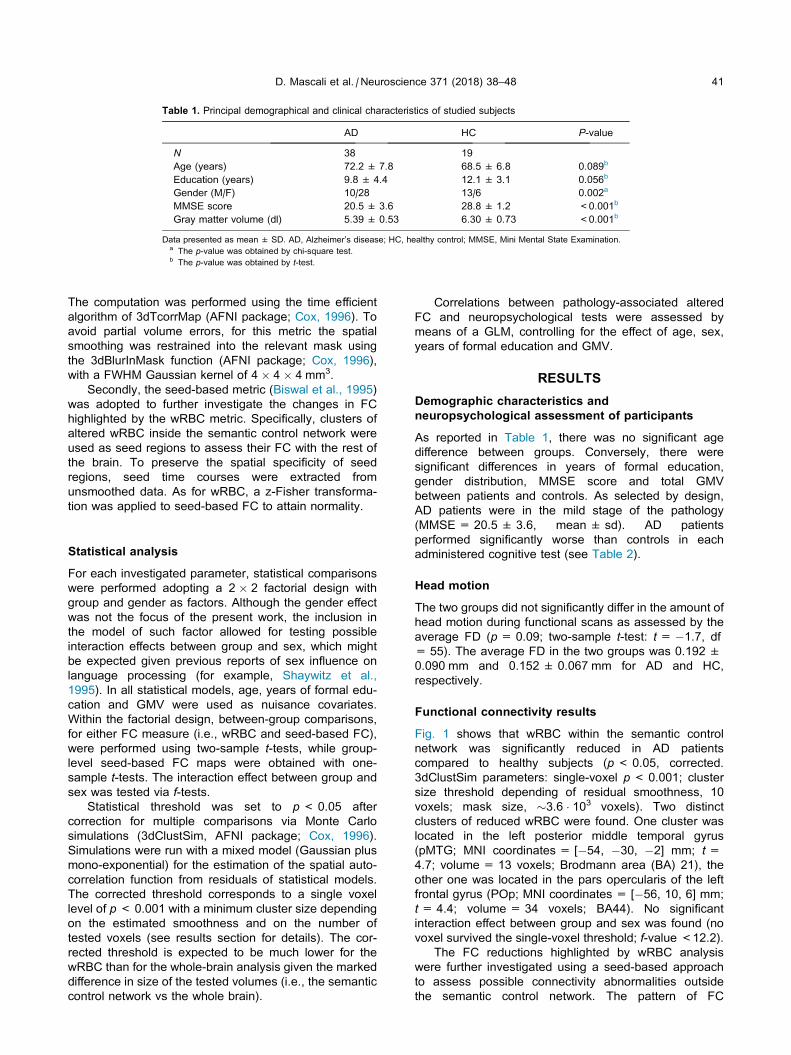
Table 1. Principal demographical and clinical characteristics of studied subjects
AD HC P-value
N 38 19
Age (years) 72.2 ± 7.8 68.5 ± 6.8 0.089b
Education (years) 9.8 ± 4.4 12.1 ± 3.1 0.056b
Gender (M/F) 10/28 13/6 0.002a
MMSE score 20.5 ± 3.6 28.8 ± 1.2 <0.001b
Gray matter volume (dl) 5.39 ± 0.53 6.30 ± 0.73 <0.001b
Data presented as mean ± SD. AD, Alzheimer’s disease; HC, healthy control; MMSE, Mini Mental State Examination.a The p-value was obtained by chi-square test.b The p-value was obtained by t-test.
D. Mascali et al. / Neuroscience 371 (2018) 38–48 41
The computation was performed using the time efficient
algorithm of 3dTcorrMap (AFNI package; Cox, 1996). To
avoid partial volume errors, for this metric the spatial
smoothing was restrained into the relevant mask using
the 3dBlurInMask function (AFNI package; Cox, 1996),
with a FWHM Gaussian kernel of 4 � 4 � 4 mm3.
Secondly, the seed-based metric (Biswal et al., 1995)
was adopted to further investigate the changes in FC
highlighted by the wRBC metric. Specifically, clusters of
altered wRBC inside the semantic control network were
used as seed regions to assess their FC with the rest of
the brain. To preserve the spatial specificity of seed
regions, seed time courses were extracted from
unsmoothed data. As for wRBC, a z-Fisher transforma-
tion was applied to seed-based FC to attain normality.
Statistical analysis
For each investigated parameter, statistical comparisons
were performed adopting a 2 � 2 factorial design with
group and gender as factors. Although the gender effect
was not the focus of the present work, the inclusion in
the model of such factor allowed for testing possible
interaction effects between group and sex, which might
be expected given previous reports of sex influence on
language processing (for example, Shaywitz et al.,
1995). In all statistical models, age, years of formal edu-
cation and GMV were used as nuisance covariates.
Within the factorial design, between-group comparisons,
for either FC measure (i.e., wRBC and seed-based FC),
were performed using two-sample t-tests, while group-
level seed-based FC maps were obtained with one-
sample t-tests. The interaction effect between group and
sex was tested via f-tests.Statistical threshold was set to p< 0.05 after
correction for multiple comparisons via Monte Carlo
simulations (3dClustSim, AFNI package; Cox, 1996).
Simulations were run with a mixed model (Gaussian plus
mono-exponential) for the estimation of the spatial auto-
correlation function from residuals of statistical models.
The corrected threshold corresponds to a single voxel
level of p< 0.001 with a minimum cluster size depending
on the estimated smoothness and on the number of
tested voxels (see results section for details). The cor-
rected threshold is expected to be much lower for the
wRBC than for the whole-brain analysis given the marked
difference in size of the tested volumes (i.e., the semantic
control network vs the whole brain).
Correlations between pathology-associated altered
FC and neuropsychological tests were assessed by
means of a GLM, controlling for the effect of age, sex,
years of formal education and GMV.
RESULTS
Demographic characteristics andneuropsychological assessment of participants
As reported in Table 1, there was no significant age
difference between groups. Conversely, there were
significant differences in years of formal education,
gender distribution, MMSE score and total GMV
between patients and controls. As selected by design,
AD patients were in the mild stage of the pathology
(MMSE = 20.5 ± 3.6, mean ± sd). AD patients
performed significantly worse than controls in each
administered cognitive test (see Table 2).
Head motion
The two groups did not significantly differ in the amount of
head motion during functional scans as assessed by the
average FD (p= 0.09; two-sample t-test: t= �1.7, df
= 55). The average FD in the two groups was 0.192 ±
0.090 mm and 0.152 ± 0.067 mm for AD and HC,
respectively.
Functional connectivity results
Fig. 1 shows that wRBC within the semantic control
network was significantly reduced in AD patients
compared to healthy subjects (p< 0.05, corrected.
3dClustSim parameters: single-voxel p< 0.001; cluster
size threshold depending of residual smoothness, 10
voxels; mask size, �3.6 � 103 voxels). Two distinct
clusters of reduced wRBC were found. One cluster was
located in the left posterior middle temporal gyrus
(pMTG; MNI coordinates = [�54, �30, �2] mm; t =4.7; volume = 13 voxels; Brodmann area (BA) 21), the
other one was located in the pars opercularis of the left
frontal gyrus (POp; MNI coordinates = [�56, 10, 6] mm;
t= 4.4; volume = 34 voxels; BA44). No significant
interaction effect between group and sex was found (no
voxel survived the single-voxel threshold; f-value <12.2).
The FC reductions highlighted by wRBC analysis
were further investigated using a seed-based approach
to assess possible connectivity abnormalities outside
the semantic control network. The pattern of FC
Table 2. Neuropsychological assessment of studied subjects
Cognitive domain Neuropsychological test Mean (SD) scores
AD HC
Verbal episodic long-term memory 15-Words List
Immediate recall (cut-off � 28.5) 15.8 (7.2)* 39.8 (11.4)
Delayed recall (cut-off � 4.6) 2.3 (5.5)* 8.3 (2.3)
Short story test
Immediate recall (cut-off � 3.1) 3.3 (6.2) 6.1 (1.4)
Delayed recall (cut-off � 2.6) 1.3 (2.0)* 6.0 (1.3)
Visuo-spatial episodic long-term memory Complex Rey’s Figure:
Immediate recall (cut-off � 6.4) 2.1 (2.9)* 14.3 (6.6)
Delayed recall (cut-off � 6.3) 1.9 (2.7)* 13.1 (5.9)
Verbal short-term memory Digit span forward (cut-off � 3.7) 15.1 (1.9)* 16.3 (1.3)
Visuo-spatial short-term memory Corsi span forward (cut-off � 3.5) 3.1 (1.5)* 4.9 (0.9)
Executive functions Phonological Word Fluency (cut-off � 17.3) 20.6 (11.0)* 37.6 (8.7)
Modified Card Sorting Test Criteria achieved (cut-off � 4.2) 1.6 (0.9)* 5.9 (0.3)
Language Naming of objects (cut-off � 22) 24.1 (5.7)* 29.1 (0.8)
Reasoning Raven’s Coloured Progressive Matrices (cut-off � 18.9) 18.8 (7.9)* 30.4 (3.9)
Constructional praxis Copy of drawings (cut-off � 7.1) 6.3 (3.9)* 10.6 (1.3)
Mini Mental State Examination (cut-off � 61.8) 50.4 (23.0)* 69.3 (1.0)
Copy of Complex Rey’s Figure (cut-off � 23.7) 18.0 (13.1)* 31.0 (4.3)
* One-way ANOVA p< 0.05.
42 D. Mascali et al. / Neuroscience 371 (2018) 38–48
between each seed region and the rest of the brain is
shown in Fig. 2 for patients and controls (p< 0.05,
corrected. 3dClustSim parameters: single-voxel p <
0.001; mask size, �2.2 � 105 voxels; cluster size
threshold depending on residual smoothness, 222 and
104 for POp and pMTG, respectively). In both groups
the POp cluster was connected to the orbito-frontal and
pre-frontal cortex bilaterally, and to the left parietal
cortex. The HC group was also connected to the left
temporal cortex. Conversely, when considering the
pMTG cluster as seed region, the HC group showed a
widespread pattern of connectivity involving bilateral
frontal and temporal regions and the left parietal cortex,
while AD patients showed a more restricted pattern
confined to the bilateral temporal lobes.
When comparing patients and controls, specific
patterns of altered FC were identified for POp and
pMTG clusters as shown in Fig. 3A, B, respectively (p
< 0.05, corrected as above). Patients showed reduced
FC to the POp cluster in brain regions mainly localized
within the semantic control network used for wRBC
computation, with the only exception of the bilateral
superior frontal gyrus. Conversely, regions of reduced
FC to the pMTG cluster extended beyond the semantic
control network, including several left-lateralized brain
regions (i.e., inferior frontal and anterior temporal
regions, the superior frontal gyrus and the angular
gyrus). The pMTG cluster was also disconnected from
the right hemisphere in the temporal pole. In addition,
increased connectivity to the pMTG cluster was found
within the Wernicke’s area (i.e., the left planum
temporale and in the left parietal operculum) of AD
patients compared to HC. Detailed cluster information is
reported in Table 3. No significant interaction effect
between group and sex was found (p> 0.5 and p >
0.2, for POp and pMTG, respectively, corrected as
above. f-value <12.2).
Finally, there was no significant correlation between
FC outcomes and neuropsychological tests (p > 0.05).
DISCUSSION
Using resting-state fMRI, here we aimed at characterizing
the brain functional correlates of semantic impairment in
AD. We thus focused on identifying functional
abnormalities in language-semantic-related regions
during the mild stage of the illness. The main result was
a reduction of FC in brain areas critically involved in
language/semantic processing in AD patients compared
to controls.
Altered connectivity within the semantic controlnetwork
Based on the neuropsychological-supported hypothesis
of early deterioration of the semantic control system in
AD (Corbett et al., 2012), we assessed the internal con-
nectivity of the semantic control network adopting a con-
strained voxel-wise FC metric, namely a modified
version of wGBC (Cole et al., 2010) that we refer to as
wRBC. Restricting the computation to the semantic con-
trol network, we were able to increase the sensitivity of
the metric, allowing the detection of even subtle changes
in FC.
In agreement with our main hypothesis, we observed
pathology-associated reductions of wRBC in the
semantic control network (Fig. 1). Degraded connectivity
in this network is consistent with the specific pattern of
semantic impairment previously reported in the mild
stage of the disease (Corbett et al., 2012). Indeed, the
poor performance of mild AD patients in semantic tasks
has been related to a failure of controlling or shaping
semantic knowledge in a task-appropriate fashion, rather
than to a degraded conceptual knowledge. The wRBC
reductions in the semantic control network were restricted

Fig. 1. Pathology-associated differences in weighted Regional Brain Connectivity (wRBC).
Patients with AD compared to HC revealed a reduction of wRBC within the semantic control
network (highlighted in green). Color-coded t-statistic map shows a pattern of significantly reduced
voxel-wise connectivity in the left pars opercularis (POp; peak MNIcoord: �56, 10, 8) and in the left
posterior middle temporal gyrus (pMTG; peak MNIcoord: �56, �30, �2). The result was obtained
via a two-sample, two-tailed t-test (|t| > 3.5, df = 50, p< 0.05, corrected. 3dClustSim param-
eters: single-voxel p < 0.001; cluster size threshold: 10 voxels; mask size: �3.6 � 103 voxels). (Forinterpretation of the references to colour in this figure legend, the reader is referred to the web
version of this article.)
D. Mascali et al. / Neuroscience 371 (2018) 38–48 43
to two small, but highly significant, clusters, one located in
the pars opercularis (referred to as POp cluster) and
another in the pMTG (referred to as pMTG cluster). The
seed-based analysis, utilizing as seed regions the POp
and the pMTG clusters, revealed a common pattern of
functional disconnection between the two regions inside
the semantic control network (Fig. 3). Such mutual dis-
connection between the POp and the pMTG clusters
strongly indicates that the reduction of wRBC was primar-
ily due to a specific fronto-temporal functional disconnec-
Fig. 2. Group-level, seed-based FC from cluster in POp and in pMTG to the rest of the brain. Color-c
level, seed-based FC from clusters of altered wRBC (Fig. 1). Panel (A) shows the pattern of FC betw
rest of the brain, which was similar in patients with AD and HC, with the only exception of temporal reg
the AD group. Panel (B) shows the pattern of FC between the posterior middle temporal gyrus (pMTG
a widespread pattern of connectivity in the frontal, parietal and temporal regions. The pattern is large
previously implicated in sentence comprehension according to a lesion study (compare B in HC w
contrast, AD patients showed a more restricted connectivity pattern limited to the temporal lobes. Res
test (|t| > 3.5, df = 50, p < 0.05, corrected. 3dClustSim parameters: single-voxel p< 0.001; mask
222 and 104 for POp and pMTG, respectively). (For interpretation of the references to colour in this fi
version of this article.)
tion rather than to an overall
weakening of the semantic control
network. In previous studies on
healthy subjects, the importance of
the coupling between frontal and tem-
poral regions for semantic control,
specifically for lexical/semantic retrie-
val, has been proven by transcranial
magnetic stimulation (TMS). Indeed,
TMS applied separately to both
regions (i.e., inferior frontal and
pMTG) was shown to disrupt execu-
tively demanding semantic judgments
(Whitney et al., 2011b) and to affect
lexical retrieval (Krieger-Redwood
and Jefferies, 2014).
Contrary to our secondary
hypothesis, we did not observe any
significant correlation between
wRBC and language
neuropsychological tests, including
phonological word fluency and object naming (p >
0.05), although AD patients performed significantly
worse than HC (Table 2). However, a caveat must be
considered regarding the extent to which our
neuropsychological tests tap the semantic control
system. Although both tests (phonemic fluency and
confrontation naming) are designed to involve several
facets of the language/semantic cognition, including the
semantic control, they might be not sufficiently specific
to grasp the exact function subserved by the two FC
oded t-static maps showing significant group-
een the pars opercularis (POp) cluster and the
ions which were not significantly connected in
) cluster and the rest of the brain. HC revealed
ly consistent with the one arising from a region
ith Fig. 5 in Turken and Dronkers, 2011). In
ult were obtained via one-sample, two-tailed t-size, �2.2 � 105 voxels; cluster size threshold,
gure legend, the reader is referred to the web

Fig. 3. Between-group differences in seed-based FC from cluster in POp and in pMTG to the rest of the brain. Color-coded t-static maps showing
significant between-group differences of seed-based FC from clusters of altered wRBC (Fig. 1). (A) Seed located in the POp cluster. (B) Seed
located in the pMTG cluster. The majority of the regions showing FC alterations belongs to the verbal-semantic network according to the meta-
analysis from Binder et al. (compare B to Fig. 7A in Binder et al., 2009). Result were obtained via two-sample, two-tailed t-test (|t| > 3.5, df = 50, p< 0.05, corrected. 3dClustSim parameters: single-voxel p< 0.001; mask size, around 2.2 � 105 voxels; cluster size threshold, 222 and 104 for POp
and pMTG, respectively). (For interpretation of the references to colour in this figure legend, the reader is referred to the web version of this article.)
Table 3. Pathology-associated differences in functional connectivity
Seed Brain Regions Side Vol (voxels) MNI coordinates Peak
t-valueX y z
POp AD patients < HC
Frontal Orbital cortex, Temporal Pole, Pars Triangularis and Opercularis,
Frontal Operculum cortex
L 1014 �40 26 �4 6.68
Superior Frontal gyrus, Supplementary motor cortex B 617 0 0 70 4.77
Posterior Middle and Superior Temporal gyrus, Temporooccipital Middle
temporal gyrus
L 378 �62 �34 �2 5.06
pMTG AD patients < HC
Pars Opercularis, Temporal Pole, Pars Triangularis, Frontal Orbital cortex,
anterior Inferior and anterior Middle Temporal gyrus
L 2415 �48 26 �12 7.75
Superior Frontal gyrus, Supplementary Motor cortex L 562 �12 14 66 5.61
Posterior and temporooccipital Middle Temporal gyrus L 425 �54 �42 �6 5.55
Angular gyrus, posterior Supramarginal gyrus L 303 �48 �56 26 4.58
Temporal Pole, anterior Inferior Temporal gyrus R 241 50 16 �36 5.33
Superior Lateral Occipital cortex, Angular gyrus, Supramarginal gyrus L 184 �38 �62 44 5.01
Middle Frontal gyrus L 111 �40 4 60 4.76
AD patients > HC
Planum temporale, Parietal Operculum Cortex, Superior Temporal gyrus. L 111 �64 �26 14 �4.10
Regions showing significant pathology-associated differences in seed-based functional connectivity with seed in pars opercularis and posterior middle temporal gyrus
clusters (two-sample t-tests). Brain regions are sorted relative to their volume contribution inside the cluster. B, bilateral; L, left; R, right.
44 D. Mascali et al. / Neuroscience 371 (2018) 38–48
impaired regions reported here. More focused
neuropsychological tests, involving, for example, the
effect of cue and miscue in name retrieval to modulate
the involvement of the semantic control system, might
give further insight in the reported pattern of functional
disconnection.
Functional alteration beyond the semantic controlnetwork
Consistent with our initial expectations, the two identified
regions, POp and pMTG, showed also reduced
connectivity beyond the semantic control network, in
several other language-semantic-related areas. In

D. Mascali et al. / Neuroscience 371 (2018) 38–48 45
particular, when considering the left POp cluster as seed
region, AD patients revealed functional disconnection
within the left orbito-frontal and superior frontal gyrus
(bilaterally), and in the posterior part of the left superior
and middle temporal gyrus. In the ‘‘dual stream” model
of language comprehension and production (Hickok and
Poeppel, 2004, 2007), these areas are part of the ‘‘dorsal
stream”, which is considered as a key structure for the
articulatory (motor) representation of the language as well
as for the processing of complex syntactic structures.
Conversely, when considering the left pMTG cluster as
seed region, a more complex pattern of functional discon-
nection was observed. Indeed, the pMTG showed
reduced connectivity in the majority of the regions
observed for the POp cluster (i.e., in the dorsal stream)
but also in the anterior temporal lobe (bilaterally) and left
angular gyrus. These areas belong to the ‘‘ventral stream”
and are implicated in semantic processing (Hickok and
Poeppel, 2004, 2007). Notably, the pattern of abnormal
FC arising from the pMTG cluster involved the majority
of the regions implicated in verbal-semantic processing
according to a meta-analysis of 120 neuroimaging studies
(Binder et al., 2009). Moreover, all these brain regions are
anatomically connected by several WM tracts, which are
traditionally considered to be implicated in language pro-
cessing (Catani et al., 2005; Dick et al., 2014).
The functional disconnection arising from the cluster
in pMTG was found to be more spatially widespread
and significant than that arising from the cluster in POp
(see Table 3). Moreover, most of the affected regions in
the POp connectivity were also compromised in the
connectivity arising from the pMTG, while the opposite
was not the case. Together, these results suggest that
the pMTG plays a key role in generating the patterns of
functional disconnection in mild AD. The left pMTG is
thought to have a pivotal role in language-semantic
processing. A growing body of evidence has supported
such notion, including the already cited meta-analyses
of functional imaging studies focusing on verbal-
semantic processing (Binder et al., 2009), meta-
analyses focusing on language comprehension and pro-
duction (Price, 2010; Indefrey, 2011), studies focusing
on the N400 (an event-related potential associated with
lexical and semantic processing; for a review see Lau
et al., 2008), lesion analyses involved in language com-
prehension (Hart and Gordon, 1990; Dronkers et al.,
2004) and name retrieval (Baldo et al., 2013). For exam-
ple, Dronkers and colleagues have shown that lesions in
the left pMTG produced language comprehension impair-
ments even for the most simple sentences, supporting the
notion that the left pMTG holds up the function to tie con-
cepts to their corresponding lexical representations
(Dronkers et al., 2004). Remarkably, a later study focus-
ing on the FC of this region in healthy subjects showed
a pattern of connectivity largely consistent with the one
we reported here in HC (Fig. 2B), indicating the consis-
tency between our pMTG region and the one from the
lesion study (Turken and Dronkers, 2011). In this context,
it is tempting to argue that the functional disconnection
arising from the pMTG here reported might underpin the
semantic impairment in AD patients specifically by affect-
ing the conceptually driven access to lexical representa-
tions. Lexical selection, the following step in word
retrieval after lexical activation, would be affected only
consequently. Indeed, the frontal regions, which have
been implicated in lexical selection (Lau et al., 2008;
Piai et al., 2014), displayed lower connectivity to pMTG
in AD patients compared to HC. Of course, such interpre-
tation requires further support from more specific neu-
ropsychological tests and possibly from the integration
with other experimental procedures, such as TMS.
We also observed an increase of FC between the
pMTG cluster and the left planum temporale and
parietal operculum in AD patients. Both these regions
fall within the Wernicke’s area. The left planum
temporale has been previously implicated in
phonological memory, playing an important role in
speech perception and production (Buchsbaum and
D’Esposito, 2008; Pillay et al., 2014). Indeed, the planum
temporale has been found frequently activated across
phonological studies and has been proposed at the base
of an audio-motor loop for phonological processing
(Vigneau et al., 2006, 2011). The reported increase in
FC might be interpreted as a compensatory mechanism
to overcome an impaired conceptually driven access to
the mental lexicon. The increase in FC between a region
involved in phonemic processing (i.e., planum temporale)
and a region involved in lexical/semantic processing (i.e.,
pMTG), might explain previous results showing less
impaired phonemic fluency than category fluency in AD
patients (Henry et al., 2004; Clark et al., 2009). However,
we did not find correlations between the increased FC in
AD patients and the phonemic fluency nor the confronta-
tion naming test. Nonetheless, in older adults, the GM vol-
ume of the left planum temporale has been found to be
inversely correlated with reaction time, but not with the
total score, on the Boston Naming test (Obler et al.,
2010). Thus, it is possible that the increased connectivity
might affect reaction times rather than scores. More
detailed neuropsychological tests and longitudinal data
are needed to clarify the origin of increased connectivity
in this region.
POp and pMTG in the progress of AD pathology
The notion that the pMTG, not the POp, might be at the
origin of the reported disconnection pattern is supported
by the characteristic pathway of neuropathological
degeneration across the brain. While the amyloid
depositions have been found of limited significance in
staging the pathology, the neurofibrillary tangles have
shown a characterized distribution pattern across the
disease progression (Braak and Braak, 1991;
Delacourte et al., 1999). First found in transentorhinal cor-
tex, neurofibrillary degeneration expands in medial tem-
poral lobe and in lateral temporal regions starting from
inferior regions until reaching superior regions. Only in
later stages, other associative cortices, including the infe-
rior frontal regions, are found to be compromised by neu-
rofibrillary degeneration (Delacourte et al., 1999). Indeed,

46 D. Mascali et al. / Neuroscience 371 (2018) 38–48
in post-mortem AD patients GM atrophy was found in
tempo-parietal regions, while inferior frontal regions (Bro-
ca’s region, specifically BA44, BA45 and 47) were found
almost free of atrophy (Harasty et al., 1999). Consistently,
AD patients are characterized by fluent speech, not com-
patible with the fragmented speech distinctive of the dis-
solution of Broca’s regions (Kempler, 1995).
Limitations of the study
The major limitation of the present study is represented by
an insufficient battery of neuropsychological tests for
assessing semantic cognition. Indeed, despite the
abnormal connectivity inside the semantic control
network was in agreement with a previous
neuropsychological study (Corbett et al., 2012), we failed
to report any significant correlation between the altered
connectivity and the performance of AD patients. Future
studies, with a detailed assessment of semantic perfor-
mance, are required to determine the cognitive correlates
of the reported abnormal connectivity.
Another caveat regards the a priori definition of the
semantic control network where the wRBC was
computed. Despite a growing body of evidence has
supported the participation of the left inferior frontal and
left posterior temporal regions (for a review see, Ralph
et al., 2017), the precise boundaries of the network have
still to be determined. Although we substantially mitigate
the issue adopting a voxelwise FC metric (i.e., wRBC),
the inclusion in the mask of regions that do not participate
in semantic processing, as well as the exclusion of
regions that do participate in the processing, might have
lowered the sensitivity of the approach. It is worth men-
tioning that the seed-based analysis, revealing functional
disconnections in mild AD, is not constrained and thus
does not suffer from this limitation.
CONCLUSIONS
In the present work, we demonstrated abnormal FC inside
the semantic control network of AD patients at the mild
stage. This finding is consistent with previous
neuropsychological results supporting an impaired
control of semantic knowledge underpinning the
semantic impairment in the mild stage of the disease. In
addition, FC deteriorations extended beyond the
semantic control network, involving several regions
critically involved in the language-semantic processing.
To our knowledge, this is the first fMRI study that
specifically assessed modifications of connectivity in
brain regions supporting language-semantic processing
in AD. Future studies with more detailed
neuropsychological assessment are warranted to clarify
the precise involvement of the reported regions in the
disease.
Acknowledgments—Funding: the present work was supported by
the Italian Ministry for Education, University and Research
(Ministero dell’Istruzione, dell’Universita e della Ricerca, MIUR)
under the grant ‘‘Progetto premiale NETFUN: NETwork FUNzion-
ali cerebrali studiati con NMR” (Functional brain networks studied
by NMR). Research reported in this publication was also sup-
ported by Regione Lazio, grant PAMINA (to F.G.) and by the
National Institutes of Health, award number R01DK099137 (to
S.M.). This project has received funding from the European
Union’s Horizon 2020 research and innovation programme under
the Marie Skodowska-Curie grant agreement No 691110
(MICROBRADAM). M.D.N. is supported by the European
Union’s Horizon 2020 research and innovation programme under
the Marie Skodowska-Curie grant agreement No 701635. The
content is solely the responsibility of the authors and does not
necessarily represent the official views of the funding bodies.
Author contributions statement: DM, MDN, MB, BM and FG
designed research. LS and MB performed experiments. DM
and LS analyzed data. DM, MDN, LS, SM, MB and FG inter-
preted results of experiments. DM wrote the main manuscript text
and prepared figures. DM, MDN, LS, SM and FG edited and
revised manuscript. All authors reviewed and approved the
manuscript.
DISCLOSURE/CONFLICT OF INTEREST
The authors declare no conflict of interest.
REFERENCES
Association AP (2013) Diagnostic and statistical manual of mental
disorders (DSM-5�): American Psychiatric Pub.
Amieva H, Le Goff M, Millet X, Orgogozo JM, Peres K, Barberger-
Gateau P, Jacqmin-Gadda H, Dartigues JF (2008) Prodromal
Alzheimer’s disease: successive emergence of the clinical
symptoms. Ann Neurol 64:492–498.
Andersson JL, Hutton C, Ashburner J, Turner R, Friston K (2001)
Modeling geometric deformations in EPI time series. NeuroImage
13:903–919.
Baldo JV, Arevalo A, Patterson JP, Dronkers NF (2013) Grey and
white matter correlates of picture naming: evidence from a voxel-
based lesion analysis of the Boston Naming Test. Cortex
49:658–667.
Bayles KA, Tomoeda CK, Kaszniak AW, Trosset MW (1991)
Alzheimer’s disease effects on semantic memory: loss of
structure or impaired processing? J Cogn Neurosci 3:166–182.
Behzadi Y, Restom K, Liau J, Liu TT (2007) A component based
noise correction method (CompCor) for BOLD and perfusion
based fMRI. NeuroImage 37:90–101.
Binder JR, Desai RH, Graves WW, Conant LL (2009) Where is the
semantic system? A critical review and meta-analysis of 120
functional neuroimaging studies. Cereb Cortex 19:2767–2796.
Biswal B, Yetkin FZ, Haughton VM, Hyde JS (1995) Functional
connectivity in the motor cortex of resting human brain using
echo-planar MRI. Magn Reson Med 34:537–541.
Braak H, Braak E (1991) Neuropathological stageing of Alzheimer-
related changes. Acta Neuropathol 82:239–259.
Buchsbaum BR, D’Esposito M (2008) The search for the phonological
store: from loop to convolution. J Cogn Neurosci 20:762–778.
Carlesimo GA, Caltagirone C, Gainotti G (1996) The Mental
Deterioration Battery: normative data, diagnostic reliability and
qualitative analyses of cognitive impairment. The Group for the
Standardization of the Mental Deterioration Battery. Eur Neurol
36:378–384.
Carlesimo GA, Buccione I, Fadda L, Graceffa A, Mauri M, Lorusso S,
Bevilacqua G, Caltagirone C (2002) Standardizzazione di due test
di memoria per uso clinico: Breve Racconto e Figura di Rey.
Nuova Rivista di Neurologia 12:1–13.
Catani M, Jones DK, others (2005) Perisylvian language networks of
the human brain. Ann Neurol 57:8–16.
Clark LJ, Gatz M, Zheng L, Chen Y-L, McCleary C, Mack WJ (2009)
Longitudinal verbal fluency in normal aging, preclinical, and
prevalent Alzheimer’s disease. Am J Alzheimers Dis Other
Demen 24:461–468.
Cole MW, Pathak S, Schneider W (2010) Identifying the brain’s most
globally connected regions. NeuroImage 49:3132–3148.

D. Mascali et al. / Neuroscience 371 (2018) 38–48 47
Corbett F, Jefferies E, Burns A, Ralph MAL (2012) Unpicking the
semantic impairment in Alzheimer’s disease: qualitative changes
with disease severity. Behav Neurol 25:23–34.
Cox RW (1996) AFNI: software for analysis and visualization of
functional magnetic resonance neuroimages. Comput Biomed
Res 29:162–173.
Damasio H, Tranel D, Grabowski T, Adolphs R, Damasio A (2004)
Neural systems behind word and concept retrieval. Cognition
92:179–229.
Deichmann R, Schwarzbauer C, Turner R (2004) Optimisation of the
3D MDEFT sequence for anatomical brain imaging: technical
implications at 1.5 and 3 T. NeuroImage 21:757–767.
Delacourte A, David JP, Sergeant N, Buee L, Wattez A, Vermersch P,
Ghozali F, Fallet-Bianco C, Pasquier F, Lebert F, others (1999)
The biochemical pathway of neurofibrillary degeneration in aging
and Alzheimer’s disease. Neurology 52. 1158-1158.
Desikan RS, Segonne F, Fischl B, Quinn BT, Dickerson BC, Blacker
D, Buckner RL, Dale AM, Maguire RP, Hyman BT, Albert MS,
Killiany RJ (2006) An automated labeling system for subdividing
the human cerebral cortex on MRI scans into gyral based regions
of interest. NeuroImage 31:968–980.
Dick AS, Bernal B, Tremblay P (2014) The language connectome
new pathways, new concepts. The Neuroscientist 20:453–467.
Dronkers NF, Wilkins DP, Van Valin JRD, Redfern BB, Jaeger JJ
(2004) Lesion analysis of the brain areas involved in language
comprehension. Cognition 92:145–177.
Duong A, Whitehead V, Hanratty K, Chertkow H (2006) The nature of
lexico-semantic processing deficits in mild cognitive impairment.
Neuropsychologia 44:1928–1935.
Folstein MF, Folstein SE, McHugh PR (1975) ‘‘Mini-mental state”. A
practical method for grading the cognitive state of patients for the
clinician. J Psychiatr Res 12:189–198.
Garrard P, Lambon Ralph MA, Patterson K, Pratt KH, Hodges JR
(2005) Semantic feature knowledge and picture naming in
dementia of Alzheimer’s type: a new approach. Brain Lang
93:79–94.
Hallquist MN, Hwang K, Luna B (2013) The nuisance of nuisance
regression: spectral misspecification in a common approach to
resting-state fMRI preprocessing reintroduces noise and
obscures functional connectivity. NeuroImage 82:208–225.
Harasty JA, Halliday GM, Kril JJ, Code C (1999) Specific
temporoparietal gyral atrophy reflects the pattern of language
dissolution in Alzheimer’s disease. Brain 122(Pt 4):675–686.
Hart J, Gordon B (1990) Delineation of single-word semantic
comprehension deficits in aphasia, with anatomical correlation.
Ann Neurol 27:226–231.
Henry JD, Crawford JR, Phillips LH (2004) Verbal fluency
performance in dementia of the Alzheimer’s type: a meta-
analysis. Neuropsychologia 42:1212–1222.
Hickok G, Poeppel D (2004) Dorsal and ventral streams: a framework
for understanding aspects of the functional anatomy of language.
Cognition 92:67–99.
Hickok G, Poeppel D (2007) The cortical organization of speech
processing. Nat Rev Neurosci 8:393–402.
Hodges JR, Salmon DP, Butters N (1992) Semantic memory
impairment in Alzheimer’s disease: failure of access or
degraded knowledge? Neuropsychologia 30:301–314.
Indefrey P (2011) The spatial and temporal signatures of word
production components: a critical update. Front Psychol 2. 255–
255.
Indefrey P, Levelt WJM (2004) The spatial and temporal signatures of
word production components. Cognition 92:101–144.
Jefferies E (2013) The neural basis of semantic cognition: converging
evidence from neuropsychology, neuroimaging and TMS. Cortex
49:611–625.
Jefferies E, Ralph MAL (2006) Semantic impairment in stroke
aphasia versus semantic dementia: a case-series comparison.
Brain 129:2132–2147.
Kempler D (1995) Language changes in dementia of the Alzheimer
type. Dementia and Communication: Research and Clinical
Implications. p. 98–114.
Kirshner HS (2012) Primary progressive aphasia and Alzheimer’s
disease: brief history, recent evidence. Curr Neurol Neurosci Rep
12:709–714.
Krieger-Redwood K, Jefferies E (2014) TMS interferes with lexical-
semantic retrieval in left inferior frontal gyrus and posterior middle
temporal gyrus: Evidence from cyclical picture naming.
Neuropsychologia 64C:24–32.
Lau EF, Phillips C, Poeppel D (2008) A cortical network for
semantics:(de) constructing the N400. Nat Rev Neurosci
9:920–933.
Lin C-Y, Chen T-B, Lin K-N, Yeh Y-C, Chen W-T, Wang K-S, Wang
P-N (2014) Confrontation naming errors in Alzheimer’s disease.
Dement Geriatr Cogn Disord 37:86–94.
Magni E, Binetti G, Padovani A, Cappa SF, Bianchetti A, Trabucchi M
(1996) The Mini-Mental State Examination in Alzheimer’s disease
and multi-infarct dementia. Int Psychogeriatr 8:127–134.
Martin A (2007) The representation of object concepts in the brain.
Annu Rev Psychol 58:25–45.
Mascali D, DiNuzzo M, Gili T, Moraschi M, Fratini M, Maraviglia B,
Serra L, Bozzali M, Giove F (2015) Intrinsic patterns of coupling
between correlation and amplitude of low-frequency fMRI
fluctuations are disrupted in degenerative dementia mainly
due to functional disconnection. PLoS ONE 10. e0120988–
e0120988.
McKhann GM, Knopman DS, Chertkow H, Hyman BT, Jack JCR,
Kawas CH, Klunk WE, Koroshetz WJ, Manly JJ, Mayeux R, Mohs
RC, Morris JC, Rossor MN, Scheltens P, Carrillo MC, Thies B,
Weintraub S, Phelps CH (2011) The diagnosis of dementia due to
Alzheimer’s disease: recommendations from the National Institute
on Aging-Alzheimer’s Association workgroups on diagnostic
guidelines for Alzheimer’s disease. Alzheimers Dement
7:263–269.
Melrose RJ, Campa OM, Harwood DG, Osato S, Mandelkern MA,
Sultzer DL (2009) The neural correlates of naming and fluency
deficits in Alzheimer’s disease: an FDG-PET study. Int J Geriatr
Psychiatry 24:885–893.
Miceli G (1994) Batteria per l’analisi dei deficit afasici BADA: Servizio
di neuropsicologia. Cuore: Universita cattolica del S.
Mickes L, Wixted JT, Fennema-Notestine C, Galasko D, Bondi MW,
Thal LJ, Salmon DP (2007) Progressive impairment on
neuropsychological tasks in a longitudinal study of preclinical
Alzheimer’s disease. Neuropsychology 21:696–705.
Monaco M, Costa A, Caltagirone C, Carlesimo GA (2013) Forward
and backward span for verbal and visuo-spatial data:
standardization and normative data from an Italian adult
population. Neurol Sci 34:749–754.
Nebes RD, Halligan EM (1996) Sentence context influences the
interpretation of word meaning by Alzheimer patients. Brain Lang
54:233–245.
Nelissen N, Vandenbulcke M, Fannes K, Verbruggen A, Peeters R,
Dupont P, Van Laere K, Bormans G, Vandenberghe R (2007)
Abeta amyloid deposition in the language system and how the
brain responds. Brain 130:2055–2069.
Nicholas M, Obler LK, Albert ML, Helm-Estabrooks N (1985) Empty
speech in Alzheimer’s disease and fluent aphasia. J Speech Hear
Res 28:405–410.
Nocentini U, Di Vincenzo S, Panella M, Pasqualetti P, Caltagirone C
(2002) La valutazione delle funzioni esecutive nella pratica
neuropsicologica: dal Modified Card Sorting Test al Modified
Card Sorting Test: Roma Version. Dati di standardizzazione.
Nuova Rivista di Neurologia 12:14–24.
Noonan KA, Jefferies E, Corbett F, Lambon Ralph MA
(2010) Elucidating the nature of deregulated semantic
cognition in semantic aphasia: evidence for the roles of
prefrontal and temporo-parietal cortices. J Cogn Neurosci
22:1597–1613.
Noonan KA, Jefferies E, Visser M, Lambon Ralph MA (2013) Going
beyond inferior prefrontal involvement in semantic control:
evidence for the additional contribution of dorsal angular gyrus
and posterior middle temporal cortex. J Cogn Neurosci
25:1824–1850.

48 D. Mascali et al. / Neuroscience 371 (2018) 38–48
Obler LK, Rykhlevskaia E, Schnyer D, Clark-Cotton MR, Spiro rA,
Hyun J, Kim DS, Goral M, Albert ML (2010) Bilateral brain regions
associated with naming in older adults. Brain Lang 113:113–123.
Patterson K, Nestor PJ, Rogers TT (2007) Where do you know what
you know? The representation of semantic knowledge in the
human brain. Nat Rev Neurosci 8:976–987.
Piai V, Roelofs A, Jensen O, Schoffelen J-M, Bonnefond M (2014)
Distinct patterns of brain activity characterise lexical activation
and competition in spoken word production. PLoS ONE 9.
e88674–e88674.
Pillay SB, Stengel BC, Humphries C, Book DS, Binder JR (2014)
Cerebral localization of impaired phonological retrieval during
rhyme judgment. Ann Neurol 76:738–746.
Power JD, Barnes KA, Snyder AZ, Schlaggar BL, Petersen SE (2012)
Spurious but systematic correlations in functional connectivity
MRI networks arise from subject motion. NeuroImage
59:2142–2154.
Price CJ (2010) The anatomy of language: a review of 100 fMRI
studies published in 2009. Ann N Y Acad Sci 1191:62–88.
Ralph MAL, Jefferies E, Patterson K, Rogers TT (2017) The neural
and computational bases of semantic cognition. Nat Rev Neurosci
18:42–55.
Serra L, Cercignani M, Lenzi D, Perri R, Fadda L, Caltagirone C,
Macaluso E, Bozzali M (2010) Grey and white matter changes at
different stages of Alzheimer’s disease. J. Alzheimers Dis.
19:147–159.
Shaywitz BA, Shaywitz SE, Pugh KR, Constable RT, others (1995)
Sex differences in the functional organization of the brain for
language. Nature 373. 607-607.
Taler V, Phillips NA (2008) Language performance in Alzheimer’s
disease and mild cognitive impairment: a comparative review. J
Clin Exp Neuropsychol 30:501–556.
Teipel SJ, Willoch F, Ishii K, Burger K, Drzezga A, Engel R,
Bartenstein P, Moller HJ, Schwaiger M, Hampel H (2006)
Resting state glucose utilization and the CERAD cognitive
battery in patients with Alzheimer’s disease. Neurobiol Aging
27:681–690.
Tulving E (1987) Multiple memory systems and consciousness. Hum
Neurobiol 6:67–80.
Turken U, Dronkers NF (2011) The neural architecture of the
language comprehension network: converging evidence from
lesion and connectivity analyses. Front Syst Neurosci 5. 1-1.
Vigneau M, Beaucousin V, Herve PY, Duffau H, Crivello F, Houde O,
Mazoyer B, Tzourio-Mazoyer N (2006) Meta-analyzing left
hemisphere language areas: phonology, semantics, and
sentence processing. NeuroImage 30:1414–1432.
Vigneau M, Beaucousin V, Herve P-Y, Jobard G, Petit L, Crivello F,
Mellet E, Zago L, Mazoyer B, Tzourio-Mazoyer N (2011) What is
right-hemisphere contribution to phonological, lexico-semantic,
and sentence processing?: Insights from a meta-analysis.
NeuroImage 54:577–593.
Whitfield-Gabrieli S, Nieto-Castanon A (2012) Conn: a functional
connectivity toolbox for correlated and anticorrelated brain
networks. Brain Connect 2:125–141.
Whitney C, Jefferies E, Kircher T (2011a) Heterogeneity of the left
temporal lobe in semantic representation and control: priming
multiple versus single meanings of ambiguous words. Cereb
Cortex 21:831–844.
Whitney C, Kirk M, O’Sullivan J, Lambon Ralph MA, Jefferies E
(2011b) The neural organization of semantic control: TMS
evidence for a distributed network in left inferior frontal and
posterior middle temporal gyrus. Cereb Cortex 21:1066–1075.
Zahn R, Juengling F, Bubrowski P, Jost E, Dykierek P, Talazko J,
Huell M (2004) Hemispheric asymmetries of hypometabolism
associated with semantic memory impairment in Alzheimer’s
disease: a study using positron emission tomography with
fluorodeoxyglucose-F18. Psychiatry Res 132:159–172.
(Received 5 July 2017, Accepted 16 November 2017)(Available online 2 December 2017)