discerning the mauve factor, part 1 - riordan clinic · mauve (hpl) kryptopyrrole i figure 1 mauve...
TRANSCRIPT

DISCERNING THE MAUVE FACTOR, PART 1 Woody R. McGinnis, MD, Tapan Audhya, P ~ D , William J. Walsh, P ~ D ; James A. Jackson, PhD; John McLaren-Howard, DSc, FACN;
Allen Lewis, MD; Peter H. Lauda, MD; Douglas M. Bibus, P ~ D ; Frances Jurnak, PhD; Roman Lietha, MD; Abram Hoffer, MD, P ~ D
" . - <.
"Mauve Factor" w a ~ ~ n r e m i s ~ k e n far &ymlt. but is the. with pfma nitric oxide (P<.OOOl), Thus, besides implyh~pm- hydmxylxhm of hemopymle, hydroxyhemopyrrolin-2-one portionate needs for vitamin Bk and zinc, HPL i q a promising
mess, . t h i ~ IF
were e: I.
view, rnarln ~arnincd in . ",.I.,.,+-r r.
(IPL). Treatment wih nutrien~s-pxticularly vitamin BE and hiomarker for oxidative stress, I-IPL is k n o w PO cause non- zinc-wduca winq excretion of I PL and improves &*me erythroid heme tlepressian, which lowers zinc, increases nitric nm1.ohehavios.4 synptoms in subje& wish elevated urinary HPZ, axide, and increases oxidative stress. Hcighteried FIPL excretion rlassicaIIv associates tlith emotional Administration of prednisoae reportedIy provoked W L
which in hlrn is known to assodate with oxidative sm.ss. For excretion in animals. Sic@ adrenocorticoid (and catecholamine) f i r nulrtritional. status md for wess strep hormones mediate intestinal pemeabrlity, minary HPL was
relationship m urh-~ary I-PL. mamined in relationship to urinary indicans, pres~mptrue nlarker 11 r ,rrluL d t h mixed diagnoses, 24-hour urinary HPT, tor; for intestinal permeability. Urinary HPL associated with higher
related ne9atively with vitamin B, act i n c c m c e n ~ a - l& of indims (P<,OOOl), Antihiolics reportedly redfice HPT, in tion in red cells CP<.0001). Ahove I17L excretion urine, suggew an enterobit role in production, Potefitially, pt cmrespc?~lded to subnormal vimmin R, actluLy ajld subnormal -is a rcsen~oir for HPL or its precursor, and stress-rdated char+@ in P;inc w able cmsistency. IIPL correlated inversely wilh interrtinal permeability mediate systemic and urinary concenm- phsrn; ue and red-cell catdase, and cormlated directlv tion?. @ltm Thr7 Ifedth Med. 2008;14(2):40-50.)
- ith remark I 21 utathiu;
ivity and r -normal I.
- -L. -L. .
Woody R. McGinnis, MD, directs the Oxidative Stress in Autism Study, Auckland, New Zealand. Tapan Audhya, P ~ D ,
is a research professor of Endocrinology a t New York University School of Medicine. William J. Walsh, P ~ D , is director of research a t Pfeiffer Treatment Center, Warrenville, Illinois. James A. Jackson, P ~ D , is a retired professor of medi- cal technology, Wichita Kansas. John McLaren-Howard, DSC,
FACN, is director of BioLab Medical Unit, London. Allen Lewis, MD, is medical director, Pfeiffer Treatment Center. Peter H. Lauda, MD, is medical director, Diagnostic and Therapy Center, Vienna, Austria. Douglas M. Bibus, P ~ D , is director of Lipid Technologies, Austin, Minnesota, and a fac- ulty member a t the Center for Spirituality and Healing, University of Minnesota, Minneapolis. Frances Jurnak, P ~ D ,
is a professor of physiology and biophysics, University of California School of Medicine, Irvine. Roman Lietha, MD, is medical director of the Institute for Applied Biology, Rapperswil, Switzerland. Abram Hoffer, MD, P ~ D , is presi- dent emeritus of the International Schizophrenia Foundation and founder and editor in chief of the Journal of Orthomolecular ~ e d i c i n e , Victoria, British Columbia.
Editor's note: The following is part 1 o f a 2-part article. Part 2 will appear in the May/Jun 2008 issue of Alternative Therapies in Health and Medicine.
"Mauve Factor," or "Mauve" (m6v) for brevity, first was detected in the urine of psychiatric patients by the Hoffer group in 195814 and named for its appearance on paper chromatograms. Irvine extracted the compound from urine,15 correctly assigned the structure to the pyrrole family,I6 and conferred the common name.' Early technology permitted only qualitative assay.268
Hoffer observed that recovery from acute schizophrenia asso- ciated with disappearance of Mauve from the urine, regression with reappearance.247 Large doses of vitamin Bj suppressed Mauve in sch~zophrenics.~~~ PfeifTer reported superior clinical results with combined vitamin B6 and zinc, which suppressed Mauve and improved symptoms in many neurobehavioral disorders.lQL7
The Pfeiffer group introduced a colorimetric quantitative assay for Mauve,I8 which utilizes kryptopyrrole (KP) as standard. Structural similarity affords the use of KP as standard for HPL assay, but the 2 molecules are distinct (Figure 1). Mauve was iden- tified mistakenly as KP by Irvine in a high-profile scientific journal in 196919 and again by Sohler in 1970.20 A flurry of research on the experimental effects of KP eventuated.3102173 Improved technology demonstrated that KP is not found in human and Mauve was identified indisputably by synthesis as HPL.3"41
"HPL" and "Mauve" are used synonymously in this article and
Disclosure The following authors afliliate with commercial laboratories that perform HPL assay: Audhya
(Vitamin Diagnostics, Inc. Cliffwood Beach, New Jersey); Jackson (Bio-Center Laboratory,
Wichita, Kansas); and McLaren-Howard (Biolab Medical Unit, London).
40 ALTERNATIVE THERAPIES, MAR/APR 2008. VOL. 14, NO. 2 Discerning the Mauve Factor, Part 1
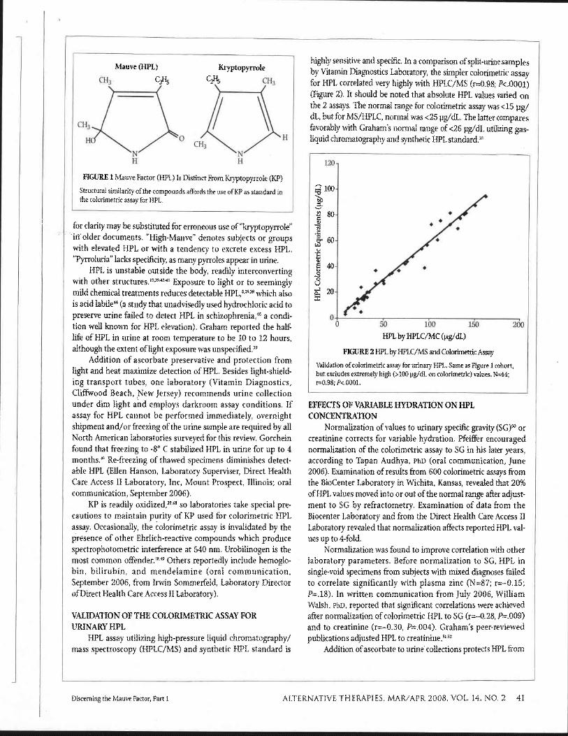
Mauve (HPL) Kryptopyrrole
I FIGURE 1 Mauve Factor (HPL) Is Distinct From Kryptopyrrole (W) 1 Structural similarity of the compounds affords the use of KP as standard in the colorimetric assay for HPL.
for clarity may be substituted for erroneous use of "kryptopyrrole" in older documents. "High-Mauve" denotes subjects or groups with elevated HPL or with a tendency to excrete excess HPL. "Pyrroluria" lacks specificity, as many pyrroles appear in urine.
HPL is unstable outside the body, readily interconverting with other structure^.'^^^^^^ 25 Exposure to light or to seemingly mild chemical treatments reduces detectable HPL,8'939 which also is acid labile" (a study that unadvisedly used hydrochloric acid to preserve urine failed to detect HPL in s~hizophren ia ,~~ a condi- tion weU known for HPL elevation). Graham reported the half- life of HPL in urine at room temperature to be 10 to 12 hours, although the extent of light exposure was un~pecif ied.~~
Addition of ascorbate preservative and protection from light and heat maximize detection of HPL. Besides light-shield- ing transport tubes, one laboratory (Vitamin Diagnostics, Cliffwood Beach, New Jersey) recommends urine collection under dim light and employs darkroom assay conditions. If assay for HPL cannot be performed immediately, overnight shipment and/or freezing of the urine sample are required by all North American laboratories surveyed for this review. Gorchein found that freezing to -8" C stabilized HPL in urine for up to 4 months4' Re-freezing of thawed specimens diminishes detect- able HPL (Ellen Hanson, Laboratory Superviser, Direct Health Care Access I1 Laboratory, Inc, Mount Prospect, Illinois; oral communication, September 2006).
KP is readily oxidized,39" so laboratories take special pre- cautions to maintain purity of KP used for colorimetric HPL assay. Occasionally, the colorimetric assay is invalidated by the presence of other Ehrlich-reactive compounds which produce spectrophotometric interference at 540 nm. Urobilinogen is the most common offender.1849 Others reportedly include hemoglo- bin, bilirubin, and mendelamine (oral communication, September 2006, from Irwin Sommerfeld, Laboratory Director ofDirect Health Care Access I1 Laboratory).
VALIDATION OF THE COLORIMETRIC ASSAY FOR URINARY HPL
HPL assay utilizing high-pressure liquid chromatography/ mass spectroscopy (HPLC/MS) and synthetic HPL standard is
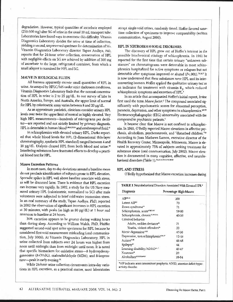
highly sensitive and specific. In a comparison of split-urine samples by Vitamin Diagnostics Laboratory, the simpler colorimetric assay for HPL correlated very highly with HPLC/MS (r=0.98; P<.0001) (Figure 2). It should be noted that absolute HPL values varied on the 2 assays. The normal range for colorimetric assay was <15 yg/ dL, but for MS/HPLC, normal was <25 pg/dL. The latter compares favorably with Graham's normal range of <26 pg/dL utilizing gas- Liquid chromatography and synthetic HPL standard.39
HPL by HPLC/MC (pg/dL)
FIGURE 2 HPL by HPLC/MS and Colorimetric Assay
Vahdation of colorimetric assay for urinary HPL. Same as Figure 1 cohort, but excludes extremely high (>I00 pg/dL on colorimetric) values. N=44; ~ 0 . 9 8 ; P<.OOOl.
EFFECTS OF VARIABLE HYDRATION ON HPL CONCENTRATION
Normalization of values to urinary specific gravity (SG)50 or creatinine corrects for variable hydration. Pfeiffer encouraged normalization of the colorimetric assay to SG in his later years, according to Tapan Audhya, P ~ D (oral communication, June 2006). Examination of results from 600 colorimetric assays from the BioCenter Laboratory in Wichita, Kansas, revealed that 20% of HPL values moved into or out of the normal range after adjust- ment to SG by refractometry. Examination of data from the Biocenter Laboratory and from the Direct Health Care Access I1 Laboratory revealed that normalization affects reported HPL val- ues up to 4-fold.
Normalization was found to improve correlation with other laboratory parameters. Before normalization to SG, HPL in single-void specimens from subjects with mixed diagnoses failed to correlate significantly with plasma zinc (N=87; r=-0.15; P=.18). In written communication from July 2006, William Walsh, P ~ D , reported that significant correlations were achieved after normalization of colorimetric HPL to SG (r=-0.28, P=.009) and to creatinine (r=-0.30, P=.004). Graham's peer-reviewed publications adjusted HPL to ~ r e a t i n i n e . ~ ~ ~ ~
Addition of ascorbate to urine couections protects HPL from
Discerning the Mauve Factor, Part 1 ALTERNATIVE THERAPIES, MARIAPR 2008, VOL. 14, NO. 2 41
degradation. However, typical quantities of ascorbate employed (250-500 mg) alter SG of urine in the usual 10 mL transport tube. Laboratories have found ways to overcome this difficulty. Vitamin Diagnostics Laboratory divides the urine at time of collection, yielding a second, unpreserved specimen for determination of SG. Vitamin Diagnostics Laboratory director Tapan Audhya, P ~ D ,
reports that for 24-hour urine collection, conservation of HPL with negligible effects on SG are achieved by addition of 500 mg of ascorbate to the large, refrigerated container, from which a small aliquot is examined for HPL and SG.
MAWE IN BIOLOGICAL FLUIDS All humans apparently excrete small quantities of HPL in
urine. As assayed by HPLC/MS under strict darkroom conditions, Vitamin Diagnostics Laboratory finds that the normal concentra- tion of HPL in urine is 2 to 25 pg/dL. In our survey of labs in North America, Europe, and Australia, the upper limit of normal for HPL by colorimetric assay varies between 8 and 20 pg/dL.
As an approximate yardstick, clinicians consider urinary HPL levels over twice the upper limit of normal as highly elevated. Very high HPL measurements-hundreds of micrograms per decili- ter-are reported and not strictly limited by primary diagnosis. HPL is detectable in human blood3,3945.53 54 and cerebrospinal
In schizophrenics with elevated urinary HPL, Durko report- ed that whole blood levels for HPL (2-dimensional thin-layer chromatography, synthetic HPL standard) ranged between 4 and 10 ~.lg/dL. Dialysis cleared HPL from both blood and urine.54 Interfering substances have frustrated efforts to develop a practi- cal blood test for HPL.
Mauve Excretion Patterns In most cases, day-to-day deviations around a baseline mean
do not preclude identification of subjects prone to HPL elevation. Sporadic spikes in HPL well above baseline associate with stress, as will be discussed later. There is evidence that HPL excretion can increase very rapidly. In 1992, a study for the US Navy mea- sured urinary HPL (colorimetric, normalized to SG) after male volunteers were subjected to brief cold-water immersion stress. In an oral summary of the study, Tapan Audhya, PhD, reported in 2002 the observation of significant increases in HPL excretion at 30 minutes, with peaks (as high as 80 pg/dL) at 1 hour and reversion to baseline at 24 hours.
HPL excretion appears to be greater during waking hours than during sleep. According to William Walsh, PhD, Pfeiffer suggested second-void spot urine specimens for HPL because he considered first-void measurement misleading (oral communica- tion, July 2006). At Vitamin Diagnostics Laboratory, HPL in urine collected from subjects over 24 hours was higher from noon until midnight than from midnight until noon. It is noted that specific biomarkers for oxidative stress-8 hydroxydeoxy- guanosine (8-OHdG), malondialdehyde (MDA), and &isopros- tane-peak in early evening.55
While 24-hour urine collection circumvents intra-day varia- tions in HPL excretion, as a practical matter, most laboratories
accept single-void urines, randomly timed. Hoffer favored sarne- time collection of specimens to improve comparability (written communication, August 2006).
HPL IN NEUROBEHAVIORAL DISORDERS The discovery of HPL grew out of Hoffer's interest in the
possible biochemical etiology of schizophrenia. In 1961 he reported for the first time that certain urinary "unknown sub- stances" on chromatograms were detectable in most schizo- phrenics hospitalized for active symptoms or relapses but not detectable after symptoms improved or abated (P<.001).785657 It is now understood that these substances were HPL and its inter- converting isomers. Hoffer applied the qualitative urinary test as a n indicator for treatment with vitamin BS, which reduced schizophrenic symptoms and excretion of HPL.4
In an article that accompanied Hoffer's initial report, Irvine first used the term Mauve factor.' The compound associated sig- nificantly with psychometric scores for abnormal perception, paranoia, depression, and other symptoms in schizophrenic^.^"^^ Electroencephalographic (EEG) abnormality associated with the compound in psychiatric patienk5
It became clear that Mauve is not confined to schizophre- nia. In 1965, O'Reilly reported Mauve elevations in affective psy- chosis, alcoholism, psychoneurosis, and "disturbed ~hildren."~" According to Joan Mathews Larson, executive director of the Health Recovery Center, Minneapolis, Minnesota, Mauve is ele- vated in approximately 75% of subjects seeking treatment for substance abuse (oral communication, July 2002). Mauve eleva- tion is documented in many cognitive, affective, and neurobe- havioral disorders (Table l).4.822 23 3032 52 5869
HPL AND STRESS O'ReiUy hypothesized that Mauve excretion increases during
TABLE 1 Neurobehavioral Disorders Associated With Elevated HPL*
Diagnosis Percentage High-Mauve
AIp22.32 100 Latent AIPS2 70 Down syndrome6' 71 Schizophrenia, acutes~s8~6'~62 59-80 Schizophrenia, chronic23,30,s3,M 40-50 Criminal behavior
Adults, sudden deviance6' 71 Youths, violent offenders6" 33
Manic depress i~n~~~ '~ 47-50 Depression, non - sch i z~ph ren i c~ ,~~ ,~~ 12-46 Auti~rn~~.~' 46-48 Epilepsys7 44 Learning di~ability/ADHD~',~~ 40-47 Neurosess9 20 ~ 1 ~ ~ h ~ l i ~ ~ 4 . 8 . 5 9 . 6 3 . 6 4 . 5 9 20-84
-
*AIP indicates acute intermittent porphyria: ADHD, attention deficit hyper- activity disorder.
I
42 ALTERNATIVE THERAPIES, MAR/APR2008, VOL. 14, NO, 2 Discerning the Mauve Factor, Part 1
I
I physical or psychosocial ("emotional") stress." Over decades, cli- nicians formed the strong opinion that, irrespective of behavioral diagnosis, stress increases associated symptoms and excretion of
Pfeiffer came to state unequivocally that Mauve is "a stress-induced fa~tor."~~(p"~' Sohler reportedly induced HPL with experimental stress.45 The effect of cold-water stress in the unpublished US Navy study was described earlier.
McCabe advocated short-term increases in B6 dosing to blunt symptomatic deterioration in high-Mauve subjects during physical or emotional stress.49 Clinicians give higher short-term "stress doses" of both B6 and zinc.16"
VITAMIN B, AND ZINC Pfeiffer discovered the clinical response of high-Mauve sub-
jects to B6 and zinc in 1971 and saw remarkable improvements in a series of 1000 high-Mauve patients.1671
Treatment with B6 and zinc reportedly reduced mean uri- nary HPL in 99 patients from 60 pg/dL to 30 pg/dL in 1 month.30 Although randomized trials have not been performed, combined B6 and zinc are now entrenched as core treatment for high-Mauve subjects. According to William Walsh, P ~ D , neu- robehavioral symptoms associated with elevated HPL may improve after only a few days of therapy with B6 and zinc (oral communication, 2006). Discontinuation may result in severe deterioration within 48 hours."
Clinicians report proportionality between Mauve excretion and symptom severityjO and according to the late Hugh Riordan, MD, former director of the Center for the Improvement of Human Functioning International, Wichita, Kansas (oral com- munication, 2000), higher Mauve excretion usually requires higher dosages of B6 and zinc for suppression. HPL in urine decreased progressively with higher B6 dosing,16 and progressive B6 dosing associates with normalization of erythrocyte glutamate oxaloacetate transaminase (EGOT).72
Initially, Pfeiffer tended to use high doses of vitamin B6 (400-3000 mg daily) and relatively modest ("dietary") doses of zinc. Later, some patients were noted to respond optimally to B6 and as much as 160 mg daily of elemental zincu In the collective experience of the authors, long-term treatment with B6 and zinc usually is needed for ongoing HPL suppression and symptom management. Optimal initial dosages may be higher than main- tenance dosages. Zinc requirements in high-Mauve subjects are noted to increase during growth spurts then decline abruptly. Pfeiffer reported that on occasion, previously high-Mauve sub- jects no longer may require high doses of B6 and zincI6; the phe- nomenon was confirmed in oral communication in 2003 with Mark Vonnegut, MD, a former high-Mauve Pfeiffer patient and now a practicing pediatrician in Quincy, Massachusetts.
Pfeiffer's claims of a "double deficiency" of B6 and zinc in association with abnormal Mauve excretionlo were based on the clinical response to supplementation and a pattern of lower blood levels of zinc and functional B6 status (pyridoxal-5-phosphate [P5P] and EGOT) among his high-Mauve patients.117374 Numerical data were not published.
Pfeiffer and Sohler proposed that functional B6 deficiency and zinc deficiency in high-Mauve subjects results from increased uri- nary loss of P5P and zinc due to complexation with Mauve, and they cited 20 pg/dL higher zinc content in spot urines of Mauve- postive subjects." The finding would extrapolate to relatively insubstantial total zinc loss, unless the effect extended to other routes of excretion. Pfefler published evidence of binding between P5P and KP" and between zinc and KPl8 but did not study HPL.
Validation of HPL as a Marker for B, Status The original data presented in this review were retrieved
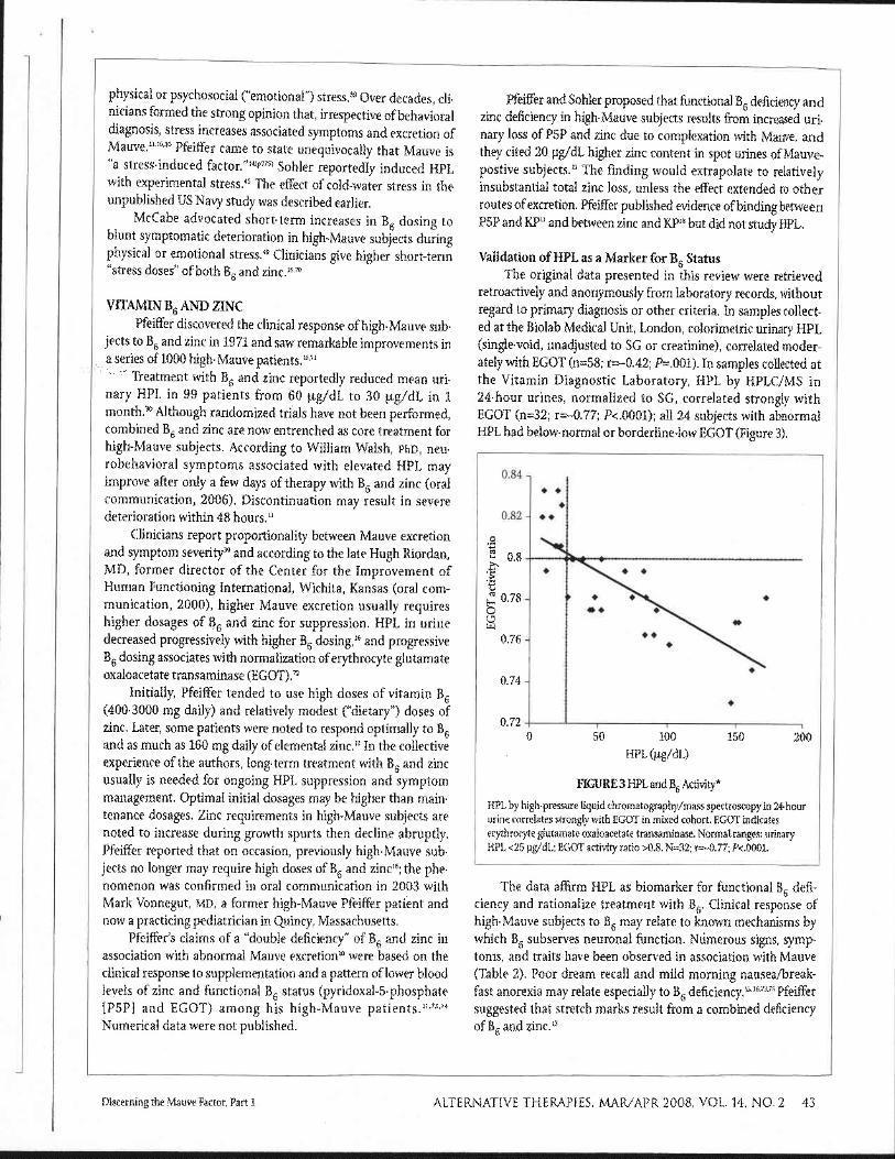
retroactively and anonymously from laboratory records, without regard to primary diagnosis or other criteria. In samples collect- ed at the Biolab Medical Unit, London, colorimetric urinary HPL (single-void, unadjusted to SG or creatinine), correlated moder- ately with EGOT (n=58; r=-0.42; P=.001). In samples collected at the Vitamin Diagnostic Laboratory, HPL by HPLC/MS in 24-hour urines, normalized to SG, correlated strongly with EGOT (n=32; r=-0.77; P<.0001); all 24 subjects with abnormal HPL had below-normal or borderline-low EGOT (Figure 3).
0 50 100 150 HPL (pg/dL) I
FIGURE 3 HPL and B6 Activity*
HPL by high-pressure Liquid chromatography/mass spectroscopy in 24-hour urine correlates strongly with EGOT in mixed cohort. EGOT indicates erythrocyte glutamate oxaloacetate transaminase. Normal ranges: urinary HPL <25 pg/dL; EGOT activity ratio >0.8. N=32; r=-0.77; P<.0001.
The data affirm HPL as biomarker for functional B6 defi- ciency and rationalize treatment with B6. Clinical response of high-Mauve subjects to B6 may relate to known mechanisms by which B, subserves neuronal function. Numerous signs, symp- toms, and traits have been observed in association with Mauve (Table 2). Poor dream recall and mild morning nausea/break- fast anorexia may relate especially to B6 deficiency.13 1673 75 Pfeiffer suggested that stretch marks result from a combined deficiency of B6 and zinc.13
Discerning the Mauve Factor, Part 1 ALTERNATIVE THERAPIES. MARIAPR 2008, VOL. 14, NO. 2 43

TABLE 2 Signs, Symptoms, and Traits Clinicians Report as More Prevalent in High-Mauve Patients*4.10 U.l3.L6.~.49.64.71.73
Poor dream recall Impotence Nail spots Eosinophilia Stretch marks (striae) B6-responsive anemia Pale skidpoor tanning Attention deficit/hyperactivity Coarse eyebrows Crime and delinquency Knee and joint pain Substance abuse Acne Alcoholism Allergy Stress intolerance Cold hands or feet Emotional lability Abdominal tenderness Explosive anger Stitch in side Anxiety Constipation Pessimism Morning nausea Dyslexia Light/sound/odor intolerance Familial or social withdrawal Tremor/shaking/spasms Depression Hypoglycernia/glucose intolerance Paranoia Obesity Hallucinations Migraine Disordered perception Delayed puberty Bipolar disorder Amenorrhea/irregular periods Autism
*The frequency of these features and their relationship to biochemical abnor- malities associated with HPL are not well-studied.
Validation of HPL as a Marker for Zinc Status White flecks in the nails (Figure 4) are responsive to ~ i n c ' " ~ ' , ~ ~
and reportedly detectable in 60% of high-Mauve s~bjec t s .~~HPL was examined in relationship to 3 different measurements for zinc. As discussed earlier, Walsh reported that plasma zinc and single-void colorimetric HPL correlated significantly once normal- ized to SG (r=0.28; P=.009) or to creatinine (r=0.30; P=.004).
FIGURE 4 Leukodynia Implies Zinc Deficit
curred after dosage was lowered to 40 mg, and
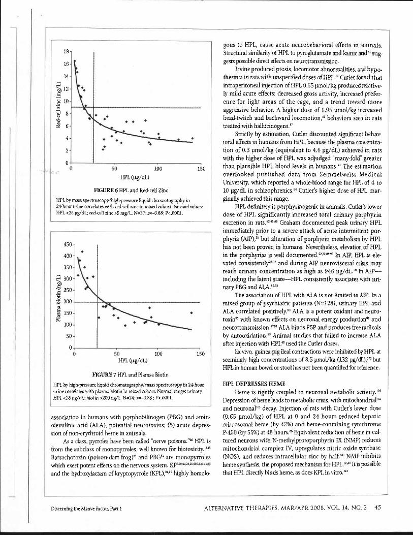
Cellular zinc levels correlated more strongly with urinary HPL. In samples at the BioLab Medical Unit, single-void colori- metric HPL (unadjusted to SG) from a mixed cohort correlated substantially with white-cell zinc (N=58; r=-0.60; P<.0001). Abnormal HPL corresponded to below-normal white-cell zinc in 42 of 58 patients (Figure 5). In samples at Vitamin Diagnostic Laboratory, stronger association existed between red-cell zinc and 24-hour urinary HPL (HPLC/MS, adjusted to SG) in a mixed cohort (N=37; r=-0.88; P<.0001). Twenty-four of 24 subjects with elevated HPL had below-normal red-cell zinc (Figure 6).
7 + ; ;4
6.5 - V) - - I
.- =:
+ 13 1 t + +
4 " I
3.5 7 I -
0 10 20 30 40 50 60 70
Colorimetric HPL equivalents (bg/dL)
FIGURE 5 Single-void HPL and White-cell Zinc Colorimetric HPL equivalents in single-void urines, unadjusted to specific gravity, correlates with white-cell zinc in mixed cohort. Normal values: HPL <8 pg/dL; white-cell zinc > 5.4 ng/106 leukocytes. N=58; r--0.60; P<.0001.
HPL AND OTHER NUTRITIONAL PARAMETERS Oscar Kruesi, MD, former academic dean for Integrative
Medicine, Capitol University, Washington, District of Columbia, reported a pattern of low plasma biotin levels in high-Mauve patients (oral communication, 2005). At the Vitamin Diagnostics Laboratory, 24-hour urinary HPL (HPLC/MS, adjusted to SG) and plasma biotin concentrations from a small, mixed cohort strongly correlated (N=24; r=-0.88, P<.0001). Elevated HPL predicted below-normal plasma biotin in 16 of 16 subjects (Figure 7). These data are the first to suggest biotin deficiency in association with HPL. Biotin deficiency causes neurological disease in animals and humans7778 and is more common than thought."
Examination oflaboratory records found no association between HPL and markers for vitamin Bg (urinary n-methyl nicotinamide), vitamin B12 (urinary methylmalonic acid), folate (urinary formimino- glutarnic acid, FIGLU), or thiamine (red-cell transketolase).
POSSIBLE NEUROTOXICITY OF HPL Several findings suggest that HPL is neurotoxic in humans:
(1) structural homology to known neurotoxin; (2) acute behav- ioral effects in animals; (3) porphyrinogenicity in animals; (4)
44 ALTERNATIVE THERAPIES, MAR/APR2008, VOL. 14. NO. 2 Discerning the Mauve Factor, Part 1 I
association in humans with porphobilinogen (PBG) and amin- olevulinic acid (ALA), potential neurotoxins; (5) acute depres- sion of non-erythroid heme in animals.
As a class, pyrroles have been called "nerve poi .son~."~WL is from the subclass of monopyrroles, well known for biotoxicity. 3,45
Batrachotoxin (poison-dart frog)81 and PBGa2 are monopyrroles which exert potent effects on the nervous system. KP3~"~22~Z4~z5~29~31-33~45583
and the hydroxylactam of kryptopyrrole (KPL),84.85 highly homolo-
gous to HPL, cause acute neurobehavioral effects in animals. Structural similarity of HPL to pyroglutamate and kainic acid4'sug- gests possible direct effects on neurotransmission.
Irvine produced ptosis, locomotor abnormalities, and hypo- thermia in rats with unspecified doses of HPL." Cutler found that intraperitoneal injection of HPL 0.65 pmol/kg produced relative- ly mild acute effects: decreased gross activity, increased prefer- ence for light areas of the cage, and a trend toward more aggressive behavior. A higher dose of 1.95 pmol/kg increased head-twitch and backward l o c ~ m o t i o n , ~ ~ behaviors seen in rats treated with hallucinogen^.^^
Strictly by estimation, Cutler discounted significant behav- ioral effects in humans from HPL, because the plasma concentra- tion of 0.3 pmol/kg (equivalent to 4.6 pg/dL) achieved in rats with the higher dose of HPL was adjudged "many-fold greater than plausible HPL blood levels in humans." The estimation overlooked published data from Semmelweiss Medical University, which reported a whole-blood range for HPL of 4 to 10 pg/dL in schizophrenics." Cutler's higher dose of HPL mar- ginally achieved this range.
HPL definitely is porphyrinogenic in animals. Cutler's lower dose of HPL significantly increased total urinary porphyrin excretion in rats.528588 Graham documented peak urinary HPL immediately prior to a severe attack of acute intermittent por- phyria (AIP),39 but alteration of porphyrin metabolism by HPL has not been proven in humans. Nevertheless, elevation of HPL in the porphyrias is well documented.Z23z8993 In AIP, HPL is ele- vated c o n ~ i s t e n t l y ~ ~ , ~ ~ and during AIP neurovisceral crisis may reach urinary concentration as high as 946 ~ g / d L . ~ ~ In AIP- including the latent state-HPL consistently associates with uri- nary PBG and ALA.5293
The association of HPL with ALA is not limited to AIP. In a mixed group of psychiatric patients (N=128), urinary HPL and ALA correlated po~it ively.~~ ALA is a potent oxidant and neuro- toxing5 with known effects on neuronal energy productiong6 and neuro t ransmi~s ion .~~~~ ALA binds P5P and produces free radicals by au tooxida t i~n .~~ Animal studies that failed to increase ALA after injection with HPLa8 used the Cutler doses.
Ex vivo, guinea-pig ileal contractions were inhibited by HPL at seemingly high concentrations of 8.5 pmol/kg (132 pg/dL),lW but HPL in human bowel or stool has not been quantfiedfor reference.
HPL DEPRESSES HEME Heme is tightly coupled to neuronal metabolic activity.lol
Depression of heme leads to metabolic crisis, with mitochondrialm2 and neuronallo3 decay. Injection of rats with Cutler's lower dose (0.65 pmol/kg) of HPL at 0 and 24 hours reduced hepatic microsomal heme (by 42%) and heme-containing cytochrome P-450 (by 55%) at 48 hours.88 Equivalent reduction of heme in cul- tured neurons with N-methylprotoporphyrin IX (NMP) reduces mitochondria1 complex IV, upregulates nitric oxide synthase (NOS), and reduces intracellular zinc by half.lol NMP inhibits heme synthesis, the proposed mechanism for HPL.SZ,88 It is possible that HPL directly binds heme, as does KPL in vitro.IQ4
Discerning the Mauve Factor, Part 1 ALTERNATIVE THERAPIES, MAFUAPR2008, VOL. 14, NO. 2 45
Non-erythroid heme in high-Mauve subjects has not been measured, but depressed levels are predictable. Besides potential depression by HPL, deficiencies of zinc, B6, and biotin (all cofac- tors for heme synthesis) independently decrease non-erythroid
And heme is degraded by stress.'" It should be men- tioned as well that heavy metals, which have not been examined in relation to Mauve, are renowned dysregulators of porphyrin metabolism and increase heme degradation.'"
Heme plays a central role in energy production and is required by a family of biomolecules needed for detoxification and antioxidant defense: catalase, cystathionine synthase, cyto- chrome, guanylate cyclase, heme-hemopexin (for production of metallothionein), NOS, pyrrolase, sulfite reductase. Ultimately, heme depression increases oxidant leak from mitochondria and oxidative damage to cells.99 lo2
HPL AND OXIDATIVE STRESS Oxidative stress clearly results from deficiency of zinc or B6,
as reviewed by McGinnis.Io5 For example, even marginal B6 defi- ciency is associated with lower glutathione peroxidase (GSHPx), lower glutathione (GSH) reductase, lower reduced/oxidized glu- tathione ratios, higher lipid peroxide levels, and mitochondria1 d e ~ a y . ' ~ ~ . ' ~ ~ The B6 vitamers are themselves highly vulnerable to damage by oxidative s p e c i e ~ . l ~ ~ - ~ P5P protects neurons from oxi- dative stress, apparently by increasing energy production and lowering excitoto~icity,"~.~~ and zinc supplementation decreases oxidized b iomole~ules .~~~ lL5 Since HPL is a marker for B6 and zinc deficiency, HPL is a potential biomarker for oxidative stress.
Biomarkers for oxidative stress are known to be higher in high-Mauve disorders such as schizophrenia,116117 a u t i ~ m , ~ ' " - ' ~ ~ ADHD,121 lZ2 Down syndrome,123125 and In schizo- phrenia, lower blood levels of glutathione and response to intra- venous glutathione were reported nearly 50 years ago.lZ9
Plasma levels of reduced GSH, the ubiquitous intracellular antioxidant, are decreased in diseases associated with greater oxidative stress,130 including Down syndrome.131 In Alzheimer's disease, in which oxidative modification of brain precedes appearance of neurofibrillary tangles and plasma GSH correlates inversely with brain levels of oxidatively-modified biomolecules.134 It is reasonable to view plasma GSH as a bio- marker for pathological effects of oxidative stress.
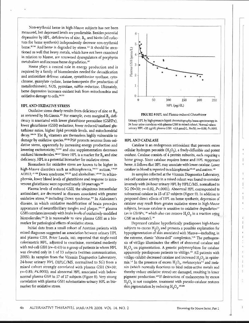
Initial data from a small cohort of Austrian patients with mixed diagnoses suggested an association between urinary HPL and plasma GSH. Peter Lauda, MD, reported that single-void colorimetric HPL, adjusted to creatinine, correlated modestly with red-cell GSH (r=-0.41) in a group of patients in whom HPL was elevated only in 1 of 13 subjects (written communication, 2005). In samples from the Vitamin Diagnostics Laboratory, 24-hour urinary HPL (HPLC/MS, normalized to SG) from a mixed cohort strongly correlated with plasma GSH (N=30; r=-0.85; P1.0001), and abnormal HPL associated with below- normal plasma GSH in 17 of 17 subjects (Figure 8). Very strong correlation with plasma GSH substantiates urinary HPL as bio- marker for oxidative stress.
0 0 50 100 150 200 250
HPL (pg/dL)
FIGURE 8 HPL and Plasma-reduced Glutathione
Urinary HPL by high-pressure liquid chromatography/mass spectroscopy in 24-hour urine correlates with plasma GSH in mixed cohort. Normal values: urinary HPL <25 pg/dl; plasma GSH >3.8 pmol/L. N=30; r=-0.88; P<.0001.
HPL AND CATALASE Catalase is an endogenous antioxidant that prevents excess
cellular hydrogen peroxide (H20z), a freely-diffusible and potent oxidant. Catalase consists of 4 protein subunits, each requiring a heme group. Since catalase requires heme and HPL suppresses heme, it follows that HPL may associate with lower catalase. Lower catalase in blood is reported in sch iz~phren ia~"~~ and autism.'36
In samples collected at the Vitamin Diagnostics Laboratory, red-cell catalase activity in a mixed cohort was found to correlate inversely with 24-hour urinary HPL by HPLC/MS, normalized to SG (N=30; r=-0.92, P<.0001). Abnormal HPL corresponded to subnormal catalase in 15 of 17 subjects (Figure 9). In addition to proposed direct effects of HPL on heme synthesis, depression of catalase may result from greater oxidative stress in high-Mauve subjects, because catalase is sensitive to oxidative degradat i~n '~~ (as is GSHPX,'~~ which also can remove H202 in a reaction using GSH as s~bs t ra te ) . '~~
Depressed catalase hypothetically predisposes high-Mauve subjects to excess H202 and presents a possible explanation for hypopigmentation of skin associated with Mauve-including, in the extreme, classic "china-doll" complexion.'011 The pathogene- sis of vitiligo illuminates the effect of abnormal catalase and H202 on pigmentation. A genetic polymorphism for catalase apparently predisposes patients to vitiligo.140 All patients with vitiligo exhibit decreased catalase and increased H202 in epider- mis.14' In the presence of excess H202, melanocytesM2 and mela- nin (which normally funct~ons to bind redox-active metals and thereby reduce oxidative stress) are damaged, resulting in lesser pigment p rodu~t ion . '~~I f destruction of melanocytes by excess H202 is not complete, treatment with pseudo-catalase restores skin pigmentation by reducing H202.143144
46 ALTERNATIVE THERAPIES. MAR/APR 2008, VOL. 14. NO. 2 Discerning the Mauve Factor, Part 1 I I
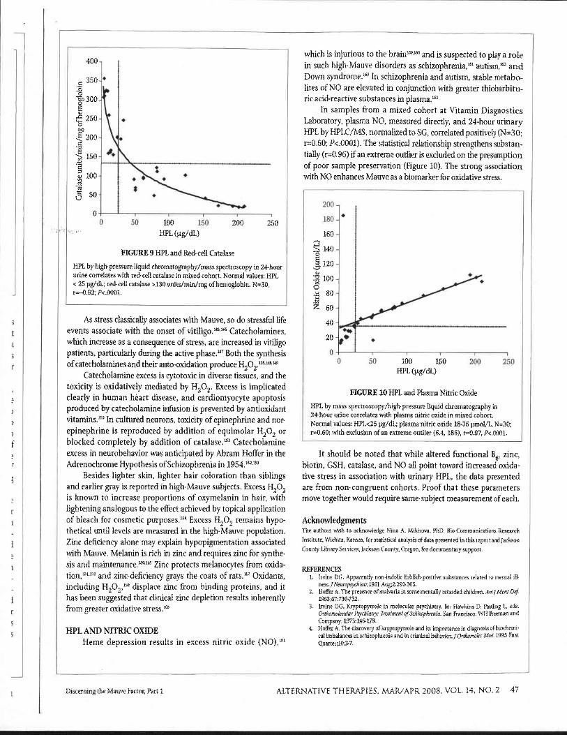
FIGURE 9 HPL and Red-cell Catalase
HPL by high-pressure liquid chromatography/mass spectroscopy in 24-hour urine correlates with red-cell catalase in mixed cohort. Normal values: HPL < 25 pg/dL; red-cell catalase >I30 units/min/mg of hemoglobin. N=30, r=-0.92; P<.0001.
As stress classically associates with Mauve, so do stressful life events associate with the onset of v i t i l i g ~ . ' " ~ ~ Catecholamines, which increase as a consequence of stress, are increased in vitiligo patients, particularly during the active pha~e ."~ Both the synthesis of catecholamines and their auto-oxidation produce H202.139Y8 149
Catecholamine excess is cytotoxic in diverse tissues, and the toxicity is oxidatively mediated by H202. Excess is implicated clearly in human heart disease, and cardiomyocyte apoptosis produced by catecholamine infusion is prevented by antioxidant vitamins.'50 In cultured neurons, toxicity of epinephrine and nor- epinephrine is reproduced by addition of equimolar H202 or blocked completely by addition of catalase.15' Catecholamine excess in neurobehavior was anticipated by Abram Hoffer in the Adrenochrome Hypothesis of Schizophrenia in 1954.152153
Besides lighter skin, lighter hair coloration than siblings and earlier gray is reported in high-Mauve subjects. Excess H202 is known to increase proportions of oxymelanin in hair, with lightening analogous to the effect achieved by topical application of bleach for cosmetic p u ~ p o s e s . ' ~ ~ Excess H202 remains hypo- thetical until levels are measured in the high-Mauve population. Zinc deficiency alone may explain hypopigmentation associated with Mauve. Melanin is rich in zinc and requires zinc for synthe- sis and maintenance.150155 Zinc protects melanocytes from oxida- tion,lS4 lS6 and zinc-deficiency grays the coats of rats.IS7 Oxidants, including H202,158 displace zinc from binding proteins, and it has been suggested that clinical zinc depletion results inherently from greater oxidative s t res~."~
HPL AND NITRIC OXIDE Heme depression results in excess nitric oxide (NO),lol
which is injurious to the and is suspected to play a role in such high-Mauve disorders as schizophrenia,16' autism,'" a n d Down syndrome.'63 In schizophrenia and autism, stable metabo- lites of NO are elevated in conjunction with greater thiobarbitu- ric acid-reactive substances in ~ 1 a s m a . l ~ ~
In samples from a mixed cohort at Vitamin Diagnostics Laboratory, plasma NO, measured directly, and 24-hour urinary HPL by HPLC/MS, normalized to SG, correlated positively (N=30; r=0.60; P<.0001). The statistical relationship strengthens substan- tially (r=0.96) if an extreme outlier is excluded on the presumption of poor sample preservation (Figure 10). The strong association with NO enhances Mauve as a biomarker for oxidative stress.
0,' 0 50 100 150 200 250
HPL (pg/dL)
FIGURE 10 HPL and Plasma Nitric Oxide
HPL by mass spectroscopy/high-pressure liquid chromatography in 24-hour urine correlates with plasma nitric oxide in mixed cohort. Normal values: HPL<25 pg/dL; plasma nitric oxide 18-36 pmol/L. N=30; r=0.60; with exclusion of an extreme outlier (6.4, 186), r=0.97, P<.0001.
It should be noted that while altered functional B6, zinc, biotin, GSH, catalase, and NO all point toward increased oxida- tive stress in association with urinary HPL, the data presented are from non-congruent cohorts. Proof that these parameters move together would require same-subject measurement of each.
Acknowledgments The authors wish to acknowledge Nina A. Mikirova, PhD, Bio-Communications Research
Institute, W~chita, Kansas, for statistical analysis of data presented in this report and Jackson
County Library Services, Jackson County, Oregon, for documentary support.
REFERENCES 1. Irvine DG. Apparently non-indolic Erhlich-positive substances related to mental ill-
ness. J Neuropsychiatr.1961 Aug;2:292-305. 2. Hoffer A. The presence of malvaria in some mentally retarded children. ArnlMentDef:
1963:67:730-732. 3. Iwine DG. Kryptopyrrole in molecular psychiatry. In: Hawkins D, Pauling L, eds.
Orthomolecular Psychiatry: Treatment ofSchizophrenia. San Francisco: WH Freeman and Company: 1973:146-178.
4. Hoffer A. The discovery of kryptopyrrole and its importance in diagnosis of biochemi- cal imbalances in schizophrenia and in criminal behavior.JOrfhomolec Med. 1995 First Quarter:10:3-7.
Discerning the Mauve Factor, Part 1 ALTERNATIVE THERAPIES, MAR/APR 2008, VOL. 14, NO. 2 47

5. Irvine DG. Mauve factor and 6-sulfatoxy skatole: two biochemical abnormalities asso- ciated with specific measures of psychiatric disease. Clin Chem. 1963;9:444-445.
6. Hoffer A. Osmond H. The association between schizophrenia and two objective tests. Can Med AssocJ 1962;87(12):641-646.
7. Hoffer A, Mahon M. The presence of unidentified substances in the urine ofpsychiatric patients.JNeuropsychiatr. 1961 Aug;2:331-362.
8 Hoffer A, Osmond H. Malvaria: a new psychiatric disease. Acta Psychiatr Scand. 1963:39:335-366.
9. Hoffer A. Malvaria, schizophrenia and the HOD test. Int J Neuropsychiatry. 1965;2:175-178.
10. Pfeiffer CC, Iliev V. Pyroluria, urinary mauve factor causes double deficiency of 86 and zinc in schizophrenics. Fed Am SocExpBiol. 1973:32:276.
11. Pfeiffer CC, Sohler A, Jenney EH, et al. Treatment ofpyroluric schizophrenia (malvaria) with large doses of pyridoxine and a dietary supplement of zinc. J Appl Nutr. 1974;26:21-28.
12. PfeifTer CC, Bacchi D. Copper, zinc, manganese, niacin and pyridoxine in the schizo- phrenias.JApp1 Nutr. 1975;27:9-39.
13. PfeifTer CC. Mental andElementa1 Nutrients. New Canaan, CT: Keats Publishing;1976. 14. Pfeiffer CC. The schizovhrenias '76. BiolPsvchiatrv. 1976:ll(6):773-775. 15. PfeifTer CC. Extra nutri'ents and mental illiess. B~l~s~chiat~'1981;16(9):797-799, 16. Pfeiffer CC, Holford P. Mental Illness and Schizophrenia: The Nutritional Connection.
Harper Collins Publishers, Great Britain; 1987. 17. PfeifTer CC. Nutrition and Mental Illness: An Orthomolecular Approach to Balancing Body
Chemistry. Rochester, VT: Healing Arts Press; 1987. 18. Sohler A, Holsztynska MS, PfeiiTer CC. A rapid screening test for pyroluria; useful in
distinguishing a schizophrenic population. JOrthomolecPsychiatr. 1974;3(4).273-279, 19. Irvine DG, Bayne W, Miyashita H, Majer JR. Identification of kryptopyrrole in human
urine and its relation to psychosis. Nature. 1969;224(5221):8U-813. 20. Sohler A, Beck R, Noval JJ. Mauve factor re-identified as 2.4-dimethyl-3-ethyl pyrrole
and its sedative effect on the CNS. Nature. 1970;228(5278):1318-1320. 21. Wetterberg L. Pharmacological and toxic effects of kryptopyrrole in mice./Orthomolec
Psychiatr. 1972;1:141-144. 22. Huszik I, Durk6, Karsai K. Experimental data to the pathogenesis of cryptopyrrole
excretion in schizophrenia. Acta Physiol Acad Scien Hung 1972;42(1):79-86. 23. Gebel L. Occurence of the mauve factor in schizophrenia [in Polish]. Psychiatr Pol.
1973;7(2):153-159. 24. Irvine DG, EU D, Crichlow EC. Anticonvulsant and EEG activities of kryptopyrrole and
a possible underlying neurochemical mechanism. Proceedings ofthe 4th International Meeting ofthe International Societyfor Neurochemisrry. Tokyo: 1973: 359.
25. Wetterberg L. Pharmacological and toxic effects of kryptopyrrole in mice. Ups J Med Sci 1973;78(1):78-80.
26. Kriicher K, PfeiiTer CC. Biochemical relationship between kryptopyrrole (mauve fac- tor) and trans-3-methyl-2-hexanoic acid (schizophrenia odor). Res Comunm Chem PatholPharmacol. 1973;5(1):9-15.
27. Berek I, Huszik I, Durk6 I. The effect of kryptopyrrole on the porphyrin auxotrophic strains of Bacillus subtilis. Eqerientia. 1975;31(2):185.
28. Durk6 I, B e ~ k I, Huszik I. Effects of kyrptopyrrole on porphyrin synthesis in Bacillus subtilii 168. Hoppe-Sqlers ZPhysiol Chem. 1975;356(1ll):1679-1684.
29. Sohler A. Letter: Urinary kryptopyrrole. BiolPsychiaby 1975;10(6):685-686. 30. Ward JL. Relationship of kryptopyrrole, zinc and pyridoxine in schizophrenics. J
OrthomolecPsychiatr. 1975;4:27-31. 31. Walker JL. Neurological and behavioral toxicity of kryptopyrrole in the rat. Pharmacol
Biochem Behav. 1975;3(2):243-250. 32. Brodie MJ, Graham DJM, Thompson GG, Moore MR, Goldberg A. The porphyrino-
genic effects of kryptopyrrole in the rat and the occurence of urinary kryptopyrrole in human hereditary hepatic porphyria. Clin SciMolMed. 1976;50(5):431-434.
33. Wetterberg L, Formgren B. Pharmacological and biochemical properties of kryptopyr- role and its oxidation products possibly related to acute intermittent porphyria. Ann Clin Res. 1976;s Suppl17:162-167.
34. Jacobson SJ, Rapoport H, Ellman GL. The nonoccurence of hemo- and kryptopyrrole in the urine of schizophrenics. BiolPsychiatry 1975;10(1):91-93.
35. Gendler PL, Duhan HA, Rapoport H. Hemopyrrole and kryptopyrrole are absent from the urine of schizophrenics and normal persons. Clin Chem. 1978;24(2):230-233.
36. Wooldrige TA, Lightner DA. Synthesis of "oxidized" hemopyrrole and kryptopyrrole: porphyric monopyrroles.JHeterocyclic Chem. 1977;14:1283-1284.
37. Irvine DG. Pyrroles in neuropsychiatric and porphyric disorders: confirmation of a metabolite structure by synthesis. Life Sci 1978;23(9):983-990.
38. Irvine DG. Hydroxy-hemopyrrolenone, not kryptopyrrole, in the urine of schizophren- ics and porphyrics. Clin Chem. 1978;24(ll):2069-2070.
39. Graham DJM. Monopyrroles in Porphyria and Related Disorders [doctoral thesis]. University of Glasgow; 1978.
40. Graham DJ. Quantitative determination of 3-ethyl-5-hydroxy-4.5-dimethyl-delta 3-pyr- rolin-2-one in urine using gas-liquid chromatography. Clin Chim Acta. 1978;85(2):205-210.
41- Cutler MG, Graham DJ, Moore MR. The mauve factor of porphyria. 3-ethyl-5hydroxy- 4,5-dimethyl-delta-3pyrroline-2-one: effects on behavior of rats and mice. Pharmacol Toxicol. 1990;66(1):66-68.
42. Irvine DG, Bayne W, Majer JR. Autotransfer chromatography combined with mass spectroscopy, for the characterization of pyrroles and indoles. J Chromatogr. 1970;48(2):334-342.
43. Lightner DA, Bisacchi GS, Norris RD. On the mechanism of the sensitized photooxi-
genation of pyrroles. ]Am Chem Soc 1976;98(2):802-807. 44. Lightner DA, Norris RD, Kirk DI, Key RM. The dye-sensitized photooxygenation of
hemopyrrole. Experientia. 1974:30(6):587-588. 45. Irvine DG. Kryptopyrrole and other monopyrroles in molecular neurobiology. Int Rev
Neurobiol. 1974;16(0):145-182. 46. Rao BS, Narayanan HS, Reddy GN. Investigations on urinary excretion of 3,4 dime-
thoxyphenylethylamine (the pink spot), the Mauve factor and aromatic compounds in patients with schizophrenia. Indian JMed Res. 1971;59(3):455-460.
47. Gorchein A. Urine concentration of 3-ethyl-5-hydroxy-4.5-dimethl-delta 3-pyrrolin-2- one ('mauve factor') is not causally related to schizophrenia or to acute intermittent porphyria. Clin Sci (Lond). 1980;58(6):469-476.
48. Lightner DA. Crandall DC. The dye sensitized photooxygenation of kryptopyrrole. Experientia. 1973;29(3):262-264.
49. McCabe DL. Kryptopyrroles.JOrthomolecPsychiatr. 1983:12:2-18. 50. Miller RC, Brindle E, Holman DJ, et al. Comparison of specific gravity and creaiiine
from normalizing urinary reproductive hormone concentrations. Clrn Chtm. 2004:50(5):924-932.
51. Graham DJ, Thompson GG, Moore MR. et al. The effects of selected monopyrroles on various aspects of heme biosynthesis and degradation in the rat. Arch Biochem Biophys. 1979:197(1):132-138.
52. Moore MR, Graham DIM. Monopyrroles in porphyria, psychosis and lead exposure. IntJBiochem. 1980;12(5-6):827-832.
53. Irvine DG, Bayne W, Miyashita H. The main form of naturally-occuring kryptopyrrole: its 5-OH-2-lactam, a product of pyrrolooxygenase. Report to the Psychiatric Research Meeting of the Saskatchewan Psychiatric Association. 1973: 46-67.
54. Durkn I, Englehardt I, SzilardJ, et al. The effect of haemodialysis on the excretion ofthe mauve factor in schizophrenia.JOrthomolecPsychiatry. 19&4;13:222-232.
55. Kanabrocki EL, Murray D. Hermida RC, et al. Circadian variation in oxidative stress markers in healthy and type I1 diabetic men. Chronobiollnt. 2002;19(2):423-439.
56. Hoffer A, Osmond H, Callbeck MJ, Kahan I. Treatment of schizophrenia with nicotinic acid and nicotinamide. JClin Exper Psychopathol. 1957;18(2):131-158.
57. Hoffer A. Megavitamin B3-therapy for schizophrenia. Can Psychiatr Assoc J. 1971:16(6):499-504.
58. Hoffer A, Osmond H. The relationship between an unknown factor ("US") in urine of subjects and HOD test results.]Neuropsychiatr. 1961 Aug;2:363-374.
59. O'Reilly PO, Ernest M, Hughes G. The incidence of malvaria. Br ]Psychiatry. 1965 Aug;lll:741-744.
60. Jackson JA, Riordan HD, Neathery S. Vitamin, blood lead, and urine pyrrole levels in Down syndrome. Am Clin Lab. 1990:Jan-Feb:8-9.
61. Sohler A, Renz RH, Smith S, Kaufman J. Significance of hydroxyskatole and mauvefac- tor excretion in schizophrenia. IntJNeuropsychiatry. 1967;3(4):327-331.
62. EUman GL, Jones RT, Rychert RC. Mauve spot and schizophrenia. Am JPsychiatry. 1968:125(6):849-851.
63. Hoffer A, Osmond H. How to Survive with Schizophrenia. Secausus. NJ.: University Books: 1974.
64. Cutler P. Pyridoxine and trace element therapy in selected clinical cases. JOrthomolec Psychiatr. 1974;89-95.
65. Hoffer A. Malvaria and the law. Psychosomatics. 1966;7(5):303-310. 66. Walsh WJ, Glab LB, Haakenson ML. Reduced violent behavior following biochemical
therapy. PhysiolBehav. 2004;82(5):835-839. 67. Audhya T. Urinary EHHPL in neurobehavioral disorders: Think-tank presentation.
Presented at: Defeat Autism Nowl (DAN!) Conference. October 24-27, 2002; San Diego, CA.
68. Isaacson HR, Moran MM. Hall A, et al. Autism: a retrospective outcome study of nutri- ent therapy. JApplNutr. 1996:Ill-US.
69. Hoffer A. A program for the treatment of alcoholism: LSD, malvaria and nicotinicacid. In: Abramson HA, ed. The Use ofLSD in Psychotherapy and Alcoholism. Indianapolis: Howard W. Sams and Company, Inc.; 1967; 343-406.
70. No authors listed. The 'mauve factor' in schizophrenia. Medical World News. Demriiber. 14,1973: 49.
71. Pfeiffer CC, Maillous R, Forsythe L. The Schizophrenias: Ours to Conquer. Wichita, Kansas: Bio-Communications Press; 1988.
72. Jaffee R, Kruesi OR. The biochemical-immunology window: a molecular view of psychi- atric case management.JApplNutr 1992;44:26-42.
73. Pfeiffer CC, Audette L. Pyroluria-zinc and B6 deficiencies. lnt Clin Nutr Rev. 1988;8:107-109.
74. Sohler A, Pfe8er CC. Vitamin B6 nutritional status of a psychiatric outpatient popula- tion.] Orthomolec Psychiatr. 1982:ll:81-86.
75. Gaby A. TheDoctorS Guide to fitamin B6. Rodale Press: Emmaus, PA; 1984. 76. Pfeiffer CC, Jenney EH. Fingernail white spots: possible zinc deficiency. JAMA.
1974;228(2):157. 77. Bregola G, Muzzolii A, Mazzari S, et al. Biotin deficiency facilitates kindling hyperex-
ciabiity in rats. Neuroreport. 1996;7(U):1745-1748. 78. Griinewald S. Champion MP, Leonard jV, SchaperJ. Morris AA. Biotinidase deficiency:
a treatable leukoencephalopathy. Neuropediatrics. 2004;35(4):2ll-216. 79. Mock DM, Henrich CL. CarneU N. Mock NI. Indicators of marginal biotin deficiency
and repletion in humans: validation of 3-hydroxyisovaleric acid excretion and a leucine challenge. AmJClin Nutr. 2002;76(5):1061-1068.
80. Cowin AM, et al. Encyclopaedia Britannzca. 1960;18:801. 81. Dumbacher JP. Wako A, Derrickson SR, Samuelson A, Spande TF, Daly JW. Melryrid
beetles (Choresine): putative source for batrachotoxin alkaloids found in poison-dart
48 ALTERNATIVE THERAPIES, MAR/APR2008, VOL. 14, NO. 2 Discerning the Mauve Factor, Part 1 I

frogs and toxic passerine birds. ProcNatlAcadScr U S A . 2004:101(45):15857-15860. 82. Becker DM. Kramer S. The neurological manifestations of porphyria: a review.
Medicine (Baltimore). 1977;56(5):411-423. 83. Irvine DG. Kryptopyrrole: some clinical, metabolic and pharmacological correlates.
Report to the Psychiatric Research Meeting of the Saskatchewan Psychiatric Association. 1972:171-178.
84. Graham DJ, Moore MR, Thompson GG, Goldberg AA. The effect of 4-ethyl-5-hydroq- 3.5-dimethyl-delta 3-pyrrolin-2-one on porphyrin synthesis in the rat. Biochem Soc Trans. 1976;4(6):1089-1091,
85. Graham DJ. Thompson GG. Moore MR. Brodie MJ, Goldberg A. The effect of 4-ethyl- 5-hydroxy-3.5-dimethyl-delta 3-pyrrolin-2-one on haem metabolism in the rat [pro- ceedings]. Biochem Soc Trans. 1977:5(5):1468-1470.
86. Irvine DG. Synthesis, reactivity and toxicity of a pyrrolic metabolite associated with porphyrias.Programme and Proceedings of the Canadian Federation of Biological Societies; Calgary, Alberta; June 21,1977:715.
87. Corne SJ, Pickering RW. A possible correlation between drug-induced hallucinations in man and a behavioral response in mice. Ps~~chopharmacology. 1967;ll(1):65-78.
88. Graham DJM, Thompson GG, Moore MR. Goldberg AA. The effects of selected monopyrroles on various aspects of heme biosynthesis and degradation in the rat. Arch Biochem Biophys. 1979;65(1):132-138.
89. Irvine DG, Bayne W. Miyashita H. Massive urinary excretion of a kyrptopyrrole-like substance in an inherited neuropsychiatric disorder.Report to the Psychiatric Research Meeting of the Saskatchewan Psychiatric Association:1971:86-91.
90. Irvine DG, Wetterberg L. Kryptopyrrole-like substance in acute intermittent porphyria. -.Y .,,.: .. Lancet. 1972;2(7788):1201.
91. Irvine DG, Wilson DL. Oxidized monopyrroles in porphyric disorders and related con- ditions. In: Doss M, ed. Porphyrins in Human Diseases: Proceedings ofthe International Porphyrin Meeting, 1st. Freiburg, Germany, May, 1975. Basel, Switzerland: Karger Publishers; 1976:217-224.
92. Graham DJM, Brodie MJ, Moore MR, et al. Quantitation of a urinary pyrrolic metabo- lite found in excess in acute intermittent porphyria. Scottish Medical Journal. 1977:22:243.
93. Graham DIM, Brodie MJ. McColl KEL, et al. Quantition of 3-ethyl-5-hydroxy-4,5-dime- thyl-[delta 31-pyrrolin-2-one in the urine of patients with acute intermittent porphyria. EurJClin Invest. 1979;9:49-53.
94. Irvine DG. Clinical, EEG and biochemical correlates of hydroxyhemopyrrolenone excretion.Proceedings of the 22nd Annual Psychiatric Research Meeting, Saskatoon Psychiatric Research Division. Saskatoon, Saskatchewan: May 1977: 59-60.
95. Martinez-Viayandre B, Paniagua MA, Fernindez-L6pez A, Calvo P. Effect of delta- aminolevulinic acid and vitamin E treatments on the N-methyl-D-aspartate receptor at diierent ages in the striatum of rat brain. Brain Res. 2006;1114(1):19-23.
96. Becker DM, Viljoen JD, Kramer S. The inhibition ofred cell and brain ATPase by delta- aminolaevulinic acid. Biochem Biophys Acta. 1971;225(1):26-34.
97. Feldman DS, Levere RD, Liberman JS, Cardinal RA, Watson CJ. Presynaptic neuromus- cular inhibition by porphobilinogen and porphobilin. Proc Nut Acad Sci, U S A. 1971;68(2):383-386.
98. Loots JM, Becker DM, Meyer BJ, Goldstuck N, Kramer S. The effect of porphyrin pre- cursors on monosynaptic reflex activity in the isolated hemisected frog spinal cord. J Neur Trans. 1975;36(1):71-81.
99. Ames BN, Atamna H, Killilea DW. Mineral and vitamin deficiencies can accelerate the mitochondrial decay of aging. Mol Aspects Med 2005;26(4-5):363-378.
100. Gorchein A, Rogers AT. The 'mauve factor' of schizophrenia and porphyria, 5-hydroxy- haemopyrrole lactam, has low pharmacological potency on guinea-pig ileum. Experientia. 1979;35(8):1078-1079.
101. Atamna H, Wiiea DW, K i e a AN, Ames BN. Heme deficiency may be a factor in the mitochondrial and neuronal decay of aging. Proc Nu t Acad Sci U S A. 2002:99(23):14807-14812.
102. Atamna H. Heme, iron, and the mitochondrial decay of ageing. Ageing Res Rev. 2004:(3)3:303-318.
103. L i R , Kispal G. Maturation of cellular Fe-S proteins: an essential function of mitochon- dria. TrendsBiochem Sci. 2000;25(8):352-356.
104. Durko I, Berek I, Huszak I. On complex formation between haem and oxidation prod- ucts of kryptopyrrole, as a possible explanation for enhanced porphyrin synthesis of bacillus subtilis 168 strain. Proceed Int Congr Biochem. New York, NY: Academic Press: 1976;10:104.
105. McGinnis W. Oxidative stress in autism. Altern Ther Health Med 2004;10(6):22-36. 106. Cabrini L, Bergami R, Fiorentini D, Marchetti M, Landi L, Tolomelli B. Vitamin B6
deficiency affects antioxidant defences in rat liver and heart. Biochem Mo1 Biol Int. 1998;46(4):689-697.
107. Park LC. Zhang H. Sheu KF, et al. Metabolic impairment induces oxidative stress, com- promises inflammatory responses, and inactivates key mitochondrial enzyme in micro- glia. J Neurochem. 1999:72(5):1948-1958.
108. Atamna H, Walter PB, Ames BN. The role of heme and iron-sulfur clusters in mito- chondria] biogenesis, maintenance, and decay with age. Archiv Biochem Biophys. 2002;397(2):345-353.
109. Moorthy PN, Hayon E. One-electron redox reactions of water-soluble vitamins. 111. Pyridoxine and pyridoxal phosphate (vitamin 86) . J A m Chem Soc. 1975;97(8):2048-2052.
ll0. Biilski P, Li MY, Ehrenshaft M. Daub ME, Chignell CF. Vitamin B6 (pyridoxine) and its derivatives are efficient singlet oxygen quenchers and potential fungal antioxidants. Photochem Photobiol 2000:71(2):129-134.
l l l . Devasagayam TP, Kamat JP. Biological significance of singlet oxygen. Indian]Exp Biol. 2002;40(6):680-692
112. Yamashima T, Zhao L, Wang XD, Tsukada T, Tonchev AB. Neuroprotective effects of pyridoxal phosphate and pyridoxal against ischemia in monkeys. Nutr Neurosci. 2001;4(5):389-397.
113. Villela GG, Calcagnotto AM. Effect of vitamin B6 on L-glutamate dehydrogenase activi- ty in mice brain.JNuhSci fitamino1 (Tokyo). 1977:23(1):19-22,
ll4. Maret W. Metallothionein/disulfide interactions, oxidatives stress, and the mobiliza- tion of cellular zinc. Neurochem Int 1995;27(1):lU-117.
115. Faure P, Benhamou PY. Perard A, Halimi S, Roussel AM. Lipid peroxidation in insulin- dependent diabetic patients with early retina degenerative lesions: effects of an oral zinc supplementation. EurJClin Nutr. 1995;49(4):282-288.
ll6. Fendri C, Mechri A, Khiari G, Othman A, Kerkeni A, Gaha L. Oxidative stress involve- ment in schizophrenia pathophysiology: a review [in French]. Encephale. 2006;32(2 Pt 1):244-252.
117. Prabakaran S, Swattoo JE, Ryan MM, et al. Mitochondria1 dysfunction in schizophre- nia: evidence for compromised brain metabolism and oxidative stress. Mol Psychlatry. 2004;9(7):684-697.
ll8. Chauhan A, Chauhan V. Oxidative stress in autism. Pathophysiology. 2006:13(3)171-181. ll9. James SJ, Melnyk S, Jernigan S, et al. Metabolic endophenotype and related genotypes
are associated with oxidative stress in children with autism. Am J Med Genet B Neuropsychiatr Genet. 2006:141(8):947-956.
120. Yao Y, Walsh WJ, McGinnis WR, Praticd D. Altered vascular phenotype in autism: cor- relation with oxidative stress. Arch Neurol. 2006;63(8):1161-ll64.
121. Zabrodina LV, Osokin W, Mikhnovich VI, Gorokhova LG, Dolgikh MI, Pin VP. Lipid peroxidation and functional state of erythrocytes in children with attentiondeficit dis- orders [in Russian]. Zh NevrolPsikhiatr lm SSKorsakova. 1999;99(9):48-49.
122. Ross BM, McKenzie I, Glen I, Bennett CP. Increased levels of ethane, a non-invasive marker of n-3 fatty acid oxidation, in breath of chiildren with attention deficient hyper- activity disorder. Nutr Neurosci 2003:6(5):277-281.
123. Pastore A, Tozzi G, Gaeta LM, et al. Glutathione metabolism and antioxidant enzymes in chiildren with Down syndrome. JPediatr. 2003;142(5):583-585.
124. Pratico D, Iuliano L, Americo G, et al. Down's syndrome is associated with increased 8.12-iso-iPF2alpha-VI levels: evidence for enhanced lipid peroxidation in vivo. Ann Neurol 2000;48(5):795-798.
125. Zitnanovi I, Korytir P, Sobotovi H, et al. Markers of oxidative stress in children with Down syndrome. Clin Chem Lab Med. 2006;44(3):306-310.
126. Zhou JF, Chen P. Studies on the oxidative stress in alcohol abusers in Ulina. Biomed Environ Sci. 2001;14(3):180-188.
127. Gotz ME, Janetzky B, Pohli S, eta]. Chronic alcohol consumption and cerebral indices of oxidative stress: is there a link? Alcohol Clin Exp Res. 2001:25(5):717-725.
128. Pemberton PW, Smith A, Warnes TW. Non-invasive monitoring of oxidant stress in alcoholic liver disease. ScandJ Gastroenterol. 2005:40(9):ll02-ll08.
129. Hoffer A, Osmond H. The Chemical Basis ofClinical Psychiatry. Springfield, IL: Charles C Thomas Publisher; 1960:220.
130. Halliwell B, Gutteridge JM. Free Radicals in Biology and Medicine. 3rd ed. New York: Oxford University Press: 1999:158.
131. Pogribna M, Melnyk S, Pogribny I, Chango A, Yi P, James SJ. Homocysteine metabo- lism in children with Down syndrome: in vitro modulation. Am J Hum Genet. 2001;69(1):88-95.
132. Campbell A, Smith MA, Sayre LM, Bondy SC, Perry G. Mechanisms by which metals promote events connected to neurodegenerative diseases. Brain Res Bull. 2001:55(2):125-132.
W3. Nunomura A, Castellani RJ, Zhu A, Moreira PI, Perry G, Smith MA. Involvement of oxidative stress in Alzheimer disease./ Neuropathol Exp Neurol. 2006;65(7):631-641.
134. Calabrese V, Sultana R, Scapagnini G, et al. Nitrosative stress, cellular stress response, and thiol homeostasis in patients with Alzheimer's disease. Antioxid Redox Signal. 2006:8:1975-1986,
135. Ranjekar PK, Hinge A, Hegde MV, et al. Decreased antioxidant enzymes and mem- brane essential poly unsaturated fatty acids in schizophrenic and bipolar mood disor- der patients. Psychiatry Res. 2003;121(2):109-122.
136. Zoroglu SS. Armutcu F, Ozen S, et al. Increased oxidative stress and altered activities of erythrocyte free radical scavenging enzymes in autism. Eur Arch Psychiatry Clin Neurosci 2004;254(3):143-147.
137. Haidara K, Moffatt P, Denizeau F. Metallothionein induction attenuates the effects of glutathione depletors in rat hepatocytes. Toxic01 SCI. 1999;49(2):297-305.
138. Das D, Bandyopadhyay D, Banejee RK. Oxidative inactivation of gastric peroxidase by site-specific generation of hydroxyl radical and its role in stress-induced gastric ulcer- ation. Free Radic Biol Med. 1998;24(3):460-469.
139. Schulz JB. Lindenau J, Seyfried J, Dichgans J. Glutathione, oxidative stress and neuro- degeneration. EurJBiochem. 2000;267(16):4904-49lL
140. Casp CB, She JX. McCormack WT. Genetic association of the catalase gene (CAT) with vitiligo susceptibility. Pigment Cell Res. 2002;15(1):62-66.
141. Schallreuter KU, Moore J, Wood JM, et al. In vivo and in vitro evidence for hydrogen peroxide (H202) accumulation in the epidermis of patients with vitiligo and its suc- cessful removal by a UVB-activated pseudocatalase. J Investlg Dermatol Symp Proc. 1999;4(1):91-96.
142. Chun WH, Hann SK. The progression of nonsegmental vitiligo: clinical analysis of 318 patients. IntJDermatol. 1997:36(12):908-910.
1.13. Schallreuter KU, Wood JM, Lemke KR, Levenig C. Treatment of vitiligo with a topical application of pseudocatlase and calcium in combination with short-term UVB expo-
Discerning the Mauve Factor, Part 1 ALTERNATIVE THERAPIES. MAR/APR2008, VOL. 14, NO. 2 49

sure: a case study of 33 patients. Dermatolo~. 1995;190(3):223-229. 144. SchaUreuter KU, Moore J, Wood JM, et al. Epidermal H(2)0(2) accumulation alters tet-
rahydrobiopterin (6BH4) recycling in vitiligo: identification of a general mechanism in regulation of all 6BHCdependent processes? /Invest Dermatol. 2001;ll6(1):167-174.
145. Papadopoulos L, Bor R, Legg C, Hawk JL. Impact of life events on the onset of vitiligo in adults: preliminary evidence for a psychological dimension in aetiology. Clin Exp Dennatal. 1998;23(6):243-248.
146. Barisi-Drusko V, Rucevi I. Trigger factors in childhood psoriasis and vitiligo. Coil Anfropol. 2004;28(1):277-285.
147. Cucchi ML, Frattini P, Santagostino G, Preda S, Orecchia G. Catecholamines increase in the urine of non-segmental vitiigo especially during its active phase. Pigrnent Cell Res. 2003;16(2):lll-U6.
148. Baez S, Segura-Aguilar I, Widersten M, Johansson AS, Mannervik B. Glutathione transferases catalyse the detoxication of oxidized metabolites (0-quinones) of cate- cholamines and may serve as an antioxidant system preventing degenerative ceuular processes. Biocheml. 1997;324(Pt 1):25-28.
149. Siraki AG, O'Brien PJ. Prooxidant activity offree radicals derived from phenol-contain- ing neurotransmitters. Toxicolog. 2002;177(1):81-90,
150. Qin F, Rounds NK, Mao W, Kawai K, Liang CS. Antioxidant vitamins prevent cardio- myocyte apoptosis produced by norepinephrine infusion in ferrets. Cardiovasc Res. 2001;51(4):736-748.
151. Noble PG, Ante1 JP, Yong VW. Astrocytes and catalase prevent toxicity of cate- cholamines to oligodendrocytes. Brain Res. 1994;633(1-2):83-90.
152. Hoffer A, OsmondH, Smythies J. Schizophrenia, a new approach. 11. Results ofa year's research. JMent Sci. 1954;100(418):29-45.
153. Altschule MD. Hegedus ZL. The adrenochrome hypothesis of schizophrenia-1972. The role of rheomelanin formation in some toxic effects of catecholamine derivatives. In: Hawkins D, Pauling L, eds. Orthomolecular Psychiatry: Treatment of Schizophrenia. San Francisco: WH Freeman and Company; 1973: 93-U9.
154. Prota G. Melanins, melanogenesis and melanocytes: looking at their functional signifi- cance from the chemist's viewpoint. Pigment Cell Res. 2000;13(4):283-293.
155. Harley-Mason J, Bu'Lock JD. Synthesis of 55-dihydroxy-indole derivatives: an oxi- doreduction rearrangement cadayzed by zinc ions. Nature. 1950;166(4233):1036-1037.
156. BorovanskJ. Zinc in pigmented cells and structures, interactions and possible roles. Sb Lek. 1994;95:309-320.
157. Davies NT, Olpin SE. Studies on the phytate: zinc molar contents in diets as a determi- nant ofZn availability to young rats. BrJNutr. 1979;41(3):590-603.
158. Chung MJ, Walker PA, Brown RW, Hogstrand C. v r s protection against H202-induced cytotoxicity. Toxic01 Appl Pharmarol. 2005:205(3):225-236.
159. Liu S, Kawai K, Tyurin VA, et al. Nitric oxide-dependent pro-oxidant and pro-apoptotic effect of metallothioneins in HL-60 cells challenged with cupric nitrilotiracetate. Biocheml. 2001;354(pt 2):397-406.
160. Smith KJ, Kapoor R, Felts PA. Demyelination: the role of reactive oxygen and nitrogen species. Brain Path. 1999:9(1):69-92.
161. Shinkai T, Ohmori 0 , Hori H, Nakamura J. AUelic association of the neuronal nitric oxide synthatse (NOS1)gene with schizophrenia. MolPsychiaby 2002;7(6):560-563.
162. Sogiit S, Zoroglu SS. Ozyurt H, et al. Changes in nitric oxide levels and antioxidant enzyme activities may have a role in the pathophysiological mechanisms involved in autism. CIin Chim Acta. 2003;331(1-2):U-U7.
163. De la Monte SM, Bloch KD. Aberrant expression of the constituitive endothelial nitric oxide synthase gene in Alzheimer disease Mol Chem Neuropathol 1997;30(1-2):139-159.