ci f nce a journal of oncology medicine & n o d l a n r u
TRANSCRIPT

Teratoid Medulloepithelioma: A Rare Intraocular Tumor of a ChildRaoudha Doghri, Lamia Charfi, Yoldez Houcine*, Nadia Boujelbene, Karima Mrad and Maha Driss
Department of Pathology, Salah Azaïz Institute, 1006 Bab Saadoun,Tunis, Tunisia*Corresponding author: Yoldez Houcine, Department of Pathology, Salah Azaïez Institute, 1006 Bab Saadoun, Tunis, Tunisia, Tel: 0021671577850; Fax:0021671574725; E-mail: [email protected] date: August 24, 2017; Accepted date: September 18, 2017; Published date: September 25, 2017
Copyright: © 2017 Doghri R, et al. This is an open-access article distributed under the terms of the Creative Commons Attribution License, which permits unrestricteduse, distribution, and reproduction in any medium, provided the original author and source are credited.
Abstract
Medulloepithelioma is a rare congenital neuroepithelial tumor commonly arising from the non-pigmented ciliarybody epithelium and rarely from iris, retina or the optic nerve. It occurs in patient under 10 years. It is a rareneuroepithelial tumor and is the second most frequent intraocular tumour in children after retinoblastoma. Unlikecases reported in the literature in which the tumour recurs rapidly, recurrence occurred in our case five years later.
Keywords Medulloepithelioma; Childhood; Ocular tumour;Differential diagnosis; Histopathology
IntroductionIntraocular medulloepithelioma is a congenital tumor of the ciliary
epithelium that typically presents during the first decade of life. Thehistologic diagnosis is based on characteristic ribbons of pseudostratified neuroepithelium admixed with loose mesenchymal tissuerich in hyaluronic acid, vaguely resembling developing retina andvitreous. Malignant medulloepitheliomas consist of a proliferation ofneuroblasts, which in areas can be indistinguishable fromretinoblastoma. Given the rarity of medulloepithelioma, there islimited information on long-term survival. The aim of this article is toreport a case of intraocular medulloepithelioma with extremely rareevolution.
Case ReportA 5-year-old girl with a history of a left eye’s congenital glaucoma
operated 3 years ago was complained of lack of her vision. Clinicalexamination found a hypervascularisation of the iris. It also revealedthe presence of a nodule in the posterior portion of the globe.
Figure 1: Well limited intra ocular tumor mass.
Standardized echo graphic examination was normal. Tomographicsections of the head and orbits confirmed the presence of a solitaryintraocular tumour situated on the posterior chamber (Figure 1). Thepatient underwent enucleation of the left eye. The specimen waspreserved in 10% formalin and submitted for histological examination.No adjuvant treatment was prescribed to the patient.
Macroscopic descriptionThe enucleated eye measured 25 × 25 × 15 mm. The eyeball was
completely filled with a whitish and partially necrotic tumour thatinfiltrated the anterior chamber, iris and ciliary body.
Microscopic descriptionSections from paraffin-embedded tissue stained with haematoxylin
and eosin (H&E) revealed in non-necrotic areas a highly cellulartumour (Figure 2).
Figure 2: Tumour proliferation with increased number of cells andnecrotic areas.
Doghri, J Oncol Med Pract 2017, 2:2
Case Report Open Access
J Oncol Med Pract, an open access journal Volume 2 • Issue 2 • 1000113
Jour
nal o
f Onc
ology Medicine and Practice
Journal of Oncology Medicine &Practice
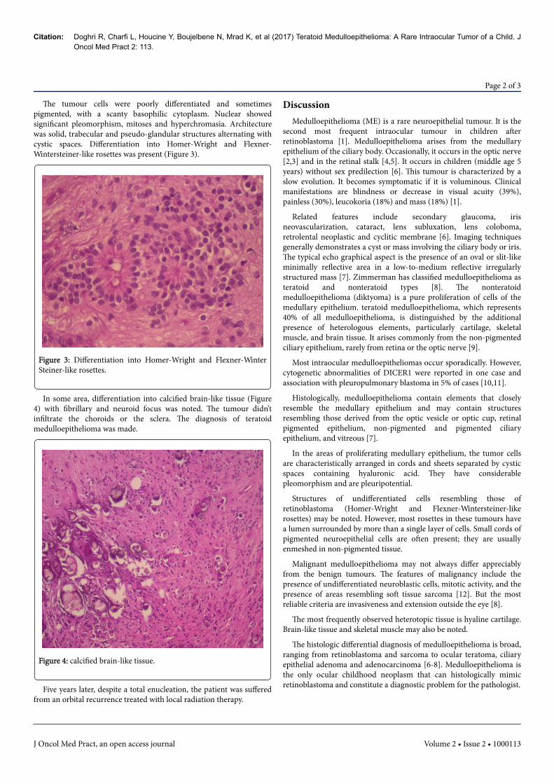
The tumour cells were poorly differentiated and sometimespigmented, with a scanty basophilic cytoplasm. Nuclear showedsignificant pleomorphism, mitoses and hyperchromasia. Architecturewas solid, trabecular and pseudo-glandular structures alternating withcystic spaces. Differentiation into Homer-Wright and Flexner-Wintersteiner-like rosettes was present (Figure 3).
Figure 3: Differentiation into Homer-Wright and Flexner-WinterSteiner-like rosettes.
In some area, differentiation into calcified brain-like tissue (Figure4) with fibrillary and neuroid focus was noted. The tumour didn’tinfiltrate the choroids or the sclera. The diagnosis of teratoidmedulloepithelioma was made.
Figure 4: calcified brain-like tissue.
Five years later, despite a total enucleation, the patient was sufferedfrom an orbital recurrence treated with local radiation therapy.
DiscussionMedulloepithelioma (ME) is a rare neuroepithelial tumour. It is the
second most frequent intraocular tumour in children afterretinoblastoma [1]. Medulloepithelioma arises from the medullaryepithelium of the ciliary body. Occasionally, it occurs in the optic nerve[2,3] and in the retinal stalk [4,5]. It occurs in children (middle age 5years) without sex predilection [6]. This tumour is characterized by aslow evolution. It becomes symptomatic if it is voluminous. Clinicalmanifestations are blindness or decrease in visual acuity (39%),painless (30%), leucokoria (18%) and mass (18%) [1].
Related features include secondary glaucoma, irisneovascularization, cataract, lens subluxation, lens coloboma,retrolental neoplastic and cyclitic membrane [6]. Imaging techniquesgenerally demonstrates a cyst or mass involving the ciliary body or iris.The typical echo graphical aspect is the presence of an oval or slit-likeminimally reflective area in a low-to-medium reflective irregularlystructured mass [7]. Zimmerman has classified medulloepithelioma asteratoid and nonteratoid types [8]. The nonteratoidmedulloepithelioma (diktyoma) is a pure proliferation of cells of themedullary epithelium. teratoid medulloepithelioma, which represents40% of all medulloepithelioma, is distinguished by the additionalpresence of heterologous elements, particularly cartilage, skeletalmuscle, and brain tissue. It arises commonly from the non-pigmentedciliary epithelium, rarely from retina or the optic nerve [9].
Most intraocular medulloepitheliomas occur sporadically. However,cytogenetic abnormalities of DICER1 were reported in one case andassociation with pleuropulmonary blastoma in 5% of cases [10,11].
Histologically, medulloepithelioma contain elements that closelyresemble the medullary epithelium and may contain structuresresembling those derived from the optic vesicle or optic cup, retinalpigmented epithelium, non-pigmented and pigmented ciliaryepithelium, and vitreous [7].
In the areas of proliferating medullary epithelium, the tumor cellsare characteristically arranged in cords and sheets separated by cysticspaces containing hyaluronic acid. They have considerablepleomorphism and are pleuripotential.
Structures of undifferentiated cells resembling those ofretinoblastoma (Homer-Wright and Flexner-Wintersteiner-likerosettes) may be noted. However, most rosettes in these tumours havea lumen surrounded by more than a single layer of cells. Small cords ofpigmented neuroepithelial cells are often present; they are usuallyenmeshed in non-pigmented tissue.
Malignant medulloepithelioma may not always differ appreciablyfrom the benign tumours. The features of malignancy include thepresence of undifferentiated neuroblastic cells, mitotic activity, and thepresence of areas resembling soft tissue sarcoma [12]. But the mostreliable criteria are invasiveness and extension outside the eye [8].
The most frequently observed heterotopic tissue is hyaline cartilage.Brain-like tissue and skeletal muscle may also be noted.
The histologic differential diagnosis of medulloepithelioma is broad,ranging from retinoblastoma and sarcoma to ocular teratoma, ciliaryepithelial adenoma and adenocarcinoma [6-8]. Medulloepithelioma isthe only ocular childhood neoplasm that can histologically mimicretinoblastoma and constitute a diagnostic problem for the pathologist.
Citation: Doghri R, Charfi L, Houcine Y, Boujelbene N, Mrad K, et al (2017) Teratoid Medulloepithelioma: A Rare Intraocular Tumor of a Child. JOncol Med Pract 2: 113.
Page 2 of 3
J Oncol Med Pract, an open access journal Volume 2 • Issue 2 • 1000113

Enucleation is generally recommended because local resection isinsufficient and the recurrent rate is high. The role of radiotherapy andchemotherapy are unknown.
The prognosis for medulloepithelioma limited to the eye is goodbecause of the slow growth rate, and rare lymphatic andhaematogenous dissemination [7].
In a series of 41 patients with ciliary body ME, systemic metastasisoccurred in 3 cases (8%) over a mean follow-up of 49 months, all ofwhom presented with extra scleral extension of tumor due to meandelay in diagnosis by 39 months [6]. Distant metastases to the lymphnodes, parotid glands, lungs and mediastinum have been described.Follow-up on the 56 patients reported by Broughton and Zimmermanshowed tumour-related deaths in 4 (12%) occurred in patients withmalignant tumours with extra ocular extension detected onhistopathological examination [8]. Deaths were preceded in three casesby orbital recurrence; 3 patients died with intracranial extension andthe fourth with distant metastasis. Of the original 56 tumours 37 werejudged histologically malignant and 10 had extra-ocular spread. Unlikecases reported in the literature in which the tumour recurs rapidly,recurrence occurred in our case five years later. Although the roles ofradiotherapy and chemotherapy are not well evaluated, a goodresponse to radiotherapy was achieved.
The major predictor of death was extra ocular extension [13]. Thevalue of radiation therapy and chemotherapy with extra ocular spreadwas too limited to draw meaningful conclusions [8,13].
ConclusionIn summary, because of the rarity of medulloepithelioma in adults,
only little information concerning its clinical evolution is available.Nevertheless this tumour should be taken into consideration in thedifferential diagnosis of retinoblastoma. Although, it is known recur ina very brief delay, recurrence occurred in our case five years later.
References1. Mojgan D (2014) Medulloepithelioma of the ciliary body. Bulletin of the
French Division of the AIP 60: 159-1642. Reese AB (1957) Medulloepithelioma (dictyoma) of the optic nerve. Am J
Ophthalmol 44: 4-6.3. Green WR, Iliff WJ, Trotter RR (1974) Malignant teratoid
medulloepithelioma of the optic nerve. Arch Ophthalmol 91: 451.4. Anderson SR (1962) Medulloepithelioma of the retina. Int Ophthalmol
Clin 2: 483-506.5. Mullaney J (1974) Primary malignant medulloepithelioma of the retinal
stalk. Am J Ophthalmol 77: 499-504.6. Saunders T, Margo CE (2012) Intraocular medulloepithelioma. Arch
Pathol Lab Med. 136: 212–216.7. Lloyd III WC, O’Hara M (2001) Malignant teratoid medulloepithelioma:
Clinical-echographic-histopathologic correlation. J AAPOS 5: 395-397.8. Zimmerman LE (1970) The remarkable polymorphism of tumors of the
ciliary epithelium. The Norman McAlister Gregg Lecture. Part 1. TransAustr Coli Ophthalmol 2: 114-125.
9. Andersen S (1971) Differentiation features in some retina tumor and indysplastic retinal conditions. Am J Ophtalmol 1: 231-241
10. Kramer GD, Arepalli S, Shields CL, Shield JA (2014) Ciliary bodymedulloepithelioma association with pleuropulmonary blastoma infamilial tumour predisposition syndrome. J Pediatr OphtalmolStrabismus 51: e48-50.
11. Priest JR, Williams GM, Manera R, Jekinson H, Brûndler MA, et al (2011)Ciliary body medulloepithelioma: Four cases associated withpleuropulmonary blastoma: A report from the Internationalpleuropulmonary blastoma registry. Br J OPhtalmol 95: 1001-1005.
12. Yanko L, Behar A (1978) Teratoid intraocular medulloepithelioma. Am JOphtalmol 85: 850-853.
13. Timothy S, Curtis EM (2012) Intraocular medulloepithelioma. ArchPathol Lab Med 136: 216-212.
Citation: Doghri R, Charfi L, Houcine Y, Boujelbene N, Mrad K, et al (2017) Teratoid Medulloepithelioma: A Rare Intraocular Tumor of a Child. JOncol Med Pract 2: 113.
Page 3 of 3
J Oncol Med Pract, an open access journal Volume 2 • Issue 2 • 1000113