cause van der woude syndrome and disrupt oral … american journal of human genetics, volume 94...
TRANSCRIPT
The American Journal of Human Genetics, Volume 94
Supplemental Data
Dominant Mutations in GRHL3
Cause Van der Woude Syndrome
and Disrupt Oral Periderm Development Myriam Peyrard-Janvid, Elizabeth J. Leslie, Youssef A. Kousa, Tiffany L. Smith, Martine Dunnwald, Måns Magnusson, Brian A. Lentz, Per Unneberg, Ingegerd Fransson, Hannele K. Koillinen, Jorma Rautio, Marie Pegelow, Agneta Karsten, Lina Basel-Vanagaite, William Gordon, Bogi Andersen, Thomas Svensson, Jeffrey C. Murray, Robert A. Cornell, Juha Kere, and Brian C. Schutte

1
SUPPLEMENTAL INFORMATION
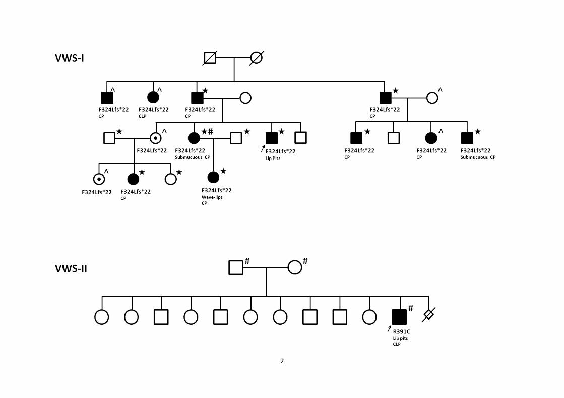
Figure S1: Pedigrees of the eight VWS families with GRHL3 mutation
In each family, the corresponding GRHL3 mutation is named under each individual where it is
detected. Mutations were detected by whole-exome sequencing (* in VWS-I only), TaqMan
genotyping (^) or Sanger sequencing (#). Mutation carriers without any detected phenotypic
characteristics of VWS are indicated with a black dot in their symbol. Phenotypical
characteristics of affected individuals are cleft palate (CP), cleft lip and palate (CLP) or
unknown (?). The proband in VWS-IV has been shown to be carrier of a rare variant in IRF6
(K80R) 1. VWS-I was previously described in
2, VWS-IV in
1 under the denomination VWS-
SM13 and VWS-VII in 3 under the denomination VWS-12.
2

3

4
VWS-V
34
4
8
IVS11+1Lip pitsCP
IVS11+1CPAnodontia
IVS11+1Lip pits
IVS11+1Lip pitsUvula anomaly
IVS11+1Lip pitsCPUvula anomaly
IVS11+1Uvula anomaly
Bifid uvula
IVS11+1Lip moundsCP
Lip pits
#
##
###
#
#
10

5

6
Figure S2: Multiple alignment and protein domains of GRHL3 gene products from
human, mouse and zebrafish
The alignment was done with the input protein sequences from human (NP_937816 for
H.s.GRHL3v2, Homo sapiens variant 2), Mouse (NP_001013778.1 for M.m.Grhl3, Mus
musculus) and zebrafish (XP_001332938.3 for D.r.Grhl3, Danio rerio) and using Clustal
Omega 1.1.0 software (www.ebi.ac.uk/Tools/msa/clustalo). Amino acids conserved in the 3
species, are denoted by a star below the alignment. In the human protein sequence, the
corresponding coding exons are numbered 1 to 16 and are identifiable by a blue or a black
protein sequence. Amino acids in red are encoded by two neighboring exons. In the human
GRHL3 protein sequence, the three known protein domains are underlined in orange for the
transactivation domain (exons 2 and 3, amino acids 25-74), in green for the DNA-binding
domain (exons 6 to 10, amino acids 230-423) and in pink for the dimerization domain (exons
13 to 16, amino acids 493-602). For each of the exonic mutations/variants detected in our set
of families, the first amino acid affected by the mutation is labeled in red (protein truncation)
or in green (missense), and a red arrow indicates the location of the splice site mutation
IVS11+1 (from VWS-V). The known repressive and activating protein domains of the murine
Grhl3 are underlined in the mouse sequence by a continuous line (repressive, amino acids 1-
102 and 296-603) or dotted line (activating, amino acids 102-296) 4. A blue arrow above
human exon 2 indicates the start of the GRHL3 protein produced by variant 4 (v4), and used
in the zebrafish experiments.

7
1 2 H.s.GHRL3v2 MSNELDFRSVRLLKNDPVNLQKFSYTSEDEAWKTYLENPLTAATKAMMRVNGDDDSVAAL
M.m.Grhl3 MSNELDFRSVRLLKNDPVSFQKFPYSNEDEAWKTYLENPLTAATKAMMRVNGDEESVAAL
D.r.Grhl3 MTKEIEAL--MVQQNESFSHIRTYESYVMDYWTNMDSGNLS---KTKPR-LASDEDLATL
* * * * * * * * *
3 Y90Hfs 4 H.s.GHRL3v2 SFLYDYYMGPKEKRILSSSTGGRNDQGKRYYHGME-----YETDLTPLESPTHLMKFLTE
M.m.Grhl3 SFLYDYYMGPKEKRILSSSTGGRNDQGKKFYHSMD-----YEPDLAPLESPTHLMKFLTE
D.r.Grhl3 NLLYDACKPSKEQKMTSCAR------ESSIYKSMERTANSSSPELAPLE-NAHIMKFLSE
*** ** * * * * *** * **** *
H.s.GHRL3v2 NVSGTPEYPDLLKKNNLMSLEGALPTPGKAAPLPAGPSKLEAGSVDSYLLPTTDMYDNGS
M.m.Grhl3 NVSGSPDYTDQLKKNNLLGLEGVLPTPGKTNTVPPGPSKLEASSMDSYLLPASDIYDNGS
D.r.Grhl3 NMSFNPSKPS----------------------------------TDSYT--TDNYDKQVN
* * * ***
5 H.s.GHRL3v2 LNSLFESIHGVPPTQRWQPDSTFKDDPQESMLFPDILKTSPEPPCPEDY---PSLKSDFE
M.m.Grhl3 LNSLFESIHGVPPTQRWQPDSTFKDDPQESLLFPDILKTSPDPPCPEDY---PGLKSDFE
D.r.Grhl3 LNNIFDSLLPQPSQKSWQSDQTFLEATPEHIGHNGFG-GQTSPVYSDSYSSPGRYRNDFQ
** * * * ** * ** * * * **
6 H.s.GHRL3v2 YTLGSPKAIHIKSGESPMAYLNKGQFYPVTLRTPAGGKGLALSSNKVKSVVMVVFDNEKV
M.m.Grhl3 YTLGSPKAIHIKAGESPMAYLNKGQFYPVTLRTPAGGKGLALSSSKVKSVVMVVFDNDKV
D.r.Grhl3 FLLGAPQASQHKTTEIPMVYLNKGQFYPITLQGVDSTAGV--PCSKVKTVIMAVFENDKS
** * * * * ** ********* ** * *** * * ** * *
R298H 7 F324Lfs 8 H.s.GHRL3v2 PVEQLRFWKHWHSRQPTAKQRVIDVADCKENFNTVEHIEEVAYNALSFVWNVNEEAKVFI
M.m.Grhl3 PVEQLRFWRHWHSRQPTAKQRVIDVADCKENFNTVQHIEEVAYNALSFVWNVNEEAKVFI
D.r.Grhl3 PEMQLKYWNHWHARQPTVKQRVIDIADYKEVFSGVSNVEEVAFNALSFIWNTNEEAKVHI
* ** * *** **** ****** ** ** * * **** ***** ** ****** *
9 R391C 10 H.s.GHRL3v2 GVNCLSTDFSSQKGVKGVPLNLQIDTYDCGLGTERLVHRAVCQIKIFCDKGAERKMRDDE
M.m.Grhl3 GVNCLSTDFSSQKGVKGVPLNLQIDTYDCGAGTERLVHRAVCQIKIFCDKGAERKMRDDE
D.r.Grhl3 GINSLSTDFSSQKGVKGLPLNLQIDTYDFSSGNNRLIHRAVCQVKIFCDKGAERKMRDEE
* * ************* ********** * ** ****** ************** *
11 H.s.GHRL3v2 RKQFRRKVKCPDSS-NSGVKGCLLSGFRGNETTYLRPETDLETPPVLFIPNVHFSSLQRS
M.m.Grhl3 RKQFRRKVKCPDSSNNAGIKGCLLSGFRGNETTYLRPETDLETQPVLFIPNLHFSSLQRP
D.r.Grhl3 RKRSKRRTKNTADSSNNNCKQALVSSSVGKDSTYFKTLDDHVTQPVLFIPEMHFSTMQRC
** * * * * * * * * ** * * ****** *** **
IVS11+1 12 13 R520Q E522Lfs V526Cfs H.s.GHRL3v2 GGAAPSAGPSSSNRLPLKRTCSPFTEEFEPLPSKQAKEGDLQRVLLYVRRETEEVFDALM
M.m.Grhl3 GGVVPSAGHSSSDRLPLKRTCSPFAEEFEPLPSKQAKEDDLQRVLLYVRRETEEVFDALM
D.r.Grhl3 GLVPPV-SLEESDRSSLKRYADANE--QSSSPPCKQPRRDEQRVLLYVRRESEEVFDALM
* * * * *** * * ********** ********
14 15 N554S 16 H.s.GHRL3v2 LKTPDLKGLRNAISEKYGFPEENIYKVYKKCKRGILVNMDNNIIQHYSNHVAFLLDMGEL
M.m.Grhl3 LKTPDLKGLRNAISEKYGLPEENICKVYKKCKRGILVNMDNNIIQHYSNHVAFLLDMGEL
D.r.Grhl3 LNTPNLKGLKEAISEKYGMQEDTIGKIFKKCKRGIFVNMDDNIIEHYSNHSAFLIEISEV
* ** **** ******* * * * ******* **** *** ***** *** *
H.s.GHRL3v2 DG-KIQIILKEL
M.m.Grhl3 DG-KIQIILKEL
D.r.Grhl3 IVNHYQVTLMEL
* * **

8
Figure S3: Molecular changes in the oral epithelium of Irf6-/-
and Grhl3-/-
embryos
Comparison of expression in E15.5 wild type (A,E), Irf6-/-
(B,F) and Grhl3-/-
(C,D,G,H)
embryos. Images in columns 3 and 4 are taken at the tooth germs sites in two different Grhl3-/-
embryos to illustrate the dynamic changes in gene expression in areas without (C,G) and with
oral adhesions (D,H), respectively. Irf6 expression is seen in epithelial cells of the tooth germ
and oral epithelium in wild type embryos (A) but absent in Irf6-/-
embryos (B). Irf6 expression
is detected in the oral epithelium (arrowhead) of Grhl3-/-
embryos, but reduced at sites of oral
adhesion (arrow) (C,D). Activated Notch1 (Act N1) is seen in the oral periderm of wild type
embryos (E) while it is undetectable in Irf6-/-
littermate embryos (F). Grhl3-/-
embryos (G,H)
show loss of Act N1 in areas of oral adhesion (arrow) but not in adjacent healthy epithelium
(arrowhead). Scale bar is 20 m (A-H). Labeled oral structures are mandible (mn) and tooth
germ (tg).

9

Supplemental References
1. Malik S, Kakar N, Hasnain S, Ahmad J, Wilcox ER, Naz S (2010) Epidemiology of Van der Woude
syndrome from mutational analyses in affected patients from Pakistan. Clin Genet 78:247-256 2. Koillinen H, Wong FK, Rautio J, Ollikainen V, Karsten A, Larson O, Teh BT, Huggare J, Lahermo P,
Larsson C, Kere J (2001) Mapping of the second locus for the Van der Woude syndrome to chromosome 1p34. Eur J Hum Genet 9:747-752
3. Peyrard-Janvid M, Pegelow M, Koillinen H, Larsson C, Fransson I, Rautio J, Hukki J, Larson O, Karsten AL, Kere J (2005) Novel and de novo mutations of the IRF6 gene detected in patients with Van der Woude or popliteal pterygium syndrome. Eur J Hum Genet 13:1261-1267
4. Kudryavtseva EI, Sugihara TM, Wang N, Lasso RJ, Gudnason JF, Lipkin SM, Andersen B (2003) Identification and characterization of Grainyhead-like epithelial transactivator (GET-1), a novel mammalian Grainyhead-like factor. Dev Dyn 226:604-617

Table S1: Human GRHL3 primers used in Sanger sequencing mutation screening
GRHL3 exons Primer Sequencea
1 and 1' F CTCACCAAGGAAGGAATTGG
R TAGCTTGAGACTGGGGCTTG
1'' F GTCTTAGCCGAGCAGCCATAG
R GTAGTGGATTTGGGAACCTCCT
2 F GTGGCAGGAAGAGGCAGTTTC
R CAAAGGCCCAGAGATGAGG
3 F AAAGCTGCAGGAGGGGATT
R TCAGCACTGTGCCTCCTGT
4 and 5 F GCATGCTGGATGGACCTAAA
R TTCATCCCCCACTTCTCATT
6 and 7 F TTTTCCAAGGTCAAACAGCA
R GACAGAGGTCAGAGCCAGGT
8 F GAGTGAGGCCCAGTTTTTAATG
R CGTCGGAGCAAATGACACTA
9, 10 and 11 F CTTGGCAGTCTAGCGGAAAC
R GAAGCCTCCTCTTTGTGTGC
12 F CTGAGCAGAATGGGCTAGAA
R AGGCGTGTGGTTGTTTCTCT
13 and 14 F TGATGGGCTAAGGGACTCAC
R GATAACATCGCAGAGGCACA
15 F GCACACCCAGATGTTAATGG
R AGAGGTGACCAGTGGCTTTG
16 F ACCACATCCCCTCTTCCATT
R TAGCCATCTCTTTCCAGCAGAC
16' F TTGCTTCTGATACTCCCCACTT
R CAGCCCTCTGCTTTTCTCTG
a All primers had a melting temperature of 61
oC

Table S2: List of the 16 SNPs spread over the genome, used for confirming the de novo
mutations
SNP Chromosomal band
rs1051614 1q21.3
rs10204426 2p11.2
rs237887 3p26.1
rs1063499 5p13.1
rs654351 6p24.3
rs1366883 7q21.13
rs4458901 8p23.2
rs2515617 9q31.1
rs2136892 10q21.1
rs1729410 11q23.3
rs1053900 14q32.2
rs140685 15q12
rs3744262 17p13.1
rs2296241 20q13.2
rs1789953 21q22.3
rs2051616 22q13.31

Table S3: Frequency of genotypes and resorbing embryos from a cross between Irf6+/-
and Grhl3+/-
mice
E13.5 E17.5 Embryos P0 P21 Pups Total
Litters 3 2 5 6 5 11 32
Irf6+/+
;Grhl3+/+
5 3 8 15 5 20 28
Irf6+/-
;Grhl3+/+
4 3 7 10 9 19 26
Irf6+/+
;Grhl3+/-
11 4 15 12 12 24 39
Irf6+/-
;Grhl3+/-
5 4 9 9 1 10 19
Resorbing 3 3 6 N/A N/A NA 6
(p=0.0008)
Total 25 14 45 46 27 73 118
p-value 0.18 0.96 0.34 0.60 0.01 0.12 0.06

We intercrossed mice heterozygous for the Irf6 genetrap allele (Irf6+/gt
here called Irf6
+/-) with
mice heterozygous for the Grhl3 knockout allele (Grhl3+/-
) to generate wild-type
(Irf6+/+
;Grhl3+/+
) embryos; Irf6+/-
or Grhl3+/-
single mutant embryos; and Irf6+/-
;Grhl3+/-
double
heterozygous embryos. We detected significant embryonic resorptions at combined E13.5 and
E17.5. Furthermore, we found a significant reduction in Irf6+/-
;Grhl3+/-
double heterozygous
mice at weaning.