case approach in glomerular disease - berlin...
TRANSCRIPT

Case Approach in Glomerular Disease
Bancha Satirapoj, MD Division of Nephrology
Department of Medicine Phramongkutklao Hospital and College of Medicine

Asymptomatic Isolated proteinuria 150 mg to 3 g/day Hematuria > 2 red blood cells (RBC)/high-power field in spun urine (RBC usually dysmorphic)
Nephritic syndrome • An abrupt onset of glomerular hematuria
(RBC cast or dysmorphic RBCs) • Proteinuria <3 g/day • Azotemia • Edema • Oliguria • Recent onset hypertension
Nephrotic syndrome • Proteinuria
• Adult >3.5 g/day • Child > 40 mg/h per m2
• Edema • Hypoalbuminemia <3.5 g/dl • Hypercholesterolemia • Lipiduria
Rapidly progressive glomerulonephritis • Glomerular disease characterized by
extensive crescents (usually >50%) • A rapid loss of renal function (usually a 50%
decline in GFR within 3 months)
Chronic glomerulonephritis • Slowing developing renal insufficiency • Proteinuria > 3 g/day and hematuria • Hypertension • Shrunken smooth kidneys
Satirapoj B. Common Problems in Internal Medicine. Bangkok 2010. p. 498-513.

Glomerular Disease
❖ Primary Glomerular Disease
❖ Idiopathic
❖ Secondary Glomerular Disease
❖ Systemic disease involving multiple organs

Secondary glomerular diseaseSystemic Diseases Infections
- Diabetes mellitus - HIV infection
- SLE/vasculitis - Hepatitis B and C
- Amyloidosis - Malaria- Carcinoma - Syphilis- Lymphoma and myeloma
- PreeclampsiaDrugs Inherited Disorders
- Gold - Alport syndrome- Antibiotics - Congenital NS- NSAIDs - Nail-patella syndrome- Penicillamine- Heroin

Primary glomerular disease
❖ Minimal change nephrotic syndrome (MCD)
❖ Focal segmental glomerulosclerosis (FSGS)
❖ Membranous nephropathy (MN)
❖ IgM nephropathy
❖ IgA nephropathy
❖ Membranoproliferative glomerulonephritis (MPGN)

Picture glomerulus

Minimal change disease (MCD) Membranous nephropathy (MN)Focal segment glomerulosclerosis (FSGS)
Membranoproliferative GN (MPGN) Mesangial proliferative GN (IgA or IgM nephropathy)

Manifestation of glomerular diseases
Disease Nephrotic features Nephritic features
Minimal change glomerulopathy ++++ -
Membranous glomerulopathy ++++ +
Focal segmental glomerulosclerosis +++ ++
Fibrillary glomerulonephritis +++ ++
Mesangioproliferative glomerulopathy (IgAN, LN) ++ ++
Membranoproliferative glomerulonephritis (MPGN) ++ +++
Proliferative glomerulonephritis (IgAN, LN) ++ +++
Acute diffuse proliferative glomerulonephritis (PSGN) + ++++
Crescentic glomerulonephritis + ++++
Adapted from Brenner & Rector’s the kidney 10th edition, 2016

Nephrotic syndromeDiseases Associations Serologic Tests
Minimal change disease Allery, atopy, NSAIDs, Hodgkin disease -
Focal segmental glomerulosclerosisAfrican American, HIV infection, heroin, pamidronate, obesity
HIV antibody
Membranous nephropathy
Drugs; gold, penicillamine, NSAIDs Infection; hepatitis B, C; malaria Lupus nephritis Malignancy; breast, lung gastrointestinal tract
Anti-PLA-R2 antibody Hepatitis B surface antigen, anti-HCV antibody ANA, Anti-DNA antibody
Diabetic nephropathy Other diabetic microangiopathy -
AmyloidosisMyeloma Rheumatoid arthritis, bronchiectasis, Crohn disease, Familial Mediterranean fever
Plasma free light chain Serum protein electrophoresis, urine immunoelectrophoresis
Adapted from Johnson RJ, Feehally, J and Floege R . Comprehensive clinical nephrology. 2015, 189.

Nephritis syndromeDiseases
Associations Serologic Tests
MPGN type I C4 nephritic factor Low C3 and C4
MPGN type II C3 nephritic factor Low C3 and normal C4
Post-streptococcal glomerulonephritis Pharyngitis, impetigo ASO titer, streptozyme antibody
Post-infectious disease -Endocarditis -Shunt
Cardiac murmur Treated hydrocephalus
Blood cultures, low C3 Blood cultures, low C3
IgA nephropathyUpper respiratory or gastrointestinal infection
-
Lupus nephritisOther multi-systemic features of lupus
ANA, anti-ds DNA antibody, low C3 and C4
Cryoglobulinemic glomerulonephritis Hepatitis CAnti-hepatitis C virus antibody, rheumatoid factor, cryoglobulinemia, low C3 and C4
Adapted from Johnson RJ, Feehally, J and Floege R . Comprehensive clinical nephrology. 2015, 189.

History/physical examination
❖ Family history of CKD, hearing loss
❖ Perimacular white dot-and-fleck retinopathy, ant lenticonus:
❖ Alport syndrome
❖ History of diabetes
❖ DR/diabetic neuropathy
❖ Diabetic nephropathy
Diabetic retinopathy
Anterior lenticonus Dot-and-fleck retinopathy

History/physical examination
❖ Photosensitivity, arthritis, alopecia, oral ulcer, malar rash, Roth spot, discoid lesion:
❖ Lupus nephritis
❖ Palpable purpura, vasculitis;
❖ Systemic vasculitis/ SLE
❖ Cryoglobulinemia
❖ Sub acute endocarditis
Malar rash
Palpable purpura
Cytoid body

History/physical examination
❖ Hepatosplenomegaly, periorbital purpura, macroglossia, carpal tunnel syndrome
❖ Amyloidosis
Periorbital purpura Macroglossia

Kidney biopsy
❖ Secondary glomerular diseases
❖ High risk for progressive disease (rising SCr, HT, proteinuria)
❖ Non response with corticosteroids (steroid resistance, steroid dependent, frequency relapse nephrotic syndrome)
❖ Longterm immunosuppressive agents (cyclophosphamide, cyclosporine)

Risk factors of Progressive Disease
❖ Male
❖ Advanced age (>50 years)
❖ Persistent heavy proteinuria (>3.5 g/d)
❖ Hypertension
❖ Impaired GFR

Grade and Quality of evidence
KDIGO. Kidney International Supplements (2012) 2, 143–153

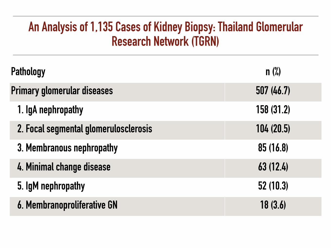
An Analysis of 1,135 Cases of Kidney Biopsy: Thailand Glomerular Research Network (TGRN)
Pathology n (%)
Primary glomerular diseases 507 (46.7)
1. IgA nephropathy 158 (31.2)
2. Focal segmental glomerulosclerosis 104 (20.5)
3. Membranous nephropathy 85 (16.8)
4. Minimal change disease 63 (12.4)
5. IgM nephropathy 52 (10.3)
6. Membranoproliferative GN 18 (3.6)

IgA nephropathy

IgA nephropathy: Pathology
❖ Immunohistology is the clue of diagnosis
❖ Mesangial cell proliferation with IgA deposit predominate
Magistroni R, et al. Kidney International (2015) 88, 974–989

Aberrant IgA1 antibody
Kidney Int 2012:81:833
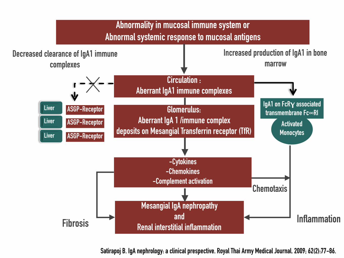
Circulation : Aberrant IgA1 immune complexes
ASGP-Receptor
ASGP-Receptor
Mesangial IgA nephropathy and
Renal interstitial inflammation
Activated Monocytes
IgA1 on FcRγ associated transmembrane Fc∞RI
Fibrosis
ASGP-Receptor
Abnormality in mucosal immune system or Abnormal systemic response to mucosal antigens
Glomerulus: Aberrant IgA 1 /immune complex
deposits on Mesangial Transferrin receptor (TfR)
Liver
Decreased clearance of IgA1 immune complexes
Chemotaxis
Inflammation
Increased production of IgA1 in bone marrow
-Cytokines -Chemokines
-Complement activation
Satirapoj B. IgA nephrology: a clinical prespective. Royal Thai Army Medical Journal. 2009; 62(2):77-86.
Liver
Liver

Multi-hit pathogenesis model of IgA nephropathy
Magistroni R, et al. Kidney International (2015) 88, 974–989

IgA nephropathy and associated disorders
Adapted from Brenner & Rector’s the kidney 10th edition, 2016
IgA vasculitis Neoplasia: Mycosis fungoides, CA lung
HIV infection Cyclic neutropenia
Toxoplasmosis Immunothrombocytopenia
Seronegative spondyloarthropathy Gluten-sensitive enteropathy
Celiac disease Scleritis
Dermatitis herpetiformis Sicca syndrome
Crohn’s disease Mastitis
Alcoholic cirrhosis Pulmonary hemosiderosis
Ankylosing spondylitis Berger’s disease
Reiter’s syndrome Leprosy

IgA nephropathy
Lupus nephritis
Crescentic glomerulonephritis
Normal glomeruli Membranoproliferative or Necrotizing
Mesangial proliferative
Sclerosing
Asymptomatic hematuria/proteinuria
Acute nephritic/ Nephrotic syndrome
Rapidly progressive Glomerulonephritis
Chronic Glomerulonephritis
Clinical presentation
Renal pathology

Clinical presentations relation to age
Adapted from Johnson RJ, Feehally, J and Floege R . Comprehensive clinical nephrology. 2015.
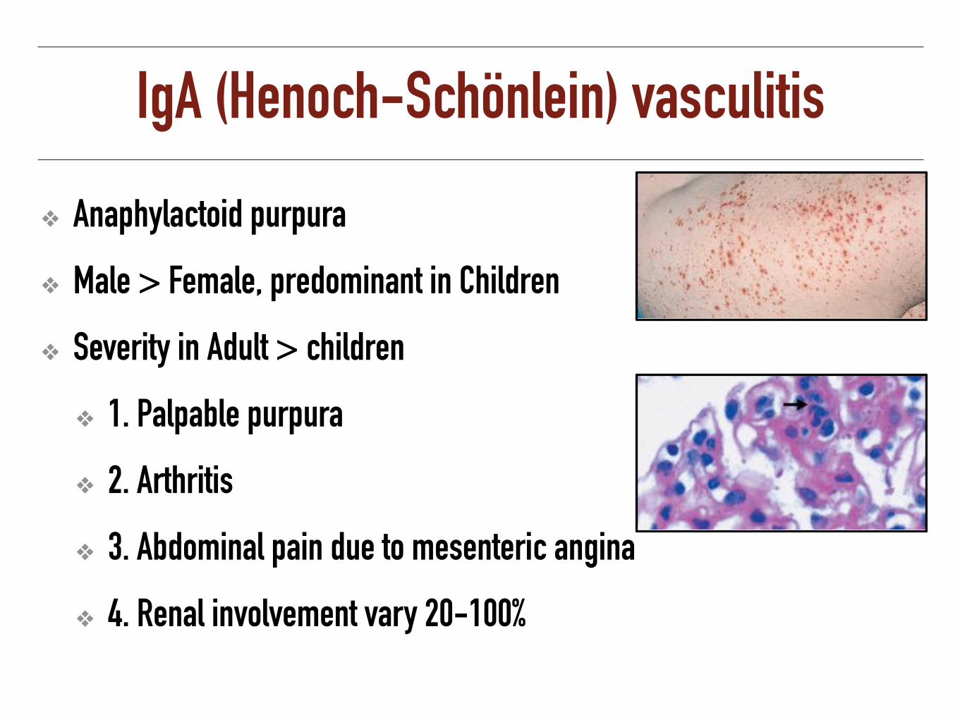
IgA (Henoch-Schönlein) vasculitis
❖ Anaphylactoid purpura
❖ Male > Female, predominant in Children
❖ Severity in Adult > children
❖ 1. Palpable purpura
❖ 2. Arthritis
❖ 3. Abdominal pain due to mesenteric angina
❖ 4. Renal involvement vary 20-100%

IgA nephropathy: Clinical feature
❖ Wide spectrum of clinical presentations
❖ Recurrent macroscopic hematuria provoke by mucosal infection (synpharyngitis) (40-50%)
❖ Microscopic hematuria with or without proteinuria (30-40%)
❖ Nephrotic syndrome (5%)
❖ RPGN (<10%)

Treatment of IgA Nephropathy
❖ Long-term ACE-I or ARB treatment: proteinuria >1 g/d, with up-titration of the drug depending on blood pressure (1B)
❖ ACE-I or ARB be titrated upwards to achieve proteinuria <1 g/d (2C)
❖ Fish oil: Persistent proteinuria >1 g/d, despite 3–6 months of optimized supportive care (including ACE-I or ARBs and BP control) (2D)
KDIGO. Kidney International Supplements (2012) 2, 143–153

Meta-analysisSubgroup analysis for the effect of corticosteroid on composite renal endpoint
❖ High-dose and short-term steroid therapy (prednisone>30 mg/d or high-dose IVMP with duration <1 year) produced significant renal protection
❖ Low-dose and long-term steroid therapy did not benefit.
❖ Steroid therapy was associated with a 55% higher risk for adverse events
Lv J, et al. J Am Soc Nephrol: 2012: 23: 1108–1116.
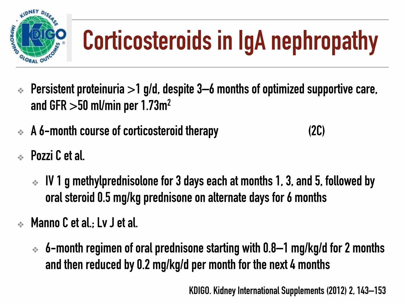
Corticosteroids in IgA nephropathy
❖ Persistent proteinuria >1 g/d, despite 3–6 months of optimized supportive care, and GFR >50 ml/min per 1.73m2
❖ A 6-month course of corticosteroid therapy (2C)
❖ Pozzi C et al.
❖ IV 1 g methylprednisolone for 3 days each at months 1, 3, and 5, followed by oral steroid 0.5 mg/kg prednisone on alternate days for 6 months
❖ Manno C et al.; Lv J et al.
❖ 6-month regimen of oral prednisone starting with 0.8–1 mg/kg/d for 2 months and then reduced by 0.2 mg/kg/d per month for the next 4 months
KDIGO. Kidney International Supplements (2012) 2, 143–153

Variants of IgA nephropathy
❖ MCD with mesangial IgA deposits
❖ Treatment as for MCD in nephrotic patients showing pathological findings of MCD with mesangial IgA deposits on kidney biopsy (2B)
KDIGO. Kidney International Supplements (2012) 2, 143–153
❖ Crescentic IgA Nephropathy
❖ Steroids and cyclophosphamide, analogous to the treatment of ANCA vasculitis (2D)

Immunosuppressive agents
❖ No treating with corticosteroids combined with cyclophosphamide or azathioprine in IgAN patients except crescentic IgAN (2D)
❖ No using immunosuppressive therapy in patients with GFR <30 ml/min per 1.73m2 except crescentic IgAN (2C)
❖ No using MMF in IgAN (2C)
KDIGO. Kidney International Supplements (2012) 2, 143–153
VALIGA-Consortium: Corticosteroids in IgAN
Tesar V, et al. J Am Soc Nephrol: 2015: 26: 2248–2258.

Response to CS and RASB compared with RASB alone in propensity-matched individuals. (A) Entire propensity-matched cohort. (B) Stratified by initial eGFR.
Tesar V, et al. J Am Soc Nephrol: 2015: 26: 2248–2258.
Steroids reduced proteinuria and the rate of renal function decline and increased renal survival extended to those with an eGFR<50 ml/min per 1.73 m2
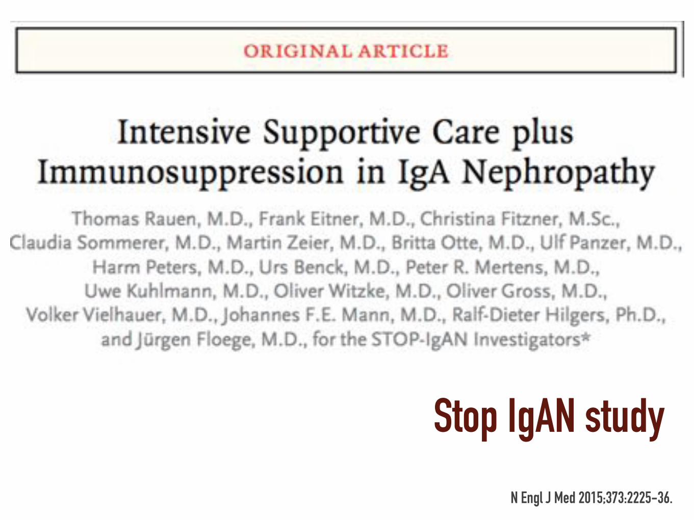
Stop IgAN study
N Engl J Med 2015;373:2225-36.

Stop IgAN studyComprehensive supportive care : RAS blocker, keep BP< 125/75 mmHg
High risk patients Persistent proteinuria > 0.75 g/day, but < 3.5 g/day
Supportive care group Immunosuppression group
eGFR> 60 ml/min/1.73m2
Glucocorticoid monotherapy IVMP 1 g/d x3 day of months 1,3, and 5) prednisolone 0.5 mg/kg AD on the other days
eGFR 30-59 ml/min/1.73m2
Cyclophosphamide 1.5 mg/kg/d for 3 months, followed by azathioprine 1.5 mg/kg/d during months 4 through 36, plus oral prednisolone
N Engl J Med 2015;373:2225-36.

After 3 years, 4 patients (5%) in the supportive-care group, as compared with 14 (17%) in the immunosuppression group, had a full clinical remission (P=0.01).
Addition of immunosuppressive therapy to intensive supportive care in patients with high-risk IgA nephropathy did not significantly improve the outcome
Full clinical remission (UPCR <0.2) Stable renal function (decrease in the eGFR of <5 ml/min/1.73m2 from baseline eGFR at the end of the 3-year trial phase)
N Engl J Med 2015;373:2225-36.

Secondary End Points on the Basis of the Analysis of Available Cases at the End of the Trial Phase
N Engl J Med 2015;373:2225-36.

Patients with AKI associated with macroscopic hematuria
KDIGO. Kidney International Supplements (2012) 2, 143–153
AKI and macroscopic hematuria Renal biopsy
Causes other than IgAN: (Crescentic GN, vasculitis, LN, postinfectious GN)
IgAN (Dominant with IgA in glomeruli by immunohistology)
ATN and intratubular erythrocytic casts
Crescentic IgAN
Supportive treatment as in
other types of ATN.
Repeated episodes of AKI accompanying macroscopic hematuria: Consider a kidney biopsy when no improvement of kidney function is observed after at
least 5 days from the onset of kidney function worsening
Steroids and cyclophosphamide as in
crescentic ANCA vasculitis

IgA nephropathy: Poor prognosis
❖ Older age
❖ Increase BMI
❖ Severity of proteinuria
❖ Persistent microscopic hematuria
❖ Hypertension
❖ Renal impairment
❖ Diffuse proliferative lesion with crescents
❖ Glomerulosclerosis
❖ Tubular atrophy, interstitial fibrosis
❖ Vascular wall thickening
❖ Capillary loop IgA deposit
Goto M, et al. Nephrol Dial Transplant 2009;24:3068-74. Cattran DC, et al. Kidney Int 2009;76:534-45.
An Analysis of 1,135 Cases of Kidney Biopsy: Thailand Glomerular Research Network (TGRN)
Pathology n (%)
Primary glomerular diseases 507 (46.7)
1. IgA nephropathy 158 (31.2)
2. Focal segmental glomerulosclerosis 104 (20.5)
3. Membranous nephropathy 85 (16.8)
4. Minimal change disease 63 (12.4)
5. IgM nephropathy 52 (10.3)
6. Membranoproliferative GN 18 (3.6)

Membranoproliferative GN

MPGN: Pathology
Endocapillary proliferation with GBM thickening/double contour
Granular deposition of IgG and C3 in the mesangium and capillary wall
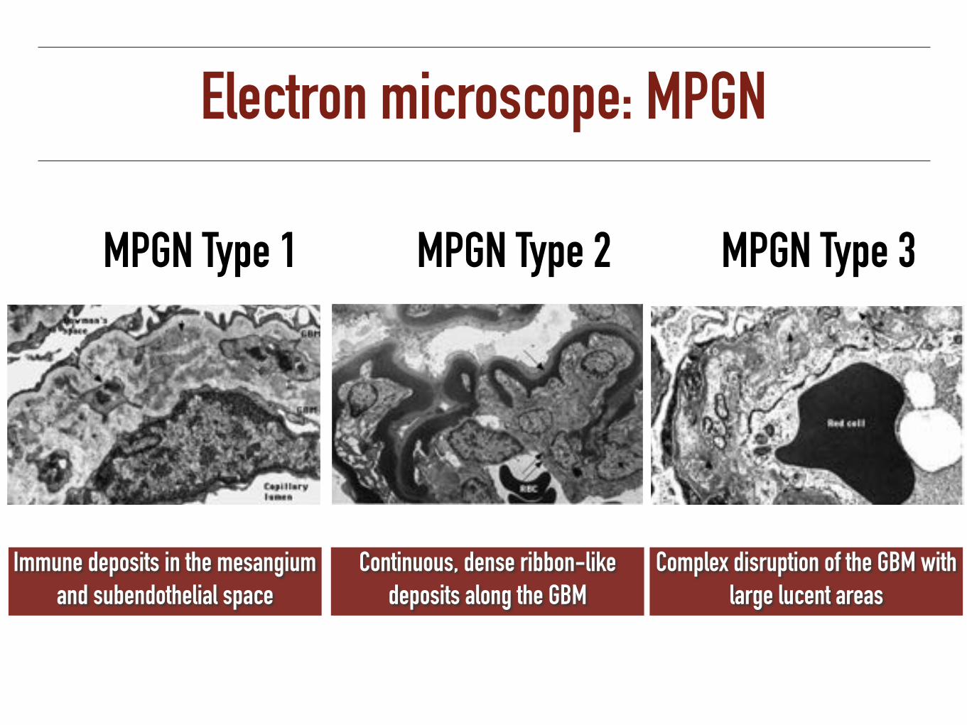
Electron microscope: MPGN
MPGN Type 1 MPGN Type 2 MPGN Type 3
Immune deposits in the mesangium and subendothelial space
Continuous, dense ribbon-like deposits along the GBM
Complex disruption of the GBM with large lucent areas

Newer classification based on immunopathology
IgG and/or C3 component

MPGN pattern (Double contour and mesangial expansion)
C3 staining No staining
❖ Infection (sub acute IE) ❖ Monoclonal gammopathy ❖ Autoimmune disease (SLE)
Dense deposit disease GN with isolated C3
Thrombotic microangiopathy
C3 and Ig staining
Immune complex: classical pathway, low C3/C4 or alternative pathway: low C3
C3 nephritic factor (C3 NeF) and Circulating IgG resulting in persistent C3 breakdown

MPGN: Clinical manifestration
❖ Focal glomerulonephritis (dysmorphic RBC, occasionally red cell casts, and proteinuria)
❖ Hypertension: 50-80%
❖ Nephritic syndrome/RPGN: 25%
❖ Non nephrotic range proteinuria: 25%
❖ Nephrotic syndrome: 50%
❖ Spontaneous improvement < 10%
KDIGO. Kidney International Supplements (2012) 2, 143–153

MPGN: treatment
❖ Optimal therapy of idiopathic MPGN remains uncertain
❖ Nephrotic syndrome and progressive decline of kidney function
❖ Oral cyclophosphamide or MMF plus low-dose alternate day or daily corticosteroids with initial therapy <6 mo (2D)
Evaluate patients with the histological (light-microscopic) pattern of MPGN for underlying diseases before considering a specific treatment regimen (Not Graded)
KDIGO. Kidney International Supplements (2012) 2, 143–153

Secondary Causes of MPGN
Associated with infection Associated with Rheumatologic disease
Hepatitis B and C Systemic lupus erythematosus
Visceral abscesses Scleroderma
Infective endocarditis Mixed essential cryoglobulinemia with or without hepatitis C infectionShunt nephritis
Quartan malaria Sarcoidosis
Schistosoma nephropathy Anti–smooth muscle syndrome
Mycoplasma infection Sjögren’s syndrome
Associated with Malignancy Associated with an Inherited Disorder
Carcinoma α1-Antitrypsin de ciencyLymphoma Complement de ciency (C2 or C3), with or
without partial lipodystrophy Leukemia
Adapted from Brenner & Rector’s the kidney 10th edition, 2016

Pathogenesis of systemic lupus erythematosus (SLE)
Harrison’s Principle of Internal Medicine 19th edition

SLE: Pathogenesis
Apoptosis cells
Immune complexC1C2b C4b
C3C3b
C3a
NeutrophilMacrophage
IgG
T cellMHC I
TCR
B cell and T cell co-operation
B cell
IgMLymphoid compartment
Kidney
Clearance hypothesis Tolerance hypothesis
Apoptosis defect
Defect clearance of apoptosis cells
Complement deficiency
Loss of tolerance of apoptosis self
Hyperactivation of self reactive B cells
Immune complex deposit

Organ Involvement in the Course of SLE
❖ Systemic (fatigue, malaise, fever) 95%
❖ Musculoskeletal 95%
❖ Cutaneous 80%
❖ Hematologic 85%
❖ Neurological 60%
❖ Cardiopulmonary 60%
❖ Kidney 30-50%
❖ Gastrointestinal 40%
❖ Thrombosis 15%
❖ Ocular 15%
❖ Vasculitis 5%
Adapted from Harrison’s Principle of Internal Medicine 19th edition

2012 American College of Rheumatology criteria for lupus nephritis
❖ Spot urine protein/creatinine ratio >0.5
❖ “Active urinary sediment” (5 RBCs/HPF, 5 WBCs/HPF in the absence of infection, or cellular casts limited to RBC or WBC casts)

Systemic Lupus International Collaborating Clinics Classification Criteria for SLE
• ANA level • Anti-dsDNA antibody • Anti-Sm antibody • Antiphospholipid antibody • Low complement • Direct Coombs’ test in the absence of
hemolytic anemia
At least one Clinical criteria At least one Immunologic criteria
Sensitivity 94% and specificity 92%, 4 item
Petri M, et al. ARTHRITIS & RHEUMATISM, 2012, 2677–268
> 4 criterion OR Biopsy-proven lupus nephritis and ANA or anti-dsDNA Ab
• Acute cutaneous lupus
• Chronic cutaneous lupus
• Oral ulcers
• Non-scarring alopecia
• Synovitis
• Serositis
• Renal
• Neurologic
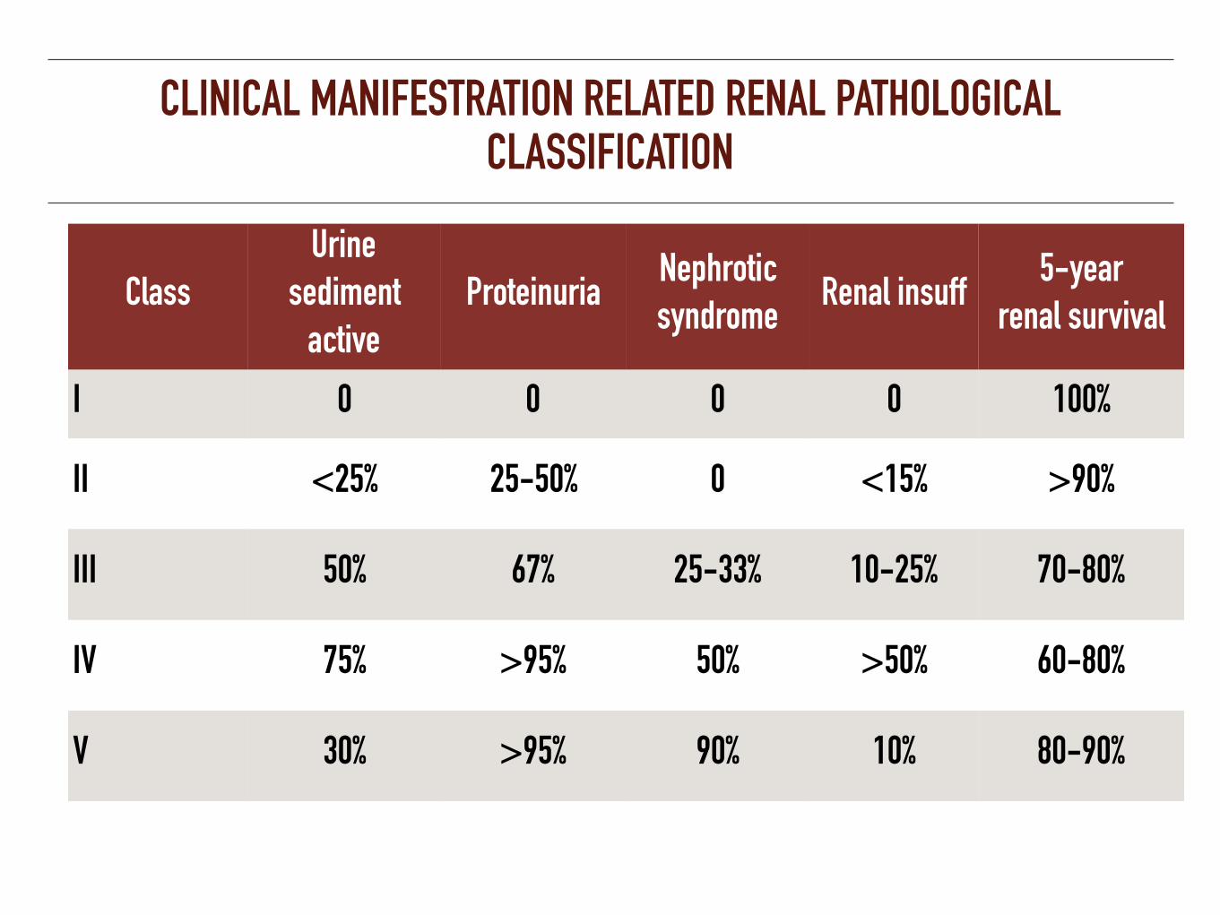
CLINICAL MANIFESTRATION RELATED RENAL PATHOLOGICAL CLASSIFICATION
Class Urine
sediment active
ProteinuriaNephrotic syndrome
Renal insuff5-year
renal survival
I 0 0 0 0 100%
II <25% 25-50% 0 <15% >90%
III 50% 67% 25-33% 10-25% 70-80%
IV 75% >95% 50% >50% 60-80%
V 30% >95% 90% 10% 80-90%

Renal Pathology Classification

Lupus nephritis biopsy ISN/RPS Classification
No endocapillary hypercellularity
Endocapillary hypercellularity
Mesangial deposits only
class I
Mesangial Hypercellularity
class II
Subepithelial deposits class V
Involving <50% glom Class III*
Involving >50% Glom
Segmental distribution Class IV S*
Global distribution Class IV G*
*Give the proportion of glomeruli with active and chronic lesions, necrosis, and crecents

ISN/RPS 2003 classificationClass % involved glomeruli Pathology of each
glomerulusActivity and chronicity
I Minimal mesangial LN (normal LM and immune-complex deposit in IF)
II Mesangial proliferation
III – focal < 50% of total glom S=segment
G=global
A=active
C=chronic
IV - diffuse > 50% of total glom S=segment
G=global
A=active
C=chronic
V Membranous
VI Diffuse glomerulosclerosis
Weening J. J., et al. Journal of the American Society of Nephrology. 2004; 241–250.
Minimum 10 glomeruli , Diagnosis of LN dominant IgG, C3 and C1q deposits are absolutely required.

Initial therapy of SLELife or organ threateningNon-life or organ threatening
Quality of life: Acceptable
Quality of life: Not-acceptable
Conservative management
Conservative treatment plus low
dose steroids Consider belimumab
High dose steroids, usually with addition of second agent
Mycofenolate mofetil (or myfortic acid)
Cyclophosphamide (Low/high dose) Do not exceed 6 months of Rx
After D/C cyclophosphamide; Maintain with MMF or azathioprine
ResponseNon-response
Taper dose of all agents especially steroids
Belimumab, rituximab Calcineurin inhibitors or Experimental therapies
Harrison’s Principle of Internal Medicine 19th edition

Treatment of lupus nephritis
❖ Class I
❖ Treated as dictated by the extrarenal clinical manifestations of lupus (2D)
❖ Class II
❖ Proteinuria <1 g/d as dictated by the extrarenal clinical manifestations of lupus (2D)
❖ Proteinuria >3 g/d be treated with corticosteroids or CNIs as described for MCD (2D)
KDIGO. Kidney International Supplements (2012) 2, 143–153

Treatment of Proliferative Lupus Nephritis (Class III-IV)
❖ Induction phase
❖ Renal remission at presentation and during follow up
❖ Maintenance phase
❖ Prevent relapse and minimizing the side effects of treatment

Regimens for initial therapy in class III/class IV LNRegimen A. NIH B. Euro-Lupus C. Oral cyclophosphamide D. MMF
Cyclophosphamidei.v. cyclophosphamide 0.5–1 g/m2; monthly
for 6 months
i.v. cyclophosphamide 500 mg; every 2 weeks
for 3 months
Oral cyclophosphamide 1.0–1.5 mg/kg/d
(maximum dose 150 mg/d) for 2–4 months
MMFMMF up to 3 g/d for
6 months
Benefit shown by RCT in proliferative LN
Yes Yes Yes Yes
Benefit shown by RCT in severe proliferative LN
Yes Untested Untested Untested
CommentsEffective in whites, blacks, Hispanics,
Chinese
Effective in whites. Untested in blacks, Hispanics, Chinese
Effective in whites, blacks, Chinese; easy to
administer and lower cost than i.v.
cyclophosphamide
Effective in whites, blacks, Hispanics, Chinese; high cost
KDIGO. Kidney International Supplements (2012) 2, 143–153

IV Pulse Cyclophosphamide: NIH regimen
Induction IVCY q 1 mo x 6
Maintenance IVCY q 3 mo x 6
❖ Initial IVCY 0.5-1.0 g/m2 (0.5 g/m2 if GFR 1/3 normal)
❖ Adjust subsequent IVCY to maximum dose of 1 g/m2 unless WBC nadir at 10-14 days after ICVY falls below 1,500/mm3
❖ Prednisolone 0.5-1 mg/kg/day for 4-8 weeks, which is subsequently tapered to low dose maintenance therapy
Boumpas DT, Austin HA, et al. Lancet 1992: 340.

KDIGO guideline: Class III-IV: initial therapy
❖ Corticosteroids (1A), combined with
❖ Cyclophosphamide (1B)
❖ OR
❖ Corticosteroids (1A), combined with MMF (1B)
KDIGO. Kidney International Supplements (2012) 2, 143–153
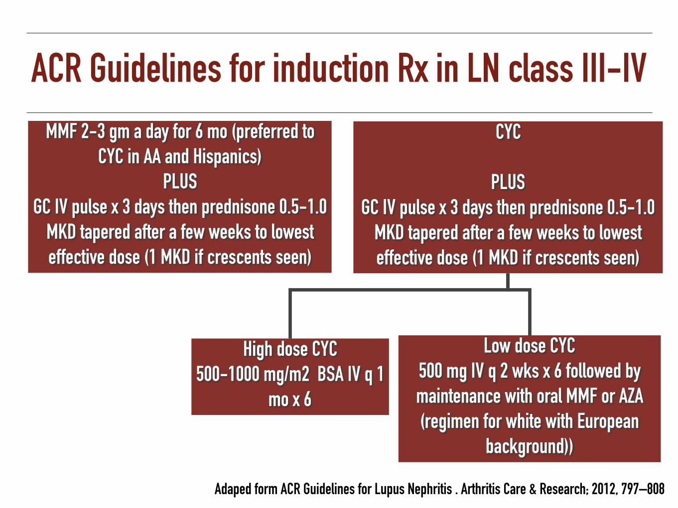
ACR Guidelines for induction Rx in LN class III-IV
Adaped form ACR Guidelines for Lupus Nephritis . Arthritis Care & Research; 2012, 797–808
MMF 2-3 gm a day for 6 mo (preferred to CYC in AA and Hispanics)
PLUS GC IV pulse x 3 days then prednisone 0.5-1.0
MKD tapered after a few weeks to lowest effective dose (1 MKD if crescents seen)
CYC
PLUS GC IV pulse x 3 days then prednisone 0.5-1.0
MKD tapered after a few weeks to lowest effective dose (1 MKD if crescents seen)
Low dose CYC 500 mg IV q 2 wks x 6 followed by maintenance with oral MMF or AZA (regimen for white with European
background))
High dose CYC 500-1000 mg/m2 BSA IV q 1
mo x 6

KDIGO guideline: Class III-IV: maintenance therapy
❖ AZA (1.5–2.5 mg/kg/d) or MMF (1–2 g/d in divided doses), and low-dose prednisolone (<10 mg/d) (1B)
❖ Maintenance therapy be continued for at least 1 year before consideration is given to tapering the immunosuppressio (2D)
KDIGO. Kidney International Supplements (2012) 2, 143–153
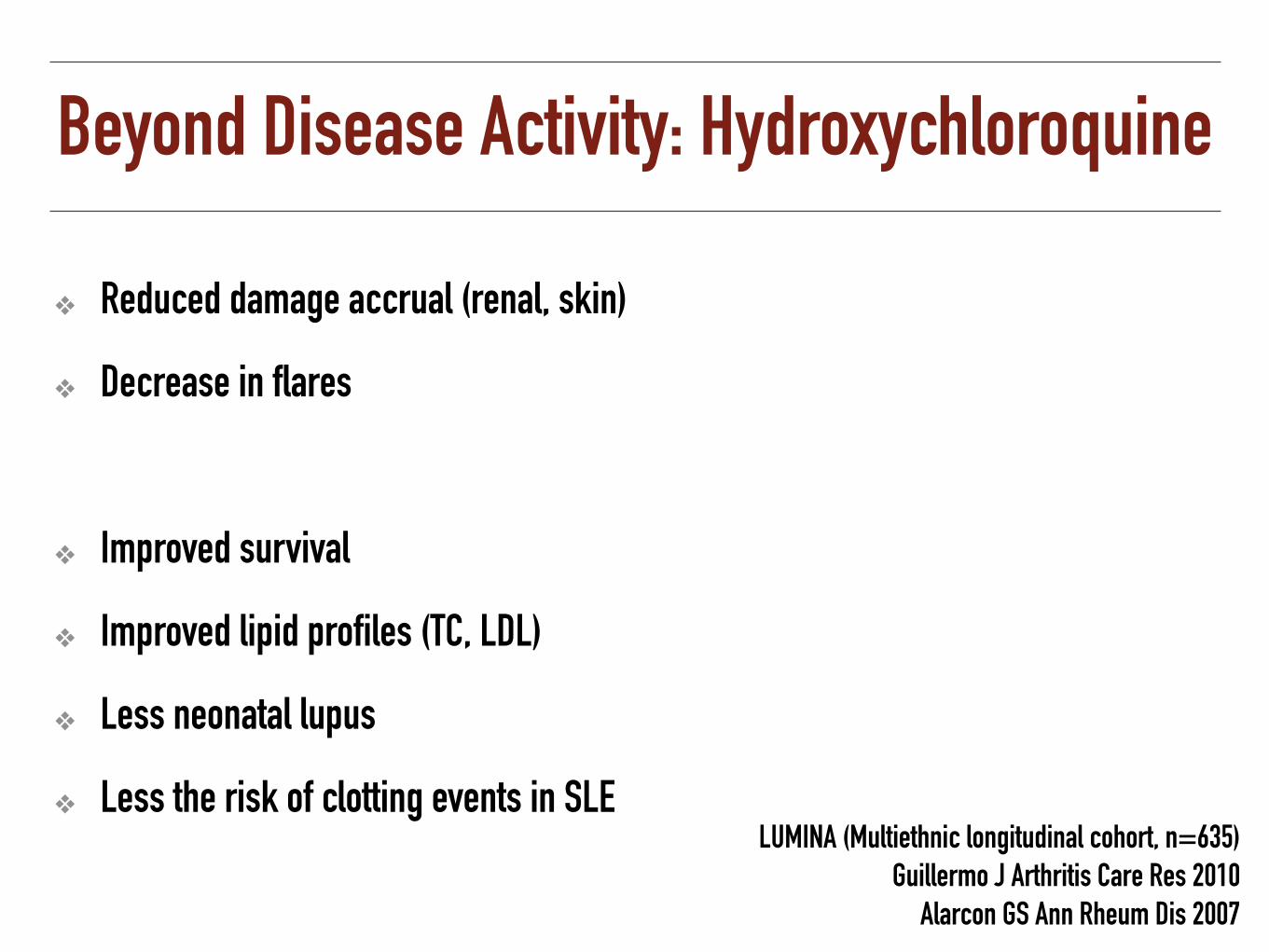
Beyond Disease Activity: Hydroxychloroquine
❖ Reduced damage accrual (renal, skin)
❖ Decrease in flares
❖ Improved survival
❖ Improved lipid profiles (TC, LDL)
❖ Less neonatal lupus
❖ Less the risk of clotting events in SLELUMINA (Multiethnic longitudinal cohort, n=635)
Guillermo J Arthritis Care Res 2010 Alarcon GS Ann Rheum Dis 2007
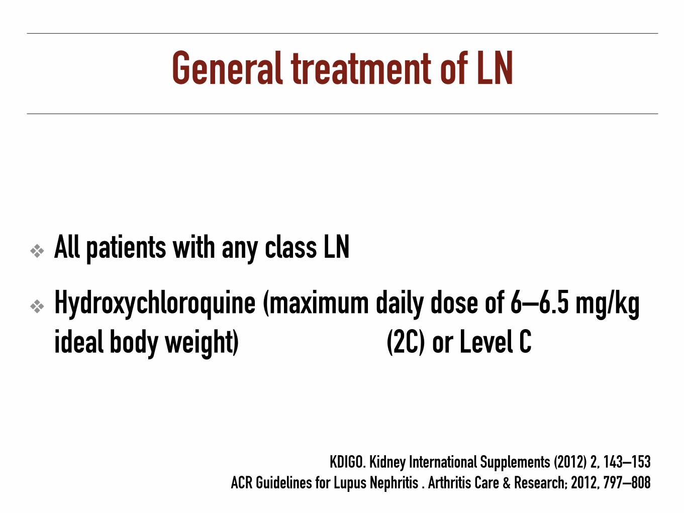
General treatment of LN
❖ All patients with any class LN
❖ Hydroxychloroquine (maximum daily dose of 6–6.5 mg/kg ideal body weight) (2C) or Level C
KDIGO. Kidney International Supplements (2012) 2, 143–153 ACR Guidelines for Lupus Nephritis . Arthritis Care & Research; 2012, 797–808
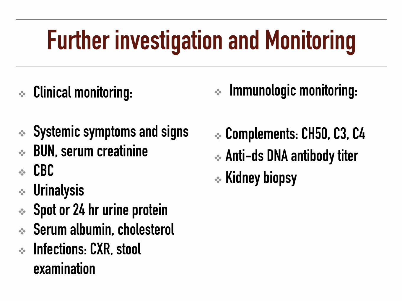
Further investigation and Monitoring
❖ Immunologic monitoring:
❖ Complements: CH50, C3, C4
❖ Anti-ds DNA antibody titer
❖ Kidney biopsy
❖ Clinical monitoring:
❖ Systemic symptoms and signs ❖ BUN, serum creatinine ❖ CBC ❖ Urinalysis ❖ Spot or 24 hr urine protein ❖ Serum albumin, cholesterol ❖ Infections: CXR, stool
examination

An Analysis of 1,135 Cases of Kidney Biopsy: Thailand Glomerular Research Network (TGRN)
Pathology n (%)
Primary glomerular diseases 507 (46.7)
1. IgA nephropathy 158 (31.2)
2. Focal segmental glomerulosclerosis 104 (20.5)
3. Membranous nephropathy 85 (16.8)
4. Minimal change disease 63 (12.4)
5. IgM nephropathy 52 (10.3)
6. Membranoproliferative GN 18 (3.6)

Minimal-change disease in adults

MCD: Pathology
❖ Normal light microscopy and IF
❖ Effacement of GEC foot processes by electron microscopy

MCD: Pathogenesis
❖ Systemic T cell dysfunction results in the production of a glomerular permeability factor
❖ Associated with HD or allergy
❖ Diminishes the heparin sulfate negative-charge barrier (anionic) properties of the GBM
Podocytopathies
Koyama A, et al. Kidney Int 1991;40:453-60.

MCD: clinical feature
❖ Nil (Nothing-In-Light microscopy) disease
❖ Children > adult (Male > female)
❖ 70% of children <10 years
❖ 10-15% of adults
❖ Bimodal with peak incidences in young children and older adults
Gesualdo L, et al. Kidney Int 2004: 66: 890. Tune BM, et al. J Am Soc Nephrol 1997: 8: 824
Cameron JS. Am J Kidney Dis 1987; 10:157

MCD: clinical feature
❖ Relatively abrupt onset of proteinuria
❖ Heavy proteinuria
❖ Hypoalbuminemia (<1.5-2.0 g/dL)
❖ Hyperlipidemia
❖ Rare signs of glomerulonephritis (HT, hematuria, rising Cr)
Waldman M, et al. Clin J Am Soc Nephrol 2007: 2: 445-53.
❖ Adult MCD
❖ HT (40%)
❖ Microscopic hematuria (29%)
❖ Reversible AKI (18%), ischemic ATN

MCD with AKI
❖ Acute tubular injury: sloughed epithelial cells, and loss of proximal tubular brush borders
❖ Risk factors
❖ Older age
❖ Hypertension
❖ Severe nephrotic syndrome
❖ Underlying arteriosclerosis of the kidney
❖ NSAIDS
Jennette JC, et al. Am J Kidney Dis 1990; 16: 432–437.
Functional renal insufficiency

Common associations with MCDInfection AllergiesVirus Food, dust
Parasitic Bee stings
Pharmaceutical agents Pollen
Nonsteroidal anti-inflammatory drugs Poison ivey and poison oak
Gold, lithium, interferon Dermatitis herpetiformis
Ampicillin, rifampin Disease and other associations
Trimethadione, tiopronin SLE
Tumors Following allogeneic stem cell transplantation for leukaemiaHodgkin’s lymphoma
Lymphoma, leukemia Following hematopoietic cell transplantationSolid tumors
Adapted from Brenner & Rector’s the kidney 10th edition, 2016

Minimal change disease
❖ Complete remission: 75 %
❖ Prednisolone 1 mg /kg/day or 2 mg/kg/AD
❖ Duration
❖ > 8 wk (remission 60%)
❖ 16-20 wk (remission 76-81%)
Nolasco F, et al. Kidney Int, 1986. 29: 1215-23.
Weeks from beginning of corticosteroid therapy
0 2 4 8 16
100-
80-
60-
40-
20-
0-
Perc
ent i
n co
mpl
ete
rem
issi
on
Children
Adults
Treatment of initial episode of adult MCD
❖ Prednisolone 1 mg/kg/day (maximum 80 mg) OD or 2 mg/kg (maximum 120 mg) AD (2C)
❖ High dose corticosteroids for a minimum period of 4 weeks, and for a maximum period of 16 weeks if complete remission is not achieved (2C)
❖ Corticosteroids be tapered slowly (5-10 mg/wk) over a total period of up to 6 months after achieving remission (2D)
KDIGO. Kidney International Supplements (2012) 2, 143–153

Treatment of initial episode of adult MCD
❖ Relative contraindications or intolerance to high-dose corticosteroids
❖ Uncontrolled diabetes, psychiatric conditions, severe osteoporosis
❖ Oral cyclophosphamide or cyclosporine (2D)
❖ Using the same initial dose and duration of corticosteroids for infrequent relapses (2D)
KDIGO. Kidney International Supplements (2012) 2, 143–153

Frequently relapsing and steroid-dependent MCD
❖ Oral cyclophosphamide 2–2.5 mg/kg/day for 8 wks (2C)
❖ Oral cyclosporine 3–5mg/kg/day or tacrolimus 0.05–0.1 mg/kg/d in divided doses for 1–2 yrs (2C)
❖ MMF 500–1000 mg twice daily for 1–2 yrs in pts who are intolerant of corticosteroids, cyclophosphamide, and CNIs (2D)
KDIGO. Kidney International Supplements (2012) 2, 143–153
MCD: prognosis
❖ Highly remission & relapse rate
❖ 50-75 % relapse within 6-12 mo
❖ 25 % frequent relapses
❖ 25 % steroid dependence
❖ Good prognosis: 5% turn to ESRD in 25 yr
7 to 12 % of adults with steroids resistance
Nakayama, M, et al. Am J Kidney Dis, 2002. 39: 503-12.
Complication of MCD
❖ Risk of mortality due to infection (peritonitis)
❖ Less commonly thromboembolism
❖ 5% develop ESRD in 9.4 yr
❖ Related to treatment
❖ Side effects of steroids
❖ Side effects of cyclophosphamie: infertility, malignancy
❖ Side effects of cyclosporine: hypertension, impair renal function

Steroid-resistant MCD
❖ Re-evaluate other causes of nephrotic syndrome
❖ Corticosteroid-resistant MCD suggests FSGS
❖ Steroid resistance may be due to undetected FSGS

An Analysis of 1,135 Cases of Kidney Biopsy: Thailand Glomerular Research Network (TGRN)
Pathology n (%)
Primary glomerular diseases 507 (46.7)
1. IgA nephropathy 158 (31.2)
2. Focal segmental glomerulosclerosis 104 (20.5)
3. Membranous nephropathy 85 (16.8)
4. Minimal change disease 63 (12.4)
5. IgM nephropathy 52 (10.3)
6. Membranoproliferative GN 18 (3.6)

Focal segmental glomerulosclerosis

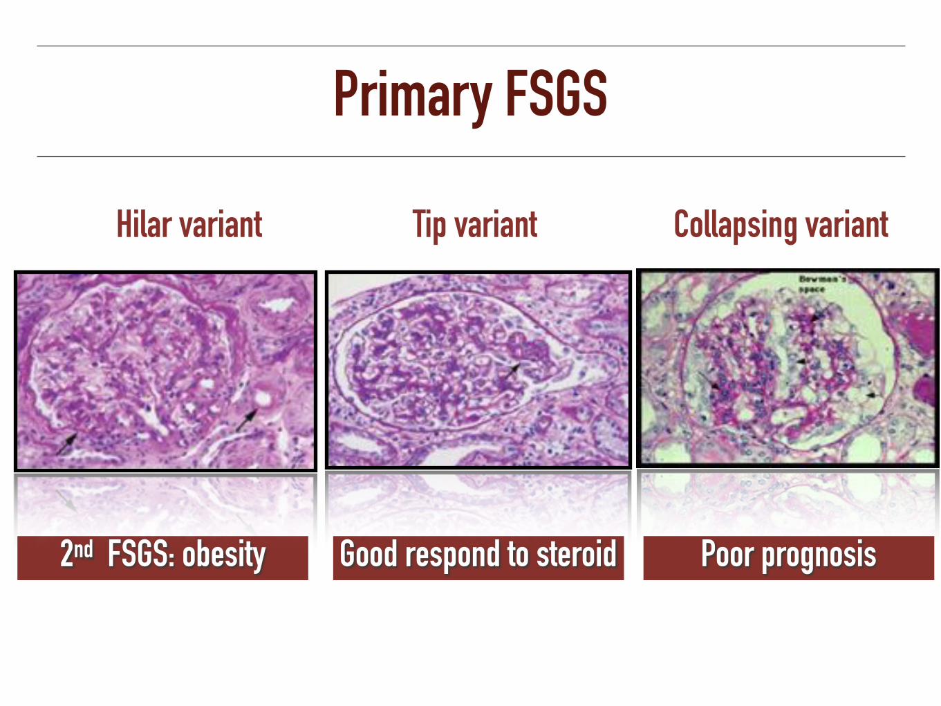
Primary FSGS Hilar variant Tip variant Collapsing variant
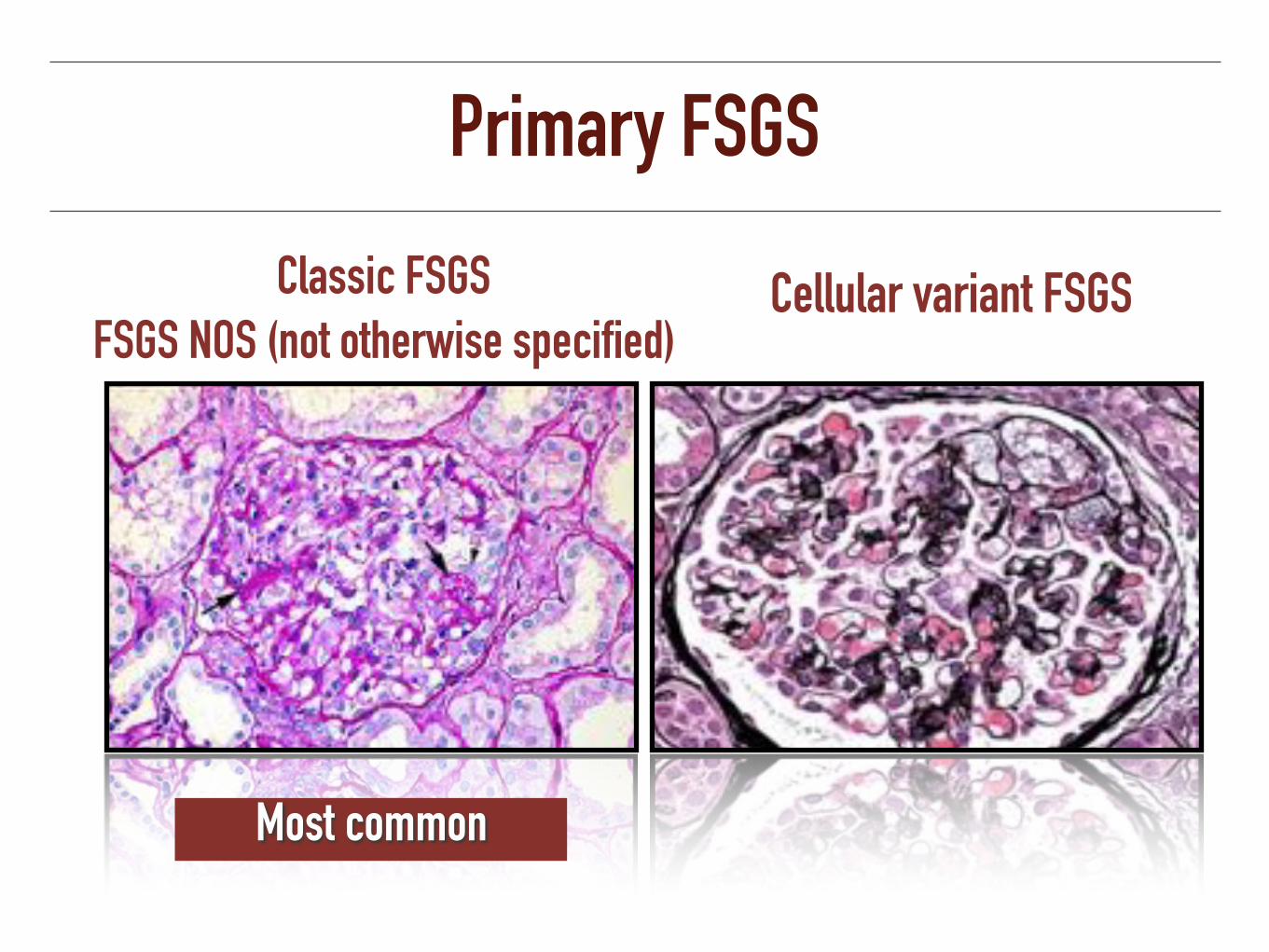
Primary FSGS
Classic FSGS FSGS NOS (not otherwise specified)
Cellular variant FSGS
Most common
Primary FSGS
Hilar variant Tip variant Collapsing variant
2nd FSGS: obesity Good respond to steroid Poor prognosis

Primary FSGS
❖ Normal IF
❖ IgM, C1q and C3 at sclerosis area
❖ EM: foot processes effacement

FSGS: Pathogenesis
❖ Alterations in T cell function and glomerular permeability factor
❖ Recurrent FSGS after KT
❖ Plasmapheresis & anti-IgG columns: absent of proteinuria
❖ Elevated circulating soluble urokinase receptors (suPAR) levels: 55-84%
Wei C, Trachtman H, Li J, et al. J Am Soc Nephrol 2012; 23:2051.

Gene mutation causally linked to FSGSNephrin: Chromosome 19q13
Congenital nephrotic syndrome of Finnish type
Focal Segmental Sclerosis; N Engl J Med 2011;365:2398-411

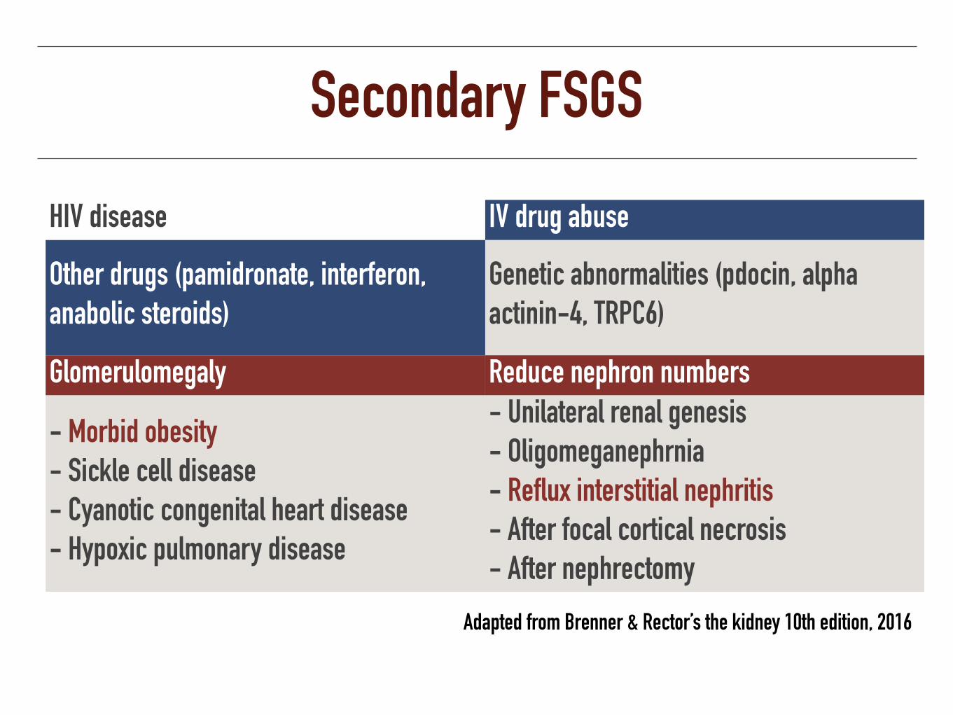
Secondary FSGS
HIV disease IV drug abuse
Other drugs (pamidronate, interferon, anabolic steroids)
Genetic abnormalities (pdocin, alpha actinin-4, TRPC6)
Glomerulomegaly Reduce nephron numbers
- Morbid obesity - Sickle cell disease - Cyanotic congenital heart disease - Hypoxic pulmonary disease
- Unilateral renal genesis - Oligomeganephrnia - Reflux interstitial nephritis - After focal cortical necrosis - After nephrectomy
Adapted from Brenner & Rector’s the kidney 10th edition, 2016
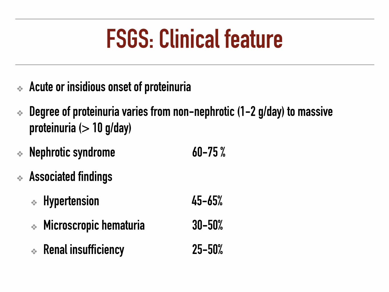
FSGS: Clinical feature
❖ Acute or insidious onset of proteinuria
❖ Degree of proteinuria varies from non-nephrotic (1-2 g/day) to massive proteinuria (> 10 g/day)
❖ Nephrotic syndrome 60-75 %
❖ Associated findings
❖ Hypertension 45-65%
❖ Microscropic hematuria 30-50%
❖ Renal insufficiency 25-50%

Clinical Features of FSGSHistologic subtype Clinical features
NOS Nephrotic syndrome or sub-nephrotic proteinuria
Perihilar More likely to present with subnephrotic proteinuria and normal serum albumin levels
Cellular Nephrotic syndrome
Tip Abrupt onset of the nephrotic syndrome Best prognosis, with response to glucocorticoids
Collapse Aggressive variant of primary FSGS with black racial predominance and severe nephrotic syndrome Worst prognosis, with poor response to glucocorticoids
N Engl J Med 2011; 365:2398-411.

Perihilar variant FSGS: Massive obesity
❖ Renal hypertrophy and increase GFR and RBF
❖ Excessive glomerulomegaly with vascular dilatation and mesangial expansion in five grossly obese individuals
Cohen AH. Am J Pathol 1975; 81:117–130. Barisoni L et al. CJASN 2007;2:529-542

Obesity related glomerulomegaly
❖ Clinical: lower incidence of nephrotic syndrome, normal serum albumin and cholesterol
❖ Natural history: more indolent progression
❖ Pathology:
❖ Glomerulomegaly
❖ Milder foot process effacement
❖ Less segmental sclerosis
Kambham N, et al. Kidney Int; 2001: 59: 1498–1509.

Primary VS Secondary FSGSPrimary Secondary
Onset Acute onset of nephrotic syndrome
Slowly increasing proteinuria and renal insufficiency
Proteinuria Nephrotic range Sub-nephrotic rangeClinical NS + +/-
Normoalbuminuria Pathology Diffuse foot process fusion Focal foot process effacement
Healed lesion - Obsolescent segment of capillary tuft
(PAS staining less intensely )
Praga, M., et al., Am J Kidney Dis, 1999. 33(1): 52-8.

Weight loss in overweight patients with proteinuric nephropathies
Proteinuria decreased by 31.2% in the diet group
Morales E, et al. Am J Kidney Dis 2003 Feb;41(2):319-27.
BMI (
kg/m
2)
- 3
- 2.5
- 2.0
- 1.5
- 1.0
Proteinuria (g/day)

Clinical interventions for obesity related glomerulomegaly
❖ Lifestyle modification
❖ Weight reduction
❖ BP-lowering medication (RAAS antagonists)
❖ Glucose-lowering medication (metformin, thiazolidinediones)
❖ Lipid-lowering medication
Prasad GV, et al. World J Nephrol. 2014: 6;3(4):210-9.

Collapsing FSGS
❖ Collapse and sclerosis of the entire glomerular tuft
❖ Marked hypertrophy and hyperplasia of podocytes
❖ Africa american
❖ HIV nephropathy
❖ Pamidonate
❖ Heroin

Treatment of idiopathic FSGS
❖ Idiopathic FSGS associated with clinical features of the nephrotic syndrome (1C)
❖ Prednisone 1 MKD OD or 2 MKD (maximum 120 mg) AD: Remission 28-74% (2C)
❖ Minimum of 4 wks; continue high-dose corticosteroids up to a maximum of 16 wks (2D)
❖ Corticosteroids be tapered slowly over a period of 6 months after achieving complete remission (2D)
KDIGO. Kidney International Supplements (2012) 2, 143–153

Intolerance to high-dose corticosteroids
❖ Cyclosporine (CNIs) be considered as first-line therapy for patients with relative contraindications or intolerance to high-dose corticosteroids
❖ Remission 50 – 60 % (steroid response)
❖ Remission 20 – 70 % (steroid non response) (2D)
KDIGO. Kidney International Supplements (2012) 2, 143–153

Treatment for steroid-resistant FSGS
❖ Cyclosporine at 3–5 mg/kg/d (initial target levels 125–175 ng/ml) in divided doses be given for at least 4–6 months (2B)
❖ Continuing cyclosporine treatment for at least 12 months, followed by a slow taper (2D)
❖ Combination of MMF and high-dose dexamethasone for not tolerate with cyclosporine (2C)
KDIGO. Kidney International Supplements (2012) 2, 143–153

FSGS: Prognosis
❖ Relatively poor outcome, with 50% reaching ESRD by 10 years
❖ Risk factors
❖ Massive proteinuria
❖ Increase serum creatinine
❖ Interstitial fibrosis and tubular atrophy
❖ Collapsing variant (ESRD within 15 months)
❖ Failure to CR/PR
Korbet, S.M. et al. Nephrol Dial Transplant, 1999. 14 S3: 68-73. Grcevska L, et al. Am J Kidney Dis 1999;33:652-7.

An Analysis of 1,135 Cases of Kidney Biopsy: Thailand Glomerular Research Network (TGRN)
Pathology n (%)
Primary glomerular diseases 507 (46.7)
1. IgA nephropathy 158 (31.2)
2. Focal segmental glomerulosclerosis 104 (20.5)
3. Membranous nephropathy 85 (16.8)
4. Minimal change disease 63 (12.4)
5. IgM nephropathy 52 (10.3)
6. Membranoproliferative GN 18 (3.6)

Idiopathic membranous nephropathy

Membranous nephropathy
Light microscopy:
GBM thickening Spike appearance

Staging of Membranous NephropathyStage I has subepithelial dense
deposits (arrow) without adjacent basement membrane reaction.
Stage II has projections of basement membrane adjacent to deposits. Stage III has deposits surrounded
by basement membrane.
Stage IV has thickened basement membrane with irregular lucent
zones

Membranous nephropathy
Diffuse granular capillary wall staining of IgG and C3
Electron dense deposits across the GBM in the subepithelial space
Immunofluorescene Electron microscopy

Membranous nephropathy: Pathogenesis
Circulating Immune complex
In situ Circulating Immune complex
“Native Ag”
In situ Circulating Immune complex
“Planted Ag”
Glassock RJ. N Engl J Med 2009; 361:81-83.
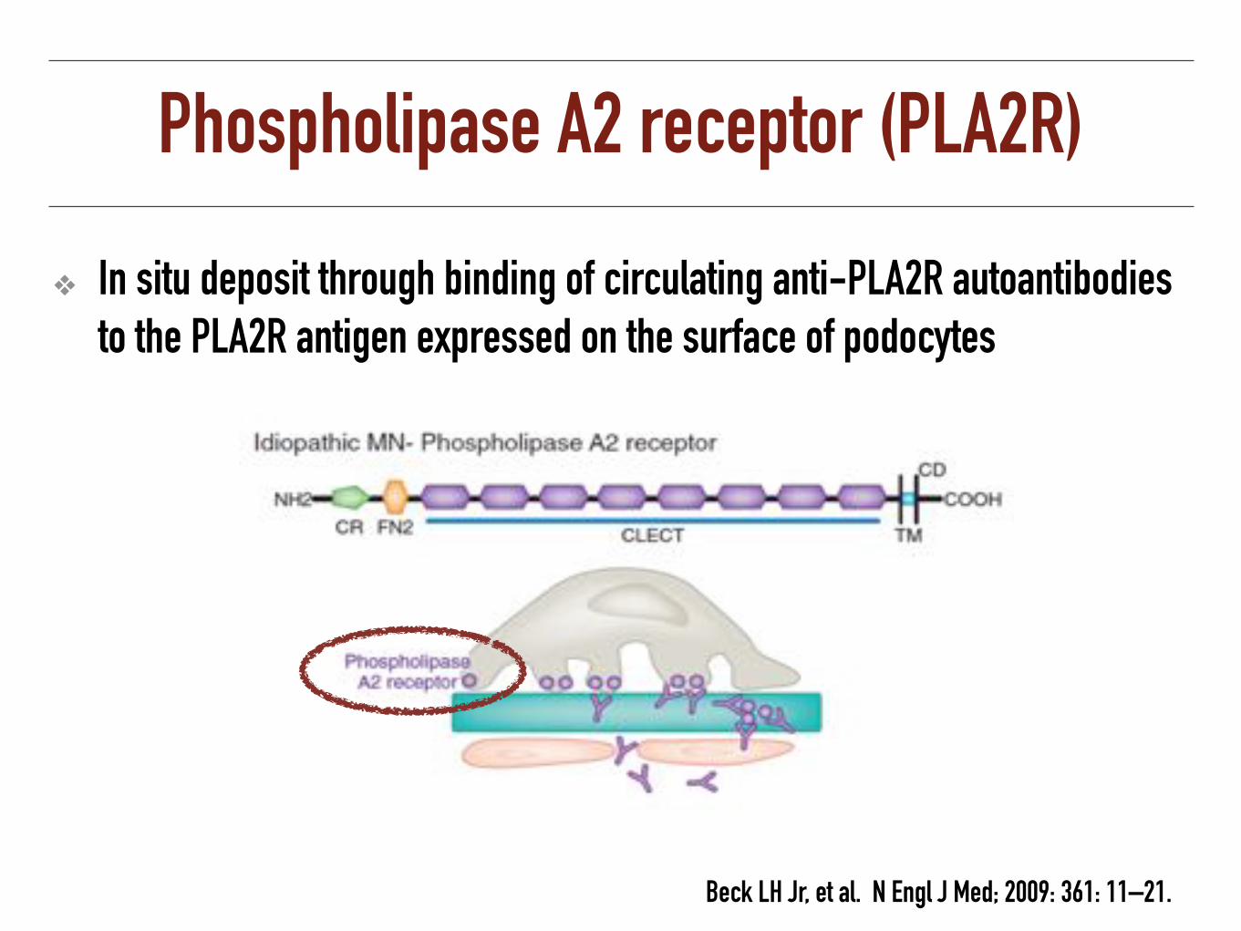
Phospholipase A2 receptor (PLA2R)
❖ In situ deposit through binding of circulating anti-PLA2R autoantibodies to the PLA2R antigen expressed on the surface of podocytes
Beck LH Jr, et al. N Engl J Med; 2009: 361: 11–21.

Membranous associated disorders
Infection: Hepatitis B and C, syphilis, malaria, schistosomiasis, leprosyCancer: Breast, colon, lung, stomach, kidney, esophagus, ovary, prostateDrugs: Gold, mercury, penicillamine, NSAIDS, probenecid, captopilAutoimmune diseases: SLE, RA, dermatitis herpetiformis, myasthenia gravis, Sjögren's syndromeSystemic diseases: Fanconi's syndrome, Crohn's disease, Sarcoidosis, Guillain-Barré syndrome
Satirapoj B. Common Problems in Internal Medicine. 2010. 487-97.

Membranous nephropathy: Clinical feature
❖ Adult > 40 yr (30-50 yr) and men: women = 2-3:1
❖ > 80% have more than 3 g/d of proteinuria
❖ Almost always insidious onset
❖ Bland urine sediment
❖ Mild microhematuria (30-40%)
❖ Hypertension (15-55%)

Membranous nephropathy: Clinical feature
❖ Normal or slightly decreased renal function (impair renal function < 10%)
❖ Hypoalbuminemia, elevated LDL and VLDL
❖ Thromboembolic manifestation (RVT, PE, DVT)
❖ Risk of malignancy increase with age
❖ Spontaneous remission 30%, stable 30% and progression 30%
Hogan SL. Am J Kidney Dis 1995; 25:862. Schieppati A. N Engl J Med 1993; 329:85.
Ponticelli C. Kidney Int 1995; 48:1600.

Predicting chronic renal insufficiency in idiopathic MN
❖ Pei and et al.; likelihood of progressing to CKD at 5-6 years
❖ 66 % in proteinuria > 8g/d for > 6 mo
❖ 55 % in proteinuria > 6g/d for > 9 mo
❖ 44 % in proteinuria > 4g/d for > 1 year
Pei Y, et al. Kidney Int. 1992;42(4): 960.
❖ Adequate assessment of proteinuria requires following patients for at least 6 to 12 months

Treatment of membranous nephropathy
❖ 35% spontaneous remission
❖ Patients with nephrotic syndrome with
❖ Persistent urinary protein >4 g/d and remains at over 50% of the baseline value, during therapy at least 6 months (1B)
❖ Presence of severe, disabling, or life-threatening symptoms related to the nephrotic syndrome (1C)
❖ Serum Cr has risen by 30% within 6 to 12 months (2C)
KDIGO. Kidney International Supplements (2012) 2, 143–153
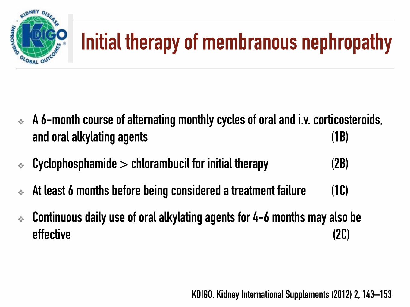
Initial therapy of membranous nephropathy
❖ A 6-month course of alternating monthly cycles of oral and i.v. corticosteroids, and oral alkylating agents (1B)
❖ Cyclophosphamide > chlorambucil for initial therapy (2B)
❖ At least 6 months before being considered a treatment failure (1C)
❖ Continuous daily use of oral alkylating agents for 4-6 months may also be effective (2C)
KDIGO. Kidney International Supplements (2012) 2, 143–153
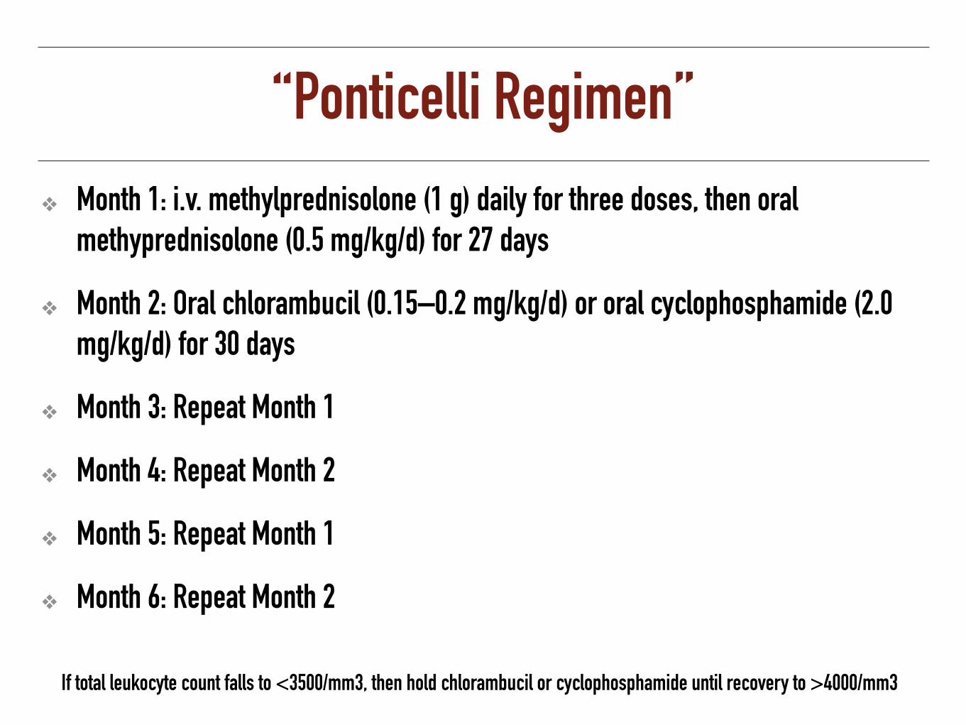
‘‘Ponticelli Regimen’’
❖ Month 1: i.v. methylprednisolone (1 g) daily for three doses, then oral methyprednisolone (0.5 mg/kg/d) for 27 days
❖ Month 2: Oral chlorambucil (0.15–0.2 mg/kg/d) or oral cyclophosphamide (2.0 mg/kg/d) for 30 days
❖ Month 3: Repeat Month 1
❖ Month 4: Repeat Month 2
❖ Month 5: Repeat Month 1
❖ Month 6: Repeat Month 2
If total leukocyte count falls to <3500/mm3, then hold chlorambucil or cyclophosphamide until recovery to >4000/mm3

UK Membranous Trial
108 subjects with proteinuria 8.5 g/day and progressive decline in eGFR (>20%) during 2 yrs, serum Cr < 3.4 mg/dL (High risk)
Howman A, et al. Lancet 2013; 381:744
Patie
nts
with
out d
eclin
e (%
)
Time to 20% decline in GFR (years)
0 1 2 3
100-
75-
50-
25-
0-
Prednisolone+ chlorambucil Cyclosporin Support
Time to 20% decline in GFR (years)

Alternative regimens
❖ Cyclosporine (3.5–5.0 MKD) or tacrolimus (0.05-0.075) MKD) at least 6 months (1C)
❖ Discontinued CNI in patients who do not achieve remission after 6 months (2C)
KDIGO. Kidney International Supplements (2012) 2, 143–153

Poor prognosis
❖ Male
❖ Advanced age (>50 years)
❖ Persistent heavy proteinuria (>3.5 g/d)
❖ Decreased serum albumin
❖ Hypertension
❖ Hyperlipidemia
❖ Impaired GFR
❖ Poor protein selectivity, or persistent excretion of β2 microglobulin or C5b-C9,C3d
❖ Advanced stage of MN
New Predictors Levels of circulating anti-PLA2R revealed a strong correlation with clinical disease activity
Zuccheli P, Oxford Medical, 1998, 570-612 Coggins CH. Semin Nephrol 2: 264-273, 1982
Ponticelli C. Oxford Medical Publications, 1997

Management of Complications

Hypertension
❖ Lifestyle modification
❖ Salt restriction, weight normalization, regular exercise, and smoking cessation
❖ ACE-I and ARB to be first-choice therapy
❖ Recommendations <130/80mmHg
KDIGO. Kidney International Supplements (2012) 2, 143–153

Proteinuria
❖ Toxic to the tubulointerstitium
❖ ACE-I or ARB may reduce proteinuria by up to 40–50% in a dose dependent manner
❖ Adequate dietary protein (0.8–1.0 g/kg daily) with a high carbohydrate intake to maximize utilization of protein
KDIGO. Kidney International Supplements (2012) 2, 143–153

Hyperlipidemia
❖ Follow the guidelines at high risk for the development of cardiovascular disease
❖ Statins are effective in correcting the lipid profile
KDIGO. Kidney International Supplements (2012) 2, 143–153

Nephrotic edema
❖ Moderate dietary sodium restriction (1.5–2 g sodium per 24 hours)
❖ Diuretic-resistant nephrotic syndrome
❖ Intestinal-wall edema
❖ Oral loop diuretics with once- or twice-daily administration are usually preferred
❖ Ease of administration and longer therapeutic effect compared to i.v. therapy
KDIGO. Kidney International Supplements (2012) 2, 143–153

Severe nephrotic edema
❖ IV diuretic, by bolus injection or infusion
❖ Combining a loop diuretic with a thiazide diuretic
❖ IV albumin infusions combined with diuretics, but unproven benefit
❖ Mechanical ultrafiltration
KDIGO. Kidney International Supplements (2012) 2, 143–153

Hypercoagulability
❖ Anti-coagulant drugs considered if serum albumin <2.0–2.5 g/dl with one or more of the following
❖ Proteinuria >10 g/d
❖ BMI> 35 kg/m2
❖ Family history of thromboembolism
❖ CHF class III or IV
❖ Recent abdominal or orthopedic surgery
❖ Prolonged immobilization
KDIGO. Kidney International Supplements (2012) 2, 143–153

Risk of infection
❖ Nephrotic children with ascites
❖ Fluid should be examined microscopically for SBP
❖ Parenteral antibiotics should treat as pneumococcal infection
❖ If repeated infections occur, serum immunoglobulins should be measured
❖ Serum IgG < 600 mg/dl, monthly administration of i.v. immunoglobulin 10–15 g to keep serum IgG >600 mg/dL
KDIGO. Kidney International Supplements (2012) 2, 143–153

Risk of infection
❖ Pneumococcal vaccination and annual influenza vaccination
❖ Live vaccines (measles, mumps, rubella, varicella, rotavirus, yellow fever) is contraindicated while on immunosuppressive or cytotoxic agents
❖ Deferred until prednisone <20 mg/d and/or immunosuppressive agents have been stopped for at least 1–3 months
KDIGO. Kidney International Supplements (2012) 2, 143–153

Thank you for your attention
Intelligence Dialysis Center Nephrology Unit
Phramongkutklao Hospital and College of Medicine