an allelic difference determines reciprocal patterns of - development
TRANSCRIPT

/. Embryol. exp. Morph. 87, 229-239 (1985) 2 2 9Printed in Great Britain © Company of Biologists Limited 1985
An allelic difference determines reciprocal patterns ofexpression of binding sites for Dolichos biflorus lectinin inbred strains of mice
BRUCE A. J. PONDER1, MICHAEL F. W. FESTING2 AND MAUREEN M.WILKINSON1
^Institute of Cancer Research, Haddow Laboratories, Sutton, Surrey, U.K.2MRC Experimental Embryology and Teratology Unit Laboratories, Carshalton,Surrey, U.K.
SUMMARY
We used staining of tissue sections by lectin conjugates to screen inbred strains of mice forpolymorphisms which could be used as histological markers of chimaerism. We found onepolymorphism, which involves reciprocal patterns of expression of binding sites for the N-acetyl-galactosamine-binding lectins from Dolichos biflorus (DBA), Helixpomatia (HPA) and Wisteriafloribunda (WFA) on intestinal epithelium and vascular endothelium. The polymorphism is dueto alleles at a single locus, designated Dlb-1 (for Dolichos lectin binding). Of 29 inbred strainsexamined, 3 are Dlb-la (type strain RHI-ro; gut epithelium - ve, vascular endothelium + ve),and 26 are Dlb-lb (type strain C57BL/6J; gut epithelium + ve, vascular endothelium — ve). InRHI-ro and C57BL/6J embryos, the polymorphic difference is not clearly present until day 11of gestation. Before then, embryos of both strains express binding sites on gut epithelium and onendothelium.
The temporal and tissue-specific patterns of expression of lectin-binding sites may result fromdifferences in expression of an N-acetyl galactosaminosyl transferase. If so, elucidation of thegenetic basis of the polymorphism might provide an insight into the mechanisms of developmentalregulation of glycosyltransferase activity.
INTRODUCTION
We screened inbred mouse strains for histological markers of chimaerism bystaining cryostat sections of tissues from different strains with a panel of peroxidase-conjugated lectins. By analogy with the blood group antigens in man (Kapadia,Feizi & Evans, 1981), we thought that polymorphisms between inbred mousestrains would most likely be carbohydrate based.
We found only one polymorphism (Ponder & Wilkinson, 1983). It is recognizedby the lectins from Dolichos biflorus (DBA), Helix pomatia (HPA) and Wisteriafloribunda (WFA), which have specificity for terminal non-reducing N-acetyl galac-tosamine residues (Etzler, 1972; Hammarstrom, 1972; Kurokawa, Tsuda &Sugino, 1976). The polymorphism is confined to intestinal epithelium and vascular
Keywords: Dolichos biflorus agglutinin, genetic polymorphism, inbred mice, lectin, mouse,glycosyltransferases

230 BRUCE A. J. PONDER AND OTHERS
endothelium, but in those tissues it can be used as a marker for studying clonaldevelopment in aggregation chimaeras (Schmidt, Garbutt, Wilkinson & Ponder,in press; Ponder et al. 1985). The polymorphism is unusual in that the patternof expression of lectin-binding sites is reciprocal in the two polymorphic types.In this paper we describe the appearance of the polymorphism during embryonicdevelopment, the reciprocal pattern in adult mice, and genetic data whichindicate that the reciprocal patterns are determined by alleles at a singlelocus.
MATERIALS AND METHODS
MiceCBA/Ca mice were bred in the animal house of the Institute of Cancer Research. DDK,
RHI-ro, C57BL/6J, BIO.A., and C57BL/10ScSn, were bred at the MRC Laboratories, Carshal-ton. Other mice of the inbred strains shown in Tables 1 and 2 were obtained from Olac (Bicester,UK) or from the Medical Research Council, Mill Hill, London and housed at the MRCLaboratories, Carshalton.
Male and female mice, 4 to 24 weeks old, were used for the screening for polymorphisms andin the study of the strain distribution of DBA binding. Embryos studied were from DDK,RHI-ro, and C57BL/6J strains, raised as above. Fl, backcross and F2 animals were bred at theMRC Laboratories, Carshalton.
Lectins, lectin conjugates and antibodiesPeroxidase conjugates of Ricinus communis agglutinin I and of lectins from Mangifera indica,
Cotoneaster and Jack fruit were a gift from Dr J. A. Forrester (Institute of Cancer Research).Other lectins were purchased as purified lectin or as peroxidase or fluorescein isothiocyanate(FITC) conjugate from Sigma, Poole, England. Peroxidase conjugates of Dolichos biflorusagglutinin (DBA) (Sigma No LI 135) were prepared in our laboratory by the periodate methodas previously described (Ponder & Wilkinson, 1983). Alkaline phosphatase conjugates ofpurified lectins from Helix pomatia, Wisteria floribunda, Vicia villosa and Codiwn fragile wereprepared by glutaraldehyde conjugation (Ponder & Wilkinson, 1983) using alkaline phosphatasefrom calf intestine (Boehringer, No 567744). Rabbit antiserum to Forssman antigen (Willison etal. 1982) was a gift from Dr K. Willison (Institute of Cancer Research). Sugars were obtainedfrom Sigma.
Histological methodsMethods were as described by Ponder & Wilkinson (1983). The initial screening of mouse
tissues for polymorphisms was carried out on cryostat sections fixed in 10 % formol saline aftercutting. Subsequent studies of the distribution of DBA, HP A and WFA-binding sites in embryonicand adult tissues were made on methacarn fixed, paraffin-embedded tissue (except that tissuefrom Fl and F2 animals used in the genetic studies and tissues used for enzyme digestions werefixed in 10 % formol saline: see below). Of the fixatives tried (10 % formol saline pH7,0-1 % to2% buffered glutaraldehyde, benzoquinone, 4°C acetone, 4°C absolute ethanol, Carnoy's,Bouin's, and methacarn), only glutaraldehyde above 0-2 % concentration caused loss of DBAbinding; but the cleanest and highest density staining was obtained with methacarn. Controls forthe specificity of lectin staining were provided in each experiment by the inclusion of slidesincubated without lectin conjugate, with lectin conjugate diluted in buffer containing theappropriate inhibiting sugar (final concentration 2 % w/v), and (for DBA) known positive andnegative tissues.
Binding of the rabbit anti-Forssman antibody was demonstrated using a goat anti-rabbit alk-aline phosphatase conjugate (Sigma, A8025).

Allelic patterns of Dolichos lectin binding in mouse tissue 231
Chemistry of the DBA-binding siteFor enzyme digestions, cryostat sections fixed in formol saline for 10 min at room temperature,
and formalin-fixed paraffin-embedded sections, were incubated as follows: (i) neuraminidase(Behring, Vibrio cholerae ljul ml"1) diluted 1:10 in 0-2M-acetate buffer pH 5-5 for 30 min at 37°C.A positive control was provided by unmasking of sites for peanut lectin-peroxidase conjugateson similarly processed sections of human kidney, (ii) Trypsin (Sigma, type III from bovinepancreas) lmg in 20 ml PBS pH7-5 for 15 min at 37°C: no control. (Hi) Glycosidases (nocontrols): a-galactosidase (Sigma) 0-025/ilml"1 in 50 mM-acetate buffer pH 4-5 for 30min atroom temperature; /3-galactosidase (Sigma) 1 mgml"1 in 50mM-Tris pH7-5 at 37°C for 15 min.Other treatments (on unfixed and formalin fixed cryostat sections): (i) NP40 (Nonidet P40)extraction: NP40 (Sigma) 1 % or 0-1 % in PBS pH7-5 for 15 min at room temperature; (ii) 10 %trichloroacetic acid (TCA): 2 min at 4°C; (iii) chloroform/methanol: 2:1 chloroform: methanolfor 2 min at room temperature followed by 1:2 chloroform: methanol for a further 2 min at roomtemperature. In each case, sections were washed in PBS after treatment and then incubated withDBA-peroxidase above.
Genetic studiesMice were scored for Dlb-1 polymorphic type by DBA-peroxidase staining of vascular endo-
thelium and intestinal epithelial cells in formalin-fixed paraffin-embedded sections of small intes-tine and adjacent mesentery.
RESULTS
Screening for polymorphismsThe mouse strains, lectins and tissues used in the initial screen for polymorphisms
are shown in Table 1.
Table 1. Screening mouse strains for lectin-binding polymorphism
Mouse strains: CBA/CaLac, C57BL/6JLac, BALB/c, GE, HTG, IF, SEA/J, 101/H, Rlll-ro,DDK
Lectins Nominal carbohydrate specificity
Bandeiraea simplicifolia IArachis hypogaeaRicinus communis 120 f D-galactoseAbrus precatoriusPhaseolus limensis II
Dolichos biflorus j N , l a c t o s a m i n e
Soybean agglutinin J J °
Wheatgerm agglutinin N-acetyl glucosamine
Concanavalin AT .. . , MannoseLens cuhnansLotus tetragonolobus \ T fll-_-tl
Ulex europaeus I J
Tissues (unfixed cryostat sections): Skin, brain, spleen, bladder, lung, thyroid and trachea,colon, salivary gland.
232 BRUCE A. J. PONDER AND OTHERS
.-•'• ,*.' ; ' - <
6Fig. 1
e

Allelic patterns of Dolichos lectin binding in mouse tissue 233
Table 2. Classification of inbred strains for DBA-binding pattern
Dlb-P DDK, SWR, RIII-ro(Gut epithelium ~ve;endothelium +ve)
Dlb-lb AKR, A/Nimr, BALB/c, C57L, C57BL/6J, B10.A,(gut epithelium +ve; C57BL/Ks, C57BL/10ScSn, C57BL/Nimr, CBA/Ca,endothelium ~ve) CBA/HN, C3H/Bi, C3H/He, DBA/1, DBA/2, GE, HTG,
IF, LP/Nimr, LT, NZB, NZW, SEA/J, SJL/J, 101/H,129/RrJ.
The screening revealed only one polymorphism, for binding of DBA-peroxidaseto intestinal epithelium and vascular endothelium (Fig. 1). In RHI-ro and DDKmice, DBA-peroxidase bound to vascular endothelium but not to intestinalepithelium: in the other 8 strains, the reverse was true. The polymorphism wasdesignated Dlb-1 for Dolichos lectin binding, RHI-ro being the type strain for Dlb-la, and C57BL/6J the type strain for Dlb-lb. Subsequently, (see below), the lectinsfrom Helix pomatia (HPA) and from Wisteria floribunda (WFA), which like DBAhave nominal specificity for terminal non-reducing N-acetyl galactosamine, werefound to recognise the same polymorphism.
A further 19 strains and substrains have been examined for their Dlb-1 type, themajority falling into the Dlb-lb group (Table 2).
In order to assess the use of the polymorphism for studying clonal developmentin gut epithelium and vascular endothelium during embryogenesis, we alsoexamined a series of embryos of C57BL/6J and RHI-ro strains at different ages (seebelow).
Distribution of DBA binding sites in RIH-ro and C57BL/6J tissues {Tables 3 and 4)The distributions reported below were consistent for 20 adult mice of each strain
(males and females, aged from 4 to 24 weeks), and four embryos of each strain atdays 8, 9, 10, 11, 13 and 18 of gestation. DBA-peroxidase (rather than HPA orWFA) was used throughout these studies.
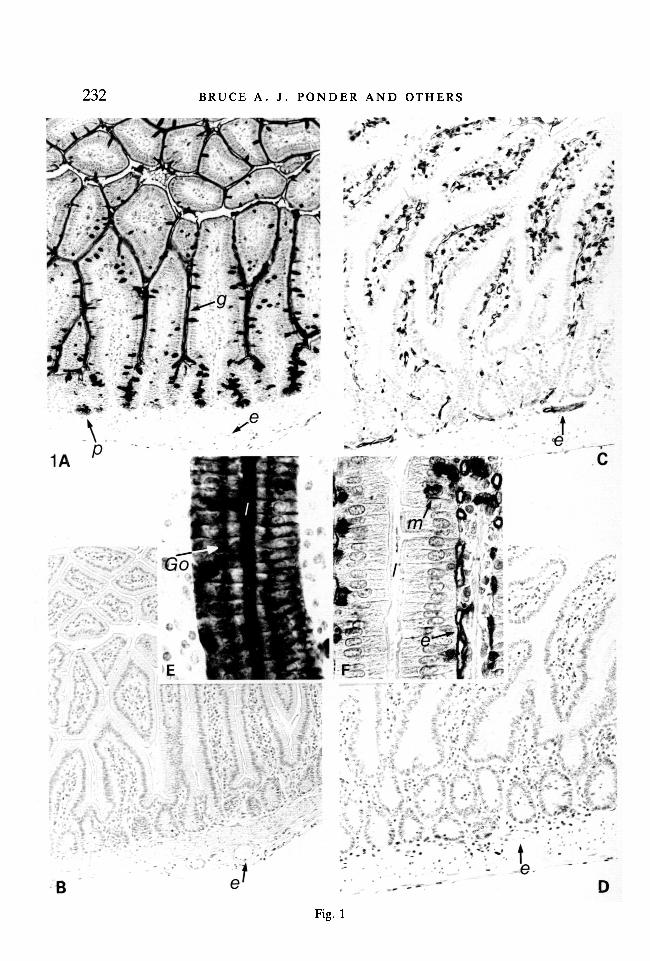
Fig. 1. Polymorphism in expression of DBA-binding sites in adult small intestine. (A,E) C57BL/6J small intestine (A, x 160; E, detail of epithelium and lamina propria ofvillus x 400). (B) adjacent section to (A): sugar control (x 160). (C, F) RHI-ro smallintestine (C, x 160; F, detail of epithelium and lamina propria of villus x 400). (D)adjacent section to (C): sugar control (x 160).
Methacarn fixed, paraffin-embedded 4fim sections of tissue from 8-week-old mice.Counterstained with haemalum. DBA-binding sites appear black, g, gut epithelium; /,lumen between villi; m, unidentified DBA-positive cells (see Table 4): p, Paneth cells;e, endothelium; Go, Golgi apparatus.
In C57BL/6J intestine, columnar, goblet and Paneth cells are DBA-positive, andvascular endothelium is DBA-negative. In RHI-ro intestine the reverse is true: theendothelium of arteries, veins and blood capillaries (but not lymphatics) is DBA-positive; intestinal epithelium is DBA-negative.

234 BRUCE A. J. PONDER AND OTHERS
Table 3. Polymorphic patterns of binding of DBA-peroxidase in tissue sections ofRHI-TO and C57BLI6J mice
RHI-ro (Dlb-la) C57BL/6J (Dlb-lb)
AdultSmall intestine
columnar cells — +goblet cells - +Paneth cells - +*entero endocrine cells — ?
colonfcolumnar cells — +goblet cells — +
vascular endotheliumt + ~
Embryo (8, 9, 10, 11, 13, and 18 days)f +ve up to ca. 11
gut epithelium < days gestation,I then —ve
patchy +ve invascular endothelium + <j major vessels up
to ca. 11 daysgestation, then — ve
* Paneth cells DBA"ve in some Dlb-lb strains (e.g. CBA/Ca, C3H/Bi)t Some Dlb-lb strains show transition to DBA"ve distally$ Consistent tissue-specific patterns of DBA+ve endothelium are seen in adult mice of
Dlb-la strains. The endothelial location of DBA binding sites has been confirmed by electronmicroscopy. Lymphatic endothelium is DBA-negative. (Ponder & Wilkinson, 1983 fordetails).
Adult miceThe polymorphism in DBA-binding sites in the adult was confined to intestinal
epithelium and vascular endothelium (Fig. 1; Table 3). Non-polymorphic bindingsites are listed in Table 4.
Notable departures from the general pattern of polymorphism in the adult micewere (i) that the colonic epithelium was DBA-negative distally in some Dlb-lb
strains (CBA/Ca, C3H/Bi, BALB/c, AKR, DBA/2, but not C57BL/6J, C57B10,NZB, NZW, 129/RrJ) which were otherwise DBA-positive in intestinalepithelium; (ii) that in otherwise DBA-positive colonic epithelium, crypts adjacentto lymphoid follicles were often DBA-negative, but this was not so for cryptsadjacent to lymphoid follicles in small intestine; and (iii) in adult Dlb-la strain mice(endothelium-positive), DBA binding was not present on endothelium in certaintissues in which endothelial cells were DBA positive in 13-day embryos (see Ponder& Wilkinson, 1983).

Allelic patterns of Dolichos lectin binding in mouse tissue 235
Table 4. Non-polymorphic patterns of binding of DBA-peroxidase in tissue sectionsofRIII-xo and C57BLI6J mice
Adult Embryo only
epithelium of yolk sacbiliary duct \ , t u \ cell population in mid/late gestation thymus*pancreatic duct J ^ a C ^ medial and dorsal quadrant of developingstomach otocystcaecumsalivary gland acinicollecting tubules of kidney (patchy)fallopian tubeoocytesunidentified cells in epithelium, lamina propria and lymphoid follicles of small intestine
(see Figure 1).
* described in detail by Kasai, Takashi, Takahashi & Tokunaga, 1983.Note: The following tissues are DBA-negative in adult mice of RIII-ro and C57BL/6J strains:
central and peripheral nervous system; skeletal and cardiac muscle; thyroid, parathyroid,adrenal, ovary (except oocytes), testis, pancreatic islets; skin and appendages; squamousepithelia; non-pregnant mammary gland ducts; lung parenchyma and bronchi; connective tissue;adipose tissue; ureter; bladder epithelium; kidney (except as above); uterine epithelium;lymphoid tissue (except as listed above); bone marrow and peripheral blood; hepatocytes:pancreatic acini.
EmbryosIn C57BL/6J and RHI-ro embryos before 11 days gestation, the polymorphism
in DBA-binding sites was not as clearly expressed as in the adult. Both endotheliumand gut epithelium were stained with DBA-peroxidase. In each strain, however,the staining was stronger and more widely distributed on the tissue which wouldremain DBA-positive in the adult. Thus, (Figure 2) the DBA-peroxidase stainingof gut epithelium in C57BL/6J embryos extended outside the basal lamina into thesurrounding mesenchyme. Conversely, endothelial staining was widespread inRHI-ro embryos, but confined in C57BL/6J embryos to patchy staining in the largevessels. By 13 days of gestation, the adult polymorphic pattern was established, andit was maintained in 18-day embryos.
Chemistry of the DBA-binding siteComplete inhibition of,DBA-peroxidase staining was obtained with 0-2 M-N-
acetyl galactosamine, and slight reduction in staining intensity with 0-2 M-L-fucose.0-2 M-D-glucose, D-galactose, D-mannose and N-acetyl glucosamine were withoutdetectable effect.
To investigate whether DBA-binding sites were borne on glycoprotein orglycolipid, we extracted unfixed cryostat sections with chloroform-methanol (seeMethods). The distribution and intensity of DBA-peroxidase staining was un-altered. To investigate whether masking by changes in membrane conformation

236 BRUCE A. J . PONDER AND OTHERS
Fig. 2. Polymorphism in expression of DBA binding sites in embryonic tissue. (A)C57BL/6J embryo: estimated 92days gestation. Transverse section through posteriorbody region (x 100). (B) RIII-ro embryo: estimated 9idays gestation. Transverse sec-tion through posterior body region (x 100).
Tissue processed as in Fig. 1. DBA-binding sites appear black, g, gut; a, dorsal aorta;e, endothelial cells; ys, yolk sac:
In the C57BL/6J embryo there is strong DBA binding to gut epithelium and sur-rounding tissue, and scattered DBA-positive endothelial cells in large vessels. In theRIII-ro embryo, the gut epithelium is weakly DBA-positive (most of the staining is atthe luminal surface), but endothelium in aorta and other vessels is strongly positive.
(Willison et al. 1982) might be responsible for the lack of binding sites in DBA-negative tissues, we treated unfixed cryostat sections with 10 % cold trichloraceticacid (TCA) and 0-1% or 1%NP4O. 10%TCA and 0-l%NP40 were withouteffect. After 1%NP4O extraction staining was lost, but again without the ap-pearance of DBA-positive areas in previously negative tissue. To investigate thepossibility of masking of DBA-binding sites by sialic acid or by galactose residues[the sequence N-acetyl galactosamine-galactose is common in carbohydrate chainsof glycoproteins (Beyer etal. 1981)], we digested tissue sections with neuraminidaseand with a and fl galactosidases. None of these altered the pattern of DBA-peroxidase staining.
Finally, as another approach to examine whether the DBA-positive/DBA-negative difference was due to addition or loss of a sugar, and if so, which, a further10 lectins [from Helix pomatia (HPA) (Hammarstrom, 1972), Wisteria floribunda

Allelic patterns of Dolichos lectin binding in mouse tissue 237(WFA) (Kurokawa et al. 1976), Vicia villosa, Codium fragile, Sofora japonica andJack fruit (all with specificity for N-acetyl galactosamine), Pisum sativum (a-D-glucose), Madura pomifera, Mangifera indica and Cotoneaster (a-D-galactose)]were examined for tissue-binding pattern. Only two of the lectins with nominalspecificity for N-acetyl galactosamine residues, HP A and WFA, gave a patternidentical to DBA. The other lectins showed different patterns, and no polymor-phism. Antibody to Forssman antigen, which contains a terminal N-acetyl galac-tosamine residue (Willison et al. 1982), also gave a different staining pattern, againwithout differences between C57BL/6J and RHI-ro intestinal epithelium orvascular endothelium.
Genetics of the polymorphismEach of the 29 inbred strains and substrains (see Table 2) tested was either gut-
epithelium-positive, endothelium-negative (G+E~) or gut-epithelium-negative,endothelium-positive (G~E+). None was G+E+, and none G~E~. Similarly, ofseven C57L x SWR recombinant inbred (RI) strains (generously provided by DrB. Taylor, Jackson Laboratory) six were G+E~ and one was G~E+.
All of 24 Dlb-r x Dlb-lb Fl animals (RHI-ro x DBA/2; RHI/ro x C57BL/6J;SWRXC57L; RUl/ro x CBA/CaLac) were G+E+. Of 36F\x Dlb-lb back-crosses , all were G+ and 15/36 were E+; and of 40 Fl x Dlb-la backcrosses, all wereE+ and 19/40 were G+. The ratio of G"xE + : G+E+ : G+E" phenotypes in 78F2animals was 16:45:17.
DISCUSSION
Our description of the distribution of DBA-binding sites in embryonic and adulttissue amplifies those of Watanabe, Muramatsu, Shirane & Ugai, (1981) andNoguchi, Noguchi, Watanabe & Muramatsu (1982), who used only Dlb-lb strains.Although Van der Valk & Hageman (1982) used mice of Dlb-P and Dlb-lb strains,they reported both intestinal epithelium and vascular endothelium to be DBA-negative. The polymorphism which we now describe is of particular interestbecause of the reciprocal pattern in the two polymorphic types, which our resultssuggest is determined by alleles at a single locus.
The Fl and backcross results are those expected if the expression of DBA-binding sites in endothelium and in gut epithelium are each determined by a singledominant gene. The results suggest further that these genes are alleles at a singlelocus. Two unlinked loci would be expected to give four phenotypes: G+G+,G+E~, G~E+ and G~E~. In this case, the likelihood of finding in inbred strains onlythe two phenotypes which we have observed would be extremely small. Discount-ing substrains, we have examined 23 inbred strains and 7 RI strains. If the loci wereunlinked and all phenotypes fully viable, the chance of observing only the G+E"and G~E+ phenotypes in these 30 strains would be (1/2)30 = < 10~9. Even ifG~E~ were lethal (we know from the Fl mice that G+E+ is viable), the chance

238 BRUCE A. J. PONDER AND OTHERS
would be only (2/3)30 = 5 x 10~6. The F2 results strengthen the argument. The16:45:17 ratio of phenotypes in the F2 animals does not depart significantly(P = 0-66) from the 1:2:1 ratio expected for a single locus. While these resultsmight also fit the 9 :3 :3 ratio that would be expected with two unlinked loci withdominance at each locus but with the double recessive homozygote (G~G~E~E~)being lethal, it would be very unusual for such a double recessive genotype tobe lethal when neither G~G~ nor E~E~ is in itself lethal. We conclude that theDlb-la/Dlb-lb polymorphism is due to an allelic difference at a single locus.
The stability of the DB A-peroxidase staining to chloroform/methanol extractionsuggests that some at least of the DBA-receptor carbohydrate is carried onglycoprotein. It is likely that the polymorphic differences and the changes in DBA-binding patterns during embryonic development are due to tissue-specific and tem-poral patterns of activity of glycosyltransferases (Hakamori, 1981; Kapadia et al.1981). The failure of detergent, trypsin or trichloroacetic acid treatment of unfixedsections to influence the DBA-staining patterns suggests that they are not due tochanges in cell membrane conformation which influence the accessibility of theDBA-binding site (Willison et al. 1982).
If differences in glycosyltransferase activity are indeed the basis for the polymor-phism, DBA-negative tissues might either contain N-acetyl galactosamine (Gal-Nac) residues masked by addition of a further sugar residue, or they might lackGalNac because of failure to transfer the sugar to its acceptor. The failure tounmask DBA-binding sites by enzyme digestion of tissue sections argues againstmasking as the basis of the DBA-binding polymorphism. Furthermore, one wouldexpect masking to be dominant, which would produce a G~E~ phenotpye in Fltissues, but this was not found. It is most probable, therefore, that the polymor-phism results from the presence or absence of tissue-specific expression of an N-acetyl galactosaminosyl transferase. This would be predicted to give the G+E+
phenotype in Fl animals which we have observed.The Dlb-la strains DDK, SWR and RIII are all of European origin or derived
from European stock, as are GRS/A, LIS/A, STS and MAS/A, which are also ofDlb-la type (G. Uiterdijk, personal communication). The Dlb-lb strains weremostly derived in the USA from mice originally from China, through Englishfanciers and then to American and English laboratories (Festing, 1979). Thereciprocal pattern of Dlb-P/Dlb-lb polymorphism may therefore have arisenfrom a single genetic event in an ancestor of one of these groups of strains. Identi-fication of the genetic mechanism of the polymorphism might provide insight intothe mechanisms of temporal and tissue-specific regulation of glycosyltransferaseactivity.
B. A. J. Ponder holds a Career Development Award from the Cancer Research Campaign.This work was supported by a programme grant to the Institute of Cancer Research jointly fromthe MRC and Cancer Research Campaign.
We thank Dr J. A. Forrester for help with the initial lectin screening, and Dr K. Willison foruseful discussions.

Allelic patterns of Dolichos lectin binding in mouse tissue 239
REFERENCESBEYER, T. A., SADLER, J. E., REARICK J. I., PAULSON, J. C. &HILL, R. L. (1981). Glycosyltrans-
ferases. Adv. Enzymology 53, 23-175.ETZLER, M. (1972). Horse Gram {Dolichos biflorus) lectin. In Enzymology, Vol XXVIII, pp.
340-344. New York/London: Academic Press.FESTING, M. F. W. (1979). Inbred Strains in Biomedical Research. London/Basingstoke: Mac-
Millan.HAKAMORI, S. (1981). Glycosphingolipids in cellular interaction, differentation and morpho-
genesis. Ann. Rev. Biochem. 50, 733-764.HAMMARSTROM, S. (1972). Snail (Helixpomatia), haemagglutinin. In Enzymology, vol. XXVIII,
pp. 368-383. New York/London: Academic Press.KAPADIA, A., FEIZI, T. & EVANS, M.J. (1981). Changes in the expression and polarisation of
blood group I and i antigens in post-implantation embryos and teratocarcinomas of mouseassociated with cell differentiation. Expl Cell. Res. 131,185-195.
KASAI, M., TAKASHI, T., TAKAHASHI, T. & TOKUNAGA, T. (1983). A new differentiation antigen(FT-1) shared with fetal thymocytes and leukemic cells in the mouse. J. exp. Med. 159,971-980.
KUROKAWA, T., TSUDA, M. & SUGINO, Y. (1976). Purification and characterisation of a lectinfrom Wisteria floribunda seeds. /. biol. Chem. 251, 5686-5693.
NOGUCHI, M., NOGUCHI, T., WATANABE, M. & MURAMATSU, T. (1982). Localisation of receptorsfor Dolichos biflorus agglutinin in early post implantation embryos in mice. /. Embryol. exp.Morph. 72, 39-52.
PONDER, B. A. J. & WILKINSON, M. M. (1983). Organ-related differences in binding of Dolichosbiflorus agglutinin to vascular endothelium. Devi Biol. 96, 535-541.
PONDER, B. A. J., SCHMIDT, G. H., WILKINSON, M. M., WOOD, M. J., MONK, M. & REID, A.(1985). Derivation of mouse intestinal crypts from single progenitor cells. Nature 313,689-691.
SCHMIDT, G. H., GARBUTT, D. J., WILKINSON, M. M. & PONDER, B. A. J. Clonal analysis ofintestinal crypt populations in mouse aggregation chimaeras. /. Embryol. exp. Morph. (inpress).
VAN DER VALK, M. A. & HAGEMAN, PH (1982). Distribution of lectin receptors in neonatal,embryonic and neoplastic mouse tissue. In Lectins - Biology, Biochemistry, ClinicalBiochemistry, vol. n , pp. 221-240. Berlin/New York: Walter de Gruyter & Co.
WATANABE, M., MURAMATSU, T., SHIRANE, H. & UGAI, K. (1981). Discrete distribution ofbinding sites for Dolichos biflorus agglutinin (DBA) and for Peanut agglutinin (PNA) in mouseorgan tissues. /. Histochem. Cytochem. 29, 779-790.
WILLISON, K. R., KAROL, R. A., SUZUKI, A., KUNDU, S. K. & MARCUS, D. M. (1982). Neutralglycolipid antigens as developmental markers of mouse teratocarcinoma and early embryo: animmunologic and chemical analysis. /. Immunol. 129, 603-609.
{Accepted 18 December 1984)
