activation of nrf2-are pathway protecting hippocampal ... · activation of nrf2-are pathway...
TRANSCRIPT

6896
Abstract. – OBJECTIVE: To explore the be-havioral changes and the expressions of the A1 receptor (A1R) and balanced nucleoside trans-porter-1 (ENT1) in the brain of epileptic rats after activating the NF-E2-related factor 2 (Nrf2)-ARE signaling pathway.
MATERIALS AND METHODS: Adult male Sprague-Dawley (SD) rats were randomly divid-ed into normal control group, epilepsy group, and t-butylhydroquinone (tBHQ) group, with 10 rats in each group. Lithium-pilocarpine induced epilepsy model in rats was established. The first epileptic latency and seizure frequency within 1 hour were observed in each group using the Racine scoring system. HE (Hematoxylin and Eosin) staining was used to observe the pathological lesions in the brain tissue of each group. The expressions of A1R, ENT1, and relative genes in Nrf2-ARE path-way in rat hippocampus was detected by immu-nohistochemistry and Western blot.
RESULTS: Compared with rats in epileptic group, the first seizure latency was prolonged and the seizure frequency decreased in tBHQ group (p<0.05). The degree of brain lesions in tHBQ group was lighter than that of epilep-sy group. ENT1 expression in rat hippocampus of epileptic group was significantly upregulat-ed than that of normal control group and tBHQ group. Besides, the protein levels of A1R, Nrf2, HO-1, and ARE in rat hippocampus of epilepsy group markedly decreased compared with those of normal control group. However, protein ex-pressions of A1R, Nrf2, HO-1, and ARE proteins in rat hippocampus of tBHQ group were marked-ly upregulated.
CONCLUSIONS: Activation of the Nrf2-ARE signaling pathway can reduce the pathological damage of rat hippocampal neurons, prolong the latency of seizures, and reduce the degree of epileptic seizure in rats.
Key Words:Epilepsy, Nrf2-ARE, A1R, ENT1.
Introduction
Epilepsy is one of the most common diseases of the nervous system1. Due to its impact on the physical and mental health of affected patients, epilepsy has become a public health issue wor-thy of the whole society’s attention2,3. However, the specific pathogenesis of epilepsy has not been fully elucidated. Therefore, investigating its pathogenic mechanism and designing the specific antiepileptic drugs have become the focus and hot topics in the field of neuroscien-ce research. Recently, Matute and Cavaliere4 summarized the previous researches and the latest developments, and proposed that adeno-sine-mediated glial interactions are pathogenic factors in epilepsy.
Adenosine is one of the important neuromo-dulators with endogenous antiepileptic effects in the central nervous system (CNS)5,6. Adeno-sine exerts brain protection and antiepileptic effects through its combination with various adenosine receptors7. At present, four adeno-sine receptors (ARs) have been studied and confirmed, namely A1 receptor (A1R), A2a re-ceptor (A2aR), A2b receptor (A2bR), and A3 receptor (A3R). These ARs all belong to the G protein-coupled receptor family8, which could bind to the corresponding G protein in vivo and act on adenylate cyclase and ion channels
European Review for Medical and Pharmacological Sciences 2018; 22: 6896-6904
Y.-N. ZHANG1, H.-T. DONG2, F.-B. YANG1, Z.-Q. WANG1, Z.-H. MA1, S.-Z. MA1, X.-D. MA1, L. DUAN1
1Department of Neurosurgery and Laboratory of Neurosurgery, Lanzhou University Second Hospital, Lanzhou, China2Department of Pediatric Neurology, Gansu Provincial Maternity and Child-care Hospital, Lanzhou, China
Yinian Zhang and Huateng Dong contributed equally to this work
Corresponding Author: Yinian Zhang, MD, Ph.D; e-mail: [email protected]
Nrf2-ARE signaling pathway regulates the expressions of A1R and ENT1 in the brain of epileptic rats

Activation of Nrf2-ARE pathway protecting hippocampal neurons of epileptic rats
6897
to exert their protection functions. A1Rs in the adenosine receptor family are widely present in CNS, especially in the cerebral cortex and hip-pocampus. As an important antiepileptic and brain protection system in CNS9, the adenosine system exerts its biological functions mainly acting through the coupling of A1R and its cor-responding G protein10. A1Rs can reduce the release of the excitatory amino acid Glu (glu-tamic acid) in the brain to reduce its excitatory neurotoxicity11. It has long been found that the balanced nucleoside transporter-1 (ENT1) is wi-dely distributed in human brain tissues, inclu-ding amygdala, caudate nucleus, hippocampus, subthalamic nucleus, and thalamus. Meanwhile, ENT1 is also widely expressed in hippocampal pyramidal cells and dentate gyrus granule cells of rat brain tissue, so as A1Rs12. ENT1 is also widely expressed in astrocytes13. The uptake of adenosine by astrocytes mainly depends on the nucleoside transporter14. Astrocytes are the main source of extracellular adenosine through exocytosis15. To regulate adenosine levels and affect the release of glutamate, astrocytes upta-ke glutamate in the synaptic cleft, thereby avoi-ding excessive excitation of neurons16. Relative researches have confirmed that glutamate upta-ke by astrocytes is mainly regulated by excita-tory amino acid transporter 2 (EAAT2). ENT1 knockdown reduces uptake of amino acids by astrocytes through downregulating EAAT217. Under basic conditions, inhibiting the function of ENT1 can lead to an increased level of extra-cellular adenosine. On the contrary, enhancing the activity of ENT1 can increase the transloca-tion of adenosine into cells18.
NF-E2-related factor 2 (Nrf2) is a key fac-tor in the cellular regulation of oxidative stress. Some studies have found that Nrf2 mediates and activates the transcription of a variety of phase II detoxification enzymes and antioxidant genes through the Nrf2-ARE signaling pathway. Hence, Nrf2 is capable of reducing cellular damage cau-sed by oxidative stress and maintaining the ho-meostasis of the oxidative-antioxidant system in the body. Reports have confirmed that activation of Nrf2-ARE signaling pathway can increase the expressions of total and nuclear Nrf2. The disrup-tion of Nrf2-ARE signaling pathway increases the body’s susceptibility to oxidative stress and toxic substances. Upregulated neuronal expres-sions of HO-1 or NQO1 can protect against oxida-tive stress and excitotoxicity effects. Previous in-vestigations19,20 have showed that activation of the
Nrf2-ARE signaling pathway has strong neuro-protective effects on neurological diseases, inclu-ding the Parkinson’s disease, cerebral hemorrha-ge, cerebral infarction, and brain trauma. Wang et al21 have indicated that activation of Nrf2-ARE signaling pathway improves oxidative stress and cognitive impairment caused by seizures in ani-mal models.
These above results indicated that the A1R, ENT1, and Nrf2-ARE pathways all play impor-tant roles in the development of epilepsy. Howe-ver, whether the Nrf2-ARE pathway could regu-late A1R and ENT1 is less studied. Therefore, we first established the rat epilepsy model to activate the Nrf2-ARE pathway. We detected the expres-sions of A1R and ENT1 in the rat hippocampus and explored the role of Nrf2-ARE signaling pa-thway in the regulation of A1R and ENT1 in the brain tissue of epileptic rats.
Methods and Materials
Experimental Animals Thirty healthy male Sprague-Dawley (SD) rats
weighing approximately 180-220 g were housed in separate cages and maintained in an environ-ment with constant temperature (24±2°C). Rats were given standard diets and purified drinking water. The Animal Ethics Committee of Lanzhou University Animal Center approved this study.
Establishment of a Rat Model of EpilepsyRats were randomly divided into 3 groups:
normal control group, epileptic group, and tBHQ group, with 10 rats in each group. Rats in epilep-tic group and tBHQ group were intraperitoneally injected with lithium chloride (127 mg/kg), fol-lowed by intraperitoneal injection of atropine sul-fate (1 mg/kg) 18-20 h later. After 30 min, rats were intraperitoneally injected with pilocarpine (50 mg/kg). Behavioral changes of the rats were observed and scored according to the Racine sco-re21. Rats with grade IV-V seizures were included in the experiment while those did not reach grade IV-V seizures were additionally intraperitoneally injected with pilocarpine (10 mg/kg).
Rats in normal control group were administe-red with isodose saline instead of lithium chloride and pilocarpine. The behavioral changes in rats were also observed.
Rats in tBHQ group were fed with chow diet containing 1% tBHQ (w/w) on the third day befo-re epilepsy was established. All other operations

Y.-N. Zhang, H.-T. Dong, F.-B. Yang, Z.-Q. Wang, Z.-H. Ma, S.-Z. Ma, X.-D. Ma, L. Duan
6898
and feeding conditions were consistent with epi-lepsy group. Behavioral changes were observed after the treatment.
HE (Hematoxylin and Eosin) Staining
After rats were sacrificed, the brain tissues were harvested and fixed in 10% neutral formal-dehyde to prepare paraffin sections. Subsequently, paraffin-embedded brain tissue was dehydrated in gradient alcohol, stained with hematoxylin-eosin, sealed with neutral resin, and photographed. The images were collected under an optical microsco-pe (Carl Zeiss, Jena, Germany).
Immunohistochemically StainingAfter decapitation, rat brain tissue underwent
gradient dehydration in 20% and 30% sucrose se-quentially. Paraffin sections were prepared after dewaxing and gradient alcohol dehydration. A1R antibody and ENT1 antibody were added for im-munohistochemical staining, and images were collected under an optical microscope.
Brain Tissue ImmunofluorescenceFrozen sections of brain tissue were prepared,
placed in 0.01 M sodium citrate solution and hea-ted in a microwave oven. After cooling, the slices were placed in antigen-repair solution (EDTA) and incubated in a 37°C incubator for 20-30 mi-nutes (Beyotime, Shanghai, China). Then, the slices were incubated with the A1R antibody and ENT1 antibody at 4°C overnight. Slices were in-cubated with secondary antibody in the dark on the next day. 50% glycerol was used as a backup and the images were collected under a laser con-focal scanning microscope.
Western BlotDifferent groups of rat brain tissue were ta-
ken and lysed by radioimmunoprecipitation assay (RIPA; Beyotime, Shanghai, China). The brain ho-mogenates were prepared using a homogenizer (all operations were performed on ice), and bicincho-ninic acid (BCA; Pierce, Rockford, IL, USA) was used for protein quantitation. Then, electrophoresis was performed and the proteins were transferred to the polyvinylidene difluoride (PVDF) membrane (Millipore, Billerica, MA, USA). After blockage, the antibody incubation was performed. Protein bands were detected by chemiluminescence (Ther-mo Fisher Scientific, Waltham, MA, USA). The software was used to statistically analyze the gray values of the proteins and the corresponding inter-nal reference proteins.
Statistical AnalysisThe data obtained were statistically analyzed
using Statistical Product and Service Solutions (SPSS) version 18.0 (SPSS Inc., Chicago, IL, USA) and expressed as mean ± SD. Rat behavio-ral results (latency and episodes), immunohisto-chemistry, and Western blot results were analyzed by One-way ANOVA, followed by Post-Hoc Test (Least Significant Difference). Difference betwe-en groups was considered statistically significant at p<0.05.
Results
The First Seizure Latency and Seizure Frequency in Each Group of Rats
Compared with epilepsy group, the first seizure latency of seizures was prolonged in tBHQ group, and the number of epileptic seizures decreased as well (Figure 1A, 1B). Intraperitoneal injection of the Nrf2 activator tBHQ significantly prolonged the first seizure latency and reduced seizure fre-quency in epileptic rats.
Observation of Pathological Changes in Brain Tissue of Normal and Epileptic Rats by HE Staining
Hippocampal neurons in the normal rats were dense and well-arranged. In addition, neurons were abundant in cytoplasm and lightly stained while the nucleus was centered, and the nucleo-li were clear (Figure 1C). However, hippocampal neurons in epileptic group were dark red with shrinking nucleus. The neurons showed expan-sion of intercellular spaces and structurally di-sordered. Part of the nerve cell lysis and nuclei lysis were also observed (Figure 1D). Although the hippocampus region also displayed part of the nerve cell degeneration and necrosis in tBHQ group, the degree was lighter than that of the epi-lepsy group (Figure 1E).
Immunohistochemistry and Immunofluorescence Detection of A1R Expression in the Hippocampus
Immunohistochemistry showed that in the hippocampus of rats, the A1Rs-positive cel-ls were mainly nerve cells. The amount of AIRs-positive cells in epilepsy group were si-gnificantly reduced compared to normal control group. Meanwhile, the color of the positive stai-ned cells was shallow (Figure 2A, 2B). Besi-des, the number of A1R-positive cells in tBHQ

Activation of Nrf2-ARE pathway protecting hippocampal neurons of epileptic rats
6899
group increased compared with epilepsy group, while the coloring was deepened (Figure 2B, 2C). Immunofluorescence results also showed that A1R expression in epilepsy group decre-ased compared to normal control group (Figu-re 2D, 2E), while A1R expression in the tBHQ group increased (Figure 2F). In addition, the difference in the number of positive cells was statistically significant (p<0.05) (Figure 2G).
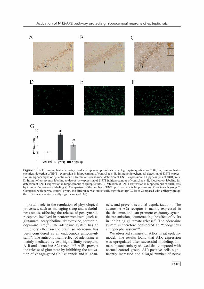
Immunohistochemical Detection of ENT1 Expression in the Hippocampus
Immunohistochemistry results showed that the ENT1-positive cells in epilepsy group si-gnificantly increased than the control group, as shown in Figure 3A and B. However, the number of ENT1-positive cells in tBHQ group decreased than normal control group (Figure 3B and 3C). Immunofluorescent labeling showed that ENT1 expression was upregulated in rat hippocampus of epileptic group (Figure 3D, 3E), while ENT1 expression was lower in tBHO group than epilep-
tic group (Figure 3F). Besides, the difference in the number of positive cells was statistically si-gnificant (p<0.05) (Figure 3G)
Western Blotting Detection of Protein Expression
The expressions of A1R and ENT1 in rat hippocampus of normal control group were de-tected (Figure 4A). However, A1R expression was significantly downregulated and ENT1 expression was upregulated in epileptic and tBHQ groups. In tBHQ group, A1R expression was markedly increased than that in epilepsy group, whereas ETN1 expression was downre-gulated (Figure 4A, 4B).
The protein expressions of Nrf2, HO-1, and ARE in hippocampus of epileptic rats were si-gnificantly downregulated compared with normal control group (p<0.05). After tBHQ treatment, protein expressions of Nrf2, HO-1, and ARE in the hippocampus of epileptic rats were signifi-cantly upregulated (Figure 4C, 4D).
Figure 1. Pathological changes, first seizure latency, and seizure frequency of rats in each group. A, Comparison of the first seizure latency of rats in each group. B, Comparison of seizure frequency in each group. C, Pathological changes in hippo-campus in each group. D, Pathological changes in hippocampus of epileptic rats. E, The pathological changes of hippocampus in tBHQ rats *: Significant difference compared with epilepsy group (p<0.05)

Y.-N. Zhang, H.-T. Dong, F.-B. Yang, Z.-Q. Wang, Z.-H. Ma, S.-Z. Ma, X.-D. Ma, L. Duan
6900
Discussion
Epilepsy is one of the most common diseases of the nervous system. It is a clinical syndrome in which high-synchronized abnormal discharges of brain neurons are caused by multiple causes22. It is characterized by episodic, transient, repetitive, and stereotypic clinical features. It is estimated that epilepsy accounts for 0.75% of the global di-sease burden, and most of epilepsy cases are in low- and middle-income countries. Epilepsy pa-tients generally need lifelong treatment. Antiepi-leptic drugs are the most commonly applied in the
epilepsy treatment23,24. The currently used antie-pileptic drugs mainly suppress epileptic seizures by inhibiting nerve hyperactivity. However, about 30% of epilepsy patients still cannot control the recurrence of epilepsy even after taking antiepi-leptic drugs. Therefore, it is still very important to study new treatment options for patients with refractory epilepsy25-27.
Adenosine exists in all cells and is involved in controlling the function of each tissue and organ. The elevation of adenosine levels plays an impor-tant role in preventing cell damage and preven-ting organ dysfunction. Adenosine also plays an
Figure 2. The results of A1R immunohistochemistry in hippocampus of rats in each group (magnification 200×). A, Im-munohistochemical detection of A1R expression in hippocampus of control rats. B, Immunohistochemical detection of A1R expression in hippocampus of epileptic rats. C, Immunohistochemical detection of A1R expression in hippocampus of tBHQ rats. D, Detection of A1R in hippocampal tissue of control rats by Immunofluorescence labeling. E, Detection of A1R in hippocampus of epileptic rats by immunofluorescence labeling. F, Detection of A1R in hippocampal tissue of tBHQ rats by immunofluorescence labeling. G, A comparison of A1R positive cells in hippocampus of rats in each group. *: Compared with normal control group, the difference was statistically significant (p<0.05); #: Compared with epilepsy group, the difference was statistically significant (p<0.05).
Activation of Nrf2-ARE pathway protecting hippocampal neurons of epileptic rats
6901
important role in the regulation of physiological processes, such as managing sleep and wakeful-ness states, affecting the release of postsynaptic receptors involved in neurotransmitters (such as glutamate, acetylcholine, dethyroxine, serotonin, dopamine, etc.)28. The adenosine system has an inhibitory effect on the brain, so adenosine has been considered as an endogenous anticonvul-sant29. The anticonvulsant effect of adenosine is mainly mediated by two high-affinity receptors, A1R and adenosine A2a receptor30. A1Rs prevent the release of glutamate by inhibiting the activa-tion of voltage-gated Ca2+ channels and K+ chan-
nels, and prevent neuronal depolarization31. The adenosine A2a receptor is mainly expressed in the thalamus and can promote excitatory synap-tic transmission, counteracting the effect of A1Rs in inhibiting glutamate release32. The adenosine system is therefore considered an “endogenous antiepileptic system”33.
We observed changes of A1Rs in rat epilepsy model. The results found that A1R expression was upregulated after successful modeling. Im-munohistochemistry showed that compared with normal control group, A1R-positive cells signi-ficantly increased and a large number of nerve
Figure 3. ENT1 immunohistochemistry results in hippocampus of rats in each group (magnification 200×). A, Immunohisto-chemical detection of ENT1 expression in hippocampus of control rats. B, Immunohistochemical detection of ENT1 expres-sion in hippocampus of epileptic rats. C, Immunohistochemical detection of ENT1 expression in hippocampus of tBHQ rats. D, Immunofluorescence labeling to detect the expression of ENT1 in hippocampus of control rats. E, Fluorescent labeling for detection of ENT1 expression in hippocampus of epileptic rats. F, Detection of ENT1 expression in hippocampus of tBHQ rats by immunofluorescence labeling. G, Comparison of the number of ENT1 positive cells in hippocampus of rats in each group. *: Compared with normal control group, the difference was statistically significant (p<0.05); #: Compared with epilepsy group, the difference was statistically significant (p<0.05).

Y.-N. Zhang, H.-T. Dong, F.-B. Yang, Z.-Q. Wang, Z.-H. Ma, S.-Z. Ma, X.-D. Ma, L. Duan
6902
cells underwent apoptosis and necrosis in epi-lepsy group. In the normal brain, homeostasis of A1R and adenosine A2a receptor maintains the inhibitory effect of the adenosine system. Other scholars found that the expressions of A1Rs were significantly upregulated after 24 hours of epi-lepsy. They pointed out that A1R expression was significantly upregulated as an adaptive response after an acute attack. Some reports have pointed out that the decreased expressions of A1Rs reflect the degeneration of neurons and increased levels of extracellular adenosine and A1Rs.
Nucleoside transporters play an important role in sleep-wakefulness, drug and alcohol addiction, nociception, and analgesia. In addition, there have been reports34,35 on the use of inhibitors of nucle-oside transporters in tumors and cardiovascular diseases. In mammals, nucleoside transporters have two major classes of equilibrium and con-centration types, and they are composed of SL-C29A and SLC28A, respectively. The balanced nucleoside transporters encoded by the SLC29A
family can be divided into ENT1, ENT2, ENT3, and ENT436. Both ENT1 and ENT2 can regulate the transfer of adenosine from intracellular to ex-tracellular or from extracellular to intracellular. The final direction of transfer entirely depends on intracellular and extracellular adenosine concen-trations. This work found that the protein content of ENT1 significantly increased in epileptic rats. According to other researchers37, mRNA level of ENT1 was expressed in pyramidal neurons and dentate gyrus granule cells in the hippocampal CA1 region of the adult human brain.
NF-E2-related factor 2 (NF-E2-related factor 2) is an important member of the transcription fac-tor leucine zipper transcriptional activator (CNC) and plays an important role in cellular defense and other stress injuries38. Nrf2 is an essential re-gulator of the induction of phase II detoxification gene expression. We found that the expressions of relative genes in Nrf2-ARE pathway in the brain tissue of epileptic rats were downregulated. After activation of Nrf2-ARE pathway by activators,
Figure 4. Protein expression in hippocampus of rats in each group. A, Expressions of A1R and ENT1 in hippocampus of rats in each group. B, Comparison of A1R and ENT1 expression levels in hippocampus of rats in each group. C, Expressions of Nrf2, HO-1 and ARE in hippocampus of rats in each group. D, Comparison of expression levels of Nrf2, HO-1, and ARE in hippocampus of rats in each group of D. *: Compared with normal control group, the difference was statistically significant (p<0.05); #: Compared with epilepsy group, the difference was statistically significant (p<0.05).

Activation of Nrf2-ARE pathway protecting hippocampal neurons of epileptic rats
6903
the pathological changes in the brain of epileptic rats were reduced, and the expression of adeno-sine receptor A1 increased, while the expression of ENT1 was downregulated. The Nrf2-ARE pa-thway may be involved in the regulation of A1R and ENT1 in the course of epilepsy. Therefore, the Nrf2-ARE pathway may serve as a new potential antiepileptic target. In other words, its selective activator can be used as a potential new antiepi-leptic drug. Our study provides novel directions for developing antiepileptic drugs, so as to impro-ve the clinical outcomes of epilepsy patients.
Conclusions
We showed that the activation of the Nrf2-A-RE signaling pathway can reduce the pathological damage of rat hippocampal neurons, prolong the latency of seizures, and reduce the degree of epi-leptic seizure in rats. The role of the Nrf2-ARE pathway in alleviating epilepsy is related to the increased A1R expression and decreased ENT1 expression.
Funding AcknowledgementsThe National Natural Science Foundation of China (81501116), and Cuiying Scientific and Technological In-novation Program of Lanzhou University Second Hospital (CY2017-MS04).
Conflict of InterestThe Authors declare that they have no conflict of interest.
References
1) StafStrom CE, Carmant L. Seizures and epilepsy: an overview for neuroscientists. Cold Spring Harb Perspect Med 2015; 5: a022426.
2) GuErrEiro Ca. Epilepsy: is there hope? Indian J Med Res 2016; 144: 657-660.
3) BEGhi E. Addressing the burden of epilepsy: many unmet needs. Pharmacol Res 2016; 107: 79-84.
4) matutE C, CavaLiErE f. Neuroglial interactions me-diated by purinergic signalling in the pathophysio-logy of CNS disorders. Semin Cell Dev Biol 2011; 22: 252-259.
5) morimoto K, fahnEStoCK m, raCinE rJ. Kindling and status epilepticus models of epilepsy: rewiring the brain. Prog Neurobiol 2004; 73: 1-60.
6) ZhanG Xf, ZhanG XQ, Wu CC, Wu hW, WEi D. Ap-plication value of procalcitonin in patients with
central nervous system infection. Eur Rev Med Pharmacol Sci 2017; 21: 3944-3949.
7) hanaya r, SaSa m, SuGata S, toKuDomE m, SEriKaWa t, KuriSu K, arita K. Hippocampal cell loss and propa-gation of abnormal discharges accompanied with the expression of tonic convulsion in the spontane-ously epileptic rat. Brain Res 2010; 1328: 171-180.
8) BroEr S, LoSChEr W. Novel combinations of phe-notypic biomarkers predict development of epilepsy in the lithium-pilocarpine model of temporal lobe epilepsy in rats. Epilepsy Behav 2015; 53: 98-107.
9) PatEL hC, roSS fm, hEEnan LE, DaviES rE, rothWELL nJ, aLLan Sm. Neurodegenerative actions of inter-leukin-1 in the rat brain are mediated through in-creases in seizure activity. J Neurosci Res 2006; 83: 385-391.
10) PoLLarD h, Charriaut-marLanGuE C, CantaGrEL S, rEPrESa a, roBain o, morEau J, BEn-ari y. Kaina-te-induced apoptotic cell death in hippocampal neurons. Neuroscience 1994; 63: 7-18.
11) honiG LS, roSEnBErG rn. Apoptosis and neurologic disease. Am J Med 2000; 108: 317-330.
12) anDErSon Cm, XionG W, GEiGEr JD, younG JD, CaSS CE, BaLDWin Sa, ParKinSon fE. Distribution of equi-librative, nitrobenzylthioinosine-sensitive nucleo-side transporters (ENT1) in brain. J Neurochem 1999; 73: 867-873.
13) PEnG L, huanG r, yu aC, funG Ky, rathBonE mP, hErtZ L. Nucleoside transporter expression and function in cultured mouse astrocytes. Glia 2005; 52: 25-35.
14) BoiSon D. The adenosine kinase hypothesis of epi-leptogenesis. Prog Neurobiol 2008; 84: 249-262.
15) hamiLton nB, attWELL D. Do astrocytes really exocytose neurotransmitters? Nat Rev Neurosci 2010; 11: 227-238.
16) miGuEL-hiDaLGo JJ. The role of glial cells in drug abuse. Curr Drug Abuse Rev 2009; 2: 76-82.
17) Wu J, LEE mr, Choi S, Kim t, Choi DS. ENT1 re-gulates ethanol-sensitive EAAT2 expression and function in astrocytes. Alcohol Clin Exp Res 2010; 34: 1110-1117.
18) Pinto-DuartE a, CoELho JE, Cunha ra, riBEiro Ja, SE-BaStiao am. Adenosine A2A receptors control the extracellular levels of adenosine through modula-tion of nucleoside transporters activity in the rat hippocampus. J Neurochem 2005; 93: 595-604.
19) JohnSon Da, JohnSon Ja. Nrf2--a therapeutic tar-get for the treatment of neurodegenerative disea-ses. Free Radic Biol Med 2015; 88: 253-267.
20) SanDBErG m, PatiL J, D’anGELo B, WEBEr SG, maLLarD C. NRF2-regulation in brain health and disease: implication of cerebral inflammation. Neurophar-macology 2014; 79: 298-306.
21) WanG W, Wu y, ZhanG G, fanG h, WanG h, ZanG h, XiE t, WanG W. Activation of Nrf2-ARE signal pathway protects the brain from damage induced by epileptic seizure. Brain Res 2014; 1544: 54-61.
22) o’muirChEartaiGh J, riCharDSon mP. Epilepsy and the frontal lobes. Cortex 2012; 48: 144-155.

Y.-N. Zhang, H.-T. Dong, F.-B. Yang, Z.-Q. Wang, Z.-H. Ma, S.-Z. Ma, X.-D. Ma, L. Duan
6904
23) WaGnEr rG, BottomLEy C, nGuGi aK, iBinDa f, Góm-EZ-oLivé fX, Kahn K, toLLman S, nEWton Cr; SEEDS WritinG GrouP, WaGnEr r, tWinE r, Connor m, CoL-LinSon m, maSanJa h, mathEW a, KaKooZa a, Pariyo G, PEtErSon S, nDyo-muGhEnyi D, oDhiamBo r, ChEn-Go E, ChaBi m, Bauni E, Kamuyu G, oDEra vm, ma-GEto Jo, aE-nGiBiSE K, aKPaLu B, aKPaLu a, aGBoKEy f, aDJEi P, oWuSu-aGyEi S, KLEinSChmiDt i, DoKu vC, oDErmatt P, nEviLLE B, SanDEr JW, WhitE S, nutman t, WiLKinS P, noh J. Incidence, remission and mor-tality of convulsive epilepsy in rural Northeast South Africa. PLoS One 2015; 10: e129097.
24) GooDriCh GS, KaBaKov ay, hamEED mQ, DhamnE SC, roSEnBErG Pa, rotEnBErG a. Ceftriaxone treatment after traumatic brain injury restores expression of the glutamate transporter, GLT-1, reduces regio-nal gliosis, and reduces post-traumatic seizures in the rat. J Neurotrauma 2013; 30: 1434-1441.
25) BoiSon D. The biochemistry and epigenetics of epilepsy: focus on adenosine and glycine. Front Mol Neurosci 2016; 9: 26.
26) Shao y, WanG C, honG Z, ChEn y. Inhibition of p38 mitogen-activated protein kinase signaling redu-ces multidrug transporter activity and anti-epilep-tic drug resistance in refractory epileptic rats. J Neurochem 2016; 136: 1096-1105.
27) KWan P, BroDiE mJ. Early identification of refractory epilepsy. N Engl J Med 2000; 342: 314-319.
28) SWiaDEr mJ, KotoWSKi J, LuSZCZKi JJ. Modulation of adenosinergic system and its application for the treatment of epilepsy. Pharmacol Rep 2014; 66: 335-342.
29) harGuS nJ, JEnninGS C, PErEZ-rEyES E, BErtram Eh, PatEL mK. Enhanced actions of adenosine in me-dial entorhinal cortex layer II stellate neurons in temporal lobe epilepsy are mediated via A(1)-re-ceptor activation. Epilepsia 2012; 53: 168-176.
30) SEBaStiao am, riBEiro Ja. Adenosine receptors and the central nervous system. Handb Exp Pharma-col 2009: 471-534.
31) frEDhoLm BB, ChEn Jf, Cunha ra, SvEnninGSSon P, vauGEoiS Jm. Adenosine and brain function. Int Rev Neurobiol 2005; 63: 191-270.
32) BoiSon D, ChEn Jf, frEDhoLm BB. Adenosine signa-ling and function in glial cells. Cell Death Differ 2010; 17: 1071-1082.
33) matutE C, CavaLiErE f. Neuroglial interactions me-diated by purinergic signalling in the pathophysio-logy of CNS disorders. Semin Cell Dev Biol 2011; 22: 252-259.
34) KinG aE, aCKLEy ma, CaSS CE, younG JD, BaLDWin Sa. Nucleoside transporters: from scavengers to novel therapeutic targets. Trends Pharmacol Sci 2006; 27: 416-425.
35) BaLDWin Sa, BEaL Pr, yao Sy, KinG aE, CaSS CE, younG JD. The equilibrative nucleoside transporter family, SLC29. Pflugers Arch 2004; 447: 735-743.
36) aymEriCh i, DufLot S, fErnanDEZ-vELEDo S, GuiLLEn-Go-mEZ E, huBEr-ruano i, CaSaDo fJ, PaStor-anGLaDa m. The concentrative nucleoside transporter family (SLC28): new roles beyond salvage? Biochem Soc Trans 2005; 33(Pt 1): 216-219.
37) anDErSon Cm, XionG W, GEiGEr JD, younG JD, CaSS CE, BaLDWin Sa, ParKinSon fE. Distribution of equi-librative, nitrobenzylthioinosine-sensitive nucleo-side transporters (ENT1) in brain. J Neurochem 1999; 73: 867-873.
38) Carmona-aPariCio L, PérEZ-CruZ C, ZavaLa-tECuaPEt-La C, GranaDoS-roJaS L, rivEra-ESPinoSa L, montESi-noS-CorrEa h, hErnánDEZ-Damián J, PEDraZa-ChavErri J, SamPiEri a 3rD, CoBaLLaSE-urrutia E, CárDEnaS-roD-ríGuEZ n. Overview of Nrf2 as therapeutic target in epilepsy. Int J Mol Sci 2015; 16: 18348-18367.