cerebellar hippocampal activation eyeblink depends ... · explore the suitability of the design for...
TRANSCRIPT
NEURAL PLASTICITY VOLUME 10, NO. 4, 2003
Cerebellar and Hippocampal Activation during Eyeblink ConditioningDepends on the Experimental Paradigm: A MEG Study
Peter Kirsch, 1’2 Caroline Achenbach, 1’3 Martina Kirsch, Matthias Heinzmann,4
Anne Schienle 1’2 and Dieter Vaitl ’2
l*Department ofClinical & Physiological Psychology and 2Bender Institute ofNeuroimaging,University ofGiessen, Germany; 3Departments ofNeurology and 4Psychology
University ofHeidelberg, Germany
SUMMARY KEYWORDS
The cerebellum and the hippocampus arekey structures for the acquisition of conditionedeyeblink responses. Whereas the cerebellumseems to be crucial for all types of eyeblinkconditioning, the hippocampus appears to beinvolved only in complex types of learning. Weconducted a differential conditioning study toexplore the suitability of the design formagnetencephalography (MEG). In addition,we compared cerebellar and hippocampalactivation during differential delay and traceconditioning. Comparable conditioning effectswere seen in both conditions, but a greaterresistance to extinction for trace conditioning.Brain activation differed between paradigms:delay conditioning provoked activation only inthe cerebellum and trace conditioning only inthe hippocampus. The results reflect differentialbrain activation patterns during the two typesof eyeblink conditioning.
This research was conducted at the Department of Clinical& Physiological Psychology, Otto-Behaghel Str. 10, D-35394Giessen, Germany. Reprint requests to: Dr. Peter Kirsche-mail: [email protected]
delay conditioning, trace conditioning, brain electric
source analysis
INTRODUCTION
Classical conditioning of eyeblink responses isone of the most frequently investigated paradigms.The experimental design consists of a tone or lightas the conditioned stimulus (CS) and a cornealairpuff as the unconditioned stimulus (US). Sincethe occurrence of the conditioned response (CR) isvery stable over time, the paradigm seems to besuitable for the investigation of magnetencephalo-graphy (MEG) correlates of human learning,where, due to an adverse signal to noise ratio, manytrials are required to average an evoked response.
Animal studies of eyeblink conditioning havebeen quite consistent in demonstrating theimportance of the cerebellum and the hippocampusfor the acquisition and initiation of the CR (seeThompson & Krupa, 1994; Woodruff-Pak &Steinmetz, 2000 for an overview). Similar findingshave been obtained for humans. In studies onpatients with cerebeilar lesions, it was shown thatthey are not able to generate conditioned eyeblinkresponses (Gerwig et al., 2003; Bracha et al.,1997; Daum et al., 1993). Brain imaging studies
(C) 2003 Freund & Pettman, U.K. 291

292 P. KIRSCH ET AL.
also point to the role the cerebellum plays inhuman eyeblink conditioning. Cerebellaractivation has been found with both, positionemission tomography (PET, Logan & Grafton,1995; Blaxton et al, 1996; Schreurs et al., 1997)and functional magnetic resonance imaging (fMRI,Ramnani et al., 2000). PET studies, however, havea number of methodological disadvantages makingthe generalization of the results difficult. Theconditioning trials have to be presented in a blockdesign, and because of the poor temporalresolution, no conclusion can be drawn about thetime course of the cerebellar activation duringlearning. Even in fMRI studies with a bettertemporal resolution, in which event-related designsallow a single trial presentation, a temporaldifferentiation of the activation is hardly possible.Therefore, a method like the whole-head MEG,which provides both an excellent temporal and asufficient spatial resolution, might allow additional
insight into the neural basis of human eyeblinkconditioning.
In contrast to the cerebellum, the role of thehippocampus in eyeblink conditioning is less clear.Although some studies found hippocampus unit
responses in CA1 as well as in CA3 neurons
(Berger & Thompson, 1978; Berger et al., 1983)during delay conditioning, most researchers agreethat delay conditioning is hippocampusindependent (Steinmetz, 2000; Thompson &Krupa, 1994). In contrast, for trace conditioning,and intact hippocampus is essential. Weiss et al.
(1999), as well as many others, found an
impairment of conditioned responses in rats after a
hippocampal lesion for the trace conditioningparadigm but no effect of the lesion of delayconditioning performance. Furthermore, Gould et
al. (1999) found an increase of the number ofhippocampal neurons in adult rats after trace
conditioning, indicating neural plasticity. For thoserats that were presented to a delay conditioningparadigm, no changes in hippocampal neuron
density could be observed. Thompson (1991)proposes that for simple types of conditioning,hippocampal processing is not necessary, whereasit is essential for complex learning involvingdeclarative memory functions. This interpretationis in good accordance with results from Kishimotoet al. (2001). The authors found an impairment oflong interval trace but not delay eyeblinkconditioning in mutant mice lacking a specificNMDA receptor. As these receptors are essentialfor long-term potentiation (LTP) in thehippocampus, the results can be interpreted as
reflecting the importance of hippocampal LTP fortrace but not delay conditioning. In accordancewith this view, others (Clark & Squire, 1998) haveshown that hippocampal functions are not
obligatory for simple delay conditioning for whichthe US occurs when the CS is still present. Incontrast, trace conditioning, with a time gapbetween CS offset and US onset, requires an intacthippocampus. This distinction is further supportedby data from studies of brain-injured humans.When delayed conditioning was applied, nodifferences could be found between patients withhippocampal lesions and healthy controls (e.g.Gabrieli et al., 1995), whereas with trace
conditioning an impaired learning of this patientgroup has been demonstrated (e.g. Daum et al.,1991, for an overview see McGlinchey-Berroth,2000.) Carrillo et al. (2001) found unaffecteddiscrimination of two tones but impaired reversallearning during a differential conditioning paradigmin patients with a medial temporal lobe lesion.Consistent with Thompson (1991), they argue thatthe hippocampus is necessary for complex types oflearning but not for simple delayed discriminationlearning.
In the present MEG study, we applied a
differential eyeblink conditioning paradigm. Thesubjects received two types of CS. The CS+ was
paired with the US during the acquisition but not
during the extinction, whereas the CS- was not
DELAY AND TRACE EYEBLINK CONDITIONING 293
paired. The degree of learning was quantified asthe difference in CR rates between the CS+ andthe CS- condition. We focused on the intensity aswell as on the time course of the cerebellar andhippocampal activation using both a trace and a
delay conditioning paradigm.As evoked neural activity from the cerebellum
and the hippocampus has been demonstratedbefore via MEG (Tesche & Karhu, 1997; Tescheet al., 1996; Tesche, 1997), we expected to findmore activation toward the CS+ than the CS-inthe cerebellum for both experimental paradigms.In contrast, the hippocampus should be active onlyduring trace conditioning. Further, in a moreexplorative approach, we studied the time courseof activation to find out whether cerebellaractivation precedes or follows hippocampalactivation.
EXPERIMENTAL
Subjects
Thirty subjects (12 males, mean age 25 years,range 20-34 years) participated in the study. Twosubjects had to be excluded because of lack ofconditioned eyeblink responses. In order toprevent a learning transfer from one experimentalparadigm to the other, we used a between-subjectdesign. Fourteen subjects (5 males) received adelayed conditioning paradigm, the other 14subjects (5 males) underwent the traceconditioning paradigm. The two groups did notdiffer with regard to age or educational level.Informed consent was obtained from all subjects.Each subject received 15 for participation.
Stimulus material and experimental design
A differential conditioning paradigm wasapplied. The acquisition phase consisted of 150 CS+
trials with a combined CS-US presentation and 150CS- trials, in which the CS- was presented alone.The subsequent extinction phase consisted of 75unpaired CS+ and 75 CS- trials. The trial sequencewas pseudo-randomized with no more than two
equal CS types following each other. The meaninter trial interval (ITI, offset of a trial to onset ofthe next trial) was 2.5 s, varying between 2 s and3 s. This short ITI was chosen to allow a highnumber of trials per subject, which is necessary toachieve an acceptable signal-to-noise ratio.
Instead of an acoustic stimulation, which istypical for eyeblink conditioning, we used a visualone because aural stimulation would haveproduced activation in temporal brain regions thatwould have been difficult to distinguish fromhippocampal activity during source localization. Inaddition, visual stimulation ensured that thesubjects would not close their eyes during theexperiment. In order to present physicallycomparable stimuli for the CS+ and CS- we usedcircles (10 cm in diameter) filled with black andwhite lines that differed only in terms of theorientation of the lines. The stimuli were presentedon a 17-inch computer monitor located 2 m infront of the subjects outside the shielded MEGroom. For one CS stimulus the lines were turned45 clockwise and for the other one 45 counterclockwise. Both stimuli were alternately used asCS+ and CS-, respectively. In the delayconditioning group, the CS was presented for 750ms and during acquisition immediately followedby the US. For the trace conditioning group, theCS was presented for 100 ms followed by a 650ms interval and then, during acquisition, by theUS. The US was a 1.48 atmospheres air puff of 50ms duration presented on one cornea (half of thesubject left, half right). The puff was presented viaspecial metal-free goggles in which the air tubewas implemented.
The whole head MEG was measured by a 122channel Neuromag-122TM MEG system (Ahonen

294 P. KIRSCH ET AL.
et al., 1993) with a sample rate of 400 Hz and a
band pass filter with a high pass cut off from. Hzto account for DC like artifacts and a standard lowpass cut off at a quarter of the sampling rate.
Eyeblinks were measured with two Ag/AgC1electrodes attached vertically above and below thestimulated eye, sampled by a NeuroscanSynamps(R) EEG system (Neuroscan Labs, Sterling,Virginia) and amplified and stored by the MEGsystem.
Before the experiment, the head of eachsubject was digitized using 30 landmarks. Thisinformation was used to define sensor positionsrelative to the individual head of tb.e subject,which is essential for group analysis of sourceactivation.
The stimulus presentation and timing wascontrolled by a PC using STIM software(Neuroscan Labs, Sterling, Virginia), which isespecially designed for event-related EEG andMEG studies providing a timing that is adjusted tothe refresh cycle of the monitor. The air puffs were
produced by a generator developed in our own
laboratory and controlled by the STIM software.Due to air compression in the tube, the delivery ofthe air puff was delayed by 50 ms, which was
explored as being constant before the experiment.
Data analysis
Each trial was manually examined for theexistence of a conditioned eyeblink response,which was defined as a reaction occurring betweenCS and US onset. This is an unusually long-timewindow to determine CRs. However, alpha or
orienting responses as often observed for visualstimuli should contribute to both conditions in thesame manner. Therefore, differences between CS+and CS- should be due to the experimentalvariation rather than to the stimulation. For eachsubject, the percentage of eye blinks wascalculated for each experimental condition
separately. To examine the course of learning foreach subject, blocks of 10 trials during acquisitionand blocks of 5 trials during extinction wereaveraged and the percentage of eye blinks perblock calculated for each experimental conditionseparately.
The results were analyzed using a three-wayanalysis of variance (ANOVA) with the factors’CS type’ (CS+ vs CS-), ’paradigm’ (trace vsdelay) and ’block’ (1 to 15), separately foracquisition and extinction. Additionally, todocument the size of extinction effects, a three-way ANOVA was computed with the factors ’CStype’ (CS+ vs CS-), ’paradigm’ (trace vs delay)and ’time’ (last 50 trials of extinction vs. first 50trials of extinction),
For all subjects, evoked magnetic activity wasaveraged for the CS+ and the CS- conditionseparately. Before averaging, the signal wasfiltered with a zero phase bandpass filter between1.5 and 20 Hz because the evoked magnetic fieldcomponents were expected within this frequencyrange. With these evoked magnetic fieldresponses, a source analysis was conducted usingBrain Electric Source Analysis (BESA, Scherg,1990). All MEG analyses were conducted with theBESA 2000 software package (MEGIS SoftwareGmbH, Grifling, Germany). With respect to thehigh amount of spread activity in the presentparadigm and to the inverse problem of sourcelocalization, a unique solution for dipolelocalization could not be found. Therefore andbecause our specific regions of interest weredefined before the experiment, we used regionalsource analysis to investigate the activity in thecerebellum and the hippocampus.
With this method, dipoles are defined inspecific anatomical regions of interest. Tomaximize the amount of variance explained,individual source fitting is conducted but restrictedto the source orientation. The software calculatesthe specific signal component of the measured
DELAY AND TRACE EYEBLINK CONDITIONING 295
data, which is explained by the particular dipolefor a predefined time window. Parameters likeamplitude (in nAmp) and the latency of theresulting signal can be analyzed. Because theBESA algorithm is sensitive to temporal andspatial interference with other active sources, we
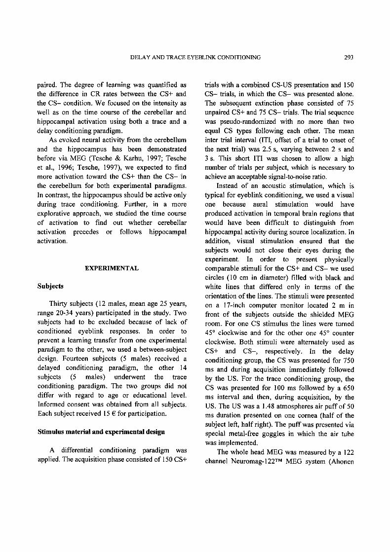
defined additional regional sources in regions inwhich activity can be expected in the presentparadigm. These additional sources were locatedin both eyes to account for eye movements, thebilateral occipital lobes to account for primary andsecondary visual processes, the parietal lobe toaccount for activation from the temporal visualsystem, and unspecific attentional processes andthe vertex to reduce a maximum of unspecificactivation.
Figure displays the position of the sourcesused in the present study. The same sourceconfiguration was used for each subject adjusted
only with respect to the individual head size andshape. The resulting signals from our four sourcesof interest (cerebellum and hippocampus, left andright) were analyzed with respect to the maximalpeak-to-peak amplitude in the time window 0 to750 ms after CS onset. Because the stimuluspresentation side was counterbalanced betweensubjects, the resulting activation from the twosources per region was averaged, resulting in oneactivation value per region. Latencies were definedas time between CS onset and the peak of themaximal amplitude. The differences betweengroups were statistically confirmed using a three-way mixed model ANOVA with the within subjectfactors ’CS-type’ (CS+ vs. CS-) and ’region’(cerebellum vs. hippocampus) and the betweensubject factor ’paradigm’ (delay vs. trace). Analysesof amplitudes and latencies were conducted foracquisition and extinction separately.
Fig. 1: Configuration of the dipoles of interest and the additional regional sources superimposed on a standardized brainimage. Abbreviations are C for Cerebellum, H for Hippocampus, E for Eyes, 0 for occipital site, V for vertexand P for parietal site. Additional characters L and R indicate left and right sources.
296 P. KIRSCH ET AL.
RESULTS
Conditioned eyeblink responses
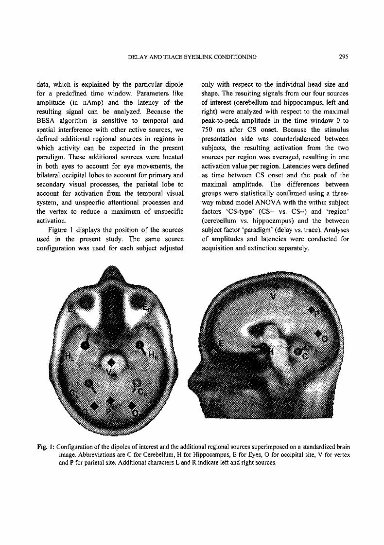
Subjects showed significantly more eyeblinksto CS+ than to CS- during acquisition (F(1/26)=2.694, p <.001). The difference between CS+ andCS- increased over time, indicated by a significantCS-type by block interaction (F(14/364)= 8.188,p < .001, e=.396, Fig. 2), but no difference wasfound between trace and delay conditioning. Thus,comparable conditioning effects were found forboth groups.
The extinction was very fast as reflected by a
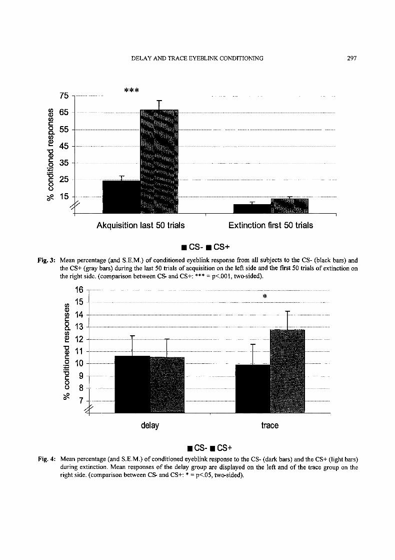
significant time main effect (F(1/26) 175.353,p < .001), as well as by a CS-type by time inter-action (F(1/26)=3.943, p<.001) for ANOVAcomparing the last 50 acquisition trials with thefirst 50 extinction trials (Fig. 3.) While there was a
highly significant CS+/CS- difference during theend of acquisition (p <.001 for the post-hoc t-
Test), the difference between CS+ and CS- nearlydisappeared for the extinction (p<.06, one-sided.).
During extinction, there were no differencesbetween the CS+ and CS- condition with respect tothe percentage of eyeblinks. However, the inter-action between CS-type and paradigm showed atrend towards significance (F(1/26) 3.281,p < .09). The post-hoc t-test revealed significantlymore eyeblinks to the CS+ than the CS- only forthe trace but not for the delay group (Fig. 4).
Magnetic brain activity
The source model used for the analysis ofbrain activation explained between 63.9% and78.2 % of the variance of the MEG signal. TheANOVA of the source activation amplitude duringacquisition revealed a significant three-wayinteraction (F(1/26)= 4.242, p .05). As displayedin Fig. 5, stronger source activation during CS+
80
70
o- 5o
g 4o
(C)
2O
-o-. CS- CS+
"0"0"0"0"0"0"0. 0-0O’
1 2 3 4 5 6 7 8 9 10 11 12 13 14 15
Blocks of 10 trials
Fig. 2" Mean percentage of conditioned eyeblink response to the CS- (dotted line) and the CS+ (solid line) duringacquisition for all subjects, averaged over 10 trials per block.
DELAY AND TRACE EYEBLINK CONDITIONING 297
75
65
o 55
" 45
35
- 25
5
Akquisition last 50 trials Extinction first 50 trials
CS- CS+Fig. 3" Mean percentage (and S.E.M.) of conditioned eyeblink response from all subjects to the CS- (black bars) and
the CS+ (gray bars) during the last 50 trials of acquisition on the left side and the first 50 trials of extinction onthe right side. (comparison between CS- and CS+: *** p<.001, two-sided).
6
. 32. 0- 9
o 87
delay trace
CS- CS+Fig. 4" Mean percentage (and S.E.M.) of" conditioned eyeblink response to the CS- (dark bars) and the CS+ (light bars)
during extinction. Mean responses of the delay group are displayed on the left and of the trace group on theright side. (comparison between CS- and CS+’ * p<.05, two-sided).
298 P. KIRSCH ET AL.
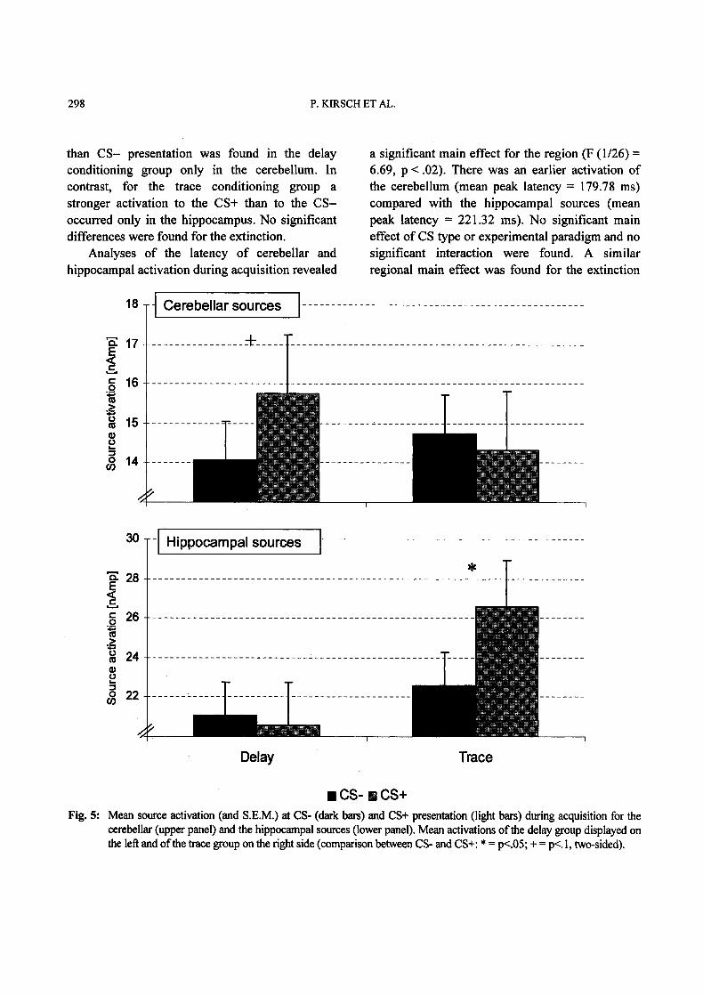
than CS- presentation was found in the delayconditioning group only in the cerebellum. Incontrast, for the trace conditioning group astronger activation to the CS+ than to the CS-occurred only in the hippoeampus. No significantdifferences were found for the extinction.
Analyses of the latency of cerebellar andhippocampal activation during acquisition revealed
a significant main effect for the region (F (1/26)=6.69, p < .02). There was an earlier activation ofthe cerebellum (mean peak latency 179.78 ms)compared with the hippocampal sources (meanpeak latency 221.32 ms). No significant maineffect of CS type or experimental paradigm and nosignificant interaction were found. A similarregional main effect was found for the extinction
18 Cerebellar sources
"E 28-E<’- 26-._._
24
22-
Hippocampal sources
Delay Trace
CS- B CS+Fig. 5: Mean source activation (and S.E.M.) at CS- (dark bars) and CS+ presentation (light bars) during acquisition for the
cerebellar (upper panel) and the hippocampal sources (lower panel). Mean activations ofthe delay group displayed onthe left and ofthe trace group on the right side (comparison between CS- and CS+: * p<.05; + p<.l, two-sided).

DELAY AND TRACE EYEBLINK CONDITIONING 299
(F(1/26)=7.44, p<.02), again with a fasteractivation of the cerebellum (mean peak latency190.92 ms) than the hippocampus (mean peaklatency 246.0 ms).
DISCUSSION
The results of the present study demonstratethat classical eyeblink conditioning can beinvestigated using MEG methodology. In bothexperimental conditions, comparable differentialeyeblink conditioning occurred. The relatively flatlearning curve (Fig. 2), could be a result of theunusual short ITI used in the present study. Thereis some evidence that in animal studies an ITI fromat least 10 s is necessary to establish stableconditioning (Nordholm, et al., 1991). However,successful eyeblink conditioning was demonstratedwith shorter ITIs in humans. Carrillo et al. (1997)reported no difference in the conditioned responserate between ITIs of 5, 10, or 30 seconds.Furthermore, our conditioned response ratesobserved for the last 50 trials during acquisition,as displayed in Fig. 3, are comparable to those ofother studies with normal participants. Therefore,we conclude that we were able to initiatedifferential eyeblink conditioning in the presentstudy.
In contrast to the acquisition, delay and traceconditioning differed in terms of resistance toextinction. While the differentiation between CS+and CS- completely disappeared in the delay group,there were slightly more CRs to the CS+ than theCS- in the trace group. This effect cannot beattributed to a sensitization to CS- during delayconditioning, since the number of CRs to CS- didnot increase and the number of CRs to CS+decreased. Even when we take into account that theabsolute number of CRs to CS+ during trace
conditioning was rather small, our results confirm
that the CS-US association learned in this conditionwas more stable than during delay conditioning.This result might be due to hippocampal processing,possibly related to awareness of the CS-USassociation (Clark, & Squire, 1998; Manns et al,,2000), leading to a longer lasting memory for thisassociation. This hippocampal involvement duringtrace conditioning was confirmed by analysis of themagnetic field activity. Only for trace conditioningmore activation to the CS+ than the CS- was foundin the hippocampus. This result is in goodaccordance with our hypothesis. Also in line withour hypothesis was the stronger cerebellar activationto the CS+ than the CS- in the delay conditioninggroup.
In contrast to our expectations was the lack ofdifferentiation between CS+ and CS- for cere-bellar activation during trace conditioning. Allcurrent models of eyeblink conditioning emphasizethe central role of the cerebellum for thisphenomenon. However, it could be argued thatsome cerebellar functions were taken over by thehippocampus during trace conditioning. Kishimotoand colleagues (2001) observed an impaired delayconditioning but an unaffected trace eyeblinkconditioning in PCLbeta4 mutant mice. Such miceshowed a reduced long-term depression (LTD) inthe rostral cerebellum, which might be essentialfor delay but not for trace conditioning. Althoughwe have no evidence for the conclusion that oursource activation reflects LTD, the resultsdemonstrate that delay and trace conditioningmight differ in terms of cerebellar involvement.
As a shortcoming of our study, we have tomention that we used only one source percerebellar hemisphere. Therefore, we cannot besure that all cerebellar processes during our
experimental procedures were registered to thesame degree. Furthermore, increases in activity ofvisual systems could have been wrongly explainedby the cerebellar sources during trace conditioning.Although we had applied occipital as well as
300 P. KIRSCH ET AL.
parietal sources to explain most of this sensoryactivity, we cannot completely exclude that visualactivity was absorbed by the cerebellar sources.While differences between CS+ and CS- duringdelay conditioning cannot be attributed to visualprocessing of physically identical stimuli andshould reflect cerebe|lar processing, the lack ofdifference between the CS conditions during traceconditioning could be based on a wronglocalization of visual activity, which in fact shouldnot differ between CS+ and CS-.
The analysis of the latency of the sourceactivation did not reveal any differences betweenthe experimental conditions. Neither the CS typenor the paradigm influenced the onset of activation.However, we found a significant main effect ofstructure with an earlier activation of cerebellar thanhippocampal sources for all conditions and foracquisition as well as extinction. These findingsmake a lot of sense for delay conditioning, as itcould be argued that the cerebellum canindependently controls the conditioned response.Hippocampal activation could reflect attentionalprocessing of the stimuli without a crucial influenceon the CR. In contrast, during trace conditioning,hippocampal processing should influence the CRinitiation through the cerebellum. Therefore,cerebellar activation should follow the hippocampalactivation rather than precede it. However, since wefound no specific cerebellar activation during traceconditioning, it might reflect more learning-independent processes or, as mentioned before,activation from other regions.
A method with a better spatial resolution likefMRI could help to find differences as well ascongruencies in cerebellar activation between delayand trace conditioning. Furthermore, it would bevery interesting to use spatial information revealedby fMRI to improve the source configuration forMEG. As mentioned before, it could also be usefulto apply different cerebellar sources for delay andtrace conditioning. Thus, the combination of fMRI
and MEG methodology promises a deeperunderstanding of temporal and spatial differences inbrain activation during human trace and delayeyeblink conditioning.
ACKNOWLEDGMENT
This work was supported by a research grantfrom the Deutsche Forschungsgemeinschaft to PK(Ki 576/4-1).
REFERENCES
Ahonen, AI. Hm,linen, MS, Knuutila, JET, Kajola,MJ, Laine, PP, Lounasmaa, OV, et al. 1993. 122-channel SQUID Instrument for Investigating theMagnetic Signals from the Human Brain. PhysicaScripta T49 198-205.
Berger TW, Rinaldi PC, Weisz DJ, Thompson RF.1983. Single-unit analysis of differenthippocampal cell types during classicalconditioning of rabbit nictitating membraneresponse. J Neurophysiol 50:1197-1219.
Blaxton TA, Zeffiro TA, Gabrieli JD, BookheimerSY, Carrillo MC, Theodore WH, et al. 1996.Functional mapping of human learning: a positronemission tomography activation study of eyeblinkconditioning. J Neurosci 16: 4032-4040.
Bracha V, Zhao L, Wunderlich DA, Morrissy SJ,Bloedel JR. 1997. Patients with cerebellarlessions cannot acquire but are able to retainconditioned eyeblink reflexes. Brain 120" 1401-1413.
Carrillo MC, Gabrieli JDE, Hopkins RO,McGlinchey-Berroth R, Fortier CB, et al. 2001.Spared discrimination and impaired reversaleyeblink conditioning in patients with temporallobe amnesia. Behav Neurosci 115’ 1171-1179.
Carrillo MC, Thompson LT, Gabrieli JDE, DisterhoftJF. 1997. Variation of the intertrial interval inhuman classical conditioning. Psychobiology 25:152-157.
Clark RE, Squire LR. 1998. Classical conditioningand brain systems" The role of awareness. Science280:77-81.

DELAY AND TRACE EYEBLINK CONDITIONING 301
Daum I, Channon S, Polkey CE, Gray JA. 1991.Classical conditioning after temporal lobe lesionsin man" impairment in conditional discrimination.Behav Neurosci 105: 396-408.
Daum I, Schugens MM, Ackermann H, LutzenbergerW, Dichgans J, Birbaumer N. 1993. Classicalconditioning after cerebellar lesions in humans.Behav Neurosci 107: 748-756.
Gabrieli JD, McGlinchey-Berroth R, Carrillo MC,Gluck MA, Cermak LS, Disterhoft JF. 1995.Intact delay-eyeblink classical conditioning inamnesia. Behav Neurosci 109:819-727.
Gerwig M, Dimitrova A, Kolb FP, Maschke M, BrolB, Kunnel A, et al. 2003. Comparison of eyeblinkconditioning in patients with superior and pos-terior inferior cerebellar lesions. Brain 126: 71-94.
Gould E, Beylin A, Tanapat P, Reeves A, Shors TJ.1999. Learning enhances adult neurogenesis in thehippocampal formation. Nat Neurosci 2: 260-265.
Kishimoto Y, Hirono M, Sugiyama T, Kawahara S,Nakao K, Kishio M, et al. 2001. Impaired delaybut normal trace eyeblink conditioning inPLCbeta4 mutant mice. NeuroReport 12: 2919-2922.
Kishimoto Y, Kawahara S, Mori H, Mishina M,Kirino Y. 2001. Long-trace interval eyeblinkconditioning is impaired in mutant mice lackingthe NMDA receptor subunit epsilonl. Eur JNeurosci 13:1221-1227
Logan CG, Grafton ST. 1995. Functional anatomy ofhuman eyeblink conditioning determined withregional cerebral glucose metabolism andpositron-emission tomography. Proc Natl AcadSci USA 92: 7500-7504.
Manns JR, Clark RE, Squire LR. 2000. Parallelacquisition of awareness and trace eyeblinkclassical conditioning. Learn Mem 7" 267-272.
McGlinchey-Berroth R. 2000. Eyeblink classicalconditioning in amnesia. In: Woodruff-Pak D,Steinmetz JE, eds, Eyeblink classicalconditioning: Volume I: Applications in humans,Boston: Kluwer Academic Press, 205-228.
Nordholm AF, Lavond DG, Thompson RF. 1991.Are eyeblink responses to tone in the decerebrate,decerebellate rabbit conditioned responses?Behav Brain Res 44: 27-34.
Ramnani N, Toni I, Josephs O, Ashbumer J, Passing-ham RE. 2000. Leaming- and expectation-relatedchanges-in the human brain during motorlearning. J Neurophysiol 84: 3026-3035.
Scherg M. Fundamentals of dipole source potentialanalysis. 1990. In: Grandori F, Hoke M, RomaniGL, eds. Auditory Evoked Magnetic Fields andPotentials. Advanced of Audiology vol 6. Basel,Switzerland: Karger 40-69.
Schreurs BG, Mclntosh AR, Bahro M, Herscovitch P,Sunderland T, Molchan SE. 1997. Lateralizationand behavioral correlation of changes in regionalcerebral blood flow with classical conditioning ofthe human eyeblink response. J Neurophysiol 77:2153-2163.
Steinmetz JE. 2000. Brain substrates of classicaleyeblink conditioning: a highly localized but alsodistributed system. Behav Brain Res 110:13-24.
Tesche CD. 1997. Non-invasive detection of ongoingneuronal population activity in normal humanhippocampus. Brain Res 749" 53-60.
Tesche CD, Karhu J. 1997. Somatosensory evokedmagnetic fields arising from sources in the humancerebellum. Brain Res 744: 23-31.
Tesche CD, Karhu J, Tissari SO. 1996. Non-invasivedetection of neuronal population activity inhuman hippocampus. Cogn Brain Res 4: 39-47.
Thompson RF. 1991. Are memory traces localized ordistributed? Neuropsychologia 29:571-582.
Thompson RF, Krupa DJ. 1994. Organization ofmemory traces in the mammalian brain. Annu RevNeurosci 17:519-549.
Weiss C, Bouwmeester H, Power JM, Disterhoft JF.1999. Hippocampal lesions prevent trace eyeblinkconditioning in the freely moving rat. BehavBrain Res 99:123-132
Woodruff-Pak DS, Steinmetz JE, eds. 2000. EyeblinkClassical Conditioning: Volume II: Animal Models.Boston, Massachusetts, USA: Kluwer Academic.