a new megastigmane glycoside from akebia quinata
TRANSCRIPT
RESEARCH ARTICLE
A new megastigmane glycoside from Akebia quinata
Hong-Guang Jin • A Ryun Kim • Hae Ju Ko •
Eun-Rhan Woo
Received: 20 December 2013 / Accepted: 9 February 2014
� The Pharmaceutical Society of Korea 2014
Abstract A new megastigmane glycoside, 8S*,9R*-
megastigman-3-one-4,6-diene-8,9-diol-9-O-b-D-glucopy-
ranoside, named akequintoside D (1), as well as six known
compounds, roseoside II (2), 3-O-caffeoylquinic acid (3),
methyl-3-O-caffeoylquinate (4), 3,4,5-trimethoxyphenyl-b-
D-glucopyranoside (5), cuneataside D (6), 3,4-dimethoxy-
phenyl-6-O-(a-L-rhamnopyranosyl)-b-D-glucopyranoside
(7) were isolated from the stem of Akebia quinata. The
structures of compounds (1-7) were identified based on 1D
and 2D NMR, including 1H–1H COSY, HSQC, HMBC and
NOESY spectroscopic analyses. The inhibitory activity of
these isolated compounds against interleukin-6 (IL-6)
production in TNF-a stimulated MG-63 cells was also
examined.
Keywords Megastigmane glycoside � IL-6 inhibitory
effect � Lardizabalaceae � Akebia quinata
Introduction
Akebia quinata DECAISENE (Lardizabalaceae) is a creeping
woody vine which is widely distributed in East Asia,
including Korea, China, and Japan (Lee 2003). Its dried
stem is used as a diuretic agent for the treatment of the
painful urinary dribbling, edema and ascites (Ahn 1998;
Bensky et al. 2004). Previous phytochemical investigations
resulted in the isolation of triterpenes, triterpene glyco-
sides, and phenylethanoid glycosides (Gao and Wang
2006; Mimaki et al. 2003; 2007). Regarding the biological
activity of A. quinata, only the cytotoxic effect of oleanane
disaccharides has been reported so far (Jung et al. 2004).
Moreover, the anti-inflammatory activity of this plant has
not been explored in detail.
In an ongoing investigation into anti-inflammatory
compounds from this plant, the methanol extract of A.
quinata was investigated. By means of repeated column
chromatography using silica gel, MCI gel, Sephadex LH-
20, and LiChroprep RP-18, a new megastigmane glycoside,
akequintoside D (1), along with six known compounds were
isolated. The structures of the known compounds were
identified as roseoside II (2), 3-O-caffeoylquinic acid (3),
methyl-3-O-caffeoylquinate (4), 3,4,5-trimethoxyphenyl-
b-D-glucopyranoside (5), cuneataside D (6), and 3,4-dime-
thoxyphenyl-6-O-(a-L-rhamnopyranosyl)-b-D-glucopyranoside
(7), by comparing their spectroscopic data with those
reported in the literature (Fig. 1). Furthermore, these six
known compounds were isolated from this plant for the first
time. The inhibitory activity of these isolated compounds
against IL-6 production in TNF-a stimulated MG-63 cells
was examined.
This paper reports the isolation and structural charac-
terization of these compounds and their inhibitory activities
against IL-6 production.
Electronic supplementary material The online version of thisarticle (doi:10.1007/s12272-014-0357-x) contains supplementarymaterial, which is available to authorized users.
H.-G. Jin � A. R. Kim � H. J. Ko � E.-R. Woo (&)
College of Pharmacy, Chosun University, 375 Seosuk-dong,
Dong-gu, Gwangju 501-759, Republic of Korea
e-mail: [email protected]
H.-G. Jin
College of Pharmacy, Jilin Medical College, Jilin 132013, China
123
Arch. Pharm. Res.
DOI 10.1007/s12272-014-0357-x
Materials and methods
General experimental procedure
Optical rotations were measured using an Autopol-IV
polarimeter. IR spectra were recorded on an IMS 85
(Bruker). CD spectra were recorded on a JASCO J-810
spectropolarimeter. HR-ESI–MS spectra were obtained on
a Q-TOF (Synapt HDMS system, Waters, USA) mass
spectrometer. NMR spectra, including NOESY, COSY,
heteronuclear multiple quantum coherence (HMQC) and
HMBC experiments, were recorded on a Varian UNITY
INOVA 500 NMR spectrometer (KBSI-Gwangju center)
operating at 500 MHz (1H) and 125 MHz (13C), respec-
tively, with chemical shifts given in ppm (d). TLC was
carried out on precoated Kieselgel 60 F254 (art. 5715,
Merck) and RP-18 F254s (art. 15389, Merck) plates. Col-
umn chromatography was performed on silica gel 60
(40–63 and 63–200 lm, Merck), MCI gel CHP20P
(75–150 lm, Mitsubishi Chemical Co.), and Sephadex LH-
20 (25–100 lm, Sigma). Silver carbonate (Ag2CO3,
Aldrich Co.) and (?)-D-glucose (C6H12O6, Sigma) were
used as neutralization reagent and standard sugar on acid
hydrolysis experiment, respectively. Low pressure liquid
chromatography was carried out over a Merck Lichroprep
Lobar�-A RP-18 (240 9 10 mm) column with a FMI
QSY-0 pump (ISCO).
Plant materials
The stem of A. quinata were collected in Gyeongju, Gy-
eongbuk province, Korea, in August 2011 and identified by
Dr. J. H. Lee, Professor of the department of Korean
Medicine, Dongguk University. A voucher specimen
(CSU-877-17) was deposited in the Herbarium of the
College of Pharmacy, Chosun University.
Extraction and isolation
The air-dried stem of A. quinata (11 kg) were cut and
extracted with MeOH three times for 4 h at 80 �C. The
resultant MeOH extract (480 g) was suspended in water
(1.5 L 9 3) and then partitioned sequentially with equal
volumes of dichloromethane, ethyl acetate, and n-butanol.
Each fraction was evaporated in vaccuo to yield the resi-
dues of CH2Cl2 (45.2 g), EtOAc (11.0 g), n-BuOH
(57.0 g), and water (150.3 g) extracts. The n-BuOH soluble
fraction (57.0 g) was subjected to column chromatography
(CC) over a diaion HP 20 column and eluted with H2O/
MeOH (100:0 ? 0:100) gradient system. The fractions
were combined based on their TLC pattern to yield sub-
fractions designated B1–B6. Fraction B2 (3.47 g) was
purified by MCI gel CC (MeOH/H2O, 1:9 ? 2:8) to yield
four subfractions (B21–B24). Subfraction B23 (0.54 g),
containing 3, 5, and 7 was purified by Lichroprep RP 18
OOH O
OHOH
HOO
OH
1
3 5
6
13
8
1'
6'
121110
O OOH
OHHO
O
OH
OH
1 2
OHHO
COORHO
O
OOH
OH
R2
R3R1
OOH
OHHO
O
OR4
3 R = H 5 R1 = OCH3 R2 = OCH3 R3 = OCH3 R4 = H4 R = CH3 6 R1 = OCH3 R2 = OH R3 = OH R4 = Rha
7 R1 = OCH3 R2 = OCH3 R3 = H R4 = Rha
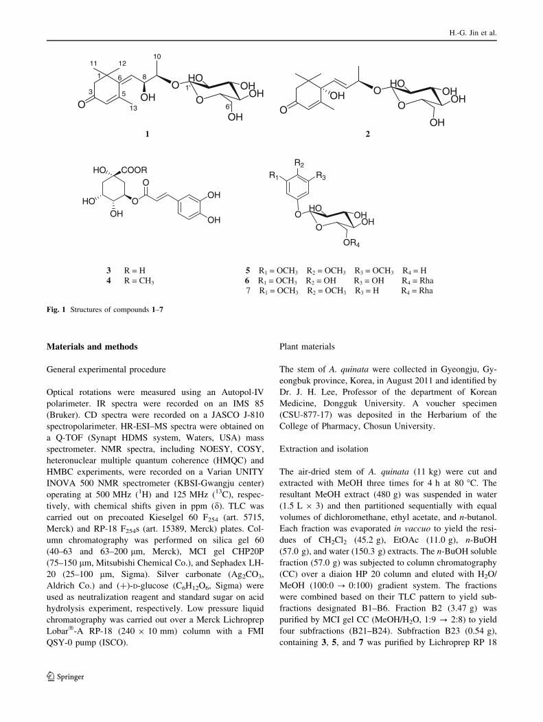
Fig. 1 Structures of compounds 1–7
H.-G. Jin et al.
123
CC (MeOH/H2O, 1:20), and finally by silica gel CC
(CHCl3/MeOH/H2O, 4:1:0.2 ? 2:1:0.2) to yield 3
(121.0 mg), 5 (26.5 mg), and 7 (17.0 mg). In addition,
subfraction B21 (2.04 g) was purified by Sephadex LH 20
CC (MeOH/H2O, 1:20), and finally by silica gel CC
(CHCl3/MeOH/H2O, 4:1:0.2 ? 2:1:0.2) to yield 6
(15.0 mg). Fraction B3 (5.6 g) was subjected to silica gel
CC eluting with a CHCl3/MeOH/H2O (6:1:0.1 ? 2:1:0.1)
in a gradient system to yield six subfractions (B31–B36).
Subfraction B31 (0.79 g) was then purified by repeated
Lichroprep RP 18 CC (MeOH/H2O, 1:3) to yield 1
(4.4 mg), 2 (3.2 mg), and 4 (3.0 mg).
Akeqintoside D (1)
Colorless gum; [a]D20 -19.6� (MeOH; c 0.37); HR-ESI–MS
(positive mode) m/z: 409.1841 [M ? Na]? (calcd for C19-
H30O8Na, 409.1838); IR mmax (film) cm-1: 3,410, 2,935,
1,655, 1,650, 1,076, 1,035; 1H, and 13C NMR: see Table 1.
Roseoside II (2)
Colorless gum; [a]D20 ?82.9� (MeOH; c 0.16); HR-ESI–MS
m/z: 385.1862 [M-H]- (calcd for C19H29O8, 385.1861);
CD (MeOH, c 8.5 9 10-6 M): 241 (?41.37), 318
(-54.25) nm; IR mmax (film) cm-1: 3,400, 2,940, 1,653,
1,076, 1,035; 1H , and 13C NMR: see Table 1.
3-O-Caffeoylquinic acid (3)
Brown powder; [a]D20 -134.6� (MeOH; c 0.44); ESI–MS
m/z: 353 [M-H]?; IR mmax (film) cm-l: 3,412, 2,928,
1,689, 1,082, 853; 1H NMR (500 MHz, CD3OD) 7.57 (1H,
d, J = 16.0 Hz, H-70), 7.05 (1H, d, J = 2.0 Hz, H-20), 6.94
(1H, dd, J = 2.0, 8.0 Hz, H-60), 6.77 (1H, d, J = 8.0 Hz,
H-50), 6.29 (1H, d, J = 16.0 Hz, H-80), 5.38 (1H, dd,
J = 5.0, 10.0 Hz, H-3), 4.13 (1H, dd, J = 3.0, 6.0 Hz,
H-5), 3.68 (1H, dd, J = 3.0, 10.0 Hz, H-4), 2.15 (1H, dd,
J = 3.0, 15.0 Hz, H-6), 2.10 (1H, m, H-2), 2.00 (1H, m,
H-2), 1.94 (1H, dd, J = 3.0, 15.0 Hz, H-6); 13C NMR
(125 MHz, CD3OD) d: 181.1 (C-7), 169.3 (C-90), 149.7 (C-
40), 147.0 (C-70), 146.9 (C-30), 127.9 (C-10), 123.1 (C-60),116.6 (C-50), 115.7 (C-20), 115.2 (C-80), 77.9 (C-1), 75.2
(C-4), 73.2 (C-5), 72.8 (C-3), 40.8 (C-2), 39.2 (C-6).
Methyl-3-O-caffeoylquinate (4)
Pale brownish powder; [a]D20 -30.7� (MeOH; c 0.15); ESI–
MS m/z: 367 [M-H]?; IR mmax (film) cm-l: 3,408, 2,930,
1,672, 1,082, 872; 1H NMR (500 MHz, CD3OD) d: 7.53
(1H, d, J = 16.0 Hz, H-70), 7.04 (1H, d, J = 2.0 Hz, H-20),6.94 (1H, dd, J = 2.0, 8.0 Hz, H-60), 6.77 (1H, d,
J = 8.0 Hz, H-50), 6.22 (1H, d, J = 16.0 Hz, H-80), 5.27
(1H, dd, J = 5.0, 7.5 Hz, H-3), 4.13 (1H, dt, J = 3.0,
7.5 Hz, H-5), 3.73 (1H, dt, J = 3.0, 7.5 Hz, H-4), 3.69
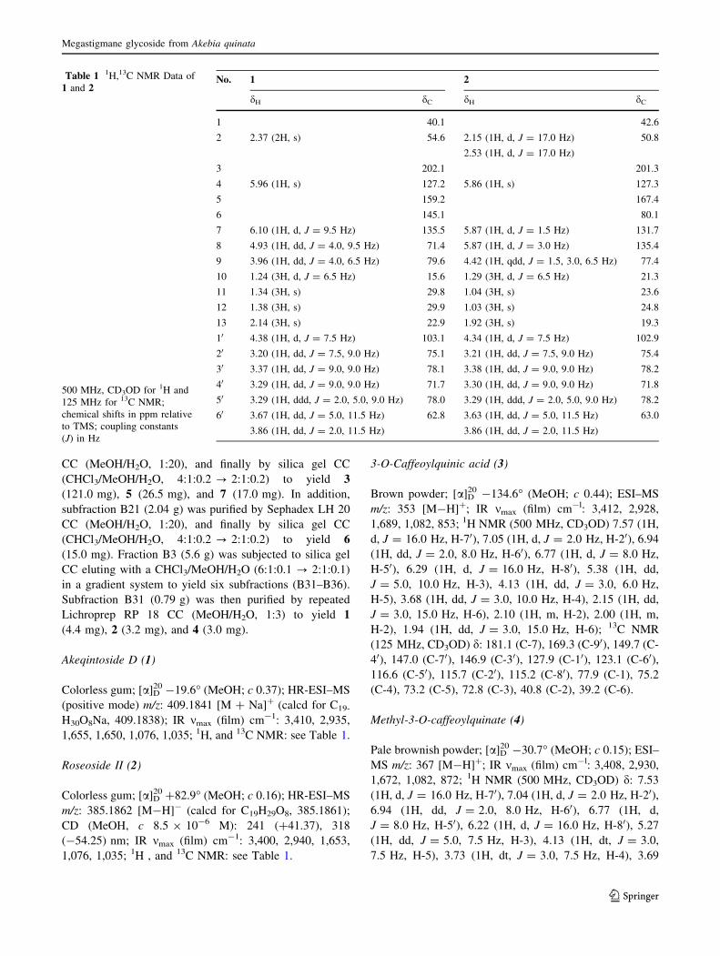
Table 1 1H,13C NMR Data of
1 and 2
500 MHz, CD3OD for 1H and
125 MHz for 13C NMR;
chemical shifts in ppm relative
to TMS; coupling constants
(J) in Hz
No. 1 2
dH dC dH dC
1 40.1 42.6
2 2.37 (2H, s) 54.6 2.15 (1H, d, J = 17.0 Hz) 50.8
2.53 (1H, d, J = 17.0 Hz)
3 202.1 201.3
4 5.96 (1H, s) 127.2 5.86 (1H, s) 127.3
5 159.2 167.4
6 145.1 80.1
7 6.10 (1H, d, J = 9.5 Hz) 135.5 5.87 (1H, d, J = 1.5 Hz) 131.7
8 4.93 (1H, dd, J = 4.0, 9.5 Hz) 71.4 5.87 (1H, d, J = 3.0 Hz) 135.4
9 3.96 (1H, dd, J = 4.0, 6.5 Hz) 79.6 4.42 (1H, qdd, J = 1.5, 3.0, 6.5 Hz) 77.4
10 1.24 (3H, d, J = 6.5 Hz) 15.6 1.29 (3H, d, J = 6.5 Hz) 21.3
11 1.34 (3H, s) 29.8 1.04 (3H, s) 23.6
12 1.38 (3H, s) 29.9 1.03 (3H, s) 24.8
13 2.14 (3H, s) 22.9 1.92 (3H, s) 19.3
10 4.38 (1H, d, J = 7.5 Hz) 103.1 4.34 (1H, d, J = 7.5 Hz) 102.9
20 3.20 (1H, dd, J = 7.5, 9.0 Hz) 75.1 3.21 (1H, dd, J = 7.5, 9.0 Hz) 75.4
30 3.37 (1H, dd, J = 9.0, 9.0 Hz) 78.1 3.38 (1H, dd, J = 9.0, 9.0 Hz) 78.2
40 3.29 (1H, dd, J = 9.0, 9.0 Hz) 71.7 3.30 (1H, dd, J = 9.0, 9.0 Hz) 71.8
50 3.29 (1H, ddd, J = 2.0, 5.0, 9.0 Hz) 78.0 3.29 (1H, ddd, J = 2.0, 5.0, 9.0 Hz) 78.2
60 3.67 (1H, dd, J = 5.0, 11.5 Hz) 62.8 3.63 (1H, dd, J = 5.0, 11.5 Hz) 63.0
3.86 (1H, dd, J = 2.0, 11.5 Hz) 3.86 (1H, dd, J = 2.0, 11.5 Hz)
Megastigmane glycoside from Akebia quinata
123
(3H, s, H-8), 2.20 (1H, dd, J = 3.0, 13.5 Hz, H-6), 2.17
(2H, dd, J = 7.5, 13.5 Hz, H-2), 2.01 (1H, dd, J = 7.5,
13.5 Hz, H-6); 13C NMR (125 MHz, CD3OD) d: 175.6 (C-
7), 168.4 (C-90), 150.0 (C-40), 147.4 (C-70), 147.1 (C-30),127.7 (C-10), 123.1 (C-60), 116.7 (C-50), 115.2 (C-20), 115.1
(C-80), 75.9 (C-1), 72.6 (C-4), 72.3 (C-3), 70.4 (C-5), 53.1
(C-8), 38.2 (C-6), 37.9 (C-2).
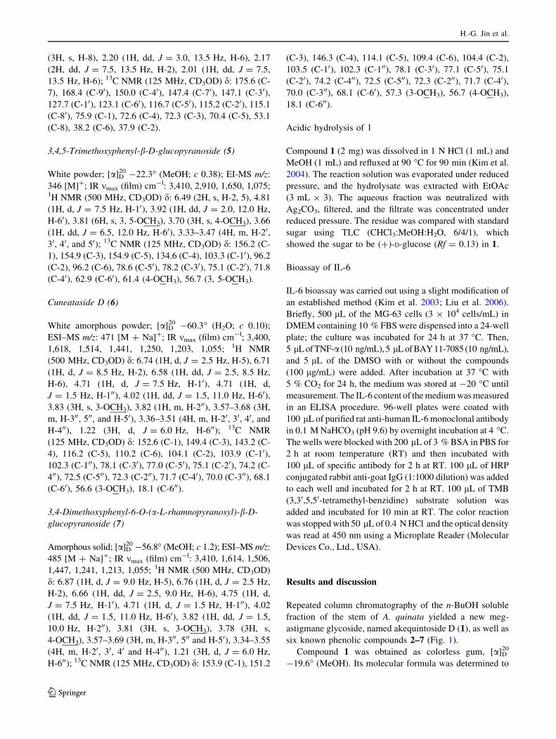
3,4,5-Trimethoxyphenyl-b-D-glucopyranoside (5)
White powder; [a]D20 -22.3� (MeOH; c 0.38); EI-MS m/z:
346 [M]?; IR mmax (film) cm-l: 3,410, 2,910, 1,650, 1,075;1H NMR (500 MHz, CD3OD) d: 6.49 (2H, s, H-2, 5), 4.81
(1H, d, J = 7.5 Hz, H-10), 3.92 (1H, dd, J = 2.0, 12.0 Hz,
H-60), 3.81 (6H, s, 3, 5-OCH3), 3.70 (3H, s, 4-OCH3), 3.66
(1H, dd, J = 6.5, 12.0 Hz, H-60), 3.33–3.47 (4H, m, H-20,30, 40, and 50); 13C NMR (125 MHz, CD3OD) d: 156.2 (C-
1), 154.9 (C-3), 154.9 (C-5), 134.6 (C-4), 103.3 (C-10), 96.2
(C-2), 96.2 (C-6), 78.6 (C-50), 78.2 (C-30), 75.1 (C-20), 71.8
(C-40), 62.9 (C-60), 61.4 (4-OCH3), 56.7 (3, 5-OCH3).
Cuneataside D (6)
White amorphous powder; [a]D20 -60.3� (H2O; c 0.10);
ESI–MS m/z: 471 [M ? Na]?; IR mmax (film) cm-l; 3,400,
1,618, 1,514, 1,441, 1,250, 1,203, 1,055; 1H NMR
(500 MHz, CD3OD) d: 6.74 (1H, d, J = 2.5 Hz, H-5), 6.71
(1H, d, J = 8.5 Hz, H-2), 6.58 (1H, dd, J = 2.5, 8.5 Hz,
H-6), 4.71 (1H, d, J = 7.5 Hz, H-10), 4.71 (1H, d,
J = 1.5 Hz, H-100), 4.02 (1H, dd, J = 1.5, 11.0 Hz, H-60),3.83 (3H, s, 3-OCH3), 3.82 (1H, m, H-200), 3.57–3.68 (3H,
m, H-300, 500, and H-50), 3.36–3.51 (4H, m, H-20, 30, 40, and
H-400), 1.22 (3H, d, J = 6.0 Hz, H-600); 13C NMR
(125 MHz, CD3OD) d: 152.6 (C-1), 149.4 (C-3), 143.2 (C-
4), 116.2 (C-5), 110.2 (C-6), 104.1 (C-2), 103.9 (C-10),102.3 (C-100), 78.1 (C-30), 77.0 (C-50), 75.1 (C-20), 74.2 (C-
400), 72.5 (C-500), 72.3 (C-200), 71.7 (C-40), 70.0 (C-300), 68.1
(C-60), 56.6 (3-OCH3), 18.1 (C-600).
3,4-Dimethoxyphenyl-6-O-(a-L-rhamnopyranosyl)-b-D-
glucopyranoside (7)
Amorphous solid; [a]D20 -56.8� (MeOH; c 1.2); ESI–MS m/z:
485 [M ? Na]?; IR mmax (film) cm-l: 3,410, 1,614, 1,506,
1,447, 1,241, 1,213, 1,055; 1H NMR (500 MHz, CD3OD)
d: 6.87 (1H, d, J = 9.0 Hz, H-5), 6.76 (1H, d, J = 2.5 Hz,
H-2), 6.66 (1H, dd, J = 2.5, 9.0 Hz, H-6), 4.75 (1H, d,
J = 7.5 Hz, H-10), 4.71 (1H, d, J = 1.5 Hz, H-100), 4.02
(1H, dd, J = 1.5, 11.0 Hz, H-60), 3.82 (1H, dd, J = 1.5,
10.0 Hz, H-200), 3.81 (3H, s, 3-OCH3), 3.78 (3H, s,
4-OCH3), 3.57–3.69 (3H, m, H-300, 500 and H-50), 3.34–3.55
(4H, m, H-20, 30, 40 and H-400), 1.21 (3H, d, J = 6.0 Hz,
H-600); 13C NMR (125 MHz, CD3OD) d: 153.9 (C-1), 151.2
(C-3), 146.3 (C-4), 114.1 (C-5), 109.4 (C-6), 104.4 (C-2),
103.5 (C-10), 102.3 (C-100), 78.1 (C-30), 77.1 (C-50), 75.1
(C-20), 74.2 (C-400), 72.5 (C-500), 72.3 (C-200), 71.7 (C-40),70.0 (C-300), 68.1 (C-60), 57.3 (3-OCH3), 56.7 (4-OCH3),
18.1 (C-600).
Acidic hydrolysis of 1
Compound 1 (2 mg) was dissolved in 1 N HCl (1 mL) and
MeOH (1 mL) and refluxed at 90 �C for 90 min (Kim et al.
2004). The reaction solution was evaporated under reduced
pressure, and the hydrolysate was extracted with EtOAc
(3 mL 9 3). The aqueous fraction was neutralized with
Ag2CO3, filtered, and the filtrate was concentrated under
reduced pressure. The residue was compared with standard
sugar using TLC (CHCl3:MeOH:H2O, 6/4/1), which
showed the sugar to be (?)-D-glucose (Rf = 0.13) in 1.
Bioassay of IL-6
IL-6 bioassay was carried out using a slight modification of
an established method (Kim et al. 2003; Liu et al. 2006).
Briefly, 500 lL of the MG-63 cells (3 9 104 cells/mL) in
DMEM containing 10 % FBS were dispensed into a 24-well
plate; the culture was incubated for 24 h at 37 �C. Then,
5 lL of TNF-a (10 ng/mL), 5 lL of BAY 11-7085 (10 ng/mL),
and 5 lL of the DMSO with or without the compounds
(100 lg/mL) were added. After incubation at 37 �C with
5 % CO2 for 24 h, the medium was stored at -20 �C until
measurement. The IL-6 content of the medium was measured
in an ELISA procedure. 96-well plates were coated with
100 lL of purified rat anti-human IL-6 monoclonal antibody
in 0.1 M NaHCO3 (pH 9.6) by overnight incubation at 4 �C.
The wells were blocked with 200 lL of 3 % BSA in PBS for
2 h at room temperature (RT) and then incubated with
100 lL of specific antibody for 2 h at RT. 100 lL of HRP
conjugated rabbit anti-goat IgG (1:1000 dilution) was added
to each well and incubated for 2 h at RT. 100 lL of TMB
(3,30,5,50-tetramethyl-benzidine) substrate solution was
added and incubated for 10 min at RT. The color reaction
was stopped with 50 lL of 0.4 N HCl and the optical density
was read at 450 nm using a Microplate Reader (Molecular
Devices Co., Ltd., USA).
Results and discussion
Repeated column chromatography of the n-BuOH soluble
fraction of the stem of A. quinata yielded a new meg-
astigmane glycoside, named akequintoside D (1), as well as
six known phenolic compounds 2–7 (Fig. 1).
Compound 1 was obtained as colorless gum, [a]D20
-19.6� (MeOH). Its molecular formula was determined to
H.-G. Jin et al.
123
be C19H30O8 by HR-ESI–MS data at m/z 409.1841
[M ? Na]? (calcd for C19H30O8Na 409.1838). In the IR
spectrum, the absorption bands for the hydroxyl
(3,410 cm-1), and carbonyl (1,655 cm-1) groups were
observed. The 1H NMR spectrum (Table 1) of 1 showed
four methyl protons at dH 1.24 (3H, d, J = 6.5 Hz, H-10),
1.34 (3H, s, H-11), 1.38 (3H, s, H-12) and 2.14 (3H, s,
H-13), two oxymethine protons at dH 4.93 (1H, dd,
J = 4.0, 9.5 Hz, H-8) and 3.96 (1H, dd, J = 4.0, 6.5 Hz,
H-9), two olefinic protons at dH 5.96 (1H, s, H-4) and 6.10
(1H, d, J = 9.5 Hz, H-7), methylene protons at dH 2.37
(2H, s, H-2), in addition to a glucosyl anomeric proton at
dH 4.38 (1H, d, J = 7.5 Hz, H-10). Acid hydrolysis of 1 in
refluxing 1 N HCl/MeOH afforded (?)-D-glucose which
was detected by direct comparison with an authentic
sample using co-TLC (Kim et al. 2004). In the 13C NMR
spectrum (Table 1), 13 carbon signals appeared besides
those of sugar unit, which included one carbonyl carbon at
dC 202.1 (C-3), two oxygenated methine carbons at dC 71.4
(C-8) and 79.6 (C-9), two olefinic carbons at dC 127.2 (C-
4) and 135.5 (C-7), four methyl carbons at dC 15.6 (C-10),
29.8 (C-11), 29.9 (C-12) and 22.9 (C-13). These spectral
data indicated that 1 was to be a megastigman derivative
(Lee et al. 2011; 2012; Jin et al. 2012). In addition, the
signals from the sugar unit appeared at dH 4.38 (1H, d,
J = 7.5 Hz, H-10), 3.20 (1H, dd, J = 7.5, 9.0 Hz, H-20),3.37 (1H, dd, J = 9.0, 9.0 Hz, H-30), 3.29 (1H, dd, J = 9.0,
9.0 Hz, H-40), 3.29 (1H, ddd, J = 2.0, 5.0, 9.0 Hz, H-50),3.67 (1H, dd, J = 5.0, 11.5 Hz, H-60a), 3.86 (1H, dd,
J = 2.0, 11.5 Hz, H-60b) [dC 103.1 (C-10), 75.1 (C-20), 78.1
(C-30), 71.7 (C-40), 78.0 (C-50), 62.8 (C-60)] and acid
hydrolysis experiment strongly supported the presence of
D-glucopyranose (Ishimaru et al. 1987). The coupling
constant (J = 7.5 Hz) of the anomeric proton of D-glucose
indicated it to be the b-form (Ishimaru et al. 1987). Based
on the 1H and 13C NMR data, the structure of 1 was closely
related to reseoside II, which was isolated from Alangium
premnifolium except for the different locations of hydroxyl
group and double bond in 1 (Otsuka et al. 1995). The
glycosidic linkage was established by a HMBC experiment
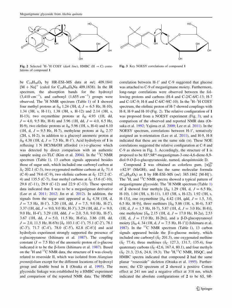
and comparison of the reported NMR data. The HMBC
correlation between H-10 and C-9 suggested that glucose
was attached to C-9 of megastigmane moiety. Furthermore,
long-range correlations were observed between the fol-
lowing protons and carbons (H-4 and C-2/C-6/C-13; H-7
and C-1/C-9; H-8 and C-6/C-9/C-10). In the 1H–1H COSY
spectrum, the olefinic proton of H-7 showed couplings with
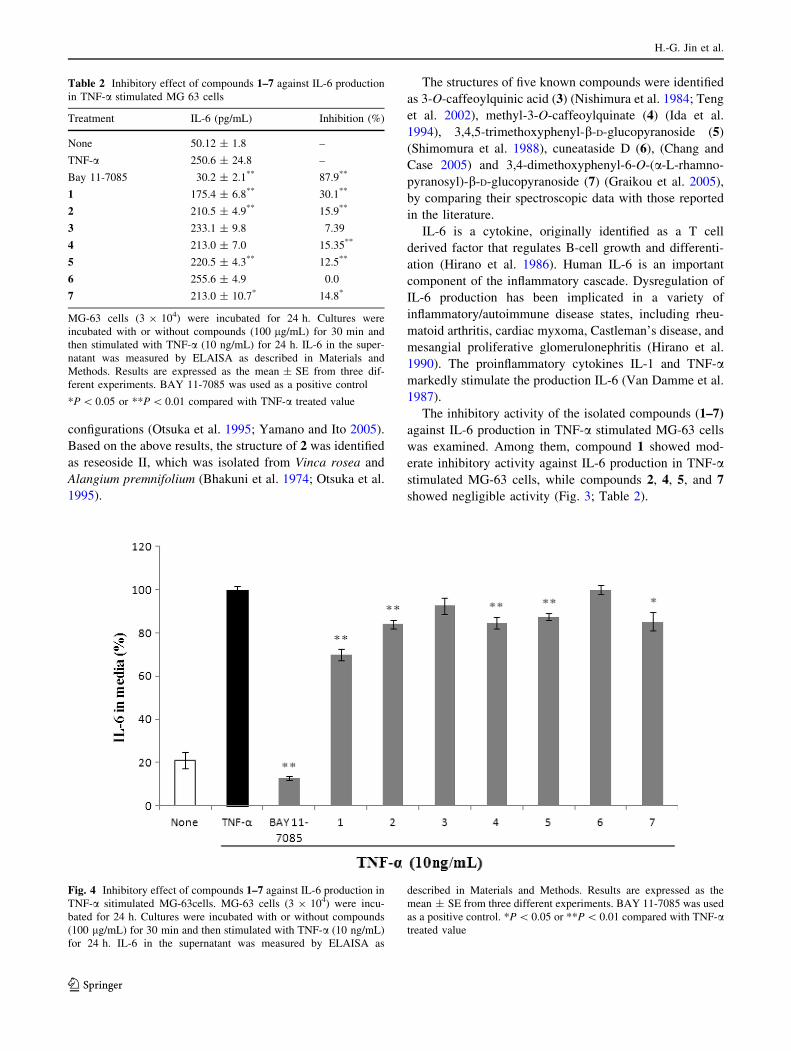
H-8, H-9 and H-10 (Fig. 2). The relative configuration of 1
was proposed from a NOESY experiment (Fig. 3), and a
comparison of the observed and reported NMR data (Ot-
suka et al. 1992; Yajima et al. 2009; Lee et al. 2011). In the
NOESY spectrum, correlations between H-10, tentatively
assigned an a-orientation (Lee et al. 2011), and H-9, H-8
indicated that these are on the same side (a). These NOE
correlations suggested the relative configuration at C-8 and
C-9 as shown in Fig. 3. Accordingly, the structure of 1 is
proposed to be 8S*,9R*-megastigman-3-one-4,6-diene-8,9-
diol-9-O-b-D-glucopyranoside, named, akequintoside D.
Compound 2 was obtained as colorless gum, [a]D20
?82.9� (MeOH), and has the same molecular formula
(C19H30O8) as 1 by HR-ESI–MS (m/z 385.1862 [M-H]-).
The 1H, and 13C NMR spectra showed a typical pattern of
megastigmane glycoside. The 1H NMR spectrum (Table 1)
of 2 showed four methyls [dH 1.29 (3H, d, J = 6.5 Hz,
H-10), 1.04 (3H, s, H-11), 1.03 (3H, s, H-12), 1.92 (3H, s,
H-13)], one oxymethine [dH 4.42 (1H, qdd, J = 1.5, 3.0,
6.5 Hz, H-9)], three methines [dH 5.86 (1H, s, H-4), 5.87
(1H, d, J = 1.5 Hz, H-7), 5.87 (1H, d, J = 3.0 Hz, H-8)],
one methylene [dH 2.15 (1H, d, J = 17.0 Hz, H-2a), 2.53
(1H, d, J = 17.0 Hz, H-2b)], and a b-D-glucopyranosyl
moiety [dH 4. 34 (1H, d, J = 7.5 Hz, H-10)] (Ishimaru et al.
1987). In the 13C NMR spectrum (Table 1), 13 carbon
signals appeared beside the b-D-glucose moiety, which
included one carbonyl (dC 201.3), one oxygenated methine
(dC 77.4), three methines (dC 127.3, 131.7, 135.4), four
quaternary carbons (dC 42.6, 167.4, 80.1), and four methyls
(dC 21.3, 23.6, 24.8, 19.3). The 1H,13C NMR, HSQC, and
HMBC spectra indicated that compound 2 had the same
planar ‘‘roseoside’’ skeleton (Otsuka et al. 1995). Further-
more, the CD spectrum of 2 showed a positive Cotton
effect at 241 nm and a negative effect at 318 nm, which
indicated the absolute configurations of 2 to be 6S, 9R-
OO
OH
OH
OH
OH
O
HO
Fig. 2 Selected 1H–1H COSY (dark line), HMBC (H ? C) corre-
lations of compound 1
OO
OH
OH
OH
OH
O
HO
Fig. 3 Key NOESY correlations of compound 1
Megastigmane glycoside from Akebia quinata
123
configurations (Otsuka et al. 1995; Yamano and Ito 2005).
Based on the above results, the structure of 2 was identified
as reseoside II, which was isolated from Vinca rosea and
Alangium premnifolium (Bhakuni et al. 1974; Otsuka et al.
1995).
The structures of five known compounds were identified
as 3-O-caffeoylquinic acid (3) (Nishimura et al. 1984; Teng
et al. 2002), methyl-3-O-caffeoylquinate (4) (Ida et al.
1994), 3,4,5-trimethoxyphenyl-b-D-glucopyranoside (5)
(Shimomura et al. 1988), cuneataside D (6), (Chang and
Case 2005) and 3,4-dimethoxyphenyl-6-O-(a-L-rhamno-
pyranosyl)-b-D-glucopyranoside (7) (Graikou et al. 2005),
by comparing their spectroscopic data with those reported
in the literature.
IL-6 is a cytokine, originally identified as a T cell
derived factor that regulates B-cell growth and differenti-
ation (Hirano et al. 1986). Human IL-6 is an important
component of the inflammatory cascade. Dysregulation of
IL-6 production has been implicated in a variety of
inflammatory/autoimmune disease states, including rheu-
matoid arthritis, cardiac myxoma, Castleman’s disease, and
mesangial proliferative glomerulonephritis (Hirano et al.
1990). The proinflammatory cytokines IL-1 and TNF-amarkedly stimulate the production IL-6 (Van Damme et al.
1987).
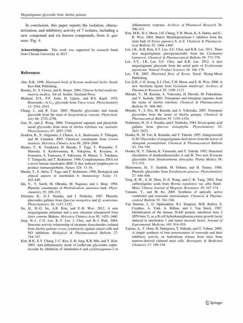
The inhibitory activity of the isolated compounds (1–7)
against IL-6 production in TNF-a stimulated MG-63 cells
was examined. Among them, compound 1 showed mod-
erate inhibitory activity against IL-6 production in TNF-astimulated MG-63 cells, while compounds 2, 4, 5, and 7
showed negligible activity (Fig. 3; Table 2).
Table 2 Inhibitory effect of compounds 1–7 against IL-6 production
in TNF-a stimulated MG 63 cells
Treatment IL-6 (pg/mL) Inhibition (%)
None 50.12 ± 1.8 –
TNF-a 250.6 ± 24.8 –
Bay 11-7085 30.2 ± 2.1** 87.9**
1 175.4 ± 6.8** 30.1**
2 210.5 ± 4.9** 15.9**
3 233.1 ± 9.8 7.39
4 213.0 ± 7.0 15.35**
5 220.5 ± 4.3** 12.5**
6 255.6 ± 4.9 0.0
7 213.0 ± 10.7* 14.8*
MG-63 cells (3 9 104) were incubated for 24 h. Cultures were
incubated with or without compounds (100 lg/mL) for 30 min and
then stimulated with TNF-a (10 ng/mL) for 24 h. IL-6 in the super-
natant was measured by ELAISA as described in Materials and
Methods. Results are expressed as the mean ± SE from three dif-
ferent experiments. BAY 11-7085 was used as a positive control
*P \ 0.05 or **P \ 0.01 compared with TNF-a treated value
Fig. 4 Inhibitory effect of compounds 1–7 against IL-6 production in
TNF-a sitimulated MG-63cells. MG-63 cells (3 9 104) were incu-
bated for 24 h. Cultures were incubated with or without compounds
(100 lg/mL) for 30 min and then stimulated with TNF-a (10 ng/mL)
for 24 h. IL-6 in the supernatant was measured by ELAISA as
described in Materials and Methods. Results are expressed as the
mean ± SE from three different experiments. BAY 11-7085 was used
as a positive control. *P \ 0.05 or **P \ 0.01 compared with TNF-atreated value
H.-G. Jin et al.
123
In conclusion, this paper reports the isolation, charac-
terization, and inhibitory activity of 7 isolates, including a
new compound and six known compounds, from A. qui-
nata. Fig. 4.
Acknowledgments This work was supported by research funds
from Chosun University in 2013.
References
Ahn, D.K. 1998. Illustrated book of Korean medicinal herbs. Seoul:
Kyo-Hak Publishing.
Bensky, D., S. Clavey, and E. Stoger. 2004. Chinese herbal medicine:
materia medica, 3rd ed. Seattle: Eastland Press.
Bhakuni, D.S., P.P. Joshi, H. Uprety, and R.S. Kapil. 1974.
Roseoside—A C13 glycoside from Vinca rosea. Phytochemistry
13: 2541–2543.
Chang, J., and R. Case. 2005. Phenolic glycosides and ionone
glycoside from the stem of Sargentodoxa cuneata. Phytochem-
istry 66: 2752–2758.
Gao, H., and Z. Wang. 2006. Triterpenoid saponins and phenyleth-
anoid glycosides from stem of Akebia trifoliata var. australis.
Phytochemistry 67: 2697–2705.
Graikou, K., N. Aligiannis, I. Chinou, A.-L. Skaltsounis, F. Tillequin,
and M. Litaudon. 2005. Chemical constituents from Croton
insularis. Helvetica Chimica Acta 88: 2654–2660.
Hirano, T., K. Yasukawa, H. Harada, T. Taga, Y. Watanabe, T.
Matsuda, S. Kashiwamura, K. Nakajima, K. Koyama, A.
Iwamatsu, S. Tsunsawa, F. Sakiyama, H. Matsui, Y. Takahara,
T. Taniguchi, and T. Kishimoto. 1986. Complementary DNA for
a novel human interleukin (BSF-2) that induced lymphocytes to
produce immunoglobulin. Nature 324: 73–76.
Hirano, T., S. Akira, T. Taga, and T. Kishimoto. 1990. Biological and
clinical aspects of interleukin 6. Immunology Today 11:
443–449.
Ida, Y., Y. Satoh, M. Ohtsuka, M. Nagasao, and J. Shoji. 1994.
Phenolic constituents of Phellodendron amurense bark. Phyto-
chemistry 35: 209–215.
Ishimaru, K., G.-I. Nonaka, and I. Nishioka. 1987. Phenolic
glucosides gallates from Quercus mongolica and Q. acutissima.
Phytochemistry 26: 1147–1152.
Jin, Q., H.-G. Jin, A.R. Kim, and E.-R. Woo. 2012. A new
megastigmane palmitate and a new oleanane triterpenoid from
Aster yomena Makino. Helvetica Chimica Acta 95: 1455–1460.
Jung, H.-J., C.O. Lee, K.-T. Lee, J. Choi, and H.-J. Park. 2004.
Structure-activity relationship of oleanane disaccharides isolated
from Akebia quinata versus cytotoxicity against cancer cells and
NO inhibition. Biological & Pharmaceutical Bulletin 27:
744–747.
Kim, B.H., E.Y. Chung, J.-C. Ryu, S.-H. Jung, K.R. Min, and Y. Kim.
2003. Anti-inflammatory mode of isoflavone glycosides sopho-
ricoside by inhibition of interleukin-6 and cyclooxygenase-2 in
inflammatory response. Archives of Pharmacal Research 26:
306–311.
Kim, M.R., H.-I. Moon, J.H. Chung, Y.H. Moon, K.-S. Hahm, and E.-
R. Woo. 2004. Matrix Metalloproteinase-1 inhibitor from the
stem bark of Styrax japonica S. et Z. Chemical & Pharmaceu-
tical Bulletin 52: 1466–1469.
Lee, I.K., K.H. Kim, S.Y. Lee, S.U. Choi, and K.R. Lee. 2011. Three
new megastigmane glucopyranosides from the Cardamine
komarovii. Chemical & Pharmaceutical Bulletin 59: 773–779.
Lee, S.Y., I.K. Lee, S.U. Choi, and K.R. Lee. 2012. A new
megastigmane glucoside from the aerial parts of Erythronium
japonicum. Natural Product Sciences 18: 166–170.
Lee, T.B. 2003. Illustrated flora of Korea. Seoul: Hyang-Moon
Publishing.
Liu, Q.H., J.-E. Jeong, E.J. Choi, Y.H. Moon, and E.-R. Woo. 2006. A
new furofuran lignan from Geranium thunbergii. Archives of
Pharmacal Research 29: 1109–1113.
Mimaki, Y., M. Kuroda, A. Yokosuka, H. Hiroshi, M. Fukushima,
and Y. Sashida. 2003. Triterpenes and triterpene saponins from
the stems of Akebia trifoliata. Chemical & Pharmaceutical
Bulletin 51: 960–965.
Mimaki, Y., S. Doi, M. Kuroda, and A. Yokosuka. 2007. Triterpene
glycosides from the stems of Akebia quinqta. Chemical &
Pharmaceutical Bulletin 55: 1319–1324.
Nishimura, H., G.-I. Nonaka, and I. Nishioka. 1984. Seven quinic acid
gallates from Quercus stenophylla. Phytochemistry 23:
2621–2623.
Otsuka, H., M. Yao, K. Kamada, and Y. Takeda. 1995. Alangionoside
G-M: Glycosides of megastigmane derivatives from the leaves of
Alangium premnifolium. Chemical & Pharmaceutical Bulletin
43: 754–759.
Otsuka, H., Y. Takeda, K. Yamasaki, and Y. Takeda. 1992. Structural
elucidation of dendranthemosides A and B: Two new b-ionone
glucosides from Dendranthema shiwogiku. Planta Medica 58:
373–375.
Shimomura, H., Y. Sashida, M. Oohara, and H. Tenma. 1988.
Phenolic glucosides from Parabenzoin praecox. Phytochemistry
27: 644–646.
Teng, R.-W., Z.-H. Zhou, D.-Z. Wang, and C.-R. Yang. 2002. Four
caffeoylquinic acids from Morina nepalensis var. alba Hand.-
Mazz. Chinese Journal of Magnetic Resonance 19: 167–174.
Yamano, Y., and M. Ito. 2005. Synthesis of optically active
vomifoliol and roseoside stereoisomers. Chemical & Pharma-
ceutical Bulletin 53: 541–546.
Van Damme, J., G. Opdenakker, R.J. Simpson, M.R. Rubira, S.
Cayphas, A. Vink, A. Billiau, and J. Van Snick. 1987.
Identification of the human 26-kD protein, interferon beta 2
(IFN-beta 2), as a B cell hybridoma/plasmacytoma growth factor
induced in interleukin 1 and tumor necrosis factor. Journal of
Experimental Medicine 165: 914–919.
Yajima, A., Y. Oono, R. Nakagawa, T. Nukada, and G. Yabuta. 2009.
A simple synthesis of four stereoisomers of roseoside and their
inhibitory activity on leukotriene release from mice bone
marrow-derived cultured mast cells. Bioorganic & Medicinal
Chemistry 17: 189–194.
Megastigmane glycoside from Akebia quinata
123