structure and function of a glycoside hydrolase family 8
TRANSCRIPT

This is a repository copy of Structure and function of a glycoside hydrolase family 8 endoxylanase from Teredinibacter turnerae.
White Rose Research Online URL for this paper:https://eprints.whiterose.ac.uk/137106/
Version: Published Version
Article:
Fowler, Claire A, Hemsworth, Glyn R orcid.org/0000-0002-8226-1380, Cuskin, Fiona et al. (5 more authors) (2018) Structure and function of a glycoside hydrolase family 8 endoxylanase from Teredinibacter turnerae. Acta crystallographica. Section D, Structural biology. pp. 946-955. ISSN 2059-7983
https://doi.org/10.1107/S2059798318009737
[email protected]://eprints.whiterose.ac.uk/
Reuse
This article is distributed under the terms of the Creative Commons Attribution (CC BY) licence. This licence allows you to distribute, remix, tweak, and build upon the work, even commercially, as long as you credit the authors for the original work. More information and the full terms of the licence here: https://creativecommons.org/licenses/
Takedown
If you consider content in White Rose Research Online to be in breach of UK law, please notify us by emailing [email protected] including the URL of the record and the reason for the withdrawal request.

research papers
946 https://doi.org/10.1107/S2059798318009737 Acta Cryst. (2018). D74, 946–955
Received 20 March 2018
Accepted 9 July 2018
Edited by C. S. Bond, University of Western
Australia, Crawley, Australia
Keywords: glycoside hydrolase; biomass;
biofuels; marine polysaccharides; cellulolytic
enzymes; shipworms; Teredinibacter turnerae.
PDB references: GH8 xylanase from
Teredinibacter turnerae, 6g00; complex with
xylobiose, 6g09; complex with xylotriose, 6g0b;
catalytic mutant, complex with xylohexaose,
6g0n
Supporting information: this article has
supporting information at journals.iucr.org/d
Structure and function of a glycoside hydrolasefamily 8 endoxylanase from Teredinibacter turnerae
Claire A. Fowler,a Glyn R. Hemsworth,b Fiona Cuskin,c Sam Hart,a Johan
Turkenburg,a Harry J. Gilbert,d Paul H. Waltone and Gideon J. Daviesa*
aYork Structural Biology Laboratory, Department of Chemistry, The University of York, York YO10 5DD, England,bSchool of Molecular and Cellular Biology, The Faculty of Biological Sciences, University of Leeds, Leeds LS2 9JT,
England, cSchool of Natural and Environmental Science, Newcastle University, Newcastle upon Tyne NE1 7RU, England,dInstitute for Cell and Molecular Biosciences, Newcastle University, Newcastle upon Tyne NE2 4HH, England, andeDepartment of Chemistry, The University of York, York YO10 5DD, England. *Correspondence e-mail:
The biological conversion of lignocellulosic matter into high-value chemicals or
biofuels is of increasing industrial importance as the sector slowly transitions
away from nonrenewable sources. Many industrial processes involve the use
of cellulolytic enzyme cocktails – a selection of glycoside hydrolases and,
increasingly, polysaccharide oxygenases – to break down recalcitrant plant
polysaccharides. ORFs from the genome of Teredinibacter turnerae, a symbiont
hosted within the gills of marine shipworms, were identified in order to search
for enzymes with desirable traits. Here, a putative T. turnerae glycoside
hydrolase from family 8, hereafter referred to as TtGH8, is analysed. The
enzyme is shown to be active against �-1,4-xylan and mixed-linkage (�-1,3,�-1,4)
marine xylan. Kinetic parameters, obtained using high-performance anion-
exchange chromatography with pulsed amperometric detection and 3,5-dinitro-
salicyclic acid reducing-sugar assays, show that TtGH8 catalyses the hydrolysis
of �-1,4-xylohexaose with a kcat/Km of 7.5 � 107 M�1 min�1 but displays
maximal activity against mixed-linkage polymeric xylans, hinting at a primary
role in the degradation of marine polysaccharides. The three-dimensional
structure of TtGH8 was solved in uncomplexed and xylobiose-, xylotriose- and
xylohexaose-bound forms at approximately 1.5 A resolution; the latter was
consistent with the greater kcat/Km for hexasaccharide substrates. A 2,5B boat
conformation observed in the �1 position of bound xylotriose is consistent with
the proposed conformational itinerary for this class of enzyme. This work shows
TtGH8 to be effective at the degradation of xylan-based substrates, notably
marine xylan, further exemplifying the potential of T. turnerae for effective and
diverse biomass degradation.
1. Introduction
The demand for biofuels is increasing amid positive shifts in
political and public opinion regarding the growing need for
more sustainable fuel sources (discussed, for example, in
Somerville, 2007; Pauly & Keegstra, 2008). A barrier towards
the sustainable and efficient usage of plant biomass for fuel
conversion lies in the complexity and recalcitrance of plant
cell walls (Himmel et al., 2007; Bomble et al., 2017). The
heterogeneous matrix of various carbohydrate compounds
hinders the enzymatic breakdown of plant cell walls into
energy-rich carbohydrate monomers. Crystalline cellulose
regions are interspersed with a web of more soluble poly-
saccharides, known as hemicelluloses. The plant cell-wall
matrix is strengthened by a hydrophobic and insoluble barrier:
a mixture of phenolic compounds known as lignin (Li et al.,
ISSN 2059-7983

2015). In nature, the breakdown of lignocellulosic material is
achieved through the synergistic action of a wide variety of
different enzymes, including glycoside hydrolases and poly-
saccharide oxygenases (Hemsworth et al., 2015; Walton &
Davies, 2016). A single organism can produce a consortium of
enzymes capable of lignocellulosic degradation or can utilize
the symbiotic behaviour of other smaller organisms such as
bacteria and fungi (Cragg et al., 2015). One organism of
increasing interest is the marine symbiont Teredinibacter
turnerae, which has been found in the gills (Fig. 1) of at least 24
species of bivalve molluscs (Ekborg et al., 2007; Horak &
Montoya, 2014).
Marine bivalve molluscs of the family Teredinidae, more
commonly referred to as shipworms (Fig. 1), are considered
pests by the maritime community as they inflict damage onto
wooden structures such as piers and ship hulls by burrowing
into the material (Cragg et al., 2015). In 1927, Boynton and
Miller observed the disappearance of 80% of cellulose and 15–
56% of hemicellulose from Douglas fir piling during its transit
through the digestive tract of Teredo navalis (Boynton &
Miller, 1927), highlighting the ability of these organisms to
degrade recalcitrant plant biomass. Much later, in 1983,
Waterbury and coworkers observed, isolated and cultured the
symbiotic bacterium T. turnerae (Waterbury et al., 1983) from
specialized cells called bacteriocytes found inside an internal
region of the shipworm’s gills. In its genome, T. turnerae
encodes >100 glycoside hydrolases, enzymes that are capable
of breaking glycosidic bonds, 54% of which have been classed
as ‘wood-specific’ (Yang et al., 2009). Furthermore, T. turnerae
enzymes have been identified in the shipworm gut (O’Connor
et al., 2014), thus establishing the resident bacteria and the
enzymes that they produce as key players in shipworm biology.
It is therefore now clear that lignocellulose degradation may
occur through the synergistic action of host enzymes and
enzymes from this community of endosymbiotic bacteria
housed within the gills of the shipworm.
In this light, we sought to mine the uncharacterized
T. turnerae glycoside hydrolases to find enzymes that would be
active under more biotechnologically relevant conditions and
on useful substrates. T. turnerae exists primarily in high-salt
conditions, and so its enzymes may be additionally stabilized
towards harsher environments such as might be experienced
in a biorefinery. As a proof of principle, we chose to focus on
the consortia of enzymes harnessed by this organism to
degrade xylans, given the interest in this substrate in the
context of biofuels (Somerville, 2007; Pauly & Keegstra, 2008;
Biely et al., 2016), and also in the pulp and paper, animal feed
and bread-making sectors. Additionally, following the recent
insights gained from studying xylan-degrading enzyme
consortia from new ecological niches (Rogowski et al., 2015),
work that revealed both new enzymes and specificities, the
study of xylan-degrading enzymes from this shipworm
symbiont represents fertile new ground for further discoveries
in this area.
Applying the CAZY classification for carbohydrate-active
enzymes (http://www.cazy.org; see Lombard et al., 2014) to
the T. turnerae genome reveals many potential xylanases,
including ten GH10 enzymes, five GH11 enzymes and two
enzymes from the potential xylanase-containing family GH30
(all of these glycoside hydrolase families are reviewed in
CAZypedia; for a review, see The CAZypedia Consortium,
2018). We were particularly drawn to family GH8, a cellulase/
xylanase-containing family, from which T. turnerae has just a
single representative, TtGH8. We sought to use this enzyme as
an exemplar for whether T. turnerae can provide enzymes with
biological and chemical utility, cast in terms of three-dimen-
sional structure and notably, in this case, in the context of a
diverse array of other potential xylanolytic enzymes.
Here, we report the cloning, expression and characteriza-
tion of this GH8 carbohydrate-active enzyme (Lombard et al.,
2014). We show that TtGH8 is a xylan-active endoxylanase
with six catalytically relevant subsites and notably a maximal
activity towards mixed-linkage (�-1,3,�-1,4) marine xylan. The
three-dimensional structure of TtGH8, determined to 1.5 A
resolution, reveals intimate details of the substrate-binding
sites and the distortions of xylose within the catalytic centre.
The work thus highlights the potential of the T. turnerae
genome for enzyme discovery and adds to the growing toolbox
of enzymes that may be used to tackle the recalcitrant hemi-
cellulose xylan (Biely et al., 2016).
2. Materials and methods
2.1. Purification of TtGH8 and catalytic mutants
The sequence of TtGH8 (TERTU_4506; UniProt C5BJ89)
was analysed, and the catalytic domain was identified
(Supplementary Table S1) and truncated to remove the
original signal peptide and linker region and to include an
N-terminal hexahistadine tag and 3C protease cleavage site.
The sequence was codon-optimized and synthesized for
expression in Escherichia coli by GenScript (New Jersey,
USA) and the plasmid was transformed into E. coli BL21
competent cells (Supplementary Table S2). Catalytic residues
were identified using the literature and structures of similar
GH8 proteins deposited in the PDB. The mutations were
designed using custom primers and implemented using the Q5
research papers
Acta Cryst. (2018). D74, 946–955 Fowler et al. � GH8 endoxylanase from Teredinibacter turnerae 947
Figure 1Basic diagram of a shipworm (image not to scale) showing the generallayout of the main organs within the animal. More information onshipworm physiology can be found in Betcher (2011), Gallager et al.
(1981) and Waterbury et al. (1983), and in-depth illustrations anddescriptions can be found in Turner (1966).

site-directed mutagenesis kit (NEB) to alter Asp281 to Asn
(TtGH8 D281N; Supplementary Table S3). Expression testing
for both constructs was carried out prior to large-scale
production. Cultures (6 � 500 ml LB, 30 mg ml�1 kanamycin)
were inoculated with 500 ml overnight culture and grown at
37�C and 200 rev min�1 until an OD of 0.6 was obtained.
IPTG (1 mM final concentration) was then added and the
cultures were cooled to 16�C and left shaking overnight. The
cultures were harvested by centrifugation and the pellet was
resuspended in 50 mM HEPES, 250 mM NaCl, 30 mM
imidazole pH 7. The cells were lysed by sonication and
centrifuged at 15g for 30 min. The supernatant was collected
and loaded onto a pre-equilibrated Nickel HiTrap Crude 5 ml
affinity column. Nickel-affinity chromatography was run on an
AKTA start, with an elution gradient of 30–300 mM imidazole
over 25 column volumes. The collected protein sample was
treated with a 1:100 ratio of 3C protease to TtGH8 and 5 mM
DTT and left shaking at room temperature overnight. The
sample was loaded onto a pre-equilibrated Nickel HiTrap
Crude 5 ml affinity column and the flowthrough and wash
were collected. The collected sample was buffer-exchanged
into 20 mM HEPES, 200 mM NaCl before being concentrated
to approximately 300 ml and loaded onto a Superdex 200 gel-
filtration column. Pure fractions were combined, concentrated
and buffer-exchanged into 10 mM HEPES. To check their
purity, samples were analysed by SDS–PAGE throughout
purification and the final sample was analysed by electrospray
ionization mass spectrometry.
2.2. Thermal shift analysis (TSA)
Samples (30 ml total) containing SYPRO Orange dye
(15 ml) and enzyme (final concentration of 1 mg ml�1) with
either buffer or substrate were prepared and thermal shift
analyses were run on a Stratagene Mx3005P qPCR device. The
substrates tested included the solid substrates (small amount,
unmeasured) tamarind xyloglucan, rye arabinoxylan, birch-
wood xylan, shrimp shell chitin and Avicel, and the soluble
oligosaccharides cellohexaose, cellobiose, xylobiose and
xylohexaose (10 mM). Samples were heated from 20 to 91�C
in increments of 1�C over 71 cycles. The fluorescence of
SYPRO Orange was monitored throughout and the data were
used to calculate the melting temperature of the protein
(Supplementary Fig. S1). The data were analysed using the
JTSA fitting program developed by Paul Bond, which is
available at http://paulsbond.co.uk/jtsa/#/input.
2.3. Thin-layer chromatography (TLC) and liquid
chromatography–mass spectrometry (LCMS)
Xylo-oligosaccharides were purchased commercially: xylo-
biose (X2) from Sigma and TCP, and xylotriose (X3), xylo-
tetraose (X4), xylopentaose (X5), xylohexaose (X6) and
polysaccharides from Megazyme (unless stated otherwise).
Overnight hydrolysis reactions with the xylo-oligosaccharides
X2–X6 (1 mM), wheat arabinoxylan (WAX), rye arabinoxylan
(RAX), corn arabinoxylan (CAX), birchwood xylan (BX),
mixed-linkage �1–3,�1–4 xylan (MLX, purchased from
Elicityl/Oligotech) and xyloglucan (XG) at 1 mg ml�1 were
incubated at 37�C with 1 mM TtGH8. The samples were
heated at 90�C prior to spotting onto a TLC plate (total 4 ml).
Standards containing X2–X6 at 1 mM each were run on the
same plate. TLC plates were placed in chromatography tanks
containing the running buffer [50%(v/v) n-butanol, 25%(v/v)
acetic acid, 25%(v/v) water]. Plates were run once, dried and
then re-run to improve the separation of sugars. The plates
were visualized using a staining solution [3%(v/v) sulfuric
acid, 75%(v/v) ethanol, 0.1%(w/v) orcinol monohydrate],
dried and then heated to approximately 100�C (Fig. 2a).
Hydrolysis samples for LCMS were prepared using 50 mM
ammonium acetate buffer pH 6, approximately 1 mg ml�1
substrate and 1 mM enzyme. Samples were incubated at 37�C
overnight and shaken at 500 rev min�1. If required, samples
were centrifuged to remove any solid materials and 100 ml was
loaded onto a Cosmosil Sugar-D HPLC column using the LC-
MS Dionex system, where the separated products were
analysed by ESI or PAD mass spectrometry. Running buffers
were a mixture of water and acetronitrile, with some test runs
also including 1% formic acid (Supplementary Figs. S2 and
S3).
2.4. Kinetics measurements using high-performance
anion-exchange chromatography with pulsed amperometric
detection (HPAEC-PAD)
Substrate-depletion kinetics measurements were performed
on TtGH8 with xylohexaose, xylopentaose and xylotetraose
and were measured using an HPAEC-PAD Dionex system.
Hydrolysis reactions were run at 37�C using different
substrate and enzyme concentrations, with aliquots removed
at set time points and boiled to inactivate TtGH8. All samples
were mixed with a fucose internal standard and run on an
anion-exchange column (CarboPac) using a sodium acetate
gradient. The depletion of substrate during the reaction can be
related to kcat/Km through
kt ¼ lnS0
St
� �
; ð1Þ
where k = (kcat/Km)[enzyme], t is time and S0 and St are the
substrate-peak areas at time 0 and t, respectively. The
substrate-peak areas observed in the HPAEC PAD traces
were normalized against both an external and internal fucose
standard and the resulting values for ln(S0/St) were plotted
against time, producing positive gradients (the change in the
substrate-peak area increases from 0, as the substrate-peak
area at t = 0 is at its maximum). Linear regression analysis was
used to measure the gradient that represents the rate of
reaction, and kcat/Km (M�1 min�1) was determined by dividing
this gradient by the enzyme concentration (Figs. 2b, 2c and
2d). The use of substrate depletion to obtain kcat/Km has been
widely used in the glycoside hydrolase field (see, for example,
Pell et al., 2004; Charnock et al., 1998; Matsui et al., 1991).
2.5. Reducing-sugar assay
The activity of TtGH8 towards several polysaccharides was
tested using the 3,5-dinitrosalicyclic acid (DNSA) reducing-
research papers
948 Fowler et al. � GH8 endoxylanase from Teredinibacter turnerae Acta Cryst. (2018). D74, 946–955

sugar assay. Hydrolysis reactions with enzyme and substrate
were run at 37�C and 150 ml aliquots were removed and
immediately mixed in a 1:1 volume ratio with the DNSA agent
[comprising 1%(w/v) DNSA, 0.2%(v/v) phenol, 1%(w/v)
NaOH, 0.002% glucose and 0.05%(w/v) Na2SO3] at set time
points to stop the reaction. The aliquots were heated at 90�C
for 20 min and cooled on ice for 10 min. The absorbance of
each sample was measured at 575 nm. A standard curve of 0–
500 mg ml�1 xylose (plus 1 mg ml�1 polysaccharide substrate)
was used to quantify the released reducing sugar. Reaction
rates determined for each different substrate-concentration
condition were plotted against the substrate concentration,
where the gradient divided by the enzyme concentration is
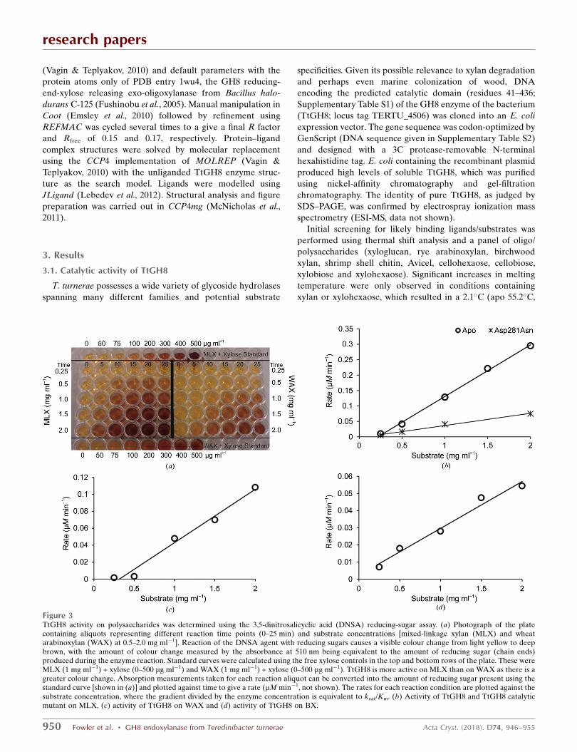
kcat/Km (Fig. 3).
2.6. Crystallization and X-ray crystallography of TtGH8 and
mutants
Initial crystallization screening was performed robotically
using a Mosquito crystal robot and commercial screens,
including Crystal Screen HT and Index from Hampton
Research and PACT from Molecular Dimensions. Several
crystal hits were obtained for TtGH8 and TtGH8 D281N. A
24-well optimization screen containing 0.1 M sodium acetate
pH 4.6–5.2, 0.2 M NaCl, 14–24% polyethylene glycol (PEG)
6000 was used to produce the final crystallization condition for
TtGH8: 0.1 M sodium acetate pH 5.0, 0.2 M NaCl, 16%
polyethylene glycol. Thin rod-shaped crystals were harvested
in nylon loops and cryoprotected by soaking them in mother
liquor plus 30%(v/v) ethylene glycol. The TtGH8 crystals
containing xylobiose and xylotriose were both soaked for 30 s
in a solution consisting of the well solution, 30% ethylene
glycol and 150 mM xylobiose/xylotriose. TtGH8 mutant crys-
tals were soaked in 20 mM xylohexose (0.1M HEPES pH 6.8,
0.2 M ammonium sulfate, 20% PEG 6000) for 15 min to
produce a product complex and for 10 s to produce a substrate
complex and were dipped in a cryosolution consisting of 30%
ethylene glycol before cooling. Crystal data sets were collected
on beamlines I02 and I04 at Diamond Light Source.
Data sets were processed using xia2/DIALS (Winter, 2010;
Winter et al., 2018), with the outer resolution limits defined by
CC1/2 > 0.5. Molecular replacement (Phaser; McCoy et al.,
2007) and refinement (REFMAC; Murshudov et al., 2011)
were carried out using the CCP4i2 pipeline (Potterton et al.,
2018). The TtGH8 structure was solved by molecular
replacement using the CCP4 implementation of MOLREP
research papers
Acta Cryst. (2018). D74, 946–955 Fowler et al. � GH8 endoxylanase from Teredinibacter turnerae 949
Figure 2(a) TtGH8 activity was explored on a range of substrates using thin-layer chromatography. Hydrolysis activity can be observed on WAX, RAX, BX andMLX, as well as on the oligosaccharides xylotetraose (X4), xylopentaose (X5) and xylohexaose (X6). Activity analysis of TtGH8 on xylohexaose (b),xylopentaose (c) and xylotetraose (d) was carried out with HPAEC-PAD by substrate depletion. The peak areas observed in the HPAEC-PAD traceswere normalized against a fucose internal standard and values for ln(S0/St), where S0 is the substrate-peak area at time 0 and St is the substrate-peak areaat time t, were plotted against time. Linear regression analysis was used to calculate kcat/Km (in M�1 min�1).
(Vagin & Teplyakov, 2010) and default parameters with the
protein atoms only of PDB entry 1wu4, the GH8 reducing-
end-xylose releasing exo-oligoxylanase from Bacillus halo-
durans C-125 (Fushinobu et al., 2005). Manual manipulation in
Coot (Emsley et al., 2010) followed by refinement using
REFMAC was cycled several times to a give a final R factor
and Rfree of 0.15 and 0.17, respectively. Protein–ligand
complex structures were solved by molecular replacement
using the CCP4 implementation of MOLREP (Vagin &
Teplyakov, 2010) with the unliganded TtGH8 enzyme struc-
ture as the search model. Ligands were modelled using
JLigand (Lebedev et al., 2012). Structural analysis and figure
preparation was carried out in CCP4mg (McNicholas et al.,
2011).
3. Results
3.1. Catalytic activity of TtGH8
T. turnerae possesses a wide variety of glycoside hydrolases
spanning many different families and potential substrate
specificities. Given its possible relevance to xylan degradation
and perhaps even marine colonization of wood, DNA
encoding the predicted catalytic domain (residues 41–436;
Supplementary Table S1) of the GH8 enzyme of the bacterium
(TtGH8; locus tag TERTU_4506) was cloned into an E. coli
expression vector. The gene sequence was codon-optimized by
GenScript (DNA sequence given in Supplementary Table S2)
and designed with a 3C protease-removable N-terminal
hexahistidine tag. E. coli containing the recombinant plasmid
produced high levels of soluble TtGH8, which was purified
using nickel-affinity chromatography and gel-filtration
chromatography. The identity of pure TtGH8, as judged by
SDS–PAGE, was confirmed by electrospray ionization mass
spectrometry (ESI-MS, data not shown).
Initial screening for likely binding ligands/substrates was
performed using thermal shift analysis and a panel of oligo/
polysaccharides (xyloglucan, rye arabinoxylan, birchwood
xylan, shrimp shell chitin, Avicel, cellohexaose, cellobiose,
xylobiose and xylohexaose). Significant increases in melting
temperature were only observed in conditions containing
xylan or xylohexaose, which resulted in a 2.1�C (apo 55.2�C,
research papers
950 Fowler et al. � GH8 endoxylanase from Teredinibacter turnerae Acta Cryst. (2018). D74, 946–955
Figure 3TtGH8 activity on polysaccharides was determined using the 3,5-dinitrosalicyclic acid (DNSA) reducing-sugar assay. (a) Photograph of the platecontaining aliquots representing different reaction time points (0–25 min) and substrate concentrations [mixed-linkage xylan (MLX) and wheatarabinoxylan (WAX) at 0.5–2.0 mg ml�1]. Reaction of the DNSA agent with reducing sugars causes a visible colour change from light yellow to deepbrown, with the amount of colour change measured by the absorbance at 510 nm being equivalent to the amount of reducing sugar (chain ends)produced during the enzyme reaction. Standard curves were calculated using the free xylose controls in the top and bottom rows of the plate. These wereMLX (1 mg ml�1) + xylose (0–500 mg ml�1) and WAX (1 mg ml�1) + xylose (0–500 mg ml�1). TtGH8 is more active on MLX than on WAX as there is agreater colour change. Absorption measurements taken for each reaction aliquot can be converted into the amount of reducing sugar present using thestandard curve [shown in (a)] and plotted against time to give a rate (mM min�1, not shown). The rates for each reaction condition are plotted against thesubstrate concentration, where the gradient divided by the enzyme concentration is equivalent to kcat/Km. (b) Activity of TtGH8 and TtGH8 catalyticmutant on MLX, (c) activity of TtGH8 on WAX and (d) activity of TtGH8 on BX.

with xylan 57.3�C) and 2.9�C (apo 57.2�C, with xylohexaose
60.1�C) increase in melting temperature, respectively
(Supplementary Fig. S1).
Given the known specificities of family GH8 members
(available at http://www.cazy.org/GH8.html), chitosanase (EC
3.2.1.132), cellulase (EC 3.2.1.4), licheninase (EC 3.2.1.73),
endo-1,4-�-xylanase (EC 3.2.1.8) and reducing-end xylose-
releasing exo-oligoxylanase (EC 3.2.1.156), the increase in
melting temperature for TtGH8 suggested activity as a
xylanase. TtGH8 was therefore incubated (18 h) with a variety
of potential substrates and the reaction products were moni-
tored by thin-layer chromatography (Fig. 2a). TtGH8 was
tested against wheat arabinoxylan (WAX), rye arabinoxylan
(RAX), corn arabinoxylan (in the aleurone layer coating the
seeds known as the bran fraction; CAX), birchwood xylan
(BX), mixed-linkage xylan (MLX), xyloglucan (XG) and also
�-1,4-linked xylo-oligosaccharides with degrees of poly-
merization (DP) from 2 to 6. TLC clearly demonstrated that
TtGH8 acts as a xylanase, with xylotriose (X3) the predomi-
nant product and with activity on WAX, RAX, BX and MLX
clearly evident. Xylotetraose (X4) was the minimal length
xylooligosaccharide that could act as a substrate for TtGH8
observable by TLC. The TLC results were confirmed using
liquid chromatography linked to mass spectrometry (Supple-
mentary Fig. S2). TtGH8 was observed to cleave X6 into X3,
X5 into X3 and X2, and X4 into X3 (X1 could not be
observed). Analysis of the soluble products removed by
centrifugation after incubation of the protein with birchwood
xylan (1 mg ml�1, 37�C, 18 h) indeed confirmed X3 as the
dominant product (Supplementary Fig. S3).
With the knowledge that TtGH8 acts as a xylanase, we next
sought to determine the kinetic parameters for the action of
TtGH8 both by substrate-depletion analysis using high-
performance anion-exchange chromatography with pulsed
amperometric detection (HPAEC-PAD) and by reducing-
sugar assays with 3,5-dinitrosalicyclic acid (DNSA). HPAEC-
PAD analysis (Figs. 2b and 2c) yielded a maximal activity,
kcat/Km, of 7.5 � 107 M�1 min�1 for X6, with X5 and X4 being
considerably worse substrates, with kcat/Km values some five
and 123 times lower, respectively (Table 1). Such values are
typical for endo-xylanases, falling as they do between the
kcat/Km values reported for GH10 enzymes such as CjXyn10A
and CyXyn10C (Pell et al., 2004). TtGH8 activity against
xylose-based polysaccharides was determined using the
DNSA reducing-sugar assay (Fig. 3a). Enzyme activity was
monitored over time at five different substrate concentrations,
allowing the determination of kcat/Km (Figs. 3b, 3c and 3d). An
apparent preference for MLX was observed (1.6 �
108 mg�1 ml min�1; Table 1).
3.2. Three-dimensional structure of TtGH8
In order to provide molecular insight into its catalytic
properties, as described above, the three-dimensional struc-
ture of TtGH8 was determined. TtGH8 was initially crystal-
lized in an apo (unliganded) form and the structure was
determined by molecular replacement at a resolution of 1.4 A
(Tables 2 and 3), using the protein coordinates only from the
structure of the GH8 reducing-end xylose-releasing exo-
oligoxylanase from B. halodurans C-125 (PDB entry 1wu4;
Fushinobu et al., 2005) as the search model.
The first three-dimensional structure of a CAZY family
GH8 member was that of Clostridium thermocellum CelA
(Alzari et al., 1996). Consistent with the defining family
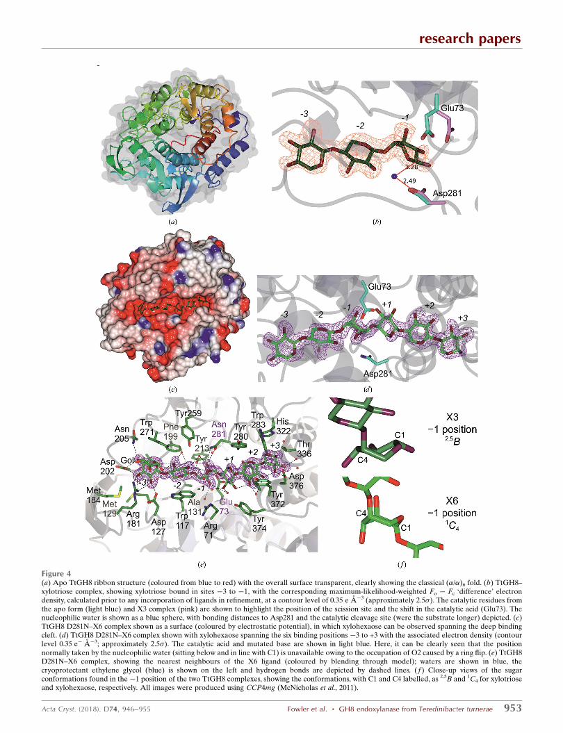
member, TtGH8 exhibits a classical (�/�)6 fold with a clear
deep substrate-binding groove (Fig. 4a). PDBeFold (Krissinel
& Henrick, 2004) analysis reveals that TtGH8 shares close
structural similarity to the reducing-end xylose-releasing exo-
oligoxylanase from B. halodurans C-125 (Fushinobu et al.,
2005). TtGH8 shares 43% sequence identity, overlaying 344
C� atoms with an r.m.s.d. of 1.1 A, a Q score of 0.7 and a
Z score of 15.9. The next closest match is the cold-adapted
GH8 xylanase from Pseudoalteromonas haloplanktis (Collins
et al., 2005) with 35% sequence identity, an r.m.s.d. of 1.4 A
over 341 C� atoms, a Q score of 0.6 and a Z score of 14.7.
3.3. Mechanism of GH8 endoxylanses
Glycoside hydrolases may act through one of two main
mechanisms, leading to retention or inversion of anomeric
configuration (reviewed, for example, in Davies & Henrissat,
1995; Henrissat & Davies, 1997; Rye & Withers, 2000). CAZY
GH8 enzymes act with inversion of anomeric configuration
(Fierobe et al., 1993) and thus feature two essential catalytic
residues: a base to activate the water for nucleophilic attack
and an acid to protonate the leaving group for departure.
Previous studies on GH8 enzymes have identified a conserved
glutamic acid found on helix �2 which functions as the cata-
lytic acid in the inverting mechanism (Alzari et al., 1996). The
equivalent in TtGH8 is Glu73. As in many inverting enzyme
families, the location of the base is less clear, and it has been
proposed that GH8 may, in fact, be subdivided into groupings
based upon the location of the catalytic base (Adachi et al.,
2004). The classical xylanases and endoglucanases (as first
reported by Alzari et al., 1996) are believed to have the base at
the end of helix �8, with the equivalent residue in TtGH8
being Asp281. In order to clarify the catalytic residues in
TtGH8, crystals were soaked in both xylobiose (X2) and
research papers
Acta Cryst. (2018). D74, 946–955 Fowler et al. � GH8 endoxylanase from Teredinibacter turnerae 951
Table 1Catalytic activities of TtGH8 on xylan substrates as determined byHPAEC-PAD (oligosaccharides) or DNSA reducing-sugar assay (poly-saccharides).
Substrate kcat/Km†
OligosaccharidesXylohexaose 7.5 � 107 � 1.1 � 106
Xylopentaose 1.4 � 107 � 1.4 � 106
Xylotetraose 6.1 � 105 � 3.1 � 104
PolysaccharidesBX 1.8 � 107 � 4 � 106
WAX 6.3 � 106 � 5 � 105
MLX (wild type) 1.6 � 108 � 4 � 106
MLX (Asp281Asn) 1.8 � 104 � 1 � 103
† Values for oligosaccharides are given in M�1 min�1 and those for polysaccharides aregiven in mg�1 ml min�1.

xylotriose (X3) and the resulting structures were refined at 1.4
and 1.8 A resolution, respectively (Tables 2 and 3).
X2 was bound in the �2 and �3 subsites (Supplementary
Fig. S4; subsite nomenclature is as discussed by Davies et al.,
1997), hinting at the strength of the �3 subsite, and consistent
with the product profiles (Fig. 2a). X3 was observed bound in
the �3 to �1 subsites (Fig. 4b) and confirms the catalytic
residue proposals, with Glu73 (which has rotated from its
position in the apo structure into this more catalytically rele-
vant position) interacting with the O1 hydroxyl and with
Asp281 interacting with a water molecule ‘below’ C1 in a
position mimicking that which would be expected for hydro-
lysis with inversion of anomeric configuration. Notably, the�1
subsite sugar is not observed in its low-energy 4C1 chair
conformation, but is instead observed distorted into a 2,5B
conformation (Fig. 4b), consistent with proposals for the
catalytic itinerary of GH8 enzymes discussed below.
Enzymatic glycoside hydrolysis involves the distortion of
the reactive, �1 subsite, sugar into a variety of skew-boat and
boat conformations, reflecting the requirements of inline
attack and the stereoelectronic requirements of an oxo-
carbenium-ion-like transition state (for reviews, see Davies &
Williams, 2016; Speciale et al., 2014; Davies et al., 2003, 2012).
The GH8 family has been proposed to go through a 2,5B-like
transition state, notably because of the observation of sugars
with 2SO and 2,5B conformations in complexes of the CelA
endoglucanase (Guerin et al., 2002) that were subsequently
analysed by QM/MM metadynamics (Petersen et al., 2009).
The previous X3 complex, that of the P. haloplanktis cold-
adapted xylanase (Collins et al., 2005; De Vos et al., 2006),
research papers
952 Fowler et al. � GH8 endoxylanase from Teredinibacter turnerae Acta Cryst. (2018). D74, 946–955
Table 2Data-collection and processing statistics for TtGH8 and the TtGH8 mutant with and without ligands, and the TtGH8 mutant–X6 complex.
Values in parentheses are for the outer shell.
Native TtGH8 TtGH8–X2 TtGH8–X3 TtGH8 mutant–X6
Diffraction source Diamond Light Source Diamond Light Source Diamond Light Source Diamond Light SourceWavelength (A) 0.98 0.98 0.98 0.98Temperature (K) 100 100 100 100Space group P212121 P212121 P212121 P212121a, b, c (A) 61.5, 73.0, 90.9 61.7, 79.0, 87.6 59.7, 80.5, 88.0 62.0, 79.7, 88.0�, �, � (�) 90.0, 90.0, 90.0 90.0, 90.0, 90.0 90.0, 90.0, 90.0 90.0, 90.0, 90.0Resolution range (A) 51.0–1.40 (1.42–14.0) 50.4–1.40 (1.42–1.40) 59.4–1.80 (1.84–1.80) 60.97–1.80 (1.84–1.80)Total No. of reflections 342816 530153 316157 313138No. of unique reflections 80610 84123 40092 40046Completeness (%) 99.4 (99.9) 99.1 (89.6) 100.0 (100.0) 99.0 (98.0)Multiplicity 4.3 (4.3) 6.3 (4.1) 7.9 (7.7) 7.8 (8.0)hI/�(I)i 9.6 (1.7) 10.1 (1.3) 8.5 (1.6) 8.2 (2.0)CC1/2 0.997 (0.670) 0.997 (0.503) 0.994 (0.528) 0.992 (0.639)Rp.i.m. 0.045 (0.414) 0.051 (0.549) 0.080 (0.611) 0.109 (0.970)Overall B factor from Wilson plot (A2) 9 9 13 12
Table 3Structure solution and refinement of TtGH8 and the TtGH8 mutant with and without ligands, and the TtGH8 mutant–X6 complex.
Values in parentheses for the outer shell.
Native TtGH8 TtGH8–X2 TtGH8–X3 TtGH8 mutant–X6
Resolution range (A) 51.0–1.40 (1.42–1.40) 50.4–1.40 (1.42–1.40) 59.4–1.80 (1.84–1.80) 60.97–1.80 (1.84–1.80)Completeness (%) 99.4 (99.9) 99.1 (89.6) 100.0 (100.0) 99.0 (98.0)No. of reflections, working set 80540 84054 40032 39993No. of reflections, test set 3924 4274 1868 1942Final Rcryst 0.15 0.16 0.17 0.17Final Rfree 0.17 0.18 0.20 0.20Cruickshank DPI 0.052 0.051 0.111 0.116No. of non-H atomsProtein 3163 3145 3125 3136Ion 1 — — —Ligand 25 23 28 61Water 310 330 164 194
R.m.s. deviationsBonds (A) 0.017 0.016 0.012 0.012Angles (�) 1.70 1.68 1.51 1.50
Average B factors (A2)Protein 14 12 17 16Ion 18 — — —Ligand 28 15 20 26Water 27 22 24 22
Ramachandran plotMost favoured (%) 98.2 97.5 98.0 97.5Allowed (%) 1.8 2.5 2.0 2.5
PDB code 6g00 6g09 6g0b 6g0n
research papers
Acta Cryst. (2018). D74, 946–955 Fowler et al. � GH8 endoxylanase from Teredinibacter turnerae 953
Figure 4(a) Apo TtGH8 ribbon structure (coloured from blue to red) with the overall surface transparent, clearly showing the classical (�/�)6 fold. (b) TtGH8–xylotriose complex, showing xylotriose bound in sites �3 to �1, with the corresponding maximum-likelihood-weighted Fo � Fc ‘difference’ electrondensity, calculated prior to any incorporation of ligands in refinement, at a contour level of 0.35 e A�3 (approximately 2.5�). The catalytic residues fromthe apo form (light blue) and X3 complex (pink) are shown to highlight the position of the scission site and the shift in the catalytic acid (Glu73). Thenucleophilic water is shown as a blue sphere, with bonding distances to Asp281 and the catalytic cleavage site (were the substrate longer) depicted. (c)TtGH8 D281N–X6 complex shown as a surface (coloured by electrostatic potential), in which xylohexaose can be observed spanning the deep bindingcleft. (d) TtGH8 D281N–X6 complex shown with xylohexaose spanning the six binding positions �3 to +3 with the associated electron density (contourlevel 0.35 e� A�3; approximately 2.5�). The catalytic acid and mutated base are shown in light blue. Here, it can be clearly seen that the positionnormally taken by the nucleophilic water (sitting below and in line with C1) is unavailable owing to the occupation of O2 caused by a ring flip. (e) TtGH8D281N–X6 complex, showing the nearest neighbours of the X6 ligand (coloured by blending through model); waters are shown in blue, thecryoprotectant ethylene glycol (blue) is shown on the left and hydrogen bonds are depicted by dashed lines. ( f ) Close-up views of the sugarconformations found in the �1 position of the two TtGH8 complexes, showing the conformations, with C1 and C4 labelled, as 2,5B and 1C4 for xylotrioseand xylohexaose, respectively. All images were produced using CCP4mg (McNicholas et al., 2011).

revealed binding in the +1 to +3 subsites, which together with
the �3 to �1 observations here approximately defined
binding through the six subsites of the enzyme, which is
consistent with the inactive-variant (Asp144Ala) complex
with X5 observed for the P. haloplanktis enzyme by De Vos et
al. (2006). In order to observe the hexasaccharide complex of
TtGH8 we first made a catalytic variant at the proposed base,
TtGH8 D281N; however, this appeared to retain around 0.1%
of the catalytic activity (kcat/Km of 1.8 � 104 mg�1 ml min�1;
Table 1) in the reducing-sugar assay with MLX as substrate,
which is consistent with similar mutants in other GH8 systems
(Collins et al., 2005; De Vos et al., 2006). In addition, structures
with X6, with long soak times, all revealed xylotriose product
complexes (not shown). A complex with unhydrolysed X6 was
therefore obtained by rapid soaking/cooling with a total time
of approximately 10 s. Thus, a TtGH8 D281N structure in
complex with X6 was obtained at 1.6 A resolution, revealing
X6 bound across the entire substrate-binding groove from
subsites �3 to +3 (Figs. 4c, 4d and 4e; Tables 2 and 3).
The complex of TtGH8 with xylotriose showed distortion of
the �1 xylose into a boat configuration, consistent with past
work by others on the conformational itinerary in this family.
To our surprise, the TtGH8 D281N–X6 complex revealed
something different; namely, the �1 subsite sugar in a
completely ring-flipped, southern hemisphere 1C4 chair
conformation (Fig. 4f). Although this allowed access to a
hexasaccharide complex structure, the ring-flipped �1 sugar is
unlikely to be representative of a catalytically relevant
conformation since its position neither allows protonation of
the leaving group by Glu73 nor is there a potential reactive
water. Indeed, in the 1C4 chair conformation the now axial
(and ‘down’) O2 occupies the position that should instead be
occupied by the nucleophilic water. Indeed, the only other
structure in this family in which a substrate spanning the �1
subsite has been observed in anything other than the boat
conformation was in the X5 complex of the P. haloplanktis
GH8 (PDB entry 2b4f; De Vos et al., 2006). Here, the �1
xylose was undistorted 4C1 (although with scant density) and
featured a position of the catalytic acid (akin to the TtGH8
apo structure) that was not commensurate with its role as a
proton donor. This work, together with our current study,
therefore highlights the difficulty in analysing substrate-
complex structures with xylose-containing oligosaccharides
which may be influenced by the integral conformational flex-
ibility of xylose. Importantly, whilst the Asp281Asn variant
has allowed definition of the interactions of the �3 to �2 and
+1 to +3 subsites well, it highlights the occasional dangers of
using inactive variants to study substrate distortion in the �1
site.
4. Discussion
As with recent studies on xylan utilization by the human
microbiota (Rogowski et al., 2015), the digestive system of
bivalve molluscs such as marine wood-boring shipworms has
the potential to provide a wealth of carbohydrate-active
enzymes with potentially beneficial applications. Here, we
have studied a potential GH8 xylanase from the shipworm
symbiont T. turnerae, the genome sequence of which (Yang et
al., 2009) unveiled a treasure chest of carbohydrate-active
enzymes. We have shown that TtGH8 is a single-domain endo-
xylanase with six catalytically relevant subsites that hydrolyses
X6 to yield predominantly xylotriose. The enzyme is active on
diverse classical �-1,4-xylans, likely reflecting its role in the
host digestion of woody biomass after the possible transloca-
tion of bacterial proteins from the gills to the connected
gastrointestinal tract of its Teredinidae shipworm host
(O’Connor et al., 2014). The enzyme thus bears similarities to
the well studied P. haloplanktis ‘cold-adapted’ xylanase, with
which TtGH8 shares 33% identity. Intriguingly, TtGH8 shows
the highest activity on mixed-linkage �-1,3,�-1,4 xylans, which
may reflect a genuine biological adaption to, or at least an
accommodation of, these marine substrates. The mixed-
linkage marine xylan used in this study is found as a compo-
nent of the red alga Palmaria palmata, and is a polysaccharide
that is involved in mechanical support, development and
defence (Viana et al., 2011). Analysis of this polysaccharide
suggests a 1:3 ratio of �-1,3:�-1,4 moieties. Whilst pure �-1,4
bonding of xylose residues would result in a threefold screw-
axis helical structure, the irregular distribution of �-1,3
sections between variable lengths of �-1,4 sections may cause
disruption of this regular conformation (Viana et al., 2011).
Optical rotation alignment further suggests that mixed-linkage
xylan exhibits a ‘random-coil’ structure; unlike linear �-1,4-
xylan, which may form interactions with other chains, the
presence of �-1,3 linkages introduces flexibility which may
assist in the solubility (Viana et al., 2011; Cerezo et al., 1971).
Flexibility may improve the fitting of the polysaccharide into
the V-shaped binding site of TtGH8. Improvement in solu-
bility owing to the flexible nature of the xylan chain may be a
factor in the increased degradation rate exhibited by TtGH8.
We would argue, therefore, that the increased activity on MLX
may not necessarily reflect the requirement for a �-1,3-linked
xylose at one or more of the subsites, but confers increased
solubility and thus enzyme access. It is also possible, given its
marine environment, that the specificity of the enzyme has
adapted to potential terrestrial xylans (�-1,4-xylans) and
marine xylans (MLXs). It should be noted, however, that only
kcat/Km values were determined and not the individual kinetic
constants. This was because the maximum soluble substrate
concentration was much lower than Km, as indicated by the
linear relationship between the rate and the substrate
concentration. It is possible, therefore, that the difference in
activity reflects a variation in Km values that may not reflect
the binding affinities but the actual concentrations of available
substrate in two very different xylans. MLX has not been
extensively used as a substrate in the analysis of xylanase
activity. Exploring whether MLXs feature as the optimum
substrate for all xylanases, or only those enzymes exposed to a
marine system, will provide insight into the environmental
selection pressures that influence glycoside hydrolase activity.
In a discussion of shipworm larvae, Turner states that whilst
shipworm larvae are quick to settle into burrows after extru-
sion from the adult, most wooden structures are covered in a
research papers
954 Fowler et al. � GH8 endoxylanase from Teredinibacter turnerae Acta Cryst. (2018). D74, 946–955

‘protective forest’ of various organisms, including algae, in
which the young shipworm larvae may swim before settlement
(Turner, 1966). Bacterial symbionts are passed onto shipworm
young, so it is possible that algal particles are digested using
enzymes such as TtGH8 before or during larvae settlement.
Such analyses highlight how shipworms and their symbionts
offer a plethora of possibilities for novel enzyme discovery
and application for biotechnology and biofuels.
Acknowledgements
The authors would also like to thank Diamond Light Source
for beamtime (proposals mx-9948 and mx-13587) and the staff
of beamlines I04 and I02 for assistance with crystal testing and
data collection.
Funding information
This project was funded by the BBSRC (grant BB/L001926/1).
GJD is a Royal Society Ken Murray Research Professor.
References
Adachi, W., Sakihama, Y., Shimizu, S., Sunami, T., Fukazawa, T.,Suzuki, M., Yatsunami, R., Nakamura, S. & Takenaka, A. (2004). J.Mol. Biol. 343, 785–795.
Alzari, P. M., Souchon, H. & Dominguez, R. (1996). Structure, 4, 265–275.
Betcher, M. A. (2011). Thesis. Oregon Health and Science University,Portland, Oregon, USA.
Biely, P., Singh, S. & Puchart, V. (2016). Biotechnol. Adv. 34, 1260–1274.
Bomble, Y. J., Lin, C.-Y., Amore, A., Wei, H., Holwerda, E. K.,Ciesielski, P. N., Donohoe, B. S., Decker, S. R., Lynd, L. R. &Himmel, M. E. (2017). Curr. Opin. Chem. Biol. 41, 61–70.
Boynton, L. C. & Miller, R. C. (1927). J. Biol. Chem. 75, 613–618.Cerezo, A. S., Lezerovich, A., Labriola, R. & Rees, D. A. (1971).Carbohydr. Res. 19, 289–296.
Charnock, S. J., Spurway, T. D., Xie, H., Beylot, M. H., Virden, R.,Warren, R. A., Hazlewood, G. P. & Gilbert, H. J. (1998). J. Biol.Chem. 273, 32187–32199.
Collins, T., De Vos, D., Hoyoux, A., Savvides, S. N., Gerday, C., VanBeeumen, J. & Feller, G. (2005). J. Mol. Biol. 354, 425–435.
Cragg, S. M., Beckham, G. T., Bruce, N. C., Bugg, T. D. H., Distel,D. L., Dupree, P., Etxabe, A. G., Goodell, B. S., Jellison, J.,McGeehan, J. E., McQueen-Mason, S. J., Schnorr, K., Walton, P. H.,Watts, J. E. M. & Zimmer, M. (2015). Curr. Opin. Chem. Biol. 29,108–119.
Davies, G. J., Ducros, V. M.-A., Varrot, A. & Zechel, D. L. (2003).Biochem. Soc. Trans. 31, 523–527.
Davies, G. & Henrissat, B. (1995). Structure, 3, 853–859.Davies, G. J., Planas, A. & Rovira, C. (2012). Acc. Chem. Res. 45, 227–234.
Davies, G. J. & Williams, S. J. (2016). Biochem. Soc. Trans. 44, 79–87.Davies, G. J., Wilson, K. S. & Henrissat, B. (1997). Biochem. J. 321,557–559.
De Vos, D., Collins, T., Nerinckx, W., Savvides, S. N., Claeyssens, M.,Gerday, C., Feller, G. & Van Beeumen, J. (2006). Biochemistry, 45,4797–4807.
Ekborg, N. A., Morrill, W., Burgoyne, A. M., Li, L. & Distel, D. L.(2007). Appl. Environ. Microbiol. 73, 7785–7788.
Emsley, P., Lohkamp, B., Scott, W. G. & Cowtan, K. (2010). ActaCryst. D66, 486–501.
Fierobe, H. P., Bagnara-Tardif, C., Gaudin, C., Guerlesquin, F., Sauve,P., Belaich, A. & Belaich, J.-P. (1993). Eur. J. Biochem. 217, 557–565.
Fushinobu, S., Hidaka, M., Honda, Y., Wakagi, T., Shoun, H. &Kitaoka, M. (2005). J. Biol. Chem. 280, 17180–17186.
Gallager, S. M., Turner, R. D. & Berg, C. J. (1981). J. Exp. Mar. Biol.
Ecol. 52, 63–77.Guerin, D. M., Lascombe, M. B., Costabel, M., Souchon, H., Lamzin,V., Beguin, P. & Alzari, P. M. (2002). J. Mol. Biol. 316, 1061–1069.
Hemsworth, G. R., Johnston, E. M., Davies, G. J. & Walton, P. H.(2015). Trends Biotechnol. 33, 747–761.
Henrissat, B. & Davies, G. (1997). Curr. Opin. Struct. Biol. 7, 637–644.Himmel, M. E., Ding, S.-Y., Johnson, D. K., Adney, W. S., Nimlos,M. R., Brady, J. W. & Foust, T. D. (2007). Science, 315, 804–807.
Horak, R. E. A. & Montoya, J. P. (2014). J. Mar. Biol. Assoc. UK, 94,177–185.
Krissinel, E. & Henrick, K. (2004). Acta Cryst. D60, 2256–2268.Lebedev, A. A., Young, P., Isupov, M. N., Moroz, O. V., Vagin, A. A. &Murshudov, G. N. (2012). Acta Cryst. D68, 431–440.
Li, C., Zhao, X., Wang, A., Huber, G. W. & Zhang, T. (2015). Chem.
Rev. 115, 11559–11624.Lombard, V., Golaconda Ramulu, H., Drula, E., Coutinho, P. M. &Henrissat, B. (2014). Nucleic Acids Res. 42, D490–D495.
Matsui, I., Ishikawa, K., Matsui, E., Miyairi, S., Fukui, S. & Honda, K.(1991). J. Biochem. 109, 566–569.
McCoy, A. J., Grosse-Kunstleve, R. W., Adams, P. D., Winn, M. D.,Storoni, L. C. & Read, R. J. (2007). J. Appl. Cryst. 40, 658–674.
McNicholas, S., Potterton, E., Wilson, K. S. & Noble, M. E. M. (2011).Acta Cryst. D67, 386–394.
Murshudov, G. N., Skubak, P., Lebedev, A. A., Pannu, N. S., Steiner,R. A., Nicholls, R. A., Winn, M. D., Long, F. & Vagin, A. A. (2011).Acta Cryst. D67, 355–367.
O’Connor, R. M. et al. (2014). Proc. Natl Acad. Sci. USA, 111, E5096–E5104.
Pauly, M. & Keegstra, K. (2008). Plant J. 54, 559–568.Pell, G., Szabo, L., Charnock, S. J., Xie, H., Gloster, T. M., Davies, G. J.& Gilbert, H. J. (2004). J. Biol. Chem. 279, 11777–11788.
Petersen, L., Ardevol, A., Rovira, C. & Reilly, P. J. (2009). J. Phys.Chem. B, 113, 7331–7339.
Potterton, L. et al. (2018). Acta Cryst. D74, 68–84.Rogowski, A. et al. (2015). Nature Commun. 6, 7481.Rye, C. S. &Withers, S. G. (2000). Curr. Opin. Chem. Biol. 4, 573–580.Somerville, C. (2007). Curr. Biol. 17, R115–R119.Speciale, G., Thompson, A., Davies, G. J. & Williams, S. J. (2014).Curr. Opin. Chem. Biol. 28, 1–13.
The CAZypedia Consortium (2018). Glycobiology, 28, 3–8.Turner, R. D. (1966). A Survey and Illustrated Catalogue of the
Teredinidae (Mollusca: Bivalvia). Cambridge: The Museum ofComparative Biology.
Vagin, A. & Teplyakov, A. (2010). Acta Cryst. D66, 22–25.Viana, A. G., Noseda, M. D., Goncalves, A. G., Duarte, M. E. R.,Yokoya, N., Matulewicz, M. C. & Cerezo, A. S. (2011). Carbohydr.Res. 346, 1023–1028.
Walton, P. H. & Davies, G. J. (2016). Curr. Opin. Chem. Biol. 31, 195–207.
Waterbury, J. B., Calloway, C. B. & Turner, R. D. (1983). Science, 221,1401–1403.
Winter, G. (2010). J. Appl. Cryst. 43, 186–190.Winter, G., Waterman, D. G., Parkhurst, J. M., Brewster, A. S., Gildea,R. J., Gerstel, M., Fuentes-Montero, L., Vollmar, M., Michels-Clark, T., Young, I. D., Sauter, N. K. & Evans, G. (2018). Acta Cryst.D74, 85–97.
Yang, J. C. et al. (2009). PLoS One, 4, e6085.
research papers
Acta Cryst. (2018). D74, 946–955 Fowler et al. � GH8 endoxylanase from Teredinibacter turnerae 955