what is it? black blood imaging - pedrad > home cardiac.pdf2013 spr cardiac session what is it?...
TRANSCRIPT

2013 SPR Cardiac Session
What is it?Black Blood Imaging
Taylor Chung, M.D.Associate Director, Body and Cardiovascular Imaging
Department of Diagnostic ImagingChildren’s Hospital & Research Center Oakland
Oakland, California, U.S.A.

Disclosure
• No financial disclosure relevant to the subject matter of this presentation

Disclosure
• No financial disclosure relevant to the subject matter of this presentation
• I am not a physicist

Acknowledgement
• All the nice slides belongs to Raja Muthupillai

SAM question
• For black blood imaging using inversion recovery technique, it is best to have a single inversion pulse to null the blood as there is enough difference between the T1 of myocardium and blood such that the myocardium will be seen well
• True or False

Black Blood Imaging
• Two main sequences– Basic Spin Echo
• Faster Variants of Basic SE (TSE / EPI)
– Double Inversion Black-Blood Imaging• Variants of Double Inversion

Spin Echo Black Blood Imaging• Simple Spin Echo Imaging
– Outflow of Spins from the imaging volume between the 90 and the 180 degree pulses

Spin Echo BB Imaging : Technique
• ECG triggered Scan• TR = 1 heart beat• Multi-Slice Acquisition

SE (SE-EPI / SE-TSE): Summary• Cardiac Triggered Scans; free-breathing • Use TE = 15 - 20 ms to achieve black blood (TOF effect)• Multi-Slice Acquisitions - Quick BB Survey of Anatomy• Use Multiple NSA ( in combination with EPI/TSE)• Use Saturation pulses ( to minimize inflow or fat ghosting)• Both T1 and T2 weighting is possible• T2W important in edema imaging
- Set TE = 60 ms
- Set TR = 2 to 4 heart beats (to achieve TR = 2000ms)

Sedated 6-day-old infant – double aortic arch
VCG-triggered Spin echo T1W EPI (5 shots) with saturation bandsFOV 240mm, 256 x 512(recon), SENSE factor 2, 8 NSA
16 slices, HR = 123 bpm; TR/TE = 480/15 ms; scan time 3:14 mins
3 mm thick / 10% gap 1.6 mm thick / 10% gap

Sedated 6-day-old infant – double aortic arch
VCG-triggered Spin echo T1W EPI (5 shots) with saturation bandsFOV 240mm, 256 x 512(recon), SENSE factor 2, 8 NSA
16 slices, HR = 123 bpm; TR/TE = 480/15 ms; scan time 3:14 mins
3 mm thick / 10% gap 1.6 mm thick / 10% gap

Sedated 6-day-old infant – double aortic arch
VCG-triggered Spin echo T1W EPI (5 shots) with saturation bandsFOV 240mm, 256 x 512(recon), SENSE factor 2, 8 NSA
16 slices, HR = 123 bpm; TR/TE = 480/15 ms; scan time 3:14 mins
3 mm thick / 10% gap 1.6 mm thick / 10% gap

Sedated 6-month-old infant with CoarctationCombining EPI, SENSE, Respiratory Triggering
VCG-triggered Spin echo EPI (5 shots); 2mm thick, no skipFOV 260mm, 256 x 256; SENSE factor 2, 4 NSA, Resp Trig
16 slices, HR = 130 bpm; TR/TE = 462/15 ms; scan time 3 - 4 mins

Sedated 6-month-old infant with CoarctationCombining EPI, SENSE, Respiratory Triggering
VCG-triggered Spin echo EPI (5 shots); 2mm thick, no skipFOV 260mm, 256 x 256; SENSE factor 2, 4 NSA, Resp Trig
16 slices, HR = 130 bpm; TR/TE = 462/15 ms; scan time 3 - 4 mins

Edema imaging Freely breathing sedated 10-year-old boy S/P cardiac arrest
Turbo spin echo T2W (Turbo factor 30) with fat suppressionRespiratory triggered, 2 NSA’s, SENSE = 1.5
1.4 x 1.8 x 8 mm; TR = 2 HBs (HR 55), TE = 60
1.5 T
Conventional Spin Echo : Limitations

Conventional Spin Echo : Limitations• Conventional SE is time consuming
– Faster Acquisition Techniques - TSE, EPI, + SENSE

Conventional Spin Echo : Limitations• Conventional SE is time consuming
– Faster Acquisition Techniques - TSE, EPI, + SENSE• Blood Signal Suppression depends on Spin Velocity
– Incomplete suppression of slow flow– Less of a problem in infants and younger patients– In-plane flow is problematic


Conventional Spin Echo : Limitations• Conventional SE is time consuming
– Faster Acquisition Techniques - TSE, EPI, + SENSE• Blood Signal Suppression depends on Spin Velocity
– Incomplete suppression of slow flow– Less of a problem in infants and younger patients– In-plane flow is problematic
• Alternative: Inversion recovery (like FLAIR, ‘STIR’)– Most optimal with breath-holding– Can use multiple NSA for free breathing scan but long scan
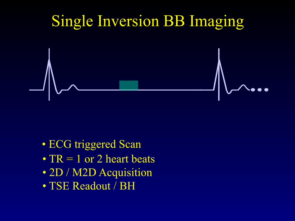
Single Inversion BB Imaging
• ECG triggered Scan• TR = 1 or 2 heart beats• 2D / M2D Acquisition• TSE Readout / BH

Single Inversion BB Imaging
• ECG triggered Scan• TR = 1 or 2 heart beats• 2D / M2D Acquisition• TSE Readout / BH

Single Inversion BB Imaging
• ECG triggered Scan• TR = 1 or 2 heart beats• 2D / M2D Acquisition
Blood
• TSE Readout / BH
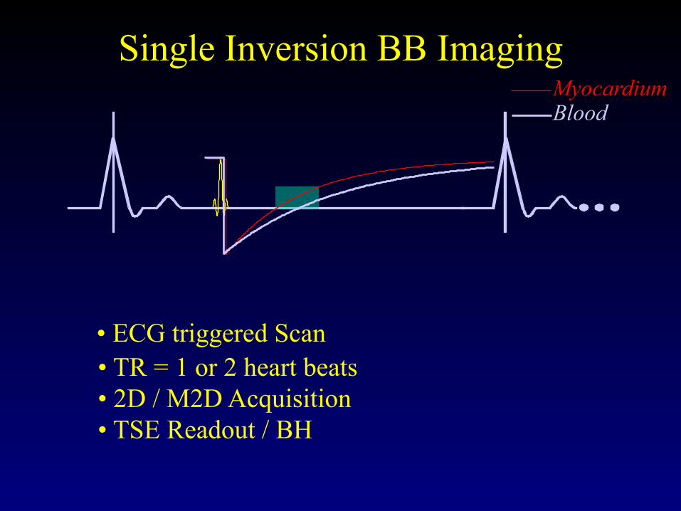
Single Inversion BB Imaging
• ECG triggered Scan• TR = 1 or 2 heart beats• 2D / M2D Acquisition
MyocardiumBlood
• TSE Readout / BH

Single Inversion BB Imaging
• ECG triggered Scan• TR = 1 or 2 heart beats• 2D / M2D Acquisition
MyocardiumBlood
• TSE Readout / BH

Double Inversion BB Imaging MyocardiumBlood

Double Inversion BB Imaging MyocardiumBlood
• The first non-selective inversion inverts everything

Double Inversion BB Imaging MyocardiumBlood
• The first non-selective inversion inverts everything

Double Inversion BB Imaging MyocardiumBlood
• The first non-selective inversion inverts everything
• The second selective inversion pulse re-inverts the signal within slice

Single Vs Double Inversion BB
Single IR Dual IRNote the increased SI in the myocardium in the Dual IR!( for the same TR/TE/TI as the Single IR sequence)

Choose correct TI:• The Inversion Delay should be adjusted for Heart Rate (or
TR) to improve nulling of blood signal
TI << TIopt TI = TIopt TI >> TIopt

Calculating the correct TI
11))/exp(1(
2ln TTTRTI ÷⎠⎞
⎜⎝⎛
−+=
0 500 1000 1500 2000 2500 30000
100
200
300
400
500
600
700
800
TR (msec)
TI (m
sec)
Blood T1 = 1200 msec
For Post Contrast Scans,TI needs to be shorter to nullCE-Blood.

DIR - T1W DIR - T1W post Gadwithout change in TI
with fat sat (inhomogeneous)
Example of incorrect TI

Choose Long enough TR!• At higher HR, keep TR > 1500 msec, for good signal
from myocardium!
TR = 800 msec TR = 1600 msec

Double IR Turbo Spin Echo
TR = 3 R-R; TE = 65; TSE factor 27; FOV = 360; 230 x 512 (r)4 mm thick / gap 2 mm; 3 slices; 4 NSA’s; scan time 2:30 min
Non-breath-hold
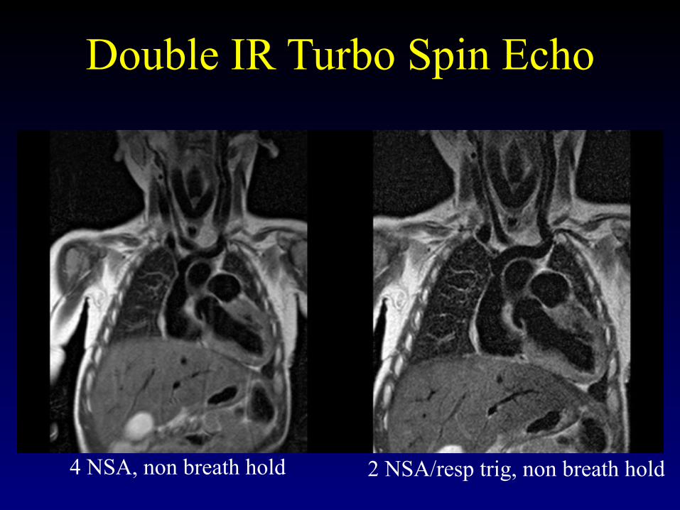
Double IR Turbo Spin Echo
4 NSA, non breath hold 2 NSA/resp trig, non breath hold

Anesthesized 3.1 kg neonate with heterotaxy? pulmonary vein anatomy
Breath-holding double inversion recovery turbo spin echo 1.8 mm thick, no skip, 0.8 x 1.1 mm, 2 NSA
TR= 2RR, TE 40, ~ 12 sec / sliceCourtesy of Andrew Powell, David Annese, Children’s Hospital, Boston

Triple inversion recovery
Cardiac fibroma – better conspicuity with triple IR

T2W TSE with fat sat Triple IR

Freely breathing sedated 3-month-old with Kawasaki disease
Vessel wall inflammation showed ontriple inversion recovery with 3 NSA’s

Freely breathing sedated 3-month-old with Kawasaki disease
Vessel wall inflammation showed ontriple inversion recovery with 3 NSA’s
LAD
RCA

Double IR TSE

Double IR TSE• TR = 2 to 3 RR depending on HR to get TR > 1500
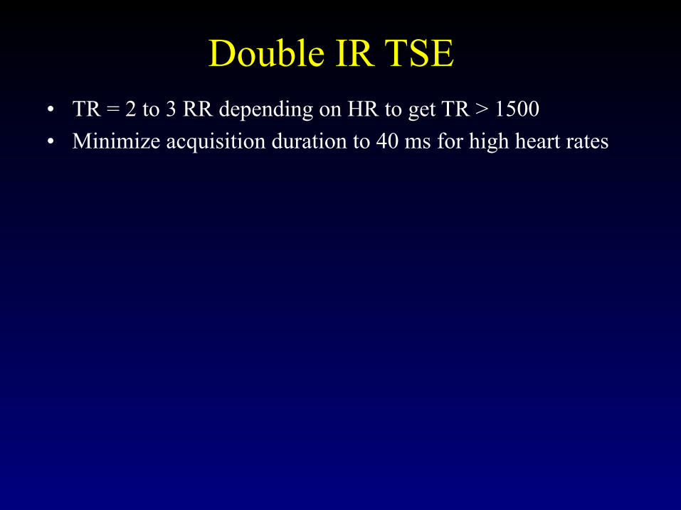
Double IR TSE• TR = 2 to 3 RR depending on HR to get TR > 1500• Minimize acquisition duration to 40 ms for high heart rates

Double IR TSE• TR = 2 to 3 RR depending on HR to get TR > 1500• Minimize acquisition duration to 40 ms for high heart rates• Set trigger delay to “longest”

Double IR TSE• TR = 2 to 3 RR depending on HR to get TR > 1500• Minimize acquisition duration to 40 ms for high heart rates• Set trigger delay to “longest”• Run a dummy scan to check blood nulling and trigger delay
and acquisition duration

Double IR TSE• TR = 2 to 3 RR depending on HR to get TR > 1500• Minimize acquisition duration to 40 ms for high heart rates• Set trigger delay to “longest”• Run a dummy scan to check blood nulling and trigger delay
and acquisition duration• Steady heart rate is essential theoretically

Double IR TSE• TR = 2 to 3 RR depending on HR to get TR > 1500• Minimize acquisition duration to 40 ms for high heart rates• Set trigger delay to “longest”• Run a dummy scan to check blood nulling and trigger delay
and acquisition duration• Steady heart rate is essential theoretically• Considering using parallel imaging to decrease acquisition
duration/heart beat if needed

Double IR TSE• TR = 2 to 3 RR depending on HR to get TR > 1500• Minimize acquisition duration to 40 ms for high heart rates• Set trigger delay to “longest”• Run a dummy scan to check blood nulling and trigger delay
and acquisition duration• Steady heart rate is essential theoretically• Considering using parallel imaging to decrease acquisition
duration/heart beat if needed• Adjust parameters for appropriate breath-hold duration

Double IR TSE• TR = 2 to 3 RR depending on HR to get TR > 1500• Minimize acquisition duration to 40 ms for high heart rates• Set trigger delay to “longest”• Run a dummy scan to check blood nulling and trigger delay
and acquisition duration• Steady heart rate is essential theoretically• Considering using parallel imaging to decrease acquisition
duration/heart beat if needed• Adjust parameters for appropriate breath-hold duration• Can use 3 NSA’s or respiratory trigger for free breathing
– Can result in long scan time

SAM question
• For black blood imaging using inversion recovery technique, it is best to have a single inversion pulse to null the blood as there is enough difference between the T1 of myocardium and blood such that the myocardium will be seen well
• True or False

SAM question
• For black blood imaging using inversion recovery technique, it is best to have a single inversion pulse to null the blood as there is enough difference between the T1 of myocardium and blood such that the myocardium will be seen well
• False: need 2 inversion pulses (non-selective and slice selective)

• Simonetti OP et al. “Black Blood” T2-weighted inversion-recovery MR imaging of the heart. Radiology 1996; 199:49-57.
• Mulkern RV, Chung T. From signal to image: magnetic resonance imaging physics for cardiac MR. Pediatr Cardiol 2000; 21:5-17

2013 SPR Cardiac Session
What is it?Black Blood Imaging
Taylor Chung, M.D.Associate Director, Body and Cardiovascular Imaging
Department of Diagnostic ImagingChildren’s Hospital & Research Center Oakland
Oakland, California, U.S.A.