vol-2, issue-1 approach to respiratory distress in the newborn
TRANSCRIPT
INTERNATIONAL JOURNAL OF HEALTH RESEARCH IN MODERN INTEGRATED MEDICAL SCIENCES, ISSN 2394-8612 (P), ISSN 2394-8620 (O), Vol-2, Issue-1, Jan-Mar 201524
Review Article
Approach to Respiratory Distress in the Newborn
Sai Sunil Kishore M, Siva Sankara Murty YV, Tarakeswara Rao P, Madhusudhan K, Pundareekaksha V, Pathrudu GB
Abstract: Respiratory distress is responsible for majority of neonatal admissions to neonatal intensive care unit. Various
pulmonary and extra-pulmonary causes are responsible for respiratory distress. Prolonged and unattended distress
leads to hypoxemia, hypercarbia and acidosis leading to pulmonary vasoconstriction and persistence of fetal circulation,
thereby aggravating hypoxemia, worsening prognosis. A discussion on general approach to identify etiology, evaluation
and management is made in this article.
Key Words : Respiratory distress, Meconium aspiration, Mechanical Ventilation, Continuous Positive airway pressure,
Surfactant.
Introduction
Respiratory distress is the commonest morbidity requiring
admission of a neonate in an intensive care unit. It is
responsible for 30-40% of the admissions in the neonatal
period 1,2. Respiratory distress occurs in 11-14% of all live
births 3. The incidence of respiratory distress on first day
of life increases with lower gestations : <30 wks- 60%,
30 to 34 wks- 43% and >34 wks: 5 to 6% 3. Respiratory
distress is defined by presence of at least 2 of the following
three features- tachypnea (respiratory rate >60 per minute),
retractions (intercostal, subcostal, sternal or suprasternal)
and noisy respiration (grunt, stridor or wheeze)1. The
distress may or may not be associated with cyanosis or
desaturation on pulse oximetry. A working diagnosis
should be made in the first few minutes of onset of
respiratory distress and immediate resuscitative measures
should be initiated till further management plans are drawn
up. The objectives of this article include a discussion on
the general approach to identify the etiology, initial
evaluation and algorithm for initial management,
interpretation of blood gases, and approach to initiation
of ventilation, titration and weaning.
Etiology
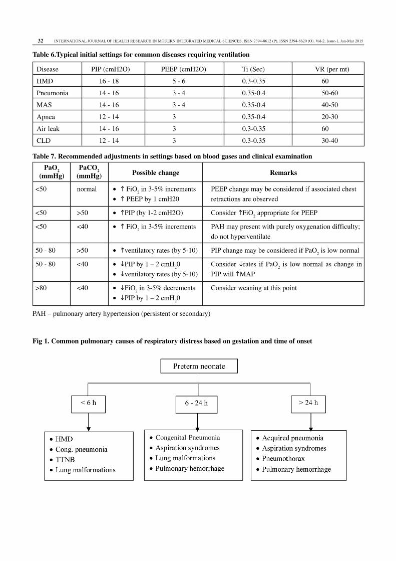
The etiology of respiratory distress is the single most
important determinant of the course and prognosis. The
common etiologies based on the time of onset of
respiratory distress and gestation are depicted in Fig1.
Respiratory distress of the neonate can be attributed either
to pulmonary or to extra-pulmonary disorders. ‘Pulmonary’
causes of respiratory distress are commoner than the ‘extra
pulmonary’. While conditions like structural anomalies and
pneumonia are common to term and preterm infants, a
condition like hyaline membrane disease (HMD) is almost
always a disease of the premature infant. Meconium
aspiration, on the other hand, is seen almost exclusively
in term infants.
Among very low birth weight (VLBW) neonates, up to
60% may develop respiratory distress soon after birth 4.
Among them the contribution by various etiologies
include: hyaline membrane disease- 36%, pneumonia-
28% and transient tachypnea of newborn (TTNB)- 27%4.
A functionally normal lung sometimes needs to work at a
capacity far exceeding normal levels, in order to
compensate for abnormalities of other systems, e.g. in the
presence of cardiac disease, shock, metabolic acidosis, or
abdominal distension. The definitive management of such
an infant would naturally be based on treating the primary
‘extra-pulmonary’ etiology.
Clinical/historical clues : Clinical clues and their possible
disease associations are presented in table 1 and the initial
examination that should be performed is shown in table 2.
Pathophysiology considerations unique to the newborn
Prolonged and unattended distress leads to hypoxemia,
hypercarbia and acidosis. This leads to pulmonary
vasoconstriction and persistence of fetal circulation with
right to left shunting through the ductus and foramen ovale,
thereby aggravating hypoxemia. An audible grunt is an
important sign of pulmonary pathology in the newborn,
indicating that the baby has a low lung volume or
Corresponding author
Sai Sunil Kishore M (Neonatologist)
Assistant Professor, Department of Paediatrics
Maharajah’s Institute of Medical Sciences, Nellimarla,
Vizianagaram - 535 217, Andhra Pradesh, India.
INTERNATIONAL JOURNAL OF HEALTH RESEARCH IN MODERN INTEGRATED MEDICAL SCIENCES, ISSN 2394-8612 (P), ISSN 2394-8620 (O), Vol-2, Issue-1, Jan-Mar 2015 25
functional residual capacity (FRC). Breathing against a
partially closed glottis increases the FRC of the baby and
helps to keep the alveoli open. This is characteristically
seen in a baby with respiratory distress syndrome (RDS)
where surfactant deficiency tends to keep the alveoli
collapsed during expiration. Indiscriminately inserting an
endotracheal (ET) tube without giving positive end
expiratory pressure (PEEP) to a neonate who is grunting
will deprive the baby of this physiological effect and
worsen the condition, instead of improving it. Hence any
baby who is grunting, depending on the severity of
respiratory distress, should be given either continuous
positive airway pressure (CPAP) or intubated and put on
ventilator support with PEEP, but never left to breathe
spontaneously with a tube in situ.
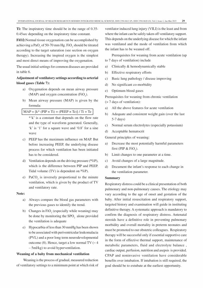
Grading of severity of respiratory distress
The severity of respiratory distress can be assessed by
Silverman-Anderson score (Fig2) and Downes’ score
(Table 3)5,6. While the Silverman Anderson retraction score
is more suited for preterms with RDS, the Downes’ score
can be applied to any gestational age and condition. Serial
monitoring of these scores with documentation will help
in determining the progression.
Investigations
Essential investigations for all cases of respiratory distress
include:
• Arterial blood gas (ABG) analysis: Blood gas is
essential because with clinical assessment and pulse
oximetry alone, one would not be able to assess PCO2
and pH.
Normal & abnormal values:
PaO2: Pre-ductal PaO
2 50–70 mmHg with an O
2 saturation
of 87-93%. A PaO2 up to 80 mmHg is acceptable in term
infants
o Hypoxemia: PaO2<50 mmHg
o Low normal oxygenation: PaO2 - 50-60mmHg
o Hyperoxemia: >80 mmHg in preterm and >90 in term.
Hyperoxia is associated with adverse effects like
retinopathy of prematurity(ROP) and
bronchopulmonary dysplasia(BPD) due to increase
in the reactive oxygen species (ROS).
PaCO2: Normal PaCO
2 is 35-45 mmHg
o Acceptable upper limit: Acute stage – 45-50mmHg,
Chronic (>72 hours of ventilation) – 55mmHg (with
pH of e”7.2). There is increasing evidence that the
strategy of permissive hypercapnia reduces the
duration of ventilation and decreases the severity of
bronchopulmonary dysplasia (BPD) 7.
o Hypocarbia: <35 mmHg. Hypocarbiawith PaCO2<30
mmHg increases the risk of periventricular
leucomalacia (PVL) in preterm neonates 8.
• Chest radiograph: Radiological findings in various
causes of respiratory distress are shown table 4.
• Electrolytes, blood glucose, hematocrit
• Sepsis work up which includes C-reactive protein
(CRP), micro ESR, total leukocyte count (TLC),
absolute neutrophil count (ANC) and Immature to
total leukocyte ratio (ITR). The parameters like TLC
and ANC should be interpreted according to the age
and gestation of the baby 9,10.
• Assessment of gas exchange: Alveolar-arterial oxygen
gradient (AaDO2), Oxygenation index (OI)
Special investigations like echocardiography and
neuro-sonogram may be required on case to case basis
depending on the clinical presentation.
Bedside investigations:
1. Gastric aspirate shake test: This is useful in
assessing the risk of RDS. This is useful in all neonates
with respiratory distress who are <34 wks of gestation
or have risk factors for RDS. In a glass test tube with
dimensions of 82 mm × 10.25 mm and 4 ml capacity,
0.5 ml of gastric aspirate is taken and mixed with 0.5
ml of absolute alcohol. The tube is shaken vigorously
for 15 seconds and allowed to stand for 15 min. If at
least one complete rim of bubbles are present all the
way round the meniscus the risk of RDS is <1% where
as complete absence of bubbles is associated with a
risk of 50-60%.
2. Gastric aspirate for polymorphs: The gastric fluid
should be aspirated preferably within one hour after
birth and mixed with one drop of heparin. A drop of
this is placed on a glass slide and a thick smear is
made and stained with Leishman’s stain. More than
5 polymorphonuclear leucocytes/HPF is suggestive
of infected amniotic fluid or chorioamniotis. The test
is not useful if the aspirate is contaminated with blood
or meconium.
INTERNATIONAL JOURNAL OF HEALTH RESEARCH IN MODERN INTEGRATED MEDICAL SCIENCES, ISSN 2394-8612 (P), ISSN 2394-8620 (O), Vol-2, Issue-1, Jan-Mar 201526
Assessment of gas exchange11
Though the blood gas parameters indicate oxygenation
and ventilation at a single point of time, these parameters
alone would not be sufficient to evaluate gas exchange.
Interpretation of PaO2 without FiO
2 is misleading. Hence
the gas exchange should be assessed using various
parameters like
1) Alveolar-arterial Oxygen gradient (A-aDO2)
2) a/A ratio
3) Oxygenation Index (OI)
Calculation and interpretation:
1) A-aDO2 (alveolar arterial oxygen diffusion gradient):
This is to be calculated as shown below. A-a DO2
=
PAO2 – PaO
2 (PAlveolar – Parterial oxygen)
= [PiO2 – PACO
2] – PaO
2
= [(P
B-P
W) × F
iO
2 – PaCO
2] – PaO
2
= [(760-47) × FiO2 – PaCO
2] – PaO
2
[PiO2=
Partial inspired oxygen pressure PB= Baromatric
pressure, PW
= water vapor pressure, FiO2 =
Fractional
inspired oxygen concentration]
Normally it ranges between 5-15, if breathing room air.
A-aDO2 is considered to be abnormal if more than 40.
2) a /A ratio: Ratio of PaO2 to PAO
2. It is considered to
be a better indicator of gas exchange as the ratio is
usually not affected by changes in FiO2
Interpretation:
a) Greater than 0.8: Normal
b) Less than 0.6: indicates need for O2 therapy
c) Less than 0.15: severe hypoxemia
3) Oxygenation Index (OI): Recommended in babies
who are mechanically ventilated as this index includes
mean airway pressure (MAP).
OI = (MAP × FiO2) / PaO
2
Interpretation:
a) OI 25 – 40: severe respiratory failure; mortality risk
is 50 – 60%
b) OI > 40: Mortality risk is >80%
Prevention: role of antenatal steroids
Antenatal steroids should be administered to pregnant
women of 24 to 34 weeks of gestation with intact
membranes or premature rupture of membranes without
chorioamnionitis, who are at high risk of preterm delivery
within next 7 days12. This strategy induces surfactant
production and accelerates maturation of lungs and other
fetal tissues. Treatment with antenatal corticosteroids is
associated with an overall reduction in neonatal mortality
(RR: 0.69, 95% CI: 0.58 to 0.81), RDS (RR 0.66, 95%CI:
0.59 to 0.73), IVH (RR 0.54, 95% CI 0.43 to 0.69) and
NEC (RR 0.46, 95% CI 0.29 to 0.74)13. Obstetricians
should be made aware of these benefits so that this
preventive strategy can be utilized optimally.
Treatment
While managing a neonate with respiratory distress,
the initial treatment is aimed at resuscitation of the neonate,
optimizing tissue oxygenation, decreasing the work of
breathing, preventing hypoxia, hypercapnia and acidosis.
The supportive measures could vary from oxygen
supplementation to various strategies of mechanical
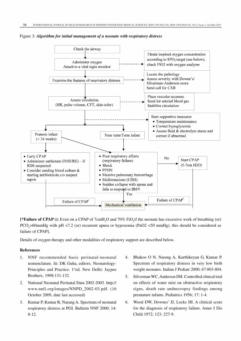
ventilation. An algorithm for initial management of a
neonate with respiratory distress is presented in Fig 3.
Subsequent management should focus on further
evaluation to identify the etiology, definitive management
and follow-up.Details of oxygen therapy and other
modalities of respiratory support are described below.
Supportive therapy: Apart from providing respiratory
support, supportive care and proper nursing care are very
crucial for success of management.
• Thermo-neutral environment : The neonate should be
nursed in a thermoneutral environment. Hypothermia
will initiate the cascade of PPHN and aggravate
hypoxemia. Baby’s temperature should be maintained
between 36.50-37.50C. VLBW neonates need
incubator for their temperature maintenance and for
providing adequate humidity
• Electrolyte balance, fluids and normal acid-base
balance should be maintained. Preterm babies have
higher insensible water loss (40-100 ml/kg). Fluid
intake should be titrated accurately by recording serial
weight, intake/ output, serum sodium and urine
specific gravity.
• Antibiotics should be started in all cases of suspected
sepsis or pneumonia.
• Calcium and glucose homeostasis should be ensured.
• Maintain normal mean arterial pressure. Hypotension
should be corrected by using appropriate fluid
volumes and inotropes if necessary.
INTERNATIONAL JOURNAL OF HEALTH RESEARCH IN MODERN INTEGRATED MEDICAL SCIENCES, ISSN 2394-8612 (P), ISSN 2394-8620 (O), Vol-2, Issue-1, Jan-Mar 2015 27
• Hypovolemia and anemia are to be treated adequately
as necessary. Hematocrit should be maintained above
40% in the acute phase of the disease 14.
• Oral feeding is withheld initially. Once the baby
stabilizes on the respiratory support and respiratory
rates are less than 70/min, gavage feeding should be
started.
Definitive management
Despite a relatively uniform approach to the initial
management, one must realize that procrastination and
delay in instituting definitive therapy may result in adverse
outcomes. For example, an infant with tension
pneumothorax could deteriorate rapidly despite the
transient improvement seen with initial therapy with
oxygen and increased ventilator support, if the
pneumothorax is not drained. RDS will progress with time
if surfactant is not administered in time. Similarly, repeated
aspiration pneumonia would contribute to poor surgical
outcome in patients with delayed diagnosis of
tracheoesophageal fistula (TEF). Therefore a definite
diagnosis and therapy is mandatory for successfully
managing infants with respiratory distress.
Surfactant replacement
Surfactant replacement therapy is the standard of care for
a baby with RDS. Surfactant should be administered early
in the course of respiratory distress, preferably within first
two hours of onset of symptoms in neonates at risk for
RDS (Early rescue therapy). Delayed rescue therapy is
less effective since the inflammatory processes and
exudation already sets in. Prophylactic administration of
surfactant may be used in neonates who are at a very high
risk of RDS and its complications. Typically, this may be
in neonates <28-30 wks of gestation depending on the local
survival rates. This approach although very effective,
would increase the costs. The dose of surfactant for RDS
is according to the phospholipid and varies between 100
to 200mg/kg depending on the manufacturer. Two or more
doses may be required especially in extremely low birth
weight babies.
Surfactant activity is altered in other respiratory
disorders also apart from RDS. Surfactant has been tried
in various conditions such as meconium aspiration
syndrome, pneumonia and pulmonary hemorrhage with
variable benefits 16. Further evidence is needed to establish
the value and limitations of surfactant therapy for these
conditions.
Oxygen therapy
Indications:
1) Clinical central cyanosis
2) Hypoxemia (O2 saturation <87% and / or PaO
2<50
mmHg in room air)
3) Presence of respiratory distress irrespective of O2
saturation
Saturation targets 17 : Preterm: 87-93%, Term: 90-95%,
Bronhopulmonary dysplasia: 90-95%, PPHN: >95%
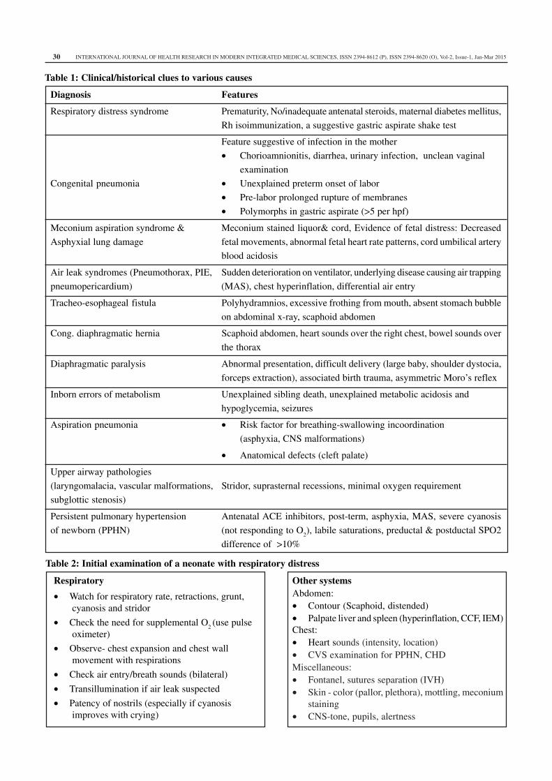
Commonly used oxygen delivery systems in neonates:
Characteristics of common oxygen delivery systems are
provided in table 5.
Note:
1) Short binasal prongs are currently available for O2
administration, which can be attached to the
humidification system to provide heated and
humidified O2
2) While using oxygen hood, change in the flow rate
will not alter the FiO2 much. If one wants to decrease
the FiO2, more mixing with air has to be allowed
through port holes. Occlusion of these holes increases
the O2 concentration by preventing air entrainment.
Approximate FiO2 delivered with a flow rate of 5-8
Lpm:
• With all port holes closed: 60-70%
• With one port hole opened – 40-50%
• With both the port holes opened – 30-40%
Precautions while administering oxygen:
1. Prewarm and humidify the oxygen especially at flow
rates > 2Lpm
2. Oxygen saturation should not cross 93% as hyperoxia
leads to wide spread free radical injury
3. Flow rate should never be crossed as excessive flow
can cause turbulence in the airflow and inadvertent
uncontrolled PEEP delivery apart from causing local
airway injury
4. Oxygen analyzer is to be used always to check the
FiO2. Desired FiO
2 can be administered by using a
blender or by using varying flow rates of oxygen and
air using a ‘Y’ piece.
Non-invasive ventilation
Continuous Positive Airway Pressure (CPAP)
Continuous distending pressure is applied throughout
the respiratory cycle in a spontaneously breathing infant.
INTERNATIONAL JOURNAL OF HEALTH RESEARCH IN MODERN INTEGRATED MEDICAL SCIENCES, ISSN 2394-8612 (P), ISSN 2394-8620 (O), Vol-2, Issue-1, Jan-Mar 201528
CPAP can be administered by applying different nasal or
nasopharyngeal interfaces. Endotracheal CPAP should not
be given. It works by allowing alveolar recruitment,
preventing atelectasis thereby improving FRC. It also
dilates the upper airways and decreases the airway
resistance. In a preterm neonate who is at risk of respiratory
distress syndrome, CPAP should be used early, to reduce
the need for mechanical ventilation unless there is a
contraindication to use CPAP. Early CPAP conserves
the neonate’s own surfactant stores and minimizes the
stimulation of inflammatory cascade. A comparison of
early versus delayed initiation of CPAP in RDS
concluded that early CPAP reduces the subsequent use
of intermittent mandatory ventilation (IMV) [typical RR
0.55, 95% CI: 0.32-0.96]18. If used early, many lives can
be saved and even upward referrals to a tertiary care unit
can be reduced 19. Considering the benefits of CPAP and
problems associated with mechanical ventilation, all efforts
should be made to manage babies on CPAP.
The level of CPAP should be individualized to the
baby’s disease. As a general guideline start CPAP of
5cmH2O and FiO
2of 0.5 to maintain normal SpO
2.
Subsequent titration can be done based on the clinical
improvement, blood gases and lung volume on chest
radiograph. The orogastric tube (OGT) should be inserted
to decompress the stomach. The signs of over inflation
should be monitored - inadequate cardiac output
(prolonged CFT, reduced urine output, metabolic acidosis)
and hyperinflated chest. Periodic inspection of the local
area is important because patient interface can lead to septal
and mucosal injury.
Nasal intermittent positive pressure ventilation
(NIPPV)
NIPPV combines nasal CPAP and intermittent
positive pressure ventilation (IPPV) via a timed cycled,
pressure limited ventilator. NIPPV has been found to be
more effective than NCPAP in weaning preterm infants
from mechanical ventilation 20 and in apnea of prematurity21. There is evidence to suggest that NIPPV as a primary
mode of respiratory support may be more effective than
CPAP in reducing the need for intubation in RDS22.
Mechanical ventilation
Mechanical ventilation needs to be started whenever
there is failure of noninvasive ventilation (see fig 2) or if
it is contraindicated in a neonate with respiratory distress.
However the decision to initiate mechanical ventilation
should be individualized for each baby and depends on
several factors. The severity of distress, severity of blood
gas abnormalities, natural history of disease and degree
of cardiovascular and other physiologic instabilities must
be taken into account. Because invasive ventilation is
associated with serious pulmonary morbidities like
subglottic stenosis, respiratory infections, ventilator
associated lung injury and BPD23,24, the decision to intubate
and ventilate should not be taken lightly. For best results
with minimum damage, it is useful to follow the laws of
ventilator efficiency (LOVE). One needs to know the baby,
the disease, the machine and have an EXIT strategy25.
Initiation of mechanical ventilation
Initial steps in starting positive pressure ventilation
include endotracheal intubation, selection of appropriate
ventilator settings and evaluation to check adequacy of
ventilatory support. Time cycled pressure limited
ventilation is the commonest type of ventilation used in
neonates. Mean airway pressure (MAP) and the delivered
volumes are dependent on the ventilator settings.
Establishing the correct ventilator settings is the key to
successful respiratory support.
Parameters in a pressure limited ventilator 26
Rate: Ventilator rate determines the minute ventilation in
combination with tidal volume (VT). This usually varies
from 20-60 depending on the disease pathology and course
of the baby on ventilator.
Flow: Initial flow rate should be 5-7 Lpm to drive the
respiratory gases and for adequate delivery of the
pressures. Flow rate above 10 Lpm can cause lot of
turbulence and increase the resistance.
PEEP: PEEP is the distending pressure in the expiratory
phase of the respiratory cycle that recruits the alveoli and
maintains FRC. Settings for PEEP range from 3-6 cm H2O
depending on the disease pathology.
PIP: Delivered VT
is dependent on the driving pressure
which is the difference between PIP and PEEP. Initial
choice of PIP is dependent on observation of chest wall
movement during hand-bag ventilation, manometer
readings during hand-bag ventilation and auscultation of
breath sounds. Subsequently PIP should be adjusted to
achieve optimal MAP and VT
and effective gas exchange
based on the blood gas parameters.
INTERNATIONAL JOURNAL OF HEALTH RESEARCH IN MODERN INTEGRATED MEDICAL SCIENCES, ISSN 2394-8612 (P), ISSN 2394-8620 (O), Vol-2, Issue-1, Jan-Mar 2015 29
Ti: The inspiratory time should be in the range of 0.35-
0.45sec depending on the inspiratory time constant.
FiO2:Normal tissue oxygenation can be accomplished by
achieving a PaO2 of 50-70 mm Hg. FiO
2 should be titrated
according to the target saturation (see section on oxygen
therapy). Increasing the inspired oxygen is the simplest
and most direct means of improving the oxygenation.
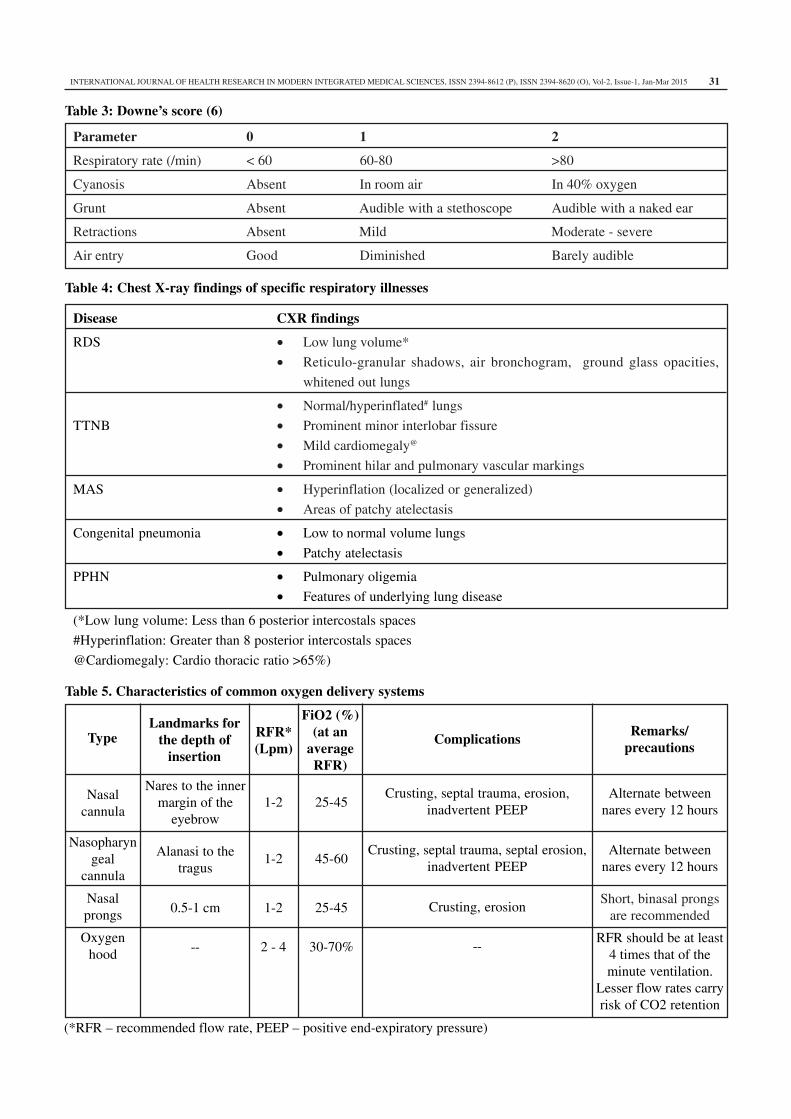
The usual initial settings for common diseases are provided
in table 6.
Adjustment of ventilatory settings according to arterial
blood gases (Table 7):
a) Oxygenation depends on mean airway pressure
(MAP) and oxygen concentration (FiO2).
b) Mean airway pressure (MAP) is given by the
formula:
MAP = [k* (PIP × Ti) + (PEEP × Te)] / Ti + Te
*‘k’ is a constant that depends on the flow rate
and the type of waveform generated. Generally,
‘k’ is ‘1’ for a square wave and ‘0.8’ for a sine
wave
c) PEEP has the maximum influence on MAP. But
before increasing PEEP, the underlying disease
process for which ventilation has been initiated
has to be considered.
d) Ventilation depends on the driving pressure (²%P),
which is the difference between PIP and PEEP.
Tidal volume (TV) is dependent on ²%P).
e) PaCO2 is inversely proportional to the minute
ventilation, which is given by the product of TV
and ventilatory rate.
Note:
a) Always compare the blood gas parameters with
the previous gases to identify the trend.
b) Changes in FiO2 (especially while weaning) may
be done by monitoring the SPO2 alone provided
the ventilation is adequate
e) Hypocarbia of less than 30 mmHg has been shown
to be associated with periventricular leukomalacia
(PVL) and a poor long term neurodevelopmental
outcome (8). Hence, target a low normal TV (~ 4
– 5ml/kg) to avoid hyperventilation.
Weaning of a baby from mechanical ventilation
Weaning is the process of gradual, measured reduction
of ventilatory settings to a minimum point at which risk of
ventilator induced lung injury (VILI) is the least and from
where the infant can be safely taken off ventilatory support.
This depends on the underlying disease for which the infant
was ventilated and the mode of ventilation from which
the infant has to be weaned off.
Prerequisites for weaning from acute ventilation (up
to 7 days of ventilation) include
a) Clinically & hemodynamically stable
b) Effective respiratory efforts
c) Basic lung pathology / disease improving
d) No significant co-morbidity
e) Optimum blood gases
Prerequisites for weaning from chronic ventilation
(> 7 days of ventilation):
a) All the above features for acute ventilation
b) Adequate and consistent weight gain (over the last
5-7 days)
c) Normal serum electrolytes (especially potassium)
d) Acceptable hematocrit
General principles of weaning:
a) Decrease the most potentially harmful parameters
first (PIP & FiO2).
b) Limit changes to one parameter at a time.
c) Avoid changes of a large magnitude.
d) Document the infant’s response to each change in
the ventilation parameter.
Summary
Respiratory distress could be a clinical presentation of both
pulmonary and non-pulmonary causes. The etiology may
vary according to the age of onset and gestation of the
baby. After initial resuscitation and respiratory support,
targeted history and examination will guide in instituting
definitive therapy. A systematic approach is mandatory to
confirm the diagnosis of respiratory distress. Antenatal
steroids have a definitive role in preventing pulmonary
morbidity and overall mortality in preterm neonates and
must be promoted to our obstetric colleagues. Respiratory
therapy will be successful only if essential supportive care
in the form of effective thermal support, maintenance of
metabolic parameters, fluid and electrolyte balance ,
cardiac output, perfusion, nutrition and asepsis is provided.
CPAP and noninvasive ventilation have considerable
benefits over intubation. If intubation is still required, the
goal should be to extubate at the earliest opportunity.
INTERNATIONAL JOURNAL OF HEALTH RESEARCH IN MODERN INTEGRATED MEDICAL SCIENCES, ISSN 2394-8612 (P), ISSN 2394-8620 (O), Vol-2, Issue-1, Jan-Mar 201530
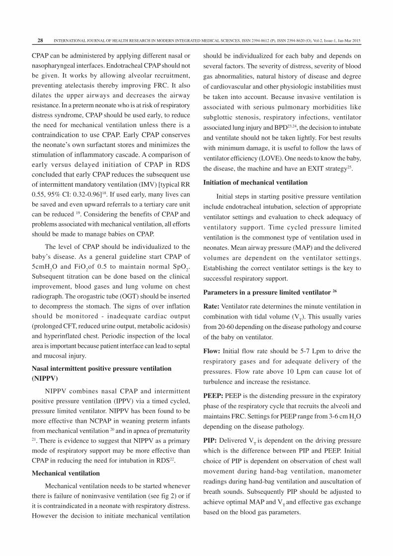
Table 1: Clinical/historical clues to various causes
Diagnosis Features
Respiratory distress syndrome Prematurity, No/inadequate antenatal steroids, maternal diabetes mellitus,
Rh isoimmunization, a suggestive gastric aspirate shake test
Feature suggestive of infection in the mother
• Chorioamnionitis, diarrhea, urinary infection, unclean vaginal
examination
Congenital pneumonia • Unexplained preterm onset of labor
• Pre-labor prolonged rupture of membranes
• Polymorphs in gastric aspirate (>5 per hpf)
Meconium aspiration syndrome & Meconium stained liquor& cord, Evidence of fetal distress: Decreased
Asphyxial lung damage fetal movements, abnormal fetal heart rate patterns, cord umbilical artery
blood acidosis
Air leak syndromes (Pneumothorax, PIE, Sudden deterioration on ventilator, underlying disease causing air trapping
pneumopericardium) (MAS), chest hyperinflation, differential air entry
Tracheo-esophageal fistula Polyhydramnios, excessive frothing from mouth, absent stomach bubble
on abdominal x-ray, scaphoid abdomen
Cong. diaphragmatic hernia Scaphoid abdomen, heart sounds over the right chest, bowel sounds over
the thorax
Diaphragmatic paralysis Abnormal presentation, difficult delivery (large baby, shoulder dystocia,
forceps extraction), associated birth trauma, asymmetric Moro’s reflex
Inborn errors of metabolism Unexplained sibling death, unexplained metabolic acidosis and
hypoglycemia, seizures
Aspiration pneumonia • Risk factor for breathing-swallowing incoordination
(asphyxia, CNS malformations)
• Anatomical defects (cleft palate)
Upper airway pathologies
(laryngomalacia, vascular malformations, Stridor, suprasternal recessions, minimal oxygen requirement
subglottic stenosis)
Persistent pulmonary hypertension Antenatal ACE inhibitors, post-term, asphyxia, MAS, severe cyanosis
of newborn (PPHN) (not responding to O2), labile saturations, preductal & postductal SPO2
difference of >10%
Table 2: Initial examination of a neonate with respiratory distress
Respiratory
• Watch for respiratory rate, retractions, grunt,
cyanosis and stridor
• Check the need for supplemental O2 (use pulse
oximeter)
• Observe- chest expansion and chest wall
movement with respirations
• Check air entry/breath sounds (bilateral)
• Transillumination if air leak suspected
• Patency of nostrils (especially if cyanosis
improves with crying)
Other systems
Abdomen:
• Contour (Scaphoid, distended)
• Palpate liver and spleen (hyperinflation, CCF, IEM)
Chest:
• Heart sounds (intensity, location)
• CVS examination for PPHN, CHD
Miscellaneous:
• Fontanel, sutures separation (IVH)
• Skin - color (pallor, plethora), mottling, meconium
staining
• CNS-tone, pupils, alertness
INTERNATIONAL JOURNAL OF HEALTH RESEARCH IN MODERN INTEGRATED MEDICAL SCIENCES, ISSN 2394-8612 (P), ISSN 2394-8620 (O), Vol-2, Issue-1, Jan-Mar 2015 31
Table 3: Downe’s score (6)
Parameter 0 1 2
Respiratory rate (/min) < 60 60-80 >80
Cyanosis Absent In room air In 40% oxygen
Grunt Absent Audible with a stethoscope Audible with a naked ear
Retractions Absent Mild Moderate - severe
Air entry Good Diminished Barely audible
Table 4: Chest X-ray findings of specific respiratory illnesses
Disease CXR findings
RDS • Low lung volume*
• Reticulo-granular shadows, air bronchogram, ground glass opacities,
whitened out lungs
• Normal/hyperinflated# lungs
TTNB • Prominent minor interlobar fissure
• Mild cardiomegaly@
• Prominent hilar and pulmonary vascular markings
MAS • Hyperinflation (localized or generalized)
• Areas of patchy atelectasis
Congenital pneumonia • Low to normal volume lungs
• Patchy atelectasis
PPHN • Pulmonary oligemia
• Features of underlying lung disease
(*Low lung volume: Less than 6 posterior intercostals spaces
#Hyperinflation: Greater than 8 posterior intercostals spaces
@Cardiomegaly: Cardio thoracic ratio >65%)
Table 5. Characteristics of common oxygen delivery systems
TypeLandmarks for
the depth of
insertion
RFR*
(Lpm)
FiO2 (%)
(at an
average
RFR)
ComplicationsRemarks/
precautions
Nasal
cannula
Nares to the inner
margin of the
eyebrow
1-2 25-45Crusting, septal trauma, erosion,
inadvertent PEEP
Alternate between
nares every 12 hours
Nasopharyn
geal
cannula
Alanasi to the
tragus1-2 45-60
Crusting, septal trauma, septal erosion,
inadvertent PEEP
Alternate between
nares every 12 hours
Nasal
prongs0.5-1 cm 1-2 25-45 Crusting, erosion
Short, binasal prongs
are recommended
Oxygen
hood-- 2 - 4 30-70% --
RFR should be at least
4 times that of the
minute ventilation.
Lesser flow rates carry
risk of CO2 retention
(*RFR – recommended flow rate, PEEP – positive end-expiratory pressure)
INTERNATIONAL JOURNAL OF HEALTH RESEARCH IN MODERN INTEGRATED MEDICAL SCIENCES, ISSN 2394-8612 (P), ISSN 2394-8620 (O), Vol-2, Issue-1, Jan-Mar 201532
Table 6.Typical initial settings for common diseases requiring ventilation
Disease PIP (cmH2O) PEEP (cmH2O) Ti (Sec) VR (per mt)
HMD 16 - 18 5 - 6 0.3-0.35 60
Pneumonia 14 - 16 3 - 4 0.35-0.4 50-60
MAS 14 - 16 3 - 4 0.35-0.4 40-50
Apnea 12 - 14 3 0.35-0.4 20-30
Air leak 14 - 16 3 0.3-0.35 60
CLD 12 - 14 3 0.3-0.35 30-40
Table 7. Recommended adjustments in settings based on blood gases and clinical examination
PaO2
(mmHg)
PaCO2
(mmHg)Possible change Remarks
<50 normal • � FiO2 in 3-5% increments PEEP change may be considered if associated chest
• � PEEP by 1 cmH20 retractions are observed
<50 >50 • �PIP (by 1-2 cmH2O) Consider �FiO2 appropriate for PEEP
<50 <40 • � FiO2 in 3-5% increments PAH may present with purely oxygenation difficulty;
do not hyperventilate
50 - 80 >50 • �ventilatory rates (by 5-10) PIP change may be considered if PaO2 is low normal
50 - 80 <40 • �PIP by 1 – 2 cmH20 Consider �rates if PaO
2 is low normal as change in
• �ventilatory rates (by 5-10) PIP will �MAP
>80 <40 • �FiO2 in 3-5% decrements Consider weaning at this point
• �PIP by 1 – 2 cmH20
PAH – pulmonary artery hypertension (persistent or secondary)
Fig 1. Common pulmonary causes of respiratory distress based on gestation and time of onset
Congenital Pneumonia
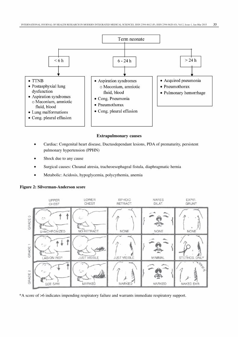
INTERNATIONAL JOURNAL OF HEALTH RESEARCH IN MODERN INTEGRATED MEDICAL SCIENCES, ISSN 2394-8612 (P), ISSN 2394-8620 (O), Vol-2, Issue-1, Jan-Mar 2015 33
*A score of >6 indicates impending respiratory failure and warrants immediate respiratory support.
Extrapulmonary causes
• Cardiac: Congenital heart disease, Ductusdependant lesions, PDA of prematurity, persistent
pulmonary hypertension (PPHN)
• Shock due to any cause
• Surgical causes: Choanal atresia, tracheoesophageal fistula, diaphragmatic hernia
• Metabolic: Acidosis, hypoglycemia, polycythemia, anemia
Figure 2: Silverman-Anderson score
INTERNATIONAL JOURNAL OF HEALTH RESEARCH IN MODERN INTEGRATED MEDICAL SCIENCES, ISSN 2394-8612 (P), ISSN 2394-8620 (O), Vol-2, Issue-1, Jan-Mar 201534
Figure 3: Algorithm for initial management of a neonate with respiratory distress
[*Failure of CPAP (): Even on a CPAP of 7cmH2O and 70% FiO
2if the neonate has excessive work of breathing (or)
PCO2>60mmHg with pH <7.2 (or) recurrent apnea or hypoxemia (PaO2 <50 mmHg), this should be considered as
failure of CPAP].
Details of oxygen therapy and other modalities of respiratory support are described below.
References
1. NNF recommended basic perinatal-neonatal
nomenclature. In: DK Guha, editors. Neonatology-
Principles and Practice. 1sted. New Delhi: Jaypee
Brothers, 1998:131-132.
2. National Neonatal Perinatal Data 2002-2003. http://
www.nnfi.org/images/NNPD_2002-03.pdf. (16
October 2009, date last accessed)
3. Kumar P, Kumar R, Narang A. Spectrum of neonatal
respiratory distress at PGI. Bulletin NNF 2000; 14:
8-12.
4. Bhakoo O N, Narang A, Karthikeyan G, Kumar P.
Spectrum of respiratory distress in very low birth
weight neonates. Indian J Pediatr 2000; 67:803-804.
5. Silverman WC, Anderson DH. Controlled clinical trial
on effects of water mist on obstructive respiratory
signs, death rate andnecropsy findings among
premature infants. Pediatrics 1956; 17: 1-4.
6. Wood DW, Downes’ JJ, Locks HI. A clinical score
for the diagnosis of respiratory failure. Amer J Dis
Child 1972; 123: 227-9.
INTERNATIONAL JOURNAL OF HEALTH RESEARCH IN MODERN INTEGRATED MEDICAL SCIENCES, ISSN 2394-8612 (P), ISSN 2394-8620 (O), Vol-2, Issue-1, Jan-Mar 2015 35
7. Miller JD, Carlo WA. Permissive hypercapnia in
neonates. Neoreviews2007;8: e345-353
8. Okumura A, Hayakawa F, Kato T, Itomi K, Maruyama
K, Ishihara N, et al. Hypocarbia in preterm infants
with periventricular leukomalacia: the relation
between hypocarbia and mechanical ventilation.
Pediatrics 2001; 107:469-475.
9. Manroe BL, Weinberg AG, Rosenfeld CR et al. The
neonatal blood count in health and disease. I.
Reference values for neutrophilic cells. J Pediatr
1979; 95: 89-93.
10. Mouzinho A, Rosenfeld CR, Sanchez PJ, Risser R.
Revised reference ranges for circulating neutrophils
in very low birth weight neonates. Pediatrics 1994;
94:76.
11. Wood BR. Physiological principles. In: Goldsmith JP,
Karotkin EH, editors. Assisted ventilation of the
neonate. 4th ed. Saunders. Philadelphia, PA 2008: 15-
40.
12. Bhakta KY. Respiratory distress syndrome. In:
Cloherty JP, Eichenwald EC, Stark AR., editors.
Manual of neonatal care. 6th ed. Lippincott Williams
& Wilkins. Philadelphia, PA 2008: 323-330.
13. Roberts D, Dalziel S. Antenatal corticosteroids for
accelerating fetal lung maturation for women at risk
of preterm birth. Cochrane Database of Systematic
Reviews 2006, Issue 3. Art. No.: CD004454. DOI:
10.1002/14651858.CD004454.pub2.
14. Mathai SS, Raju CU, Kanitkar CM. Management of
Respiratory Distress in the Newborn. MJAFI 2007;
63: 269-272.
15. Soll RF, Morley CJ. Prophylactic versus selective use
of surfactant in preventing morbidity and mortality
in preterm infants. Cochrane Database of Systematic
Reviews 2001, Issue 2. Art. No.: CD000510. DOI:
10.1002/14651858.CD000510.
16. Lacaze-Masmonteil T. Expanded use of surfactant
therapy in newborns. ClinPerinatol 2007; 34: 179–
189
17. Ambalavanan N, Carlo WA. Ventilatory strategies in
the prevention and management of
Bronchopulmonary Dysplasia. SeminPerinatol 2006;
30: 192-199.
18. Ho JJ, Henderson-Smart DJ, Davis PG. Early versus
delayed initiation of continuous distending pressure
for respiratory distress syndrome in preterm infants.
Cochrane Database Syst Rev. 2002;(2):CD002975.
19. Buckmaster AG, Arnolda G, Wright IM, Foster JP,
Henderson-Smart DJ. Continuous positive airway
pressure therapy for infants with respiratory distress
in non tertiary care centers: a randomized, controlled
trial. Pediatrics 2007;120:509-18
20. Lemyre B, Davis PG, De Paoli AG. Nasal intermittent
positive pressure ventilation (NIPPV) versus nasal
continuous positive airway pressure (CPAP) for
preterm neonates after extubation (Cochrane
review).Cochrane Database Syst Rev. 2001;
(3):CD003212.
21. Lemyre B, Davis PG, De Paoli AG. Nasal intermittent
positive pressure ventilation (NIPPV) versus nasal
continuous positive airway pressure (CPAP) for apnea
of prematurity (Cochrane review). Cochrane Database
Syst Rev. 2002;(1):CD002272. Review.
22. Kishore MSS, Dutta S, Kumar P. Early nasal
intermittent positive pressure ventilation versus
continuous positive airway pressure for respiratory
distress syndrome. ActaPaediatr 2009; 98: 1412-1415.
23. Greenspan JS, Shaffer TH. Ventilator-induced airway
injury: a critical consideration during mechanical
ventilation of the infant. Neonatal Netw 2006; 25:
159–166.
24. Hutchison AA, Bignall S. Non-invasive positive
pressure ventilation in the preterm neonate: reducing
endotrauma and the incidence of bronchopulmonary
dysplasia. Arch Dis Child Fetal Neonatal Ed 2008;
93: F64 – 68.
25. Goldsmith JP, Karotkin EH. Preface. In: Goldsmith
JP, Karotkin EH, editors. Assisted ventilation of the
neonate. 4th ed. Saunders. Philadelphia, PA 2008: 149-
170.
26. Spitzer AR,Greenspan JS, Fox WW. Positive-pressure
ventilation: pressure-limited and time cycled
ventilation. In: Goldsmith JP, Karotkin EH, editors.
Assisted ventilation of the neonate. 4th ed. Saunders.
Philadelphia, PA 2008: 149-170.