pathophysiology of acute respiratory · pdf filethe top ten causes of neonatal ......
TRANSCRIPT
29
Acute Respiratory Care of the Neonate
2 Pathophysiology of Acute Respiratory Distress
Susan Orlando, DNS, APRN, NNP-BC
Considering the complex series of cardiorespira-tory changes that occurs at birth, it is not sur-
prising that the transition to extrauterine life does not always proceed smoothly. Neonatal respiratory disorders account for the majority of admissions to intensive care units and result in significant morbidity and mortality.
Once the infant shows signs of respiratory distress, prompt diagnosis is essential. Respiratory distress may be related to structural problems such as poor lung development or defects of the chest wall or dia-phragm. Biochemical and physical immaturity may exist. Abnormalities in the central nervous system may cause alterations in the respiratory regulatory appara-tus. Perfusion abnormalities may impair gas exchange. Aspiration and infection can also occur.
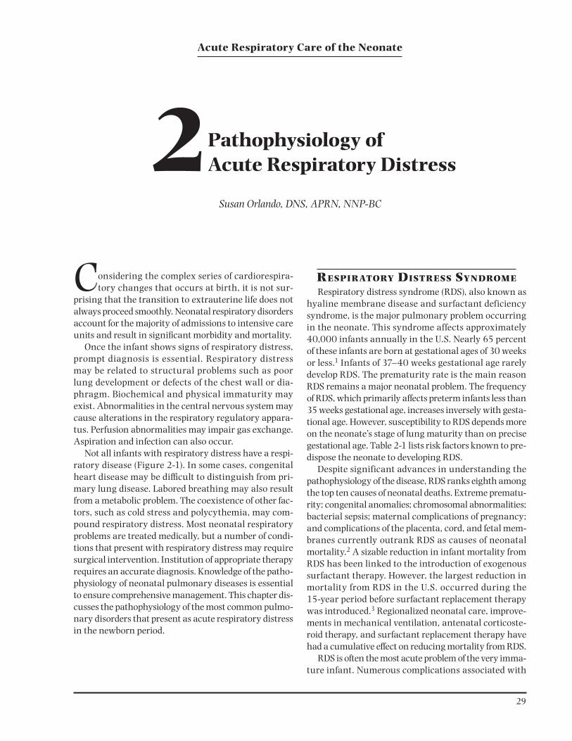
Not all infants with respiratory distress have a respi-ratory disease (Figure 2-1). In some cases, congenital heart disease may be difficult to distinguish from pri-mary lung disease. Labored breathing may also result from a metabolic problem. The coexistence of other fac-tors, such as cold stress and polycythemia, may com-pound respiratory distress. Most neonatal respiratory problems are treated medically, but a number of condi-tions that present with respiratory distress may require surgical intervention. Institution of appropriate therapy requires an accurate diagnosis. Knowledge of the patho-physiology of neonatal pulmonary diseases is essential to ensure comprehensive management. This chapter dis-cusses the pathophysiology of the most common pulmo-nary disorders that present as acute respiratory distress in the newborn period.
RESPIRATORY DISTRESS SYNDROMERespiratory distress syndrome (RDS), also known as
hyaline membrane disease and surfactant deficiency syndrome, is the major pulmonary problem occurring in the neonate. This syndrome affects approximately 40,000 infants annually in the U.S. Nearly 65 percent of these infants are born at gestational ages of 30 weeks or less.1 Infants of 37–40 weeks gestational age rarely develop RDS. The prematurity rate is the main reason RDS remains a major neonatal problem. The frequency of RDS, which primarily affects preterm infants less than 35 weeks gestational age, increases inversely with gesta-tional age. However, susceptibility to RDS depends more on the neonate’s stage of lung maturity than on precise gestational age. Table 2-1 lists risk factors known to pre-dispose the neonate to developing RDS.
Despite significant advances in understanding the pathophysiology of the disease, RDS ranks eighth among the top ten causes of neonatal deaths. Extreme prematu-rity; congenital anomalies; chromosomal abnormalities; bacterial sepsis; maternal complications of pregnancy; and complications of the placenta, cord, and fetal mem-branes currently outrank RDS as causes of neonatal mortality.2 A sizable reduction in infant mortality from RDS has been linked to the introduction of exogenous surfactant therapy. However, the largest reduction in mortality from RDS in the U.S. occurred during the 15-year period before surfactant replacement therapy was introduced.3 Regionalized neonatal care, improve-ments in mechanical ventilation, antenatal corticoste-roid therapy, and surfactant replacement therapy have had a cumulative effect on reducing mortality from RDS.
RDS is often the most acute problem of the very imma-ture infant. Numerous complications associated with
2 Pathophysiology of Acute Respiratory Distress ARC
30
shortened gestation and preterm birth can prolong hos-pitalization and add enormous costs. Most infants with RDS do not die from primary lung disease but from com-plications directly associated with RDS, such as air leak syndrome, intraventricular hemorrhage, pulmonary hemorrhage, or chronic lung disease, or from extreme prematurity itself. Chronic lung disease in infants with birth weights of less than 1,000 g has been identified as a significant predictor of later neurodevelopmental impairment.4 Efforts aimed at preventing RDS can be expected to improve morbidity and mortality, leading to significant cost savings and improved health for low birth weight infants.
Maternal antenatal steroid therapy reduces neo-natal mortality and the incidence of RDS in preterm infants. Additional short-term benefits of this type of therapy include a decreased incidence of intraventricu-lar hemorrhage, lower oxygen and ventilatory support requirements, and improved circulatory stability.5 A single course of antenatal steroids is currently rec-ommended for women at risk of delivery between the 24th and 34th week of gestation. Initiation of maternal treatment at least 24 hours before delivery produces the greatest bene fit for the infant. Treated infants born at 24–28 weeks gestation experience less severe RDS than untreated infants, and disease incidence and mortality are reduced in treated infants born at 29–34 weeks ges-tation. The benefits of antenatal corticosteroids are addi-tive to those gained from surfactant therapy. Risk and benefit data are insufficient to support the use of higher or repeat doses of antenatal corticosteroids, however.6,7
Other factors thought to produce a “sparing effect”—that is, to lessen the severity of RDS in the at-risk popu-lation—include maternal toxemia, heroin addiction,
prolonged rupture of membranes, and chronic intra-uterine stress leading to fetal growth restriction. Chronic fetal stress increases production of endogenous cortico-steroids and results in accelerated lung maturity because the effect on surfactant production is similar to that seen with antenatal steroid therapy.
ETIOLOGY AND PATHOPHYSIOLOGY
Normal postnatal pulmonary adaptation requires the presence of adequate amounts of surface-active mate-rial to line the air spaces. In the normal lung, surfactant is continually formed, oxidized during breathing, and replenished. Surfactant provides alveolar stability by decreasing the forces of surface tension and preventing alveolar collapse at expiration. This allows more com-plete gas exchange between the air space and the capil-lary blood. Additional advantages of surfactant include increased lung compliance, decreased work of breathing, decreased opening pressure, and enhanced alveolar fluid clearance. (More detailed discussions of surfactant can be found in Chapters 1 and 11.)
The development of RDS is thought to begin with sur-factant deficiency (Figure 2-2). This deficiency results from insufficient surfactant quantity, abnormal surfactant composition and function, or disruption of surfactant pro-duction. A combination of these factors may be present. The phospholipid composition of surfactant changes with gestational age.
Inability to maintain a residual volume of air in the alveoli on expiration results in extensive atelectasis. The reduced volume at the end of expiration requires the generation of high pressures to re-expand the lung with each breath (Figure 2-3).
FIGURE 21
Differential diagnosis of respiratory distress in the newborn period.
Presentation with ± cyanosis, ± grunting, ± retractions, ± tachypnea, ± apnea, ± shock, ± lethargy
Respiratory Extrapulmonary
Common
Respiratory distress syndrome(hyaline membranedisease)
Transient tachypnea
Meconium aspiration
Primary pulmonary hyper-tension (persistent fetalcirculation)
Pneumonia, especially GroupB Streptococcus
Less Common
Pulmonaryhemorrhage
Pneumothorax
Immature lungsyndrome
Rare
Airway obstruction(upper), e.g.,choanal atresia
Space-occupyinglesion, e.g.,diaphragmatichernia, lung cysts,etc.
Hypoplasia of thelung
Heart
Congenitalheartdisease
Patent ductusarteriosus(acquired)
Metabolic
Metabolicacidosis
Hypoglycemia
Hypothermia
Septicemia
Brain
Hemorrhage
Edema
Drugs
Trauma
Blood
Acute bloodloss
Hypovolemia
Twin–twintransfusion
Hyperviscosity
Adapted from: Martin RJ, Sosenko I, and Bancalari E. 2001. Respiratory problems. In Care of the High-Risk Neonate, 5th ed., Klaus MH, and Fanaroff AA, eds. Philadelphia: Saunders, 251. Reprinted by permission.

ARC Pathophysiology of Acute Respiratory Distress 2
31
Infants with RDS have abnormal ventilation-per-fusion relationships. Hypoxia results from right-to-left shunting of blood through the foramen ovale, causing significant venous admixture of arterial blood. The duc-tus arteriosus relaxes in response to hypoxia, allowing left-to-right shunting of blood. In addition, intrapulmo-nary shunting occurs as blood is directed away from areas of the lung that are ventilated, resulting in hyper-carbia. Acidemia, hypercapnia, and hypoxia increase pulmonary vasoconstriction.
The presence of large amounts of fetal lung fluid in preterm infants contributes to early alveolar flooding. The development of alveolar edema adds to the compro-mised lung function as protein-rich interstitial fluid fills the alveolar air spaces. When ventilation is initiated, dis-tal lung units tend to remain fluid filled and undistended while more proximal airways dilate to accommodate the ventilatory volume. With expiration, the fluid moves to the proximal airways as the lung collapses. The cyclic movement of fluids erodes the bronchiolar epithelium. Within hours of birth, hyaline membranes are formed from serum proteins such as fibrinogen and albumin, and cell debris is created from bronchiolar and epithelial damage.1
CLINICAL PRESENTATION
Infants with RDS develop typical signs of respiratory distress immediately after birth or within the first six hours of life. The usual presentation includes a combi-nation of grunting, intercostal retractions, cyanosis, nasal flaring, and tachypnea. In the very small infant, the disease usually manifests itself as respiratory failure at birth. The presence of apnea in the early stage of the disease is an ominous sign: It usually indicates hypox-emia and respiratory failure; it may also reflect thermal instability or sepsis.
The clinical course is variable in terms of severity. There is usually a pattern of increasing oxygen depen-dence and poor lung function in which surfactant
TABLE 21
Risk Factors for Development of RDS
Prematurity
Male sex
Maternal diabetes
Perinatal asphyxia
Second-born twin
Familial predisposition
Cesarean section without labor
FIGURE 22
Pathophysiology of respiratory distress syndrome.
ATELECTASIS
Hypoxemia + Hypercarbia
Epithelial and endothelial injury
RDS/Lung Injury
SurfactantDeficiency
StructuralImmaturity
HypoventilationV/Q Inequality
High inspired O2and barotrauma
Respiratory andmetabolic acidosis
Inflammatory cell,cytokine influx
Pulmonaryvasoconstriction
From: Martin RJ, Sosenko I, and Bancalari E. 2001. Respiratory problems. In Care of the High-Risk Neonate, 5th ed., Klaus MH, and Fanaroff AA, eds. Philadelphia: Saunders, 254. Reprinted by permission.
FIGURE 23
Pressure-volume curves of normal newborn lung and RDS lung.
Comparison of the pressure-volume curve of a normal infant (solid line) with that of a newborn with respiratory distress syndrome (dotted line). Note that very little hysteresis (i.e., the difference between the inspiratory and expiratory limbs) is observed in the respiratory distress syndrome curve because of the lack of surfactant for stabilization of the alveoli after inflation. The wide hysteresis of the normal infant’s lung curve reflects changes (reduction) in surface tension once the alveoli are opened and stabilized.
Normalnewborn
lung
RDS lung
Pressure (cmH2O)
Vo
lum
e (m
L)
From: Keszler M, and Abukaker MK. 2011. Physiologic principles. In Assisted Ventilation of the Neonate, 5th ed., Goldsmith JP, and Karotkin EH, eds. Philadelphia: Saunders, 23. Reprinted by permission.
2 Pathophysiology of Acute Respiratory Distress ARC
32
use exceeds the rate of surfactant production. After 48–72 hours of age, most infants begin to show signs of recovery. Oxygenation and ventilation improve, while retractions and respiratory rate decreases. The timing of clinical improvement coincides with a spontaneous diuresis.
A different clinical course may be seen in infants treated with surfactant therapy. These infants often have rapid improvements in oxygenation and a decreased need for ventilator support.8 Despite surfactant therapy, some extremely low birth weight infants may experi-ence a worsening in their respiratory distress after an initial period of improvement. A postsurfactant slump has been described after the first week of life in infants who require increased oxygen and ventilatory support. Repeat doses of surfactant resulted in improvement in oxygenation and ventilation.9
Infants with RDS are predisposed to developing a symptomatic patent ductus arteriosus (PDA)—left-to-right shunting through the ductus arteriosus causing compromised cardiovascular or pulmonary function relative to the magnitude of the shunt. The incidence of a symptomatic PDA in infants less than 30 weeks gesta-tional age with RDS is 75–80 percent.10 In infants with the most severe RDS, a large left-to-right shunt may be present on the first day of life without the characteristic ductal murmur.
A significant degree of shunting through the patent ductus results in diminished blood flow to the lower aorta and systemic hypoperfusion. Most of the left ven-tricular output is diverted back to the lungs. The brain, gut, kidneys, and myocardium may not receive adequate perfusion. Tissue mottling, diminished capillary filling, acidemia, and oliguria may result, mimicking the clini-cal picture of septicemia, intracranial hemorrhage, or a metabolic disorder. In very small infants, pharmaco-logic measures may fail to close the PDA, resulting in a prolonged recovery phase and ventilator dependence. Surgical intervention becomes necessary for these infants.
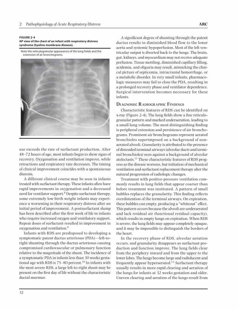
DIAGNOSIS: RADIOGRAPHIC FINDINGS
Characteristic features of RDS can be identified on x-ray (Figure 2-4). The lung fields show a fine reticulo-granular pattern and marked underaeration, leading to a small lung volume. The most distinguishing finding is peripheral extension and persistence of air broncho-grams. Prominent air bronchograms represent aerated bronchioles superimposed on a background of non-aerated alveoli. Granularity is attributed to the presence of distended terminal airways (alveolar ducts and termi-nal bronchioles) seen against a background of alveolar atelectasis.11 These characteristic features of RDS prog-ress as the disease worsens, but initiation of mechanical ventilation and surfactant replacement therapy alter the natural progression of radiologic changes.
Treatment with positive-pressure ventilation com-monly results in lung fields that appear coarser than before treatment was instituted. A pattern of small bubbles replaces the granularity. This finding reflects overdistention of the terminal airways. On expiration, these bubbles can empty, producing a “whiteout” effect. This pattern occurs because the alveoli are under aerated and lack residual air (functional residual capacity), which results in empty lungs on expiration. When RDS is severe, the lung fields may appear completely opaque, and it may be impossible to distinguish the borders of the heart.
In the recovery phase of RDS, alveolar aeration occurs, and granularity disappears as surfactant pro-duction and function improve. The lung fields clear from the periphery inward and from the upper to the lower lobes. The lungs become large and radiolucent and frequently appear hyperaerated.12 Surfactant therapy usually results in more rapid clearing and aeration of the lungs for infants at 32 weeks gestation and older. Uneven clearing and aeration of the lungs result from
FIGURE 24
AP view of the chest of an infant with respiratory distress
syndrome (hyaline membrane disease).
Note the reticulogranular appearance of the lung fields and the extension of air bronchograms.
ARC Pathophysiology of Acute Respiratory Distress 2
33
uneven distribution of the surfactant preparation.11 Some infants with RDS develop chronic lung disease following treatment with supplemental oxygen, posi-tive pressure ventilation, and surfactant replacement therapy. It may be difficult to distinguish the early x-ray findings in these infants from those of an infant in the recovery stages of RDS.
TREATMENT AND NURSING CARE
Therapy for infants with RDS begins with anticipation of the preterm birth and administration of antenatal cor-ticosteroids. Once the infant is born, therapy is directed at providing support for respiratory and cardiovascular insufficiency. Surfactant replacement therapy is rou-tinely used in many infants requiring intubation and mechanical ventilation. Immediate administration of appropriate therapy can be life saving. Preventing alveo-lar atelectasis, hypoxia, and hypercarbia are the main goals of therapy. General supportive measures must also be maximized. (See Chapter 4 for a detailed discussion of nursing care.) Maintenance of adequate oxygenation and ventilation are nursing care priorities. Meticulous attention must be paid to ensuring a thermoneutral environment. Fluid intake must be carefully balanced to avoid overload and complications related to a PDA. Acid-base disturbances, such as metabolic acidosis and respiratory acidosis, are frequently present in infants with RDS and require careful monitoring. Prophylactic antibiotic therapy may be used until the possibility of infection is ruled out.
Oxygen must be administered carefully to provide adequate amounts to tissues without risk of oxygen toxicity. (See Chapter 10 for a detailed discussion of complications of therapy.) An arterial oxygen tension (PaO2) between 50 and 70 torr is satisfactory for most infants. A high inspired oxygen concentration may be required to maintain the arterial oxygen tension within an acceptable range. Frequent or continuous monitor-ing of arterial blood gases is essential during the acute phase of the disease. Pulse oximeters provide noninva-sive means of obtaining immediate information on the infant’s oxygenation status. Surfactant replacement is a major component of treatment for infants with RDS. Natural surfactant preparations are administered via an endotracheal tube using a side port or catheter to deliver the drug into the trachea. (See Chapter 11 for a discus-sion of surfactant preparations.) Prophylactic exogenous surfactant replacement may be initiated shortly after birth in infants at risk for RDS. This approach means that some infants receive therapy when their disease is
mild or never develops. Prophylactic administration of surfactant is associated with a decreased risk of pneumo-thorax, pulmonary interstitial emphysema, and death. However, the risk of the infant’s developing a PDA and pulmonary hemorrhage increases.13 Some clinicians administer surfactant therapy as a rescue treatment once the diagnosis of RDS is confirmed. Infants requiring mechanical ventilation for respiratory distress shortly after birth have demonstrated a decreased incidence of chronic lung disease when surfactant was administered within the first two hours of life.14 Some infants with severe RDS may require multiple doses of surfactant. Others may be intubated only for administration of sur-factant and then extubated to nasal continuous positive airway pressure (NCPAP). The combination of surfactant therapy followed immediately by institution of NCPAP has been shown to shorten the duration of respiratory support and eliminate the need for later mechanical ven-tilation in some infants.15
Timely transfer of infants with RDS to a special care unit should be considered when the infant is born in a facility where staff lack experience in caring for low birth weight infants with multisystem problems. Surfactant replacement therapy requires a person skilled in intu-bation and management of mechanical ventilation. Nursing and respiratory therapy personnel must be available to monitor the infant constantly. Institutional protocols for surfactant therapy should be available. Routine use of surfactant replacement therapy in facili-ties without a full range of services and expertise is not recommended.16 Survival rates for very low birth weight infants are higher for those born in hospitals providing a high level of care to a high volume of sick infants.17
The decision to initiate ventilator therapy should be made on an individual basis. Variables that must be considered include birth weight, gestational age, post-natal age, results of the chest x-ray, progression of dis-ease, and blood gas values. More immature and smaller infants, who will have a greater incidence of fatigue and apnea, are more likely to require mechanical ventila-tion even when their oxygen requirements are low. The goal of ventilator therapy is to provide the most effec-tive gas exchange with the least risk of lung damage. Complications such as barotrauma, air leaks, oxygen toxicity, subglottic stenosis, pulmonary infections, cere-bral hemorrhage, and retinopathy of prematurity are known to occur with intubation and ventilation. (See Chapter 10.)
Approximately one-third of preterm infants with RDS develop chronic lung disease.18 However, rates
2 Pathophysiology of Acute Respiratory Distress ARC
34
of chronic lung disease vary widely among neonatal intensive care units.19 Use of conventional mechanical ventilation predisposes the infant with RDS to chronic lung disease as a result of lung injury from overdisten-tion. Elective high-frequency oscillatory ventilation as initial ventilatory support has been studied, but no significant overall reduction in chronic lung disease has been identified. Adverse effects on short- and long-term neurologic outcomes remain a concern with this approach.20 High-frequency oscillatory ventilation has been used to rescue preterm infants with severe RDS when conventional ventilation techniques have failed. However, there is concern that the benefit gained in terms of decreasing chronic lung disease is offset by the risk of an increase in the number and severity of intra-ventricular hemorrhages and the incidence of periven-tricular leukomalacia.21
A less severe form of chronic lung disease may be seen in low birth weight infants with only mild RDS. The cause of chronic lung disease in these infants is related to factors other than severity of the initial lung disease and need for mechanical ventilation with high inspired oxygen concentrations. Patent ductus arteriosus, noso-comial infection, and high fluid intake in the first days of life contribute to the development of chronic lung disease in infants with only mild RDS.22,23 Although
treatment options have increased since the mid-1980s, RDS continues to be a major problem for preterm infants. Advances in assisted reproductive technology have resulted in more multiple gestations. Since 1990, the rate of twin births has increased by 25 percent.24 The rising multiple-birth rate is contributing to an increase in the number of infants born preterm. Use of tocolytic agents coupled with antenatal steroid therapy is reduc-ing mortality, morbidity, and RDS in premature infants. However, preterm birth remains a major contributing factor for RDS. More research is needed to determine the best combination approach to treating RDS at spe-cific gestational ages and degrees of disease severity. Surfactant type, timing of surfactant administration, and ventilatory support options are key elements in developing better protocols for practice that will improve outcomes for infants with RDS.
TRANSIENT TACHYPNEA OF THE NEWBORN
Transient tachypnea of the newborn (TTN) represents one of the most common causes of respiratory distress in the immediate newborn period. Other names for TTN include wet lung disease and Type II respiratory distress syndrome.
ETIOLOGY AND PATHOPHYSIOLOGY
Delayed postnatal resorption of normal lung fluid is the most likely explanation for the clinical findings in infants with TTN. In utero, the fetus’s potential airways and air spaces are filled with fluid formed by the fetal lung (active Cl– [f luid] secretion). Resorption of fetal lung fluid begins with the onset of labor and its accom-panying catecholamine surge. In the mature lung, this catecholamine surge also initiates Na+ absorp-tion, which is enhanced by the increase in oxygen ten-sion.25 Lung fluid is also cleared before the first breath by the “thoracic squeeze” that occurs during vaginal delivery and by the pulmonary veins and lympha tics. Factors that predispose infants to wet lung disease include prematurity, cesarean section delivery without labor, breech delivery, hypervolemia, hypoproteinemia, maternal asthma, and prolonged maternal hypotonic fluid administration. Premature infants undergo less thoracic compression than term infants because their thoraxes are smaller. The normal thoracic squeeze is absent in infants delivered by cesarean section, result-ing in an increased volume of interstitial and alveolar fluid and a decreased thoracic gas volume during the first few hours after birth.26 More lung fluid is present in
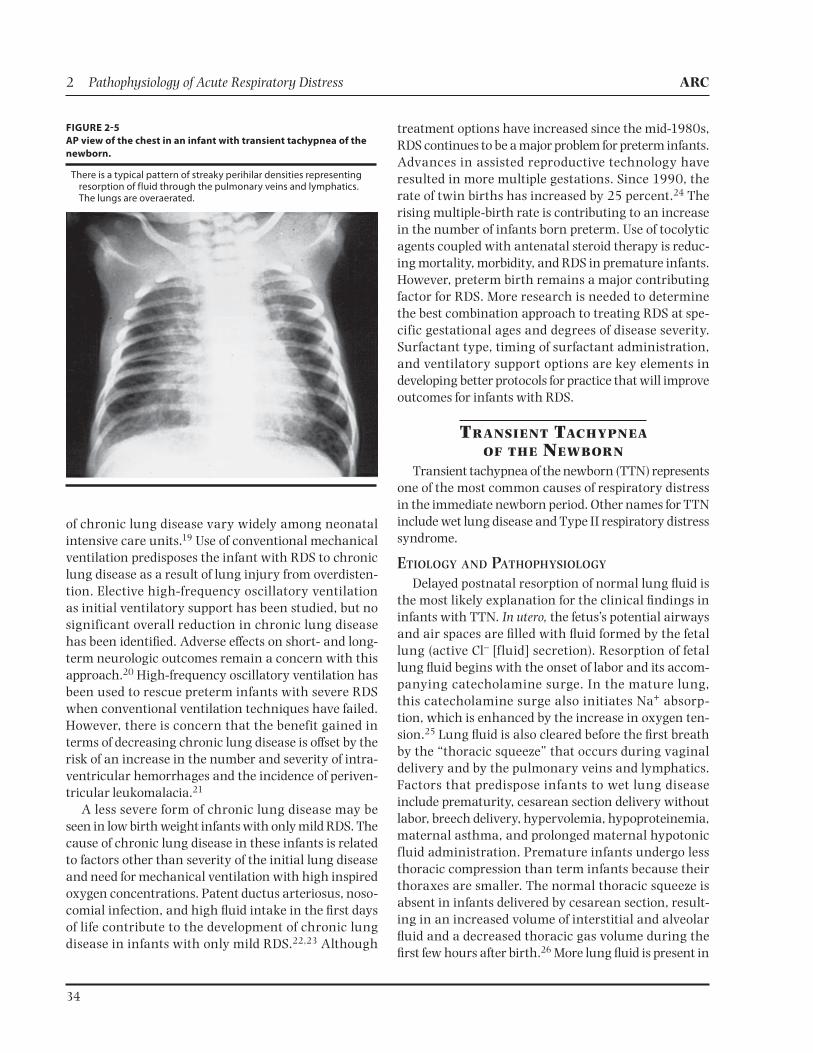
FIGURE 25
AP view of the chest in an infant with transient tachypnea of the
newborn.
There is a typical pattern of streaky perihilar densities representing resorption of fluid through the pulmonary veins and lymphatics. The lungs are overaerated.
ARC Pathophysiology of Acute Respiratory Distress 2
35
infants born by cesarean section without labor because lung fluid absorption begins in early labor. Premature infants are more hypoproteinemic than term infants. A lower plasma oncotic pressure may result in delayed resorption of lung fluid. Hypervolemia may increase capillary and lymphatic hydrostatic pressures. Elevated central pressure may result from placental transfusion and delayed clearance of lung fluid through the tho-racic duct. Maternal asthma is thought to affect the infant’s response to circulating catecholamines and to alter sodium transport and fluid resorption in the lung epithelium.27 Administration of hypotonic fluid to the mother results in a smaller osmotic gradient, reducing fluid resorption in the infant because less fluid is pulled from the lung.
An excess of interstitial fluid in the lung causes air trapping. The resulting hyperinflation is one mechanism that can raise pulmonary vascular resistance. When pulmonary vascular resistance is higher than systemic vascular resistance, the fetal pattern of circulation can occur, with shunting through the ductus arteriosus and foramen ovale. Severe hypoxemia results.28
CLINICAL PRESENTATION
Term and late preterm infants with TTN usually pres-ent with an increased respiratory rate and mild cyano-sis. Many of these infants are born by cesarean section. There is often a history of maternal sedation resulting in mild depression at birth. Substernal retractions and expiratory grunting may be present in varying degrees of severity. The clinical signs and symptoms of TTN may mimic those seen in the early phase of RDS or Group B streptococcal pneumonia. The diagnosis of TTN is usually made by excluding other, less benign, causes of respiratory distress. (See Figure 2-1 and DIAGNOSIS: RADIOGRAPHIC FINDINGS.)
The most common presentation of TTN is one in which the respiratory rate is normal for the first hour of life and gradually increases during the next 4–6 hours. The rate usually peaks between 6 and 36 hours of life, then gradually returns to normal by 48–72 hours. The maximum rate may reach 120 breaths per minute. Mild hypercarbia, hypoxemia, and acidosis may be present at 2–6 hours.29
Blood gases most frequently show a mild respiratory acidosis, which resolves within 8–24 hours. Retained lung fluid interferes with alveolar ventilation, resulting in hypercarbia. Maldistribution of ventilation and ongo-ing perfusion of nonventilated areas of the lung cause mild to moderate hypoxemia. Some infants may show
signs of mild pulmonary vascular lability; others may demonstrate more severe hypoxemia.30 Two distinct clinical presentations of TTN may be seen based on oxy-gen requirements. Infants with mild, or classical, TTN typically require less than 40 percent oxygen. Infants with severe disease need more than 60 percent oxygen and will have echocardiographic findings of pulmonary hypertension and right-to-left shunting.31
Physical examination may reveal a barrel-shaped chest. Consequently, subcostal retractions may be less prominent. As the respiratory symptoms improve, the chest resumes a more normal size. Retained lung fluid may obstruct the lower airways, resulting in overdisten-tion from a ball-valve effect. Grunting in infants with TTN may be associated with forced expiration as a result of partial airway obstruction from retained lung fluid rather than a means of increasing intra-alveolar pres-sure as lung compliance worsens.26
DIAGNOSIS: RADIOGRAPHIC FINDINGS
Because the presenting signs of TTN are commonly found in other neonatal respiratory diseases, the radio-graphic pattern becomes the key to diagnosis. The char-acteristic finding is prominent perihilar streaking and fluid in the interlobar fissures. The prominent perihilar streaking may represent engorgement of the periarte-rial lymphatics that function in the clearance of alveolar fluid. There may be small collections of liquid, particu-larly at the costophrenic angles. There is progressive clearing of lung fluid from the periphery to centrally and from upper to lower lung fields. Within 48–72 hours, the chest x-ray is normal.12
Hyperaeration of the lungs is evidenced by flattened hemidiaphragms and an increased anterior-posterior (AP) chest diameter. One factor differentiating infants with RDS from those with TTN is lung size. The lungs appear small and granular in infants with RDS; in those with TTN, the lungs are usually large and granular (Figure 2-5).
Clinicians rely on radiographic findings and the clinical presentation of the infant to diagnose TTN. A new approach to early diagnosis of TTN includes use of ultrasound. Differences in lung echogenicity between the upper and lower lung fields have been described in infants with TTN in the first hour after birth. In a study done by Copetti and Cattarossi, very compact comet-tail artifacts were seen in the inferior lung fields, but these were rare in the superior fields. This unique finding of “double lung point” was not observed in other common causes of neonatal respiratory distress such as RDS,
2 Pathophysiology of Acute Respiratory Distress ARC
36
pneumothorax, atelectasis, pneumonia, or pulmonary hemorrhage.32 Additional tools for diagnosis of TTN may include the early use of ultrasound as a means of ruling out other causes of neonatal respiratory distress.
TREATMENT AND NURSING CARE
TTN is usually a self-limited condition requiring supplemental oxygen and supportive care. Continuous positive airway pressure may be used in severe cases. (See Chapter 8.) Pulse oximetry allows noninvasive assessment of oxygenation. The infant should be care-fully monitored and maintained in a thermoneutral environment.
Fluid and electrolyte requirements should be met with intravenous fluids during the acute phase of the disease. Oral feedings are contraindicated because of rapid respiratory rates. If pneumonia is suspected ini-tially, antibiotics may be administered prophylactically. When hypoxemia is severe and tachypnea continues, persistent pulmonary hypertension may complicate the infant’s clinical condition, and aggressive medical man-agement may be required to break the cycle of hypox-emia (Figure 2-6).
NEONATAL PNEUMONIAPneumonia must be considered in every newborn
infant with asphyxia or respiratory distress at birth. Pneumonia is the most common neonatal infection, resulting in significant morbidity and mortality. Nearly 20 percent of all stillborn infants autopsied have a
congenitally acquired pulmonary infection.33 The mortality rate is approximately 20 percent for infants who have perinatally acquired pneumonia; the rate approaches 50 percent for those who acquire the infec-tion in the postnatal period.34 Recent declines in the inci-dence of Group B streptococcal disease have been linked to perinatal prevention strategies implemented in the 1990s. Although clinical guidelines for screening and treating colonized mothers have reduced the incidence of this disease, it remains a leading cause of morbidity and mortality.35
ETIOLOGY AND PATHOPHYSIOLOGY
Neonatal pneumonia can occur as part of a general-ized septicemia or as a primary infection. It is often dif-ficult to distinguish the two. Infectious agents include bacteria, viruses, protozoa, mycoplasmas, and fungi.
Pneumonia may be acquired in utero, during labor or delivery, or postnatally. Examination of the placenta and umbilical cord may provide the first evidence suggest-ing the presence of congenital pneumonia, which may result from the transplacental passage of organisms such as cytomegalovirus, herpes, varicella, and enterovirus. Listeria, Mycobacterium tuberculosis, and Treponema pallidum are less common agents.
Ascending infection from the maternal genital tract before or during labor is the more common route of contamination. The major predisposing factor is pro-longed rupture of fetal membranes, although bacteria can enter the amniotic fluid through intact membranes. Rupture of the membranes for more than 24 hours, excessive obstetric manipulation, prolonged labor with intact membranes, maternal urinary tract infection, and maternal fever have all been linked to congenital pneumonia.36 Fetal tachycardia and loss of beat-to-beat variability in the fetal heart rate pattern during labor may reflect the fetal response to infection.
Organisms that normally inhabit the maternal geni-tal tract are responsible for infecting the neonate at risk. Bacterial contamination of the infant always occurs dur-ing vaginal delivery. Organisms enter the oropharynx and gastrointestinal tract in utero when the fetus swal-lows contaminated amniotic fluid. Aspiration of con-taminated secretions present in the oropharynx may follow a complicated labor and delivery. Group B Strepto coccus (GBS), Escherichia coli, Klebsiella pneumoniae, and Enterococcus commonly cause infection in the neonate. The likelihood of neonatal pneumonia caused by GBS increases when an untreated, colo-nized mother has other risk factors such as prolonged
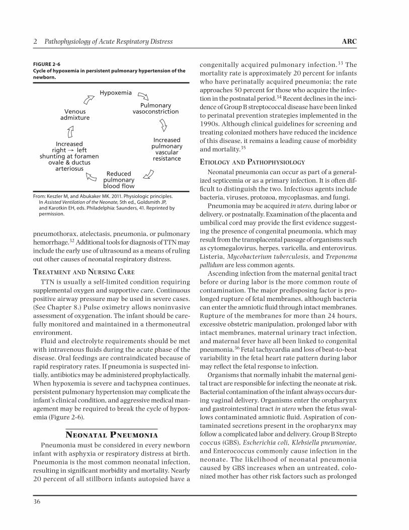
FIGURE 26
Cycle of hypoxemia in persistent pulmonary hypertension of the
newborn.
Hypoxemia
Venousadmixture
Increasedright left
shunting at foramenovale & ductus
arteriosusReduced
pulmonaryblood flow
Increasedpulmonary
vascularresistance
Pulmonaryvasoconstriction
From: Keszler M, and Abukaker MK. 2011. Physiologic principles. In Assisted Ventilation of the Neonate, 5th ed., Goldsmith JP, and Karotkin EH, eds. Philadelphia: Saunders, 41. Reprinted by permission.
ARC Pathophysiology of Acute Respiratory Distress 2
37
rupture of membranes, intrapartum fever, and signs of chorioamnionitis.37
Chlamydia, herpes simplex, and Candida albicans can infect the fetus during passage through the birth canal; however, manifestations of pneumonia may not appear until days after birth. Genital mycoplasmas are gaining increased recognition as a significant cause of perinatal infection.38
Ureaplasma urealyticum and Mycoplasma hominis may be transmitted vertically from the mother to the develop-ing fetus in utero or at delivery. U. urealyticum has been the agent most commonly linked with histologic cho-rioamnionitis and is also linked to the development of chronic lung disease in the low birth weight infant.39,40
Pneumonia during the postnatal period can also result from a nosocomial infection. The neonate may acquire pathogenic organisms by droplets spread from hospital personnel, other infected infants, or parents. Unwashed hands, contaminated blood products, infected human milk, and open skin lesions are recognized modes of transmitting various pathogens to the suscep-tible neonate.
Viral pneumonia caused by respiratory syncytial virus or adenovirus may occur in epidemic proportions in the intensive care unit. The most common nosocomial fungal infection is caused by C. albicans. Widespread use of broad-spectrum antibiotics and central lines places the very low birth weight infant at high risk for pulmo-nary candidiasis.
Immaturity of the lungs and immune system causes the neonate to be more susceptible to pulmonary infec-tion. An immature ciliary apparatus leads to suboptimal removal of inflammatory debris, mucus, and pathogens. In addition, the neonatal lung has an insufficient num-ber of pulmonary macrophages for intrapulmonary bacterial clearance.41 This is evidenced by a lack of sig-nificant pulmonary neutrophil accumulation, observed at postmortem examination, in neonates with pneumo-nia. The newborn infant has deficiencies in the neutro-phil inflammatory system, as shown by the frequency of neutropenia during serious infection, a high bacterial attack rate, and a high mortality rate.42
Infants who require admission to intensive care units are at higher risk for colonization of the upper respira-tory tract with pathogenic organisms than are those who are not admitted. Factors predisposing the NICU patient to pneumonia include liberal use of antibiotics, overcrowding and understaffing, invasive procedures such as endotracheal intubation and suctioning, con-taminated respiratory support equipment, and frequent
invasion of the protective skin barrier for blood sam-pling and parenteral fluid administration.43 The specific organisms that colonize the respiratory tracts of NICU infants are influenced by the choice of antibiotics rou-tinely used in that neonatal intensive care unit and the resident flora of the nursery. Airway colonization with organisms such as Pseudomonas aeruginosa, Klebsiella pneumoniae, Enterobacter cloacae, and Escherichia coli may be seen in very low birth weight infants requiring pro-longed mechanical ventilation. Some infants become col-onized with multiple Gram-negative and Gram-positive organisms. Asymptomatic infants may be colonized and not infected. However, ventilator-associated pneumo-nia (VAP) is more common in symptomatic, colonized infants with positive blood cultures.44 VAP leads to increased length of stay in the NICU and high mortality rates. The frequency of infection and the risk of death from VAP increase with decreasing gestational age.45
CLINICAL PRESENTATION
Clinical signs characteristic of neonatal pneumonia are nonspecific. Some infants with pneumonia demon-strate no pulmonary symptoms. More often, the pre-sentation includes subtle neurologic signs. The key to early diagnosis is a high index of suspicion. Temperature instability, lethargy, poor peripheral perfusion, apnea, tachycardia, and tachypnea are common early signs. The presence of tachypnea, cyanosis, grunting, retrac-tions, and nasal flaring focuses attention on the pulmo-nary system. These clinical signs indicative of possible pneumonia are also present in other causes of respira-tory distress. (See Figure 2-1)
More specific clinical signs, such as characteristic skin lesions, may be found in association with congeni-tal pneumonia caused by Candida, herpes simplex, or T. pallidum. Hepatosplenomegaly and jaundice suggest a congenital viral infection. Symptoms of intrapartal infection may be delayed for hours following aspira-tion of infected amniotic fluid because of the incubation period necessary before the onset of infection. In pre-term infants, it is often difficult initially to distinguish between pneumonia and RDS. Some at-risk infants may have pneumonia in combination with RDS or TTN.
DIAGNOSIS
The chest radiograph is important in detecting pneu-monia; however, appropriate bacterial and viral cultures are needed to identify the specific organism. Rapid screening tests allow earlier initiation of appropriate therapy.
2 Pathophysiology of Acute Respiratory Distress ARC
38
Laboratory TestsLatex agglutination assay of body fluids detects spe-
cific antigens and aids in rapid diagnosis of early neona-tal sepsis and pneumonia. It is recommended, however, that antigen test kits be used only as an adjunct to other diagnostic tests and not as a substitute for bacterial cul-ture.46 Blood cultures, which are usually positive in infants with congenital pneumonia, should be obtained on all infants with suspected pneumonia.47
The best indirect indication of congenital infection and pneumonia is the presence of bacteria on a Gram’s stain of a tracheal aspirate obtained during the first 8 hours of life.48 A culture of tracheal secretions obtained through a newly inserted endotracheal tube or by tra-cheal aspiration through a catheter under direct laryn-goscopy during the first 12 hours of life has proved useful in diagnosing neonatal bacterial pneumonia.47 Because of rapid colonization, results of tracheal cul-tures obtained later may be difficult to interpret. The most definitive method of diagnosis is culture and Gram’s stain of pleural fluid, but the procedural risks may result in increased morbidity and outweigh any benefits.
The neutrophil count is valuable in identifying infants with congenital pneumonia or septicemia. Neutropenia in the presence of respiratory distress during the first 72 hours of life suggests bacterial disease. In addition, an increase in the ratio of immature to total neutrophils on the leukocyte differential is frequently observed during neonatal infection.49
Radiographic FindingsChest x-ray examinations are required to support the
diagnosis of pneumonia and to distinguish it from other causes of respiratory distress. In some cases, no abnor-malities will be found if the studies are performed soon after the onset of symptoms, but radiologic diagnosis should be possible within 24–72 hours. Patchy opaci-fications become more impressive during subsequent days. In some infants, an area of radiopacification is present but may be attributed to atelectasis. Bilateral homogenous consolidation is a common finding when the pneumonia has been acquired in utero.
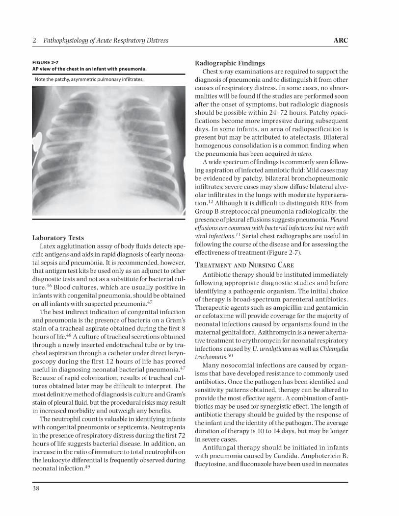
A wide spectrum of findings is commonly seen follow-ing aspiration of infected amniotic fluid: Mild cases may be evidenced by patchy, bilateral bronchopneumonic infiltrates; severe cases may show diffuse bilateral alve-olar infiltrates in the lungs with moderate hyperaera-tion.12 Although it is difficult to distinguish RDS from Group B streptococcal pneumonia radiologically, the presence of pleural effusions suggests pneumonia. Pleural effusions are common with bacterial infections but rare with viral infections.11 Serial chest radiographs are useful in following the course of the disease and for assessing the effectiveness of treatment (Figure 2-7).
TREATMENT AND NURSING CARE
Antibiotic therapy should be instituted immediately following appropriate diagnostic studies and before identifying a pathogenic organism. The initial choice of therapy is broad-spectrum parenteral antibiotics. Therapeutic agents such as ampicillin and gentamicin or cefotaxime will provide coverage for the majority of neonatal infections caused by organisms found in the maternal genital flora. Azithromycin is a newer alterna-tive treatment to erythromycin for neonatal respiratory infections caused by U. urealyticum as well as Chlamydia trachomatis.50
Many nosocomial infections are caused by organ-isms that have developed resistance to commonly used antibiotics. Once the pathogen has been identified and sensitivity patterns obtained, therapy can be altered to provide the most effective agent. A combination of anti-biotics may be used for synergistic effect. The length of antibiotic therapy should be guided by the response of the infant and the identity of the pathogen. The average duration of therapy is 10 to 14 days, but may be longer in severe cases.
Antifungal therapy should be initiated in infants with pneumonia caused by Candida. Amphotericin B, flucytosine, and fluconazole have been used in neonates
FIGURE 27
AP view of the chest in an infant with pneumonia.
Note the patchy, asymmetric pulmonary infiltrates.
ARC Pathophysiology of Acute Respiratory Distress 2
39
to treat fungal infections. Amphotericin B and flucyto-sine used in combination have a synergistic antifungal effect.51 Careful monitoring of renal and hepatic function is required during therapy.
Viral pathogens respond to a limited number of drugs. When herpes simplex infection is suspected, acyclovir or vidarabine should be used. A small number of infants with cytomegalovirus infection (CMV) have been treated with ganciclovir. Infants with congenital CMV infec-tion may have irreversible damage; those with acquired infection may show clinical improvements in respiratory status following treatment.52,53 More research is needed to determine the best treatment strategy given available antiviral drugs.54
In addition to antimicrobial therapy, the neonate with pneumonia requires careful monitoring of oxy-genation and acid-base status. Supplemental oxygen and ventilatory assistance are often necessary. Volume expanders, blood products, and buffers may be needed for the infant with cardiovascular collapse from septic shock. Exchange transfusion, granulocyte transfusion, and administration of intravenous gamma globulin have all been utilized in cases of overwhelming sepsis when conventional therapy has failed.55 Extracorporeal membrane oxygenation (ECMO) has also been used in attempts to improve survival rates in neonates with little chance of survival.56
MECONIUM ASPIRATION SYNDROMEThe passage of meconium by the fetus in utero is
estimated to occur in 8–29 percent of all deliveries.57 However, meconium passage is seen primarily in fetuses born at or beyond term and among those who are small for gestational age or have umbilical cord complications and compromised uteroplacental circulation. During breech deliveries, meconium passage is common and is often ignored.
When meconium-stained amniotic fluid is detected, careful and continuous monitoring of fetal well-being is required during labor. The passage of meconium into the amniotic fluid is considered a sign of fetal distress when accompanied by fetal heart rate abnormalities.58 Increased stillbirth and neonatal mortality rates have been associated with meconium staining. In the U.S., approximately 520,000 infants are born meconium stained annually. Five percent of these (about 26,000) develop meconium aspiration syndrome, and more than 4 percent (about 1,000) die from the disease. Approximately 30 percent of infants with meconium
aspiration syndrome (about 7,800) require mechanical ventilation. Pneumothoraces occur in at least 2,900 of those infants requiring mechanical ventilation.59 A decline in the number of postterm births has been iden-tified as the most important factor in reducing the inci-dence of meconium aspiration syndrome by one-third.60
ETIOLOGY AND PATHOPHYSIOLOGY
Meconium is first produced during the fifth month of gestation. It is free of bacteria and contains residuals of gastrointestinal secretions. The pathophysiologic stimuli that trigger the fetal passage of meconium are not clearly understood.
The following theories have been proposed to explain the relationship between fetal hypoxia and the passage of meconium in utero.57
-sion during the “diving reflex”
ischemia-
pression, resulting in increased peristalsis and anal sphincter dilation
Meconium passage in utero is considered by some to be a normal physiologic function of term and postterm fetuses, indicating fetal maturity.58 It is rarely observed in fetuses of less than 37 weeks gestation.
Fetal breathing movements occur in the healthy fetus at a rate of 30–70 times per minute. Normally, f luid from the airways moves out into the amniotic fluid with fetal respiratory movements. During an episode of fetal asphyxia, these movements cease, and apnea occurs. As the asphyxial episode continues, apnea is replaced by deep gasping. Amniotic fluid containing particulate material may be inhaled into the trachea and large bron-chi, and the infant may demonstrate airway obstruc-tion at birth. After the onset of air breathing, meconium migrates rapidly to the distal airways.
The amount of meconium passed into the amniotic fluid affects the appearance and viscosity of the fluid. Amniotic fluid containing meconium may have a light green tinge or the consistency and appearance of thick pea soup. Yellow, or “old,” meconium-stained fluid indi-cates prolonged fetal hypoxia and is an ominous sign.61
Mechanical obstruction of the airways with meco-nium particles results in a ball-valve phenomenon. Complete obstruction of the smaller airways results in atelectasis of alveoli distal to the obstruction. Partial airway obstruction results in areas of overexpansion as air passes around the obstruction to inf late the
2 Pathophysiology of Acute Respiratory Distress ARC
40
alveoli. As the airway collapses around the obstruc-tion during expiration, residual air becomes trapped distally. Pneumothorax occurs when the overdistended alveoli rupture and air leaks into the pleural space. Pneumomediastinum results when extra-alveolar air moves through interstitial tissue to the mediastinum.
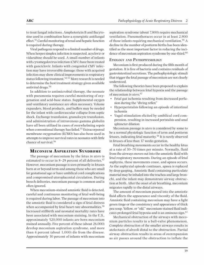
The chemical composition of meconium causes local toxic effects. Bile salts, pancreatic enzymes, desquamated intestinal epithelium, and biliverdin in meconium initi-ate a chemical pneumonitis that further compromises pulmonary function (Figure 2-8).62 Surfactant function is disrupted by serum and nonserum proteins and fatty acids, leading to atelectasis, decreased lung compliance, and hypoxia.63
CLINICAL PRESENTATION
Typically, an infant with meconium aspiration syn-drome has a history of fetal distress and meconium-stained amniotic fluid. The classic postmature infant shows signs of weight loss with little subcutaneous fat remaining. The umbilical cord may be thin, with mini-mal Wharton’s jelly. The nails, umbilical cord, and skin may be meconium stained. Respiratory distress at birth may be mild, moderate, or severe.
Tracheal occlusion by a meconium plug causes severe, gasping respirations; marked retractions; and poor air exchange. The severity of meconium aspiration syn-drome is related to the amount of aspirated meconium. In mild cases, hypoxemia is present but easily corrected with minimal oxygen therapy; tachypnea is present but usually resolves within 72 hours. A low partial pressure of carbon dioxide in arterial blood (PaCO2) and normal pH may be seen. Infants with moderate disease gradually worsen during the first 24 hours.
Severely affected infants have neurologic and respi-ratory depression at birth resulting from the hypoxic insult that precipitated the passage of meconium. They develop respiratory distress with cyanosis, nasal flaring, grunting, retracting, and tachypnea. The chest appears overinflated. Coarse crackles are common. Diminished breath sounds or heart tones may indicate a pulmonary air leak. Arterial blood gases typically show hypoxemia and acidosis. These infants have combined respiratory and metabolic acidosis secondary to respiratory failure and asphyxia. Because of large intrapulmonary shunts and persistence of fetal circulation patterns, hypoxemia is often profound despite administration of 100 percent oxygen.
DIAGNOSIS: RADIOGRAPHIC FINDINGS
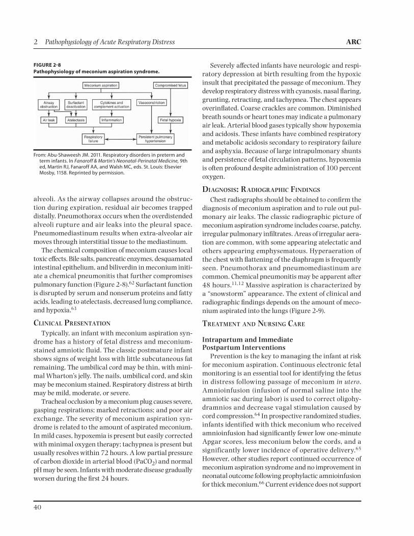
Chest radiographs should be obtained to confirm the diagnosis of meconium aspiration and to rule out pul-monary air leaks. The classic radiographic picture of meconium aspiration syndrome includes coarse, patchy, irregular pulmonary infiltrates. Areas of irregular aera-tion are common, with some appearing atelectatic and others appearing emphysematous. Hyperaeration of the chest with flattening of the diaphragm is frequently seen. Pneumothorax and pneumomediastinum are common. Chemical pneumonitis may be apparent after 48 hours.11,12 Massive aspiration is characterized by a “snowstorm” appearance. The extent of clinical and radiographic findings depends on the amount of meco-nium aspirated into the lungs (Figure 2-9).
TREATMENT AND NURSING CARE
Intrapartum and Immediate Postpartum Interventions
Prevention is the key to managing the infant at risk for meconium aspiration. Continuous electronic fetal monitoring is an essential tool for identifying the fetus in distress following passage of meconium in utero. Amnioinfusion (infusion of normal saline into the amniotic sac during labor) is used to correct oligohy-dramnios and decrease vagal stimulation caused by cord compression.64 In prospective randomized studies, infants identified with thick meconium who received amnioinfusion had significantly fewer low one-minute Apgar scores, less meconium below the cords, and a significantly lower incidence of operative delivery.65 However, other studies report continued occurrence of meconium aspiration syndrome and no improvement in neonatal outcome following prophylactic amnioinfusion for thick meconium.66 Current evidence does not support
FIGURE 28
Pathophysiology of meconium aspiration syndrome.
From: Abu-Shaweesh JM. 2011. Respiratory disorders in preterm and term infants. In Fanaroff & Martin’s Neonatal-Perinatal Medicine, 9th ed, Martin RJ, Fanaroff AA, and Walsh MC, eds. St. Louis: Elsevier Mosby, 1158. Reprinted by permission.
ARC Pathophysiology of Acute Respiratory Distress 2
41
routine use of amnioinfusion to dilute meconium stained amniotic fluid. Furthermore, the intervention requires systematic study in clinical trials.67 Several studies have demonstrated decreased mortality and morbidity when meconium is removed from the mouth, pharynx, and trachea before the onset of breathing.68–70 More recent evidence from a multicenter trial failed to show a positive effect from oropharyngeal and nasopharyngeal suction-ing before the delivery of the shoulders in meconium-stained infants.71 Current recommended practice does not include routine intrapartum suctioning of infants delivered through meconium-stained amniotic fluid.72,73
Some investigators have questioned the need for routine tracheal suctioning at the birth of meconium-stained infants who are delivered vaginally and have a one-minute Apgar score of more than 8. In a prospec-tive study, meconium-stained but vigorous infants who made their first inspiratory effort before being handed to the pediatrician did not benefit from immediate tra-cheal suctioning.74 Furthermore, case reports have dem-onstrated that aggressive airway management during and immediately after birth does not always prevent aspiration of meconium.75 The Neonatal Resuscitation Program guidelines recommend no tracheal suctioning for infants with strong respiratory efforts, good muscle tone, and a heart rate greater than 100 beats per minute. Direct tracheal suctioning is recommended for the meco-nium-stained infant with depressed respiratory effort, poor muscle tone, and a heart rate less than 100 beats per minute. This procedure should be accomplished before the infant makes repeated inspiratory efforts.
Universal precautions should be taken. Suctioning should always precede positive-pressure ventilation. Meconium aspirator devices and regulated wall suction should be utilized to effectively clear meconium from the airway. The urgent need for oxygenation and ventilation in these infants should not be ignored.76,77
Nursery ManagementSupportive respiratory therapy is required for infants
who develop meconium aspiration syndrome. The infant should be monitored continuously for tachypnea. Frequent assessment of blood gases is essential. The need for oxygen and assisted ventilation is dictated by arterial blood gas values. Continuous monitoring of oxygenation by pulse oximetry will alert the nurse to early deteriora-tion. Ventilatory assistance is indicated when adequate oxygenation cannot be achieved or maintained in a high concentration of oxygen. Respiratory failure commonly occurs in severe cases of meconium aspiration and may
necessitate prolonged assisted ventilation. Once the infant requires assisted ventilation, morbidity and mor-tality increase. Sedatives and neuromuscular blocking agents may be added to the therapeutic regime when the infant’s own ventilatory efforts interfere with the effec-tiveness of mechanical ventilation.
Gastric lavage is used to remove meconium-stained fluid from the stomach and reduce the chance of fur-ther aspiration with vomiting. There is no evidence from studies to support this practice.78 As noted under DIAGNOSIS: RADIOGRAPHIC FINDINGS, chest radiographs should be obtained to confirm the diagnosis of meco-nium aspiration and rule out pulmonary air leaks.
Chest physiotherapy (CPT) is used in many neonatal units to assist in mobilization of secretions and prevent accumulation of debris in the airway of neonates with respiratory distress. Percussion, vibration, and tracheal instillation of saline followed by suctioning are com-monly performed in the delivery room and nursery fol-lowing aspiration of meconium-stained amniotic fluid. There are no randomized controlled trials demonstrat-ing positive short- or long-term effects of this therapy in neonates. Some infants may show signs of acute clinical deterioration with further hypoxemia and the need for increased oxygen following chest physiotherapy. There
FIGURE 29
AP view of the chest in an infant with meconium aspiration
syndrome.
There are areas of patchy, asymmetric alveolar consolidation and volume loss in addition to areas of overexpansion resulting from obstruction (ball-valve effect). The lung fields are hyperexpanded.
2 Pathophysiology of Acute Respiratory Distress ARC
42
is insufficient evidence to support the use of chest phys-iotherapy for meconium aspiration syndrome.79
Broad-spectrum antibiotic therapy is indicated when infection is suspected. Appropriate cultures should be obtained before starting therapy. Prophylactic use of antibiotics is a common practice in infants with meco-nium aspiration syndrome because it is difficult to dis-tinguish on the chest radiograph from superimposed bacterial pneumonia. However, there is no evidence to suggest that prophylactic antibiotic therapy improves outcomes in nonventilated infants with meconium aspiration syndrome. No difference in duration of tachy-pnea, oxygen requirement, or need for NCPAP has been reported in a group of untreated, nonventilated infants with meconium aspiration syndrome. In the absence of perinatal risk factors for infection, these infants did not receive antibiotic therapy and had no evidence of bacte-remia, pneumonia, or meningitis.80
There is no reported increase in bacteremia among meconium-stained infants when compared to non-stained infants. The decision to use antibiotic therapy for these infants is based on each infant’s course.81
Surfactant replacement therapy early in the course of respiratory failure may reduce the severity of the dis-ease in some infants. Surfactant therapy has been shown to reduce pulmonary air leaks, duration of mechanical ventilation and oxygen therapy, as well as length of hos-pital stay.82 Further research is needed to determine the optimal timing, preparation, and method of surfactant administration for infants with meconium aspiration syndrome.
The infant should be carefully monitored for signs of seizure activity reflecting anoxic cerebral injury. Anticonvulsant therapy may be required. Metabolic derangements such as hypoglycemia and hypocalcemia require appropriate therapy and monitoring. Fluid bal-ance is critical in these infants because cerebral edema and inappropriate secretion of antidiuretic hormone often occur following an asphyxial insult. Fluid restric-tion may be initiated early in the course of the disease. Careful monitoring of urine output is essential in the postasphyxial stage. Hematuria, oliguria, and anuria may indicate anoxic renal damage.
Recovery from meconium aspiration syndrome usu-ally occurs within three to seven days in infants who do not require assisted ventilation. Those requiring assisted ventilation are usually ventilator dependent for three to seven days. Although the infant may be weaned success-fully from assisted ventilation, tachypnea may persist for weeks. Pulmonary air leaks, persistent pulmonary
hypertension, and pulmonary barotrauma often com-plicate the course of the disease. Prolonged ventilator therapy predisposes these infants to bronchopulmonary dysplasia with resulting oxygen dependency. More long-term deficits may be seen as sequelae of asphyxia.
The major cause of death in infants with meconium aspiration syndrome is respiratory failure. As noted earlier, surfactant replacement therapy may improve oxygenation and reduce the incidence of pulmonary air leaks. In some cases, however, the infant cannot be adequately oxygenated and ventilated with conventional respiratory support. Timely transfer to a tertiary level neonatal intensive care unit is essential. High-frequency ventilation and inhaled nitric oxide have been used for infants with respiratory failure and severe hypoxemia unresponsive to conventional mechanical ventilation. The combined use of surfactant, inhaled nitric oxide, and high-frequency oscillatory ventilation has resulted in a significant decrease in the need for the most invasive therapies such as ECMO.83,84
Careful consideration should be given before initiat-ing treatment with high-frequency oscillatory ventila-tion and inhaled nitric oxide in facilities where ECMO is not available. Collaborative agreements with an ECMO center and a mechanism for timely transport of the infant are recommended.85 Once nitric oxide therapy is initiated, transfer should take place without interruption of the treatment. A transport incubator equipped with a nitric oxide delivery system is required for these infants. Abrupt discontinuation of therapy can cause acute dete-rioration, with severe hypoxemia and possible death.86,87 When all other treatment options fail to reverse respi-ratory failure, ECMO has been used in many of these infants to improve survival.88
PERSISTENT PULMONARY HYPERTENSION OF THE NEWBORN
Persistent pulmonary hypertension of the newborn (PPHN) is a clinical syndrome characterized by cya-nosis secondary to shunting of unoxygenated blood through the ductus arteriosus and foramen ovale. Gersony and colleagues originally described this con-dition in infants with no parenchymal lung disease or cardiac lesion who developed central cyanosis shortly after birth; they applied the term “persistence of the fetal circulation” to these infants.89 Other terms have also been used to describe infants who, during the first few days of life, have cyanosis and respiratory disease, but no structural cardiac lesion. These monikers include
ARC Pathophysiology of Acute Respiratory Distress 2
43
progressive pulmonary hypertension, persistence of fetal cardiopulmonary circulation, and pulmonary vascular obstruction.
Because of the variable criteria used to define the syn-drome, the true incidence of PPHN is unknown. It was reported in 1.9 infants per 1,000 live births in a multi-center study, although rates as high as 6.8 per 1,000 live births were documented in one of the centers. Half of the infants had high-risk factors, including abnormal fetal heart rate tracings, meconium-stained amniotic fluid, and low Apgar scores. These infants frequently required delivery room interventions.90
ETIOLOGY AND PATHOPHYSIOLOGY
Although elevated pulmonary vascular resistance is the key pathophysiologic element in the syndrome, there is a wide spectrum of etiologies. Classification according to etiology helps us understand the pathophysiology and manage the condition.
Pulmonary artery pressure is the product of pulmo-nary blood flow and pulmonary vascular resistance. Most of infants with PPHN have elevated pulmonary vascular resistance; few have increased pulmonary blood flow as an important component of their PPHN. Pulmonary artery pressure may be equal to or greater than systemic arterial pressure in infants with PPHN. Right ventricular and right atrial pressures rise.
When right atrial pressure exceeds left atrial pressure and pulmonary arterial pressure is greater than sys-temic pressure, blood flow changes to follow the path of least resistance. Desaturated blood returning to the right heart is shunted into the systemic circulation through the foramen ovale and ductus arteriosus. This right-to-left shunt causes hypoxemia secondary to venous admixture. Hypoxemia increases pulmonary vasocon-striction, and the cycle continues (see Figure 2-6).
Persistent pulmonary hypertension may occur in association with a wide spectrum of neonatal diseases (Table 2-2). Gersony classifies the causes of pulmonary hypertension in terms of cardiopulmonary pathophysi-ology as follows: (1) pulmonary venous hypertension, (2) functional obstruction of the pulmonary vascular bed, (3) pulmonary vascular constriction, (4) decreased pulmonary vascular bed, and (5) increased pulmonary blood flow.91
The time period during which pulmonary vaso-constriction occurs may clarify the pathophysiol-ogy of PPHN. Etiologies can be categorized into intra uterine, intrapartum, and postpartum periods. The terms primary, or idiopathic, and secondary have also been
used to describe PPHN. Regardless of the classification used, it is essential to understand that a combination of etiologies may be responsible for PPHN. Many infants with PPHN also have a parenchymal lung disease caus-ing intrapulmonary shunting. Meconium aspiration syndrome, bacterial pneumonia, or surfactant deficiency syndrome may be the primary disease leading to the development of PPHN.
CLINICAL PRESENTATION
Infants presenting with clinical evidence of PPHN are usually more than 32 weeks gestational age and born following complications of pregnancy, labor, or delivery. The syndrome occurs most commonly in term or post-term infants following an intrauterine or intrapartum asphyxial episode. Onset of symptoms is usually imme-diate in infants with congenital diaphragmatic hernia or severe asphyxia. Others may have a more subtle pre-sentation, but most infants at risk have clinical manifes-tations before 24 hours of age. Clinical symptoms may initially be indistinguishable from those of cyanotic con-genital heart disease.
There is marked variability in the clinical course of PPHN. Evidence of respiratory distress may be mild to severe. Signs of heart failure may be present in more adversely affected infants. Central cyanosis may be present despite a high inspired oxygen concentration. Arterial blood gases reveal severe hypoxemia and meta-bolic acidosis. Arterial PaO2 values or oxygen saturation values may fluctuate widely when the infant is handled or stressed. The PaCO2 is usually normal but may be mildly elevated. Physical examination reveals varying degrees of respiratory distress and cyanosis.
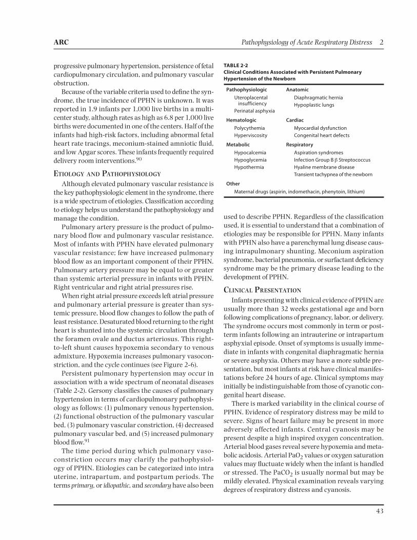
TABLE 22
Clinical Conditions Associated with Persistent Pulmonary
Hypertension of the Newborn
Pathophysiologic
Uteroplacental insufficiency
Perinatal asphyxia
Anatomic
Diaphragmatic herniaHypoplastic lungs
Hematologic
PolycythemiaHyperviscosity
Cardiac
Myocardial dysfunctionCongenital heart defects
Metabolic
HypocalcemiaHypoglycemiaHypothermia
Respiratory
Aspiration syndromesInfection Group B StreptococcusHyaline membrane diseaseTransient tachypnea of the newborn
Other
Maternal drugs (aspirin, indomethacin, phenytoin, lithium)
2 Pathophysiology of Acute Respiratory Distress ARC
44
A single, loud, second heart sound or a narrowly split second heart sound with a loud pulmonic component is heard. A long, harsh systolic murmur may be heard at the lower left sternal border. This murmur is the result of tricuspid insufficiency. Inspection of the chest reveals a hyperactive precordium with a prominent right ven-tricular impulse that is visible or easily palpable at the lower left sternal border.
The chest may be barrel-shaped following aspira-tion of meconium or the use of high positive inflating pressures with mechanical ventilation. Retractions are present when pulmonary compliance is decreased. Peripheral perfusion is often poor, and pulses are diminished.
DIAGNOSIS
Radiographic FindingsThere is no classic x-ray finding in PPHN because
the etiologies are varied. The chest radiograph may show normal or decreased pulmonary vascular mark-ings. When the syndrome is complicated by pulmonary disease, such as meconium aspiration, pneumonia, or hyaline membrane disease, the x-ray findings will reflect the primary pulmonary disorder. Cardiomegaly is a fre-quent finding on the initial chest x-ray and may be pres-ent without clinically detectable cardiac dysfunction.92 The more severely affected infants with PPHN may show signs of heart failure. Pleural effusions, pulmonary venous congestion, and marked cardiomegaly may be seen when there is myocardial dysfunction.
Diagnostic WorkupPPHN should be suspected in any infant who has
hypoxemia that is out of proportion to the severity of lung disease present. Parenchymal lung disease is the most common etiology of hypoxemia. However, per-sistent pulmonary hypertension often complicates the clinical course of infants with primary lung disease.
Differential diagnosis includes, most importantly, cyanotic heart disease. A series of noninvasive bedside tests can be performed using arterial blood gas determi-nations to differentiate between cyanotic heart disease and pulmonary parenchymal disease. These include the hyperoxia test, preductal and postductal arterial blood sampling, and echocardiography.93 Pulse oximetry monitors can also be used to follow trends in oxygen saturation levels.
The hyperoxia test is used in term infants to differ-entiate between the fixed right-to-left shunt in congeni-tal heart disease or PPHN and a ventilation-perfusion mismatch as seen in parenchymal lung disease. The infant is placed in a 100 percent oxygen concentration for five to ten minutes before an arterial oxygen pres-sure is determined. If a ventilation-perfusion problem is the cause of the hypoxemia, oxygen will diffuse into the poorly ventilated areas of the lung, and the PaO2 will usually rise above 100 mmHg. A right-to-left shunt is demonstrated when the PaO2 remains low in 100 per-cent oxygen. However, this shunt may be secondary to congenital heart disease or PPHN. Further evaluation is needed to determine if the right-to-left shunt is occur-ring at the ductal level.
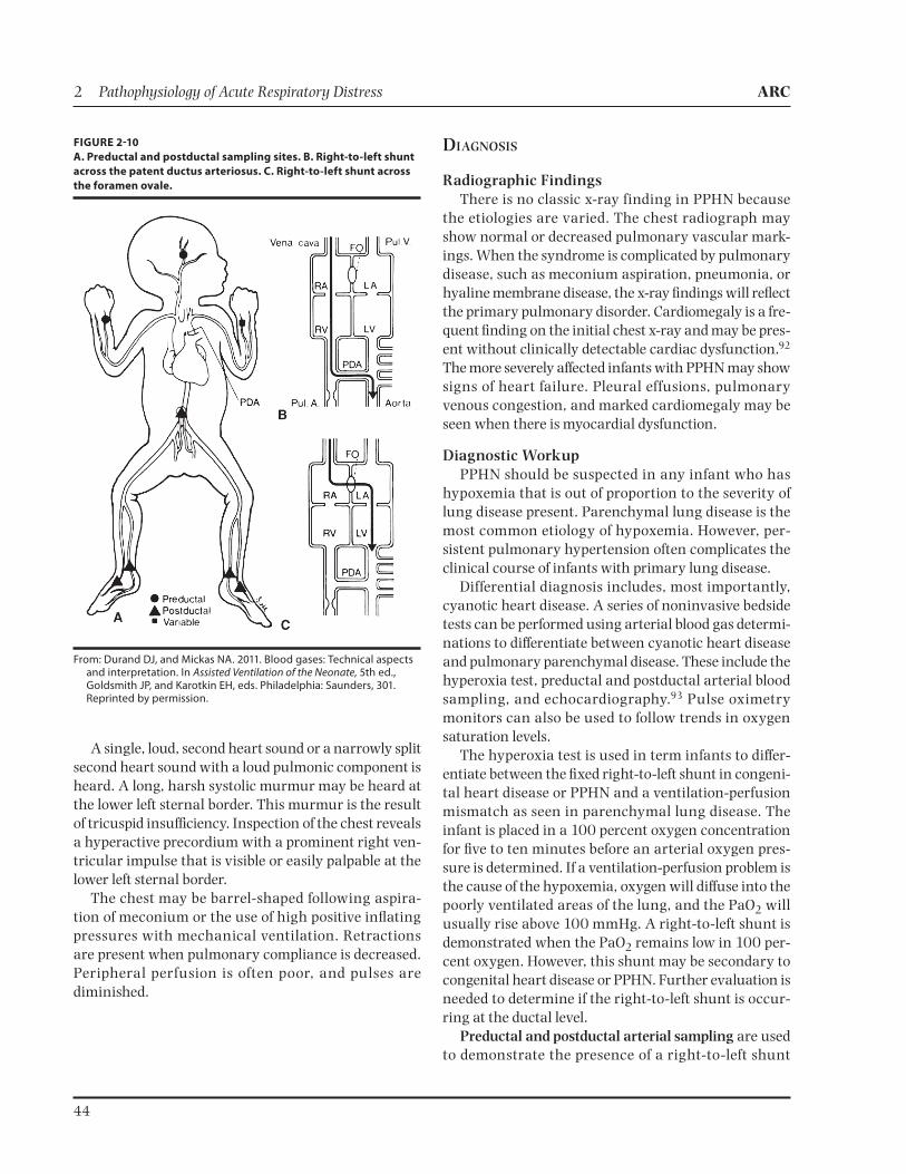
Preductal and postductal arterial sampling are used to demonstrate the presence of a right-to-left shunt
FIGURE 210
A. Preductal and postductal sampling sites. B. Right-to-left shunt
across the patent ductus arteriosus. C. Right-to-left shunt across
the foramen ovale.
From: Durand DJ, and Mickas NA. 2011. Blood gases: Technical aspects and interpretation. In Assisted Ventilation of the Neonate, 5th ed., Goldsmith JP, and Karotkin EH, eds. Philadelphia: Saunders, 301. Reprinted by permission.
ARC Pathophysiology of Acute Respiratory Distress 2
45
through the ductus arteriosus. Preductal samples can be obtained from the right radial or either temporal artery; postductal sites most frequently sampled include the umbilical, femoral, and posterior tibial arteries (Figure 2-10). The left radial artery may represent a mixture of preductal and postductal blood because of the proxim-ity of the left subclavian artery to the ductus arteriosus.
Preductal and postductal arterial blood samples must be obtained simultaneously from the quiet infant if they are to be considered reliable. Strategic placement of two pulse oximeters can aid in determining the presence of a shunt. Preductal pulse oximeter readings can be obtained by placing the probe on the right hand; either foot can be used to obtain postductal oxygen saturation readings. In the hypoxemic infant, a PaO2 difference greater than 15–20 mmHg indicates significant right-to-left shunting at the ductal level. If the test reveals no difference in PaO2 between preductal and postductal sites, pulmonary hypertension cannot yet be ruled out because shunting may be primarily at the atrial level (see Figure 2-10). Additional testing is needed to differentiate between PPHN and cyanotic heart disease.
Echocardiography is used to confirm the presence of a structurally normal heart in infants with PPHN. It can also be used to measure the ratio of the systolic time intervals of the right ventricle: The ratio of the right ventricular pre-ejection period to the right ventricular ejection time is elevated in infants with pulmonary hypertension.94 Two-dimensional echocardiography with color Doppler flow can be used to define the direc-tion and location of shunting through the foramen ovale or the ductus arteriosus. The degree of pulmonary hypertension can also be estimated. When myocardial ischemia is present, an electrocardiogram shows ST segment depression. Invasive diagnostic tests such as cardiac catheterization and pulmonary artery pressure monitoring are rarely needed to make the diagnosis of PPHN.
TREATMENT AND NURSING CARE
When the fetus has been identified to be at risk for persistent pulmonary hypertension, the first step in prevention is skilled resuscitation and stabilization. Preventing hypoxemia, acidosis, and hypothermia dur-ing the immediate newborn period is essential. The time, site, and delivery route of the fetus with a known risk factor for PPHN, such as congenital diaphragmatic hernia, may be scheduled to minimize intrapartum and postnatal stress.
The aim of therapy for infants with PPHN is to cor-rect hypoxemia by reversing right-to-left shunting. This is accomplished by decreasing pulmonary artery pres-sure or elevating the systemic arterial blood pressure. Treatment is often complex and includes mechanical ventilation, drug therapy, and supportive care.
Mechanical VentilationTreatment with mechanical ventilation should be
individualized based on the underlying cause of pul-monary hypertension. The main goal is to improve oxygenation. Initially, the fraction of inspired oxygen (FiO2) should be increased until the PaO2 is greater than 50 mmHg postductally. In most cases, the infant will require an FiO2 of 0.70 or more to maintain a PaO2 of 50 mmHg or greater. Mechanical ventilation is most effec-tive when it is begun early in the course of the disease. Ventilator management strategies may include use of conventional mechanical ventilation, high-frequency jet ventilation, or high-frequency oscillatory ventilation. Ventilation strategies are modified when parenchymal lung disease is identified as the etiology of the infant’s pulmonary hypertension. If the infant fails to respond to initial ventilator therapy, adjunct therapies may include surfactant administration or inhaled nitric oxide. Surfactant deficiency as well as surfactant inactivation can lead to pulmonary hypertension. A combination of surfactant therapy and high-frequency ventilation may be required for some infants with pulmonary hypertension caused by respiratory distress syndrome or meconium aspiration syndrome. A different ventila-tor strategy may be required for those with pulmonary hypertension due to lung hypoplasia.
Mechanical hyperventilation using high rates and high inspiratory pressure to induce hypocarbia has been widely used in infants to improve oxygen transfer into the blood, when there is evidence of pulmonary hyper-tension. Each infant has a critical level of PaCO2 at which optimum oxygenation occurs because of a decrease in pulmonary vascular resistance and in right-to-left shunting.93 However, significant reductions in carbon dioxide levels in the blood can have adverse effects on the neonate, and there are many unanswered ques-tions regarding the risk to benefit ratio of this therapy. Induced hypocarbia and alkalosis shift the oxygen-hemoglobin dissociation curve farther to the left, which reduces oxygen release at the tissue level. Venous blood return to the heart is impeded, and cardiac output is reduced when extremely high inspiratory pressure and ventilatory rates are used. Hypotension and reduced
2 Pathophysiology of Acute Respiratory Distress ARC
46
cardiac output cause a further reduction in oxygen-ation. Induced hypocarbia can diminish cerebral blood flow and increase cerebrospinal fluid lactate levels.95 The degree and duration of hypocarbia have been linked to poor neurologic outcomes in preterm and term infants managed by hyperventilation. The worst outcomes were seen in infants who were hyperventilated twice as long as infants without abnormalities. Affected infants spent a significantly greater time in an alkalotic state with their PaCO2 less than 25 mmHg.96 Periventricular leu-komalacia, cerebral palsy, abnormal cognitive develop-ment, hearing loss, and chronic lung disease have been identified as detrimental effects of hyperventilation in neonates.97 Many clinicians have abandoned this treat-ment strategy because of ventilator-induced lung injury and adverse cerebral effects.98,99
The use of hyperventilation in management of the infant with PPHN has also decreased since the introduc-tion of nitric oxide therapy. However, when nitric oxide therapy is not available, a variety of ventilation tech-niques may be used in attempts to stabilize the infant until transport can occur.
Some clinicians prefer a gentle ventilation approach over hyperventilation. The goal of this approach is to minimize barotrauma while maintaining a PaO2 between 50 and 70 mmHg. PaCO2 is maintained in the 40–60 mmHg range. The appropriate peak inspiratory pressure is determined by clinical assessment of chest excursion. This conservative approach has been used successfully to manage a group of infants with PPHN and severe respiratory failure.100 Others have success-fully treated infants with a combined approach using gentle ventilation and inhaled nitric oxide.101
The risk of barotrauma and pulmonary air leaks during conventional ventilator therapy may lead some clinicians to choose high-frequency ventilators as an alternative method to achieve adequate lung inflation. (See Chapter 12.) The goal is to optimally inflate the lungs and lower carbon dioxide levels while using lower proximal airway pressures. However, the risk of extreme hyperventilation is present. Once adequate lung volume is achieved with high-frequency ventilation, improved response to supplemental therapies such as surfactant and inhaled nitric oxide may be seen.102
Infants with PPHN often demonstrate extreme labil-ity. Regardless of the ventilation strategy used, weaning should be done cautiously while the pulmonary vascula-ture is reactive because aggressive decreases in FiO2 may result in pulmonary vasospasm and sudden hypoxemia. Oxygen concentration is decreased cautiously, 1 percent
at a time. If the PaO2 remains greater than 120 mmHg, weaning should continue. High peak inflating pres-sure should also be decreased cautiously, provided that carbon dioxide levels remain within the desired range and adequate oxygenation is maintained. Many infants receiving inhaled nitric oxide therapy show a significant decrease in the amount of ventilator support required to maintain adequate oxygenation.103
The transitional phase of PPHN is the point in the dis-ease process when the hypoxemia no longer results from pulmonary artery hypertension but from chronic paren-chymal lung disease.104 This change usually occurs at 3 to 5 days of age. During the transition phase, PaCO2 can be allowed to rise by decreasing ventilator settings. Caution must be taken to ensure that no sudden decline in PaO2 occurs. Failure to wean from high pressures and rates during the transition phase can result in severe barotrauma. Each infant must be carefully evaluated to determine the best ventilatory approach to reverse the severe hypoxemia while causing the least amount of harm.
Drug TherapyA variety of pharmacologic agents has been used
in managing PPHN. Most infants receive some combi-nation of analgesia and sedation during the course of mechanical ventilation; however, there is no accepted standard to guide clinical practice. Sedation may be utilized early in the course of treatment if the infant’s spontaneous respiratory efforts are not synchronous with the ventilator. Intravenous midazolam infusions for sedation of infants requiring mechanical ventilation are commonly used in the NICU. Data from a review of ran-domized, controlled trials indicate infants treated with midazolam had a statistically higher incidence of hypo-tension, adverse neurologic events, and a longer length of stay in the NICU.105 There are no studies comparing outcomes of preterm and term infants with a specific diagnosis of PPHN who were sedated with midazolam. Further research is needed to determine whether identi-fied risks outweigh the benefits infants may receive from this therapy.
Opioids are effective in reducing behavioral and physiologic indicators of pain and stress in mechani-cally ventilated infants.106 Morphine or fentanyl is commonly administered in bolus doses followed by a continuous infusion. Morphine sulfate is administered to decrease the infant’s spontaneous activity and resis-tance to controlled ventilation. Careful blood pressure monitoring is essential because systemic hypotension is
ARC Pathophysiology of Acute Respiratory Distress 2
47
a known adverse effect and can worsen PPHN. Fentanyl is useful in infants with PPHN because it prevents pain-induced increases in pulmonary vascular resistance. Hemodynamic stability is maintained in infants receiv-ing fentanyl because the drug causes less histamine release than morphine.107
Skeletal muscle paralysis may be pharmacologically induced with agents such as pancuronium bromide when sedation fails to produce a desired improvement in oxygenation and ventilation. Meticulous nursing care and continuous assessment of all bodily functions are required when neuromuscular blocking agents are used.
Volume expanders and pressor agents may be required to maintain normal blood pressure. When vascular volume has been restored and hypotension still exists, dopamine may be utilized to increase myo-cardial contractility, cardiac output, and blood pres-sure. Improvement in oxygenation may be seen when systemic blood pressure is greater than pulmonary blood pressure. Dobutamine is sometimes used in combination therapy with dopamine.
Surfactant replacement therapy is used when the PPHN is complicated by the coexistence of parenchy-mal lung disease. Early administration of surfactant may reduce the need for more invasive interventions.
Before inhaled nitric oxide was available, success with pharmacologic therapy for infants with PPHN was varied and unpredictable. No available drug had selective pulmonary vasodilator effects. Side effects such as systemic hypotension caused further deterioration. Critically ill infants with severe hypoxemia and evidence of pulmonary hypertension respond to inhaled nitric oxide with significant improvement in oxygenation. (See Chapter 13.) The need for further treatment with ECMO is reduced when inhaled nitric oxide is used to interrupt the cycle of hypoxemia.84 Other novel therapies have been studied in centers where nitric oxide therapy was not available. A pilot study of sildenafil use in infants with PPHN demonstrated steady improvements in oxy-genation with no detrimental effect on blood pressure.108 However, there have been no large clinical trials to deter-mine the safety, efficacy, and long-term effects of silde-nafil use in the neonatal population.109 More research is needed before sildenafil can be considered as standard therapy in treatment of PPHN in countries where nitric oxide is available.
Supportive CareProtocols for minimal stimulation are utilized for
infants with PPHN in many neonatal centers. The infant
may be secluded in a quiet, darkened room with restric-tions on caregivers and visitors. The infant’s sensitiv-ity to noise and handling during the acute stage of the disease is manifested by sudden and prolonged periods of hypoxia. Nursing care should be organized and coor-dinated to prevent unnecessary disturbances.
Pulse oximetry may be utilized simultaneously at pre-ductal and postductal sites. Continuous arterial blood pressure monitoring is imperative. In term infants, the mean systemic arterial pressure should be main-tained above 50 mmHg. Maintaining systolic pressures between 60 and 80 mmHg reduces the systemic to pulmonary pressure gradient, resulting in a decreased right-to-left shunt.72 Vasopressor therapy is usually required to attain this.
Fluid balance must be maintained to ensure ade-quate intravascular volume and blood pressure. Central venous pressure monitoring may aid in determining adequacy of fluid replacement. General nursing care measures to ensure maintenance of skin integrity are essential because these infants may not tolerate frequent position changes.
Despite ventilatory, pharmacologic, and supportive therapies, some infants do not survive. Others have been saved with ECMO therapy.110 Long-term follow-up care is necessary for all survivors because they are at increased risk for abnormal neurodevelopmental outcomes, includ-ing cognitive delays and hearing loss.111 Whatever the treatment, the outcome for infants with PPHN varies according to the etiology and severity of the disease. The optimal approach to treating the infant with PPHN remains to be determined through clinical trials.
SUMMARYMost infants admitted to neonatal intensive care units
have respiratory disorders. Understanding the patho-physiology associated with each disease process is essen-tial to ensure timely and comprehensive management. Knowledge of clinical presentation and etiology in rela-tion to gestational age assists in differential diagnosis. The goal of therapy for neonatal respiratory disorders is the maintenance of adequate oxygenation and ventila-tion. Advances in antenatal care affect the prematurity rate and lessen the incidence of respiratory disorders related to immaturity. Technologic advances including exogenous surfactant, new modes of ventilation, ECMO, and inhaled nitric oxide therapy have improved out-comes for many infants with severe respiratory disease.
2 Pathophysiology of Acute Respiratory Distress ARC
48
REFERENCES 1. Raja JU. 1988. Hyaline membrane disease. In Neonatal Cardiopulmonary
Distress, Emmanouilides GC, and Baylen G, eds. Chicago: Year Book Medical Publishers, 54–55.
2. Heron M. 2011. Deaths: Leading causes for 2007. National Vital Statistics Reports 59(8): 1–95.
3. Lee K, et al. 1999. Trend in mortality from respiratory distress syndrome in the United States, 1970–1995. Journal of Pediatrics 134(4): 434–440.
4. Hack M, et al. 2000. Neurodevelopment and predictors of outcomes of children with birth weights of less than 1000 g: 1992–1995. Archives in Pediatric and Adolescent Medicine 154(7): 725–731.
5. National Institutes of Health. 1994. Effect of corticosteroids for fetal maturation on perinatal outcomes. NIH Consensus Statement 12(2): 1–24.
6. National Institutes of Health. 2000. Antenatal corticosteroids revisited: Repeat courses. NIH Consensus Statement 17(2): 1–18.
7. Crowther CA, and Harding JE. 2003. Repeat doses of prenatal corticosteroids for women at risk of preterm birth for preventing neonatal respiratory disease. Cochrane Database of Systematic Reviews (2): CD003935.
8. Suresh GK, and Soll RF. 2005. Overview of surfactant replacement trials. Journal of Perinatology 25(supplement 2): S40–S44.
9. Katz LA, and Klein JM. 2006. Repeat surfactant therapy for postsurfactant slump. Journal of Perinatology 26(7): 414–422.
10. Cotton RB. 1987. The relationship of symptomatic patent ductus arteriosus to respiratory distress in preterm newborn infants. Clinics in Perinatology 14(3): 621–633.
11. Swischuk LE. 2004. Imaging of the Newborn Infant and Young Child, 5th ed. Philadelphia: Lippincott Williams & Wilkins, 31, 41, 45.
12. Wesenberg RL. 1973. The Newborn Chest. Hagerstown, Maryland: Harper & Row, 45–46, 62, 74–83, 119–124.
13. Soll RF. 1998. Prophylactic synthetic surfactant for preventing morbidity and mortality in preterm infants. Cochrane Database of Systematic Reviews (3): CD001079.
14. Yost CC, and Soll RF. 1999. Early versus delayed selective surfactant treatment for neonatal respiratory distress syndrome. Cochrane Database of Systematic Reviews (3): CD001456.
15. Dani C, et al. 2004. Early extubation and nasal continuous positive airway pressure after surfactant treatment for respiratory distress syndrome among preterm infants <30 weeks’ gestation. Pediatrics 113(6): e560–e563.
16. American Academy of Pediatrics, Committee of Fetus and Newborn. 1999. Surfactant replacement therapy for respiratory distress syndrome. Pediatrics 103(3): 684–685.
17. Phibbs CS, et al. 2007. Level and volume of neonatal intensive care and mortality in very-low-birth-weight infants. New England Journal of Medicine 356 (21): 2165–2175.
18. Northway WH Jr. 1992. An introduction to bronchopulmonary dysplasia. Clinics in Perinatology 19(3): 489–495.
19. Payne NR, et al. Reduction of bronchopulmonary dysplasia after participation in the Breathsavers Group of the Vermont Oxford Network Neonatal Intensive Care Quality Improvement Collaborative. Pediatrics 118(supplement 2): S73–S77.
20. Henderson-Smart DJ, et al. 2003. Elective high frequency oscillatory ventilation versus conventional ventilation for acute pulmonary dysfunction in preterm infants. Cochrane Database of Systematic Reviews (4): CD000104.
21. Bhuta T, and Henderson-Smart DJ. 1998. Rescue high frequency oscillatory ventilation versus conventional ventilation for pulmonary dysfunction in preterm infants. Cochrane Database of Systematic Reviews (3): CD000438.
22. Rojas MA, et al. 1995. Changing trends in the epidemiology and pathogenesis of neonatal chronic lung disease. Journal of Pediatrics 126(4): 605–610.
23. Marshall DD, et al. 1999. Risk factors for chronic lung disease in the surfactant era: A North Carolina population-based study of very low birth weight infants. Pediatrics 104(6): 1345–1350.
24. Martin JA, and Park MM. 1999. Trends in twin and triplet births: 1980–1997. National Vital Statistics Reports 47(24): 1–16.
25. Bland RD. 2005. Lung fluid balance during development. NeoReviews 6(6): e255–e267.
26. Milner AD, Saunders RA, and Hopkin IE. 1978. Effects of delivery by caesarean section on lung mechanics and lung volume in the human neonate. Archives of Disease in Childhood 53(7): 545–548.
27. Demissie K, et al. 1998. Maternal asthma and transient tachypnea of the newborn. Pediatrics 102(1 part 1): 84–90.
28. Bucciarelli RL, et al. 1976. Persistence of fetal cardiopulmonary circulation: One manifestation of transient tachypnea of the newborn. Pediatrics 58(2): 192–197.
29. Sundell H, et al. 1971. Studies on infants with Type II respiratory distress syndrome. Journal of Pediatrics 78(5): 754–764.
30. Bonta BW. 1988. Transient pulmonary vascular lability: A form of mild pulmonary hypertension of the newborn not requiring mechanical ventilation. Journal of Perinatology 8(1): 19–23.
31. Halliday HL, McClure G, and Reid MM. 1981. Transient tachypnoea of the newborn: Two distinct clinical entities? Archives of Disease in Childhood 56(5): 322–325.
32. Copetti R, and Cattarossi L. 2007. The “double lung point”: An ultrasound sign diagnostic of transient tachypnea of the newborn. Neonatology 91(3): 203–209.
33. Merritt TA. 1984. Respiratory distress. In Assessment of the Newborn: A Guide for the Practitioner, Ziai M, Clark T, and Merritt TA, eds. Boston: Little, Brown, 168.
34. Dennehy PH. 1987. Respiratory infections in the newborn. Clinics in Perinatology 14(3): 667–682.
35. Puopolo KM, Madoff LC, and Eichenwald EC. 2005. Early-onset group B streptococcal disease in the era of maternal screening. Pediatrics 115(5): 1240–1246.
36 Schrag S, and Schuchat A. 2005. Prevention of neonatal sepsis. Clinics in Perinatology 32(3): 601–615.
37. Benitz WE, Gould JB, and Druzin ML. 1999. Risk factors for early-onset Group B streptococcal sepsis: Estimation of odds ratios by critical literature review. Pediatrics 103(6): e77.
38. Waites KB, Katz B, and Schelonka, RL. 2005. Mycoplasmas and ureaplasmas as neonatal pathogens. Clinical Microbiology Reviews 18(4): 757–789.
39. Cassell GH, et al. 1988. Association of Ureaplasma urealyticum infection of the lower respiratory tract with chronic lung disease and death in very-low-birth-weight infants. Lancet 2(8605): 240–245.
40. Sanchez PJ, and Regan JA. 1988. Ureaplasma urealyticum colonization and chronic lung disease in low birth weight infants. Pediatric Infectious Disease JournaI 7(8): 542–546.
41. Reid L. 1977. Inf luence of the pattern of structural growth of lung on susceptibility to specific infectious diseases in infants and children. Pediatric Research 11(3 part 2): 210–215.
42. Christensen RD, Thibeault DW, and Hall RT. 1986. Neonatal bacterial and fungal pneumonia. In Neonatal Pulmonary Care, 2nd ed., Thibeault DW, and Gregory GA, eds. Norwalk, Connecticut: Appleton & Lange, 580.
43. Hudome SM, and Fisher MC. 2001. Nosocomial infections in the neonatal intensive care unit. Current Opinion in Infectious Diseases 14(3): 303–307.
44. Cordero L, et al. 2000. Ventilator-associated pneumonia in very-low-birth-weight infants at the time of nosocomial bloodstream infection and during airway colonization with Pseudomonas aeruginosa. American Journal of Infection Control 28(5): 333–339.
45. Apisarnthanarak A, et al. 2003. Ventilator-associated pneumonia in extremely preterm neonates in a neonatal intensive care unit: Characteristics, risk factors, and outcomes. Pediatrics 112(6 part 1): 1283–1289.
46. U.S. Food and Drug Administration. 1997. FDA safety alert: Risks of devices for direct detection of Group B streptococcal antigen. Rockville, Maryland: Department of Health and Human Services.
47. Sherman MP, Chance KH, and Goetzman BW. 1984. Gram’s stains of tracheal secretions predict neonatal bacteremia. American Journal of Diseases of Children 138(9): 848–850.
48. Sherman MP, et al. 1980. Tracheal aspiration and its clinical correlates in the diagnosis of congenital pneumonia. Pediatrics 65(2): 258–263.
49. Manroe BL, et al. 1979. The neonatal blood count in health and disease. Part I: Reference values for neutrophilic cells. Journal of Pediatrics 95(1): 89–98.
50. Asmar BI, and Abdel-Haq NM. 2005. Macrolides, chloramphenicol, and tetracyclines. In Neonatal and Pediatric Pharmacology: Therapeutic Principles in Practice, 3rd ed., Yaffe SJ, and Aranda JV, eds. Philadelphia: Lippincott Williams & Wilkins, 403–408.
51. Steinbach WJ, and Perfect JR. 2005. Antifungal agents. In Neonatal and Pediatric Pharmacology: Therapeutic Principles in Practice, 3rd ed., Yaffe SJ, and Aranda JV, eds. Philadelphia: Lippincott Williams & Wilkins, 459–474.
52. Schleiss MR. 2005. Antiviral therapy of congenital cytomegalovirus infection. Seminars in Pediatric Infectious Disease 16(1): 50–59.
53. Brayer C, et al. 2004. Bronchopulmonary dysplasia and cytomegalovirus pneumonia. Archives de Pédiatrie 11(3): 223–225.
54. Smets K, et al. 2006. Selecting neonates with congenital cytomegalovirus infection for ganciclovir therapy. European Journal of Pediatrics 165(12): 885–890.
55. Wasserman RL. 1983. Unconventional therapies for neonatal sepsis. Pediatric Infectious Disease Journal 2(6): 421–423.
ARC Pathophysiology of Acute Respiratory Distress 2
49
56. Short BL, Miller MK, and Anderson KD. 1987. Extracorporeal membrane oxygenation in the management of respiratory failure in the newborn. Clinics in Perinatology 14(3): 737–748.
57. Bacsik RD. 1977. Meconium aspiration syndrome. Pediatric Clinics of North America 24(3): 463–479.
58. Fenton AN, and Steer CM. 1962. Fetal distress. American Journal of Obstetrics and Gynecology 83(1): 354–362.
59. Wiswell TE, and Bent RC. 1993. Meconium staining and the meconium aspiration syndrome. Pediatric Clinics of North America 40(5): 955–981.
60. Yoder BA, et al. 2002. Changing obstetric practices associated with decreasing incidence of meconium aspiration syndrome. Obstetrics and Gynecology 99(5 part 1): 731–739.
61. Brown CA, et al. 1957. Meconium staining of the amniotic fluid: A marker of fetal hypoxia. Obstetrics and Gynecology 9(1): 91–103.
62. Tyler DC, Murphy J, and Cheney FW. 1978. Mechanical and chemical damage to lung tissue caused by meconium aspiration. Pediatrics 62(4): 454–459.
63. Whitsell J, et al. 2005. Acute respiratory disorders. In Neonatology: Pathophysiology and Management of the Newborn, 6th ed., MacDonald MG, Seshia MM, and Mullett MD, eds. Philadelphia: Lippincott Williams & Wilkins, 553–577.
64. Miyazaki FS, and Nevarez F. 1985. Saline amnioinfusion for relief of repetitive variable decelerations: A prospective randomized study. American Journal of Obstetrics and Gynecology 153(3): 301–306.
65. Pierce J, Gaud ier FL , a nd Sa nchez-Ra mos L . 2000. I nt rapa r t u m amnioinfusion for meconium-stained f luid: Meta-analysis of prospective clinical trials. Obstetrics and Gynecology 95(6 part 2): 1051–1056.
66. Xu H, Hofmeyr J, Roy C, and Fraser W. Intrapartum amnioinfusion for meconium stained amniotic f luid: A systematic review of randomized controlled trials. British Journal of Obstetrics and Gynaecology 114(4): 383–390.
67. ACOG Committee on Obstetric Practice. 2006. ACOG Committee Opinion Number 346. Amnioinfusion does not prevent meconium aspiration syndrome. Obstetrics and Gynecology 108(4): 1053–1055.
68. Carson BS, et al. 1976. Combined obstetric and pediatric approach to prevent meconium aspiration syndrome. American Journal of Obstetrics and Gynecology 126(6): 712–715.
69. Ting P, and Brady JP. 1975. Tracheal suction in meconium aspiration. American Journal of Obstetrics and Gynecology 122(6): 767–771.
70. Gregory GA, et al. 1974. Meconium aspiration in infants—A prospective study. Journal of Pediatrics 85(6): 848–852.
71. Vain NE, et al. 2004. Oropharyngeal and nasopharyngeal suctioning of meconium-stained neonates before delivery of their shoulders: Multicentre, randomized controlled trial. Lancet 364(9434): 597–602.
72. Aguilar AM, and Nestor NE. 2011. The suctioning in the delivery room debate. Early Human Development 87(supplement 1): S13–S15.
73. Perlman JM, et al. 2010. Part 11: Neonatal resusitation: 2010 international consensus on cardiopulmonary resusitation and emergency cardiovascular care science with treatment recommendations. Circulation 122(16 supplement 2): S516–S538.
74. Linder N, et al. 1988. Need for endotracheal intubation and suction in meconium-stained neonates. Journal of Pediatrics 112(4): 613–615.
75. Davis RO, et al. 1985. Fatal meconium aspiration syndrome occurring despite airway management considered appropriate. American Journal of Obstetrics and Gynecology 151(6): 731–736.
76. Wiswell TE, et al. 2000. Delivery room management of the apparently v igorous meconium-stained neonate: Results of the mult icenter, international collaborative trial. Pediatrics 105(1 part 1): 1–7.
77. Kattwinkel J, ed. 2011. Textbook of Neonatal Resuscitation, 6th ed. Elk Grove Village, Illinois: American Academy of Pediatrics and American Heart Association, 37–70.
78. Narchi H, and Kulaylat N. 1999. Is gastric lavage needed in neonates with meconium-stained amniotic f luid? European Journal of Pediatrics 158(4): 315–317.
79. Hough JL, Flenady, V, Johnson L, Woodgate PG. 2008. Chest physiotherapy for reducing respiratory morbidity in infants requiring ventilatory support. Cochrane Database of Systematic Reviews (3): CD006445.
80. Lin HC, et al. 2005. Role of antibiotics in management of non-ventilated cases of meconium aspiration syndrome without risk factors for infection. Biology of the Neonate 87(1): 51–55.
81. Wiswell TE, and Henley MA. 1992. Intratracheal suctioning, systemic infection, and the meconium aspiration syndrome. Pediatrics 89(2): 203–206.
82. Soll RF, and Dargaville P. 2000. Surfactant for meconium aspiration syndrome in full term infants. Cochrane Database of Systematic Reviews (2): CD002054.
83. Finer NN, and Barrington KJ. 2006. Nitric oxide for respiratory failure in infants born at or near term. Cochrane Database of Systematic Reviews (4): CD000399.
84. Hintz SR, et al. 2000. Decreased use of neonatal extracorporeal membrane oxygenation (ECMO): How new treatment modalities have affected ECMO utilization. Pediatrics 106(6): 1339–1343.
85. American Academy of Pediatrics, Committee on Fetus and Newborn. 2000. Use of inhaled nitric oxide. Pediatrics 106(2 part 1): 344–345.
86. Kinsella JP, et al. 2002. Use of inhaled nitric oxide during interhospital transport of newborns with hypoxemic respiratory failure. Pediatrics 109(1): 158–161.
87. Davidson D, et al. 1999. Safety of withdrawing inhaled nitric oxide therapy in persistent pulmonary hypertension of the newborn. Pediatrics 104(2 part 1): 231–235.
88. Heiss KF, and Bartlett RH. 1989. Extracorporeal membrane oxygenation: An experimental protocol becomes a clinical service. Advances in Pediatrics 36: 117–136.
89. Gersony WM, Duc GV, and Sinclair JC. 1969. “PFC” syndrome (persistence of the fetal circulation). Circulation 40(supplement 3): S87.
90. Walsh M, and Stork E. 2001. Persistent pulmonary hypertension of the newborn: Rational therapy based on pathophysiology. Clinics in Perinatology 28(3): 609–627.
91. Gersony WM. 1984. Neonatal pulmonary hypertension: Pathophysiology, classification, and etiology. Clinics in Perinatology 11(3): 517–524.
92. Henry GW. 1984. Noninvasive assessment of cardiac function and pulmonary hypertension in persistent pulmonary hypertension of the newborn. Clinics in Perinatology 11(3): 627–640.
93. Fox WW, and Duara S. 1983. Persistent pulmonary hypertension of the neonate: Diagnosis and management. Journal of Pediatrics 103(4): 505–514.
94. Riggs T, et al. 1977. Neonatal circulatory changes: An echocardiographic study. Pediatrics 59(3): 338–344.
95. Plum F, and Posner JB. 1967. Blood and cerebrospinal fluid lactate during hyperventilation. American Journal of Physiology 212(4): 864–870.
96. Bifano EM, and Pfannenstiel A. 1988. Duration of hyperventilation and outcome in infants with persistent pulmonary hypertension. Pediatrics 81(5): 657–671.
97. Ambalavanan N, and Carlo WA. 2001. Hypocapnia and hypercapnia in respiratory management of newborn infants. Clinics in Perinatology 28(3): 517–531.
98. Gannon CM, Wiswell TE, and Spitzer A. 1998. Volutrauma, PaCO2 levels, and neurodevelopmental sequelae following assisted ventilation. Clinics in Perinatology 25(1): 159–174.
99. Walsh MC, and Stork EK. 2001. Persistent pulmonary hypertension of the newborn: Rational therapy based on pathophysiology. Clinics in Perinatology 28(3): 609–627.
100. Wung JT, et al. 1985. Management of infants with severe respiratory failure and persistence of the fetal circulation, without hyperventilation. Pediatrics 76(4): 488–494.
101. Gupta A, et al. 2002. Inhaled nitric oxide and gentle ventilation in the treatment of pulmonary hypertension of the newborn—A single-center, 5-year experience. Journal of Perinatology 22(6): 435–441.
102. Kinsella JP, and Abman SH. 1998. Inhaled nitric oxide and high frequency oscillatory ventilation in persistent pulmonary hypertension of the newborn. European Journal of Pediatrics 157(supplement 1): S28–S30.
103. Sadiq HF, et al. 2003. Inhaled nitric oxide in the treatment of moderate persistent pulmonary hypertension of the newborn: A randomized controlled, multicenter trial. Journal of Perinatology 23(2): 98–103.
104. Sosulski R, and Fox WW. 1982. Hyperventilation therapy for persistent pulmonary hypertension of the neonate and occurrence of a transition phase. Pediatric Research 16: 309A.
105. Ng E, Taddio A, and Ohlsson A. 2003. Intravenous midazolam infusion for sedation of infants in the neonatal intensive care unit. Cochrane Database of Systematic Reviews (1): CD002052.
106. Aranda JV, et al. 2005. Analgesia and sedation during mechanical ventilation in neonates. Clinical Therapeutics 27(6): 877–899.
107. Zenk KE, Sills JH, and Koeppel RM. 2003. Neonatal Medications and Nutrition: A Comprehensive Guide, 3rd ed. Petaluma, California: NICU INK, 240–241, 411–414.
108. Baquero H, et al. 2006. Oral sildenafil in infants with persistent pulmonary hypertension of the newborn: A pilot randomized blinded study. Pediatrics 117(4): 1077–1083.
109. Shan PS, and Ohlsson A. 2011. Sidenafil for pulmonary hypertension in neonates. Cochrane Database of Systematic Reviews (8): CD005494.

2 Pathophysiology of Acute Respiratory Distress ARC
50
NOTES
110. O’Rourke PP, et al. 1989. Extracorporeal membrane oxygenation and conventional medical therapy in neonates with persistent pulmonary hypertension of the newborn: A prospective randomized study. Pediatrics 84(6): 957–963.
111. Lipkin PH, et al. 2002. Neurodevelopmental and medical outcomes of persistent pulmonary hypertension in term newborns treated with nitric oxide. Journal of Pediatrics 140(3): 306–310. (Comment in Journal of Pediatrics, 2002, 140(3): 284–287.)