respiratory function - · pdf fileapply that information to the care of children with...
TRANSCRIPT

CD-ROM
AnimationsCO2 and O2 TransportGas Exchange
VideosPediatric Respiratory Emergency ManagementSIDS
Skill 11-12: Using a Metered Dose InhalerSkill 14-2: Oxygen Saturation: Pulse OximetrySkill 14-5: Peak Expiratory Flow MeterSkill 14-11: Tracheostomy CareSkill 14-25: Performing Chest
Physiotherapy/Postural DrainageNCLEX-RN® ReviewAudio Glossary
Companion Website
Thinking CriticallyMediaLink Applications
Teaching Plan: Metered Dose InhalerTeaching Plan: Discharge Instructions for a Child
with a Tracheostomy TubeNursing Care Plan: The Child with Cystic Fibrosis
NCLEX-RN® ReviewAudio Glossary
www.prenhall.com/londonMEDIALINK
47The Child withAlterations inRespiratory Function
CHAPTER
Emily gets sick so much faster than my other children. I guess thebronchopulmonary dysplasia and her tracheostomy make her more susceptibleto infections. I really get concerned because she struggles so hard to breathewhen she gets all these extra secretions. I have learned to suction and changeher tracheostomy, but I am afraid that one day she will completely obstructher airway. I just hope I remember all the things I have been taught if thathappens, and that the emergency medical personnel come quickly.
—Father of Emily, 8 months
■ Describe unique characteristics of the pediatricrespiratory system anatomy and physiology andapply that information to the care of children withrespiratory conditions.
■ List the different respiratory conditions and injuriesthat can cause respiratory distress in infants andchildren.
■ Assess the child’s respiratory signs and symptomsto distinguish between respiratory distress and
respiratory failure and describe the appropriatenursing care.
■ Develop a nursing care plan for a child withcommon acute respiratory conditions.
■ Develop a nursing care plan for the child with achronic respiratory condition.
LEARNING OBJECTIVES
1388
lon23944_ch47.qxd 2/17/06 2:39 PM Page 1388

Adventitious, 1389
Airway remodeling, 1415
Airway resistance, 1390
Alveolar hypoventilation, 1394
Apnea, 1396
Cor pulmonale, 1397
Dysphagia, 1403
Dysphonia, 1403
Dyspnea, 1392
Hypercapnia, 1394
Hypoxemia, 1394
Hypoxia, 1394
Laryngospasm, 1401
Paradoxical breathing, 1389
Periodic breathing, 1396
Polysomnography, 1397
Retractions, 1391
Stridor, 1400
Tachypnea, 1389
Trigger, 1414
This chapter explores several special factors in the child’s respiratorysystem that create ongoing threats to respiratory function and over-all health. Most respiratory problems in children produce mild symp-
toms, last a short time, and can be managed at home. Nevertheless, acuterespiratory problems are the most common cause of illness requiring hospi-talization in infants and children under 10 years of age and a leading causeof hospitalization in children between 10 and 15 years of age (Health Re-sources and Services Administration, 2002).
Pediatric respiratory conditions may occur as a primary problem or as acomplication of nonrespiratory conditions and may be life threatening orhave long-term implications. Nurses must learn to assess the child’s currentrespiratory status quickly, monitor progress, and anticipate potential com-plications (Table 47–1).
Respiratory problems may result from structural problems, functionalproblems, or a combination of both. Structural problems involve alterations inthe size and shape of parts of the respiratory tract.Functional problems involve
1389
KEY TERMS
Quality of Respirations■ Inspect the rate, depth, and ease of respirations. See Table 35–8 for expected respiratory
rate ranges by age.∞■ Identify the signs of respiratory distress: tachypnea (abnormally rapid rate of respirations),
retractions, nasal flaring, inspiratory stridor, expiratory grunting.■ Note lack of simultaneous chest and abdominal rise with inspiration (paradoxical
breathing).■ Auscultate breath sounds to see if they are bilateral, diminished, or absent, and for
presence of adventitious sounds (wheezes, crackles, rhonchi).
Quality of Pulse■ Assess the rate and rhythm: tachycardia may indicate hypoxia.■ Compare pulse sites (apical to brachial) for strength and rate.
Color■ Observe overall color: with respiratory distress, color progresses from pallor to mottled to
cyanosis; central cyanosis is a late sign of respiratory distress.■ Compare peripheral and central color: assess capillary refill and nailbed color and inspect
mucous membranes; central cyanosis in mucous membranes is more ominous.■ Note whether crying improves or worsens color.
Cough■ Quality: note whether it is dry (nonproductive), wet (productive, mucousy), brassy (noisy,
musical), or croupy (barking, seal-like).■ Effort: note whether it is forceful or weak; weak cough may indicate an airway obstruction
or fatigue from prolonged respiratory effort (not valid in newborns).
Behavior Change■ Note level of consciousness: alert or lethargic; lethargy may indicate hypoxia.■ Restlessness and irritability are associated with hypoxia.■ Watch for abrupt behavior changes; restlessness, irritability, and lowered level of
consciousness may indicate increasing hypoxia.
Signs of Dehydration■ Inspect for dry mucous membranes, lack of tears, poor skin turgor, and decreased urine
output, which indicate that fluid needs are not being met.
aRefer to Chapter 35 for the actual techniques of assessment mentioned in this table. ∞
TABLE 47–1 Assessment Guidelines for a Child inRespiratory Distressa
lon23944_ch47.qxd 2/17/06 2:39 PM Page 1389

1390 CHAPTER 47
alterations in gas exchange and threats to this normalprocess from irritants (such as large particles and chemi-cals) or invaders (such as viruses or bacteria). Alterationsin other organ systems, especially the immune and neu-rologic systems, may also threaten respiratory function.When reading this chapter, keep the distinction betweenstructural and functional problems in mind to help dis-tinguish between what is normal and what is abnormalabout the child’s maturing respiratory system. SeeChapter 46 for upper respiratory conditions such as colds,otitis, sinusitis, and pharyngitis. ∞ANATOMY AND PHYSIOLOGY OF PEDIATRIC DIFFERENCESThe child’s respiratory tract constantly grows and changesuntil about 12 years of age. The young child’s neck is
shorter than an adult’s, resulting in airway structures thatare closer together.
UPPER AIRWAY DIFFERENCESThe child’s airway is shorter and narrower than an adult’s.These differences create a greater potential for obstruction(see “As Children Grow: Airway Development”). The in-fant’s airway is approximately 4 mm in diameter, about thewidth of a drinking straw, in contrast to the adult’s airwaydiameter of 20 mm. The trachea primarily increases inlength rather than diameter during the first 5 years of life.The child’s little finger is a good estimate for the child’s tra-cheal diameter and can be used for a quick assessment ofairway size. The trachea in a child is higher and at a differ-ent angle than the adult’s (see “As Children Grow: TracheaPosition”). The child’s narrower airway causes an increasein airway resistance, the effort or force needed to move
Smaller nasopharynx, easily occluded during infection.
Lymph tissue (tonsils, adenoids) grows rapidly inearly childhood; atrophies after age 12.
Smaller nares, easily occluded.
Small oral cavity and large tongue increase risk ofobstruction.
Long, floppy epiglottis vulnerable to swelling with resulting obstruction.
Larynx and glottis are higher in neck, increasing riskof aspiration.
Because thyroid, cricoid, and tracheal cartilages areimmature, they may easily collapse when neck is flexed.
Because fewer muscles are functional in airway, it isless able to compensate for edema, spasm, and trauma.
The large amounts of soft tissue and looselyanchored mucous membranes lining the airwayincrease risk of edema and obstruction.
It is easy to see that a child’s airway is smaller and less developed than an adult’s airway, but why is this important? An upper respiratory tractinfection, allergic reaction, positioning of the head and neck during sleep, and the small objects children play with can have serious consequencesin the child.
Airway DevelopmentAirway Development
lon23944_ch47.qxd 2/17/06 2:39 PM Page 1390

The Child with Alterations in Respiratory Function 1391
GROWTH AND DEVELOPMENT
At birth the lung tissue contains only 25 million alveoli, which arenot fully developed. The number of alveoli increases to 300 millionby 8 years of age, after which these structures begin increasing insize and complexity until puberty (Froh, 2002).
oxygen through the trachea to the lungs (see “Pathophysi-ology Illustrated: Airway Diameter”).
Physiologically, the upper airway is the port for inspi-ration of oxygen and expiration of carbon dioxide. Infants,children, and adults can breathe through either the nose orthe mouth. Until 4 weeks of age, newborns are obligatorynose breathers. The coordination of mouth breathing iscontrolled by maturing neurologic pathways; thus, younginfants do not automatically open the mouth to breathewhen the nose is obstructed. The only time a newbornbreathes through the mouth is when he or she is crying.Nasal patency in newborns is therefore essential for suchactivities as breathing and eating.
Lower Airway DifferencesThe developing alveoli change size and shape, and theirnumbers increase until respiratory maturity is attained atpuberty (Froh, 2002). Alveolar growth increases the areaavailable for gas exchange. At birth the distal (peripheral)bronchioles that extend to the alveoli are narrow andfewer in number than in an adult. The child’s overallgrowth can be correlated to the increased branching ofthe peripheral bronchioles as the alveoli continue to mul-tiply. The taller the child, the greater the lung surface area.
The bronchi and bronchioles are lined with smoothmuscle. The newborn does not have enough smooth mus-
cle bundles to help trap airway invaders. By 5 months ofage, however, a baby has enough muscles to react to irri-tants by bronchospasm and muscle contraction. Smoothmuscle development is complete and comparable to that ofan adult by 1 year of age (Webster & Huether, 1998).
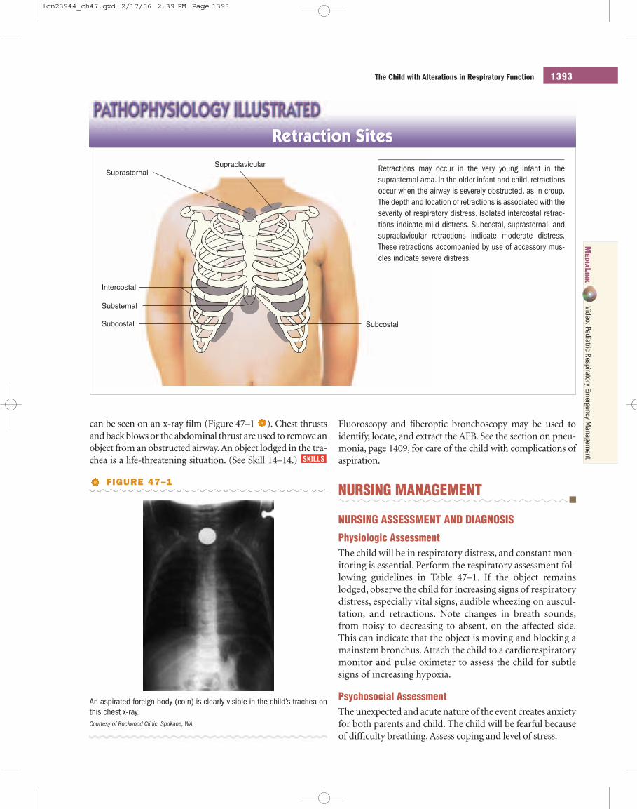
Ventilation is the movement of air in and out of thelungs and alveoli. The lungs rely on the diaphragm and in-tercostal muscles to power respiration. Children under 6years of age use the diaphragm to breathe as the intercostalmuscles are immature. The negative pressure caused by thedownward movement of the diaphragm draws in air. By 6years of age, the child uses the intercostal muscles more ef-fectively. The ribs are primarily cartilage and very flexible,and in cases of respiratory distress, the negative pressurecaused by the diaphragm movement causes the chest wallto be drawn inward, causing retractions (see “Pathophysi-ology Illustrated: Retraction Sites”).
Bifurcation of trachea inchildren is at T3 level.
Bifurcation in adultsis at T6 level.
Right mainstem bronchusin children has a steeperslope than in adults.
In children, the trachea is shorter and the angleof the right bronchus at bifurcation is more acutethan in the adult. When you are resuscitating orsuctioning, you must allow for the differences. Doyou think that this difference is significant in res-piratory infection? Why?
Trachea PositionTrachea Position
MED
IALIN
KAnim
ation: CO2
and O2 Transport
lon23944_ch47.qxd 2/17/06 2:39 PM Page 1391

1392 CHAPTER 47
RESPIRATORY DISTRESS AND RESPIRATORY FAILUREMany respiratory conditions associated with difficultybreathing can progress to respiratory distress. If the condi-tion is not managed effectively, it can progress to respira-tory failure. Foreign-body aspiration is a common cause ofairway obstruction and respiratory distress.
FOREIGN-BODY ASPIRATIONForeign-body aspiration is the inhalation of any object(solid or liquid, food or nonfood) into the respiratorytract. Aspiration occurs most often during feeding andreaching activities, while crawling, or during playtime inchildren 6 months to 4 years of age. These young childrenhave a tendency to put small objects in the mouth. How-ever, aspiration may occur in children of any age.
Etiology and PathophysiologyIn infants over 6 months of age and young children, anynumber of small objects that make their way into the child’smouth may cause aspiration. Foods such as nuts, popcorn,or small pieces of raw vegetables or hot dog; small, loose toy
parts such as small wheels and bells; or household objectsand substances such as beads, safety pins, coins, buttons, la-tex balloon pieces, and colorful liquids (mouthwash, per-fume) in enticing packages (screw-top bottles) are frequentcauses of airway obstruction. Partial and sometimes com-plete airway obstruction can occur.
The severity of the obstruction depends on the size andcomposition of the object or substance and its locationwithin the respiratory tract. Most aspirated foreign bodies(AFBs) usually cause bronchial, not tracheal, obstruction.An object lodged high in the airway above the vocal cordsis frequently coughed out.
The right lung is the most common site of lower airwayaspiration because of the sloped angle of its bronchus (see“As Children Grow: Trachea Position” on page 1391). Ob-jects may migrate from higher to lower airway locations.An object may also move back up to the trachea, creatingextreme respiratory difficulty. If oxygen is depleted for anextended time, brain damage may occur.
Clinical ManifestationsChildren are usually brought to the hospital after a suddenepisode of coughing or gagging. The child may have signsof increased respiratory effort such as dyspnea (difficultybreathing), tachypnea (rapid respiratory rate), nasal flar-ing, and retractions. If the child cannot say the “P” in wordslike Pluto or Peter Pan, the foreign body has noticeably di-minished expiratory effort. As respiratory distress pro-gresses, the child may have a concentrated focus onbreathing, have an anxious expression, and sit in a forwardposition with the neck extended. As the child becomes in-creasingly hypoxic, behavior changes such as irritabilityand decreased responsiveness are seen.
Coughing, choking, gagging, dysphonia, and wheezingmay be brief or may persist for several hours if the objectdrops below the trachea into one of the mainstem bronchi.In some cases the child may become asymptomatic aftercoughing for 15 to 30 minutes. If the foreign body dropsinto the lower airway and is not removed, the child maypresent with a chronic cough, persistent or recurrent pneu-monia, or a lung abscess weeks later.
Clinical TherapyClinical therapy focuses on taking a careful history to deter-mine whether aspiration has indeed occurred.Coughing,gag-ging, or choking associated with feeding or crawling on thefloor is usually a confirming event for aspiration. The physicalexamination often reveals decreased breath sounds, stridor,and respiratory distress in the child without a witnessed aspi-ration. A special radiograph, called a forced expiratory film,may be ordered.This shows local hyperinflation (air trapping)and a mediastinal shift away from the affected side (Hazinski,1999). Sometimes, when the object aspirated is radiopaque, it
Newborn
20 mmdiameter of airway
18 mmdiameter of airway
4 mm 2 mm
Adult
1 mmswelling
1 mmswelling
Airway DiameterAirway Diameter
The diameter of an infant’s airway is approximately 4 mm, in contrastto an adult’s airway diameter of 20 mm.An inflammatory process inthe airway causes swelling that narrows the airway, and airway resis-tance increases. Note that swelling of 1 mm reduces the infant’s air-way diameter to 2 mm, but the adult’s airway diameter is onlynarrowed to 18 mm. Air must move more quickly in the infant’s nar-rowed airway to get the same amount of air to the lungs. The frictionof the quickly moving air against the side of the airway increases air-way resistance. The infant must use more effort to breathe andbreathe faster to get adequate oxygen.
lon23944_ch47.qxd 2/17/06 2:39 PM Page 1392
The Child with Alterations in Respiratory Function 1393
Fluoroscopy and fiberoptic bronchoscopy may be used toidentify, locate, and extract the AFB. See the section on pneu-monia, page 1409, for care of the child with complications ofaspiration.
NURSING MANAGEMENT
NURSING ASSESSMENT AND DIAGNOSIS
Physiologic AssessmentThe child will be in respiratory distress, and constant mon-itoring is essential. Perform the respiratory assessment fol-lowing guidelines in Table 47–1. If the object remainslodged, observe the child for increasing signs of respiratorydistress, especially vital signs, audible wheezing on auscul-tation, and retractions. Note changes in breath sounds,from noisy to decreasing to absent, on the affected side.This can indicate that the object is moving and blocking amainstem bronchus. Attach the child to a cardiorespiratorymonitor and pulse oximeter to assess the child for subtlesigns of increasing hypoxia.
Psychosocial AssessmentThe unexpected and acute nature of the event creates anxietyfor both parents and child. The child will be fearful becauseof difficulty breathing. Assess coping and level of stress.
Intercostal
SuprasternalSupraclavicular
Subcostal Subcostal
Substernal
Retraction SitesRetraction Sites
Retractions may occur in the very young infant in thesuprasternal area. In the older infant and child, retractionsoccur when the airway is severely obstructed, as in croup.The depth and location of retractions is associated with theseverity of respiratory distress. Isolated intercostal retrac-tions indicate mild distress. Subcostal, suprasternal, andsupraclavicular retractions indicate moderate distress.These retractions accompanied by use of accessory mus-cles indicate severe distress.
An aspirated foreign body (coin) is clearly visible in the child’s trachea onthis chest x-ray.Courtesy of Rockwood Clinic, Spokane, WA.
FIGURE 47–1
can be seen on an x-ray film (Figure 47–1 ). Chest thrustsand back blows or the abdominal thrust are used to remove anobject from an obstructed airway. An object lodged in the tra-chea is a life-threatening situation. (See Skill 14–14.) SKILLS
MED
IALIN
KVideo: Pediatric Respiratory Em
ergency Managem
ent
lon23944_ch47.qxd 2/17/06 2:39 PM Page 1393

1394 CHAPTER 47
Developmental AssessmentAs the child’s condition stabilizes, observe how well thechild’s abilities match the parents’ understanding of age-appropriate behaviors. See developmental abilities by ageChapter 33. ∞
Common nursing diagnoses for a child with an AFBinclude the following:
■ Ineffective Airway Clearance related to foreign bodyaspiration
■ Impaired Spontaneous Ventilations related to foreignbody aspiration and respiratory muscle fatigue
■ Fear (Parent or Child) related to uncertainty ofprognosis, unfamiliar surroundings and procedures
■ Risk for Injury related to developmental age and smallobjects in environment
PLANNING AND IMPLEMENTATIONThe period right after aspiration until AFB removal is critical.Promptly document and report any subtle changes in thechild’s respiratory status. The nurse must remain with thechild who has a significant obstruction,and have resuscitationequipment at the bedside. Permit the child to stay in a posi-tion of comfort. Avoid performing procedures that increasethe child’s anxiety as sudden movements and increased respi-ratory efforts may cause the obstruction to move and com-pletely obstruct the airway. Be prepared to perform backblows and chest thrusts for an infant or abdominal thrusts forthe child with complete obstruction. (See Skill 14–9. )
After the AFB is removed, the child is stabilized and ob-served for a few hours in a short-stay unit.
Discharge Planning and Home Care TeachingDischarge planning centers on anticipatory guidance aboutchildproofing the home (see Chapter 33 ∞). Encouragethe parents to learn CPR, choking-prevention techniques,and back blows, chest thrusts, or abdominal thrusts.
EVALUATIONExpected outcomes of nursing care include the following:
■ The child regains the ability to ventilate spontaneouslyafter removal of the foreign body.
SKILLS
■ Parents complete a safety check of the home to preventfuture aspiration incidents.
RESPIRATORY FAILURERespiratory failure occurs when the body can no longermaintain effective gas exchange. The physiologic processthat ends in respiratory failure begins with hypoventilationof the alveoli. Hypoventilation occurs when the body’s needfor oxygen exceeds actual oxygen intake, the airway is par-tially occluded, or the transfer of oxygen and carbon dioxidein the alveoli is disrupted. This disruption may occur eitherbecause of a malfunction of respiratory center stimulation(the alveoli do not receive the message to diffuse) or becausethe alveolar membrane is defective (a structural problem).
Alveolar hypoventilation (poor ventilation of the alve-oli) results in hypoxemia (lower than normal blood oxygenlevel) and hypercapnia (an excess of carbon dioxide in theblood).Arterial blood gas levels indicative of respiratory fail-ure are a PaO2 level less than 50 mm Hg and a PaCO2 levelgreater than 50 mm Hg in a patient breathing room air(Grant & Curley, 2001). See Appendix B for expected labo-ratory values by age and Skill 10–6. When the bloodlevels of oxygen and carbon dioxide reach abnormal levels,hypoxia (lower than normal oxygen in the tissues) occursand respiratory failure begins. Hypoxemia that persists whensupplemental oxygen is given is a sign of respiratory failure.
Signs of impending respiratory failure include irri-tability, lethargy, cyanosis, and increased respiratory effortsuch as dyspnea, tachypnea, nasal flaring, and intercostalretractions. Grunting slows the expiratory flow and in-creases the lung volume and alveolar pressures. This is asign of severe disease and suggests the onset of respiratoryfailure (Margolis & Gadomski, 1998).
Nursing ManagementEarly recognition of impending respiratory failure is themost important aspect of care for a child with any signs ofrespiratory compromise. When the child has a chronic res-piratory condition, development of respiratory failure maybe gradual. Signs will be subtle. Be particularly alert to be-havior changes in addition to respiratory signs. Serialblood gases may be needed to monitor the child.
Place a child who has respiratory distress in an uprightposition (by elevating the head of the bed). Assess respira-tory quality and rate, followed by apical pulse rate and tem-perature. Monitor oxygen saturation with pulse oximetry.Administer oxygen as ordered and keep respiratory emer-gency equipment at the child’s bedside. Monitor the childfor changes in vital signs, respiratory status, and level of re-sponsiveness. Be prepared to assist ventilations if respira-tory status deteriorates. (See Skills 9–8 to 9–10. )SKILLS
SKILLS
NURSING PRACTICE
These signs and symptoms signal the body’s response to increasedmetabolic demands for oxygenation as a result of airway stress, ob-struction, or impending illness:
■ Increasing restlessness, irritability, unexplained suddenconfusion
■ Rapid heart rate accompanied by rapid respiratory rate
MED
IALI
NK
Anim
atio
n: G
as E
xcha
nge
lon23944_ch47.qxd 2/17/06 2:39 PM Page 1394

The Child with Alterations in Respiratory Function 1395
CLINICAL MANIFESTATIONS
RESPIRATORY FAILURE AND IMMINENT RESPIRATORY ARREST
PHYSIOLOGIC CAUSE CLINICAL MANIFESTATIONS
Respiratory failureThese signs occur because the child is trying to compensate for oxygen deficit and airway blockage. Oxygen supply is inadequate; behavior and vital signs reflect compensation and beginning hypoxia.
The child tries to use accessory muscles to assist oxygen intake; hypoxia persists and efforts now waste more oxygen than is obtained.
Imminent respiratory arrestThese signs occur because oxygen deficit is overwhelming and beyond spontaneous recovery. Cerebral oxygenation is dramatically affected; central nervous system changes are ominous.
Initial signsRestlessnessTachypneaTachycardiaDiaphoresis
Early decompensationNasal flaringRetractionsGruntingWheezingAnxiety and irritabilityMood changesHeadacheHypertensionConfusion
Severe hypoxiaDyspneaBradycardiaCyanosisStupor and coma
Excessive crying and anxiety deplete metabolic re-serves. External ventilatory support and vigorous cryingmay impede diaphragm function if the stomach becomesdistended with air. Because the child’s metabolic rate isabout double that of an adult, the child has a greater needfor oxygen. Respiratory distress, anxiety, and even fever candramatically add to the child’s oxygen demand.
Using Artificial AirwaysRespiratory problems that do not respond to oxygen therapy,medications, or position changes require the insertion of anartificial airway. As the child’s level of responsiveness deteri-orates, the ability to keep the airway open decreases. Endo-tracheal intubation is a short-term, emergency measure tostabilize the airway by placing a tube in the trachea. The tubemust be protected and stabilized to prevent its displacement.(See Skills 14–8 and 14–9. ) A tracheostomy is the cre-ation of a surgical opening into the trachea through the an-terior neck at the cricoid cartilage. Surgeons prefer toperform this procedure in the operating room; however, atracheostomy may also be performed in an emergency de-partment or other setting when immediate intervention isneeded. These children usually require admission to the in-tensive care unit (ICU) for monitoring and ventilatory sup-
SKILLS
port. Suction airway secretions as needed and provide tra-cheostomy care if present. See Skills 14–20—14–24 in theClinical Skills Manual.
Because endotracheal and tracheostomy tubes preventvocal cord vibration, intubated children cannot cry or talk.Infants and young children often express initial frustrationwhen they realize they cannot communicate verbally.When the child is alert, give suggestions for ways to makenoise and gain attention, such as striking the mattress. Acommunication board can be used with older children.
Many children are discharged from the hospital andcared for at home for an extended period with a tra-cheostomy tube in place. It is essential to teach parents how
SKILLS
THINKING CRITICALLY
OXYGEN DELIVERY DEVICESOxygen delivery devices are selected to match the concentration ofoxygen needed by the child. In respiratory failure, a higher concen-tration of oxygen is needed to reverse the hypoxemia. Which oxygendelivery device should be used? Are there any contraindications tooxygen use in a child who is hypoxic?
lon23944_ch47.qxd 2/17/06 2:39 PM Page 1395

1396 CHAPTER 47
to maintain the airway, clean the tracheostomy site, andchange the tube. A home healthcare nurse can providefollow-up care and support for the child and family. (SeeSkill 14–11. )
APNEAInfants have periodic breathing, an irregular rhythm, andmay have pauses of up to 20 seconds between breaths. Thisbreathing pattern is not apnea. Apnea is the cessation ofrespiration lasting longer than 20 seconds, or any pause inrespiration associated with cyanosis, marked pallor, hypo-tonia, or bradycardia. Apnea may be the first major sign of
SKILLS
respiratory dysfunction in the newborn (see respiratorydistress in Chapter 30 ∞).
APPARENT LIFE-THREATENING EVENT (ALTE)Apparent life-threatening event (ALTE) is defined as anepisode of apnea accompanied by a color change (cyanosis,pallor, or occasionally ruddiness), limp muscle tone, chok-ing, or gagging in a near-term or term infant who is greaterthan 37 weeks’ gestation. The majority of these events oc-cur in infants under 4 months of age, with a peak incidencebetween 1 week and 2 months (Davies & Gupta, 2002).These episodes may occur during sleep, wakefulness, or
CLINICAL MANIFESTATIONS
CAUSES OF APPARENT LIFE-THREATENING EVENTS
CAUSE CLINICAL MANIFESTATIONS DIAGNOSTIC TESTS
Functional or structural airway problem orimmaturity
Aspiration as a result of dysfunctionalswallowing or gastroesophageal reflux
Cardiac problems
Drug toxicity or poisoning; maternal historyof ingestion
Environmental, thermoregulation problem
Impaired oxygenation, respiratory disease(pulmonary edema, atelectasis,pneumonia)
Acute infection (sepsis, meningitis,necrotizing enterocolitis)
Intracranial pathology (intraventricularhemorrhage, ventricular dilation, centralnervous system anomalies, meningitis)
Metabolic disorders
Apnea of 20 sec or longer; accompaniedby bradycardia or cyanosis
Choking, coughing, cyanosis, vomiting
Tachycardia, tachypnea, dyspnea
Central nervous system depression,hypotonia
Lethargy, tachypnea, hypothermia orhyperthermia
Cyanosis, tachypnea, respiratory distress,anemia, choking, coughing
Feeding intolerance, lethargy, temperatureinstability
Abnormal neurologic examination, seizures
Jitteriness, poor feeding, lethargy, centralnervous system depression or irritability,hypotonia
Cardiorespiratory monitoring, sleep study,pneumogram, sepsis workup
Barium swallow, esophageal pH probe
Cardiorespiratory monitoring,electrocardiogram, echocardiogram,arterial blood gases
Serum magnesium level, toxicity screen
Cardiorespiratory and temperaturemonitoring, environmental temperaturelevel (ambient air temperature)
Oximetry, chest radiograph, arterial bloodgases, complete blood count, upper airwayevaluation, sleep study, serum electrolytes
Complete blood count, cultures whenappropriate, C-reactive protein, chest andabdominal radiographs
Cranial ultrasound, computed tomographyscan, electroencephalogram, magneticresonance imaging, cerebrospinal fluidevaluation
Serum electrolytes (potassium, sodium,chloride), glucose, calcium, arterial bloodgases
Data from Theobald, K., Botwinski, C., Albanna, S., & McWilliam, P. (2000). Apnea of prematurity: Diagnosis, implications for care, and pharmacologic management. NeonatalNetwork, 19(6), 17–24; and Eichenwald, E., & Stark, A. (1992). Apnea of prematurity: Etiology and management. Tufts University School of Medicine Reports on NeonatalRespiratory Diseases, 2(1), 1–11. Adapted.
MED
IALI
NK
Teac
hing
Pla
n: D
isch
arge
Inst
ruct
ions
for a
Chi
ld w
ith a
Trac
heos
tom
y Tu
beM
ED
IAL I
NK
Skill
s 14
–11:
Trac
heos
tom
y Ca
re
lon23944_ch47.qxd 2/17/06 2:39 PM Page 1396

The Child with Alterations in Respiratory Function 1397
feeding. ALTE should not be confused with sudden infantdeath syndrome (SIDS) (see page 1398).
A variety of identifiable diseases and conditions cancause ALTE, such as infection, gastroesophageal reflux,seizures, cardiac arrhythmias, and metabolic or endocrineproblems. In some cases, no cause is identified. ALTE canfrighten the parent or observer, who often fears the infanthas died. Emergency resuscitation is usually required.
Nursing ManagementAfter ALTE, infants are usually admitted to the hospital forevaluation and cardiorespiratory monitoring. Nursing careincludes collecting a detailed history of the event, observingand monitoring cardiorespiratory status,providing support-ive care to the infant and family,and anticipating the need foremergency resuscitation and for the diagnostic process.
Monitor Cardiorespiratory Status. Cardiorespiratory monitoringrecords heart rate and respiratory rate while the infant isawake and asleep.Pulse oximetry provides a noninvasive con-tinuous evaluation of the infant’s oxygenation status.A pulseoximetry reading (SpO2) less than 95% indicates hypox-emia. (See Skills 14–2 to 14–4. )
Provide Emotional Support. Establishing rapport and open com-munication with the parents is essential for creating a senseof trust. To obtain further information about the episode, useopen-ended questions and active listening skills. Parents arefearful and anxious about the infant’s prognosis. Explana-tions of tests and treatment help to decrease their anxiety andincrease their understanding of the situation.
During hospitalization the infant should be held andcuddled to provide a sense of security and well-being. En-couraging parents’ participation in the infant’s care helpsto meet these needs and promotes family bonding. Oftenparents are afraid to touch the infant because they mightdisconnect the monitoring cable. Wrapping the cable in-side the infant’s blanket helps secure the wires, increasingparents’ feelings of confidence in handling the infant.
Support the mother to continue breastfeeding andmaintaining a supply of breast milk by pumping, if neces-sary. Ensure that the mother gets adequate fluids and nu-trition. Provide privacy for breast pumping, and storebreast milk for future feedings.
Anticipate Emergency Resuscitation. Because the infant who hashad ALTE continues to be at risk for cardiopulmonary arrest,keep emergency resuscitation equipment and drugs readilyaccessible at all times.
Discharge Planning and Home Care Teaching. Identify and addresshome care needs well in advance of discharge. Teach parentshow to operate an apnea monitor, what to do when the infanthas an apneic episode, and how to perform cardiopulmonaryresuscitation (CPR) and choking-prevention techniques (seeSkill 14–13 ).SKILLS
SKILLS
OBSTRUCTIVE SLEEP APNEAObstructive sleep apnea syndrome (OSA) is a disorder ofbreathing during sleep that involves prolonged partial up-per airway obstruction and/or intermittent complete ob-struction that disrupts normal ventilation during sleep andnormal sleep patterns (American Academy of Pediatrics,2002). This results in labored breathing and snoring whenthe child tries to move air past the obstruction. Its inci-dence peaks between 2 and 6 years of age when tonsils andadenoids are at their largest in contrast to the airway’s size.
The upper airway contains about 30 muscles that per-mit the pharynx to collapse, enabling the child to talk andswallow, but also maintain airway patency. When the childis awake, muscle tone is maintained and the airway re-mains patent even when obstructions such as enlargedadenoids and tonsils, craniofacial anomalies, or obesity arepresent. During sleep, the airway muscles relax and thepharynx becomes obstructed. When the airway musclesare relaxed, the airway resistance is increased. Reduced up-per airway tone and obstruction then results in apneaepisodes that lead to hypoventilation, hypoxia, hypercap-nia, and an elevated blood pressure. Hypertrophy of theadenoids and tonsils is the most common cause of OSA,followed by craniofacial abnormalities. Without treat-ment, complications develop that can include failure tothrive, pulmonary hypertension, cor pulmonale (obstruc-tion of pulmonary blood flow that leads to right ventricu-lar hypertrophy and heart failure), systemic hypertension,and cognitive impairment.
Children with OSA snore and have signs of laboredbreathing during sleep such as retractions and paradoxicalbreathing. After pauses in snoring or lack of airflow, thechild may be noted to snort, gasp, choke, move, or arouseto take a breath. Sleep is restless and the child may sleep inunusual positions to hyperextend the neck and airway.Daytime sleepiness and other symptoms of sleep depriva-tion (poor attention, increased activity, aggression, acting-out behavior, poor school performance) may be noted.
Diagnosis is made by polysomnography, a sleep studythat simultaneously records the brain activity, eye move-ment, apnea episodes, oxygen desaturation and sleep dis-turbances. Adenotonsillectomy is the most commontreatment for OSA, and resolution of the condition occursin the majority of children. Weight loss strategies may beimplemented for obese children. Continuous positive air-way pressure (CPAP) is used for children with surgical con-traindications or those with persistent OSA.
Nursing ManagementIn the community setting, all children should be screenedfor snoring as part of their routine healthcare. Assess thechild for signs of nasal obstruction, mouth breathing, andenlarged tonsils. Determine if the child has symptoms of
MED
IALIN
KSkill 14–2
lon23944_ch47.qxd 2/17/06 2:39 PM Page 1397

1398 CHAPTER 47
Apnea Equipment■ Understand monitor type, lead wires, placement of skin
electrodes or chest belt, battery power, manual fortroubleshooting.
Emergency Preparation■ Notify telephone company, electric company, local rescue squad,
local emergency department (establishes priority status).■ Post phone numbers of rescue squad, physician, equipment
company, power company, emergency number, cardiopulmonaryresuscitation (CPR) guidelines, other important numbers(neighbor, parents’ work numbers) in at least two places in thehome; have at least one added extension phone.
■ Keep the apnea monitor battery fully charged.
Safety Precautions■ Place monitor on firm surface; keep away from other appliances
(television, microwave oven) and water.■ Ensure that alarms are audible from all locations.■ Double-check that monitor is on before going to bed.■ Thread cable and wires through lower end of infant’s clothes.■ Ensure integrity of leads, monitor cable, and power cord (replace
if frayed).
Routine Care■ Understand reasons for apnea monitor and frequency of use.■ Be able to attach and detach infant chest leads and belt.■ Evaluate skin for irritation or breakdown from electrode
placement and give skin care (no oils or lotion; move patchescorrectly).
HOME CARE INSTRUCTIONS FOR THE INFANT REQUIRING APNEA MONITORING
TEACHING HIGHLIGHTS
Emergency Care■ Develop plan for respiratory failure and power failure.■ Demonstrate CPR and back blows and chest thrusts for airway
obstruction.■ Understand how to respond to alarms for apnea, bradycardia, or
loose lead.
Apnea Alarm■ Observe infant’s respiratory movement.■ If respiration is absent or infant is lethargic, stimulate by calling
name and gently touching, proceeding to vigorous touch ifneeded.
■ If no response, proceed with CPR.
Bradycardia Alarm■ Stimulate infant; infant should respond quickly.
Loose Lead■ Check electrode patch. Is it loose? Dirty? Belt loose?■ Check wires from electrode or monitor cable.■ Check power supply. Is battery low? Power failure? Monitor
malfunctioning?
sleep deprivation or if a condition is present that places thechild at high risk for OSA. Coordinate referral to a sleepcenter for polysomnogram evaluation and explain the pur-pose of the test. Discuss how to prepare the child for thestrange setting and wires that will be attached during thesleep study. Most pediatric centers will allow the parent tostay with the child during the study.
Following adenotonsillectomy, the hospital nursemonitors the child for bleeding and respiratory distress,such as obstructive sleep apnea and pulmonary edema.Continuous pulse oximetry is used to detect oxygen desat-uration. See Chapter 46 for care of the child having adeno-tonsillectomy. ∞
Sleep center nurses provide education and support tofamilies of children who need to use CPAP to treat theOSA.The nurse helps identify the best fitting mask or nasalprong system for CPAP delivery. Parents may need guid-ance about helping children to go to sleep wearing themask until they are accustomed to it.
SUDDEN INFANT DEATH SYNDROME (SIDS)Sudden infant death syndrome (SIDS) has been defined asthe sudden unexpected death of an infant under 1 year ofage with onset of the fatal episode during sleep that re-mains unexplained after a thorough investigation, includ-ing an autopsy, a review of the circumstances of death, anda review of the clinical history (Krous, 2004). It remains aleading cause of death in infants between 1 month and 1year of age, with 90% of cases occurring before 6 monthsof age (American Academy of Pediatrics Committee onChild Abuse and Neglect, 2001). SIDS occurs rarely in in-fants younger than 2 weeks. It is currently unpredictableand in some cases unpreventable.
SIDS is referred to as a “syndrome” because of themany and varied autopsy and clinical findings that char-acterize most infants who die of the disorder. The autopsytypically does not identify a disease process that causedthe death. Current evidence suggests a genetic susceptibil-ity to SIDS (American Academy of Pediatrics Committee
lon23944_ch47.qxd 2/17/06 2:39 PM Page 1398

The Child with Alterations in Respiratory Function 1399
1. Provide parents with a private area and a support person whoreinforces that the infant’s death was not their fault. Parents needto be able to express their grief in their own way and hear thatthey are not being blamed for the infant’s death.
2. Prepare the family for the viewing of the infant. Describe how theinfant will look and feel. You can say, “Paul’s [use the infant’sname] skin will feel cool. He will be very still and his eyes will beclosed.” They probably know this, but a gentle explanationdemonstrates empathy. Explain that pooling of blood on thedependent areas will look like bruises.
3. Allow parents to hold, touch, and rock the infant if desired.Viewing the infant allows parents a chance to say good-bye.Before bringing the infant to parents, wrap in a clean blanket,comb the hair, wash the face, swab the mouth clean, and applyVaseline to lips.
4. Reinforce the physician’s explanation about the need for anautopsy. An autopsy is required for all unexplained deaths. Youcan say to parents, “It is the only way we can be sure of whatcaused your baby’s death.”
5. Answer parents’ questions and provide them with sources forfurther information. Provide literature and a name of the localcontact for a SIDS support group, as well as for the nationalfoundation. Parents may not be able to take in all of your answers.Many emergency departments and pediatric units have a socialworker who provides ongoing contact with the family. Providenames of resource people and phone numbers for SIDS supportgroups.
6. Advise parents that surviving siblings may benefit from psychologicsupport. Siblings often require emotional support in the weeksand months after the death. Social workers can help the familyobtain counseling and support for all members.
7. Provide parents with a lock of hair, footprints, and handprints, ifthey desire. Personal items can be placed in a memory book. Thisreaffirms the child’s existence for many parents.
TABLE 47–3 Supportive Care for theFamily of an Infant withSudden Infant DeathSyndrome (SIDS)
on Fetus and Newborn, 2003). The current thinking isthat vulnerable infants have a defect in the arcuate nu-cleus, a brain structure that plays a role in regulatingbreathing, heartbeat, body temperature, and arousal (Kato,Franco, Groswasser et al., 2003; Parnigrahy, Filiano,Sleeper et al., 2000). Child abuse and homicide may be as-sociated with 1% to 5% of suspected SIDS cases (AmericanAcademy of Pediatrics Committee on Child Abuse and Ne-glect, 2001). Other proposed causes include H. pylori gas-trointestinal infection and long QT syndrome, a cardiacarrhythmia. (See cardiac arrhythmias in Chapter 48. ∞)SIDS has not been found to be associated with newbornapnea or immunizations for diphtheria, tetanus, and per-tussis (DTaP). See Table 47–2 for infant and maternal fac-tors that place infants at risk for SIDS.
The first symptom is a cardiac arrest. Clinical findingsinclude evidence of a struggle or change in position duringsleep and the presence of frothy, blood-tinged secretionsfrom the mouth and nares. Typically parents find the infantdead in the crib in the morning or after a nap and reporthaving heard no cries or disturbances during the night.
Nursing ManagementThe sudden, unexpected nature of the infant’s death is con-firmed in the emergency department. The nurse’s role is tobe empathetic and provide support during one of the great-est crises a family must face. The focus is on supporting thefamily during the acute grieving period. See Table 47–3.
Reassure the parents that they are not responsible forthe infant’s death and help them contact other familymembers and mobilize support. Older children may needreassurance that SIDS will not happen to them. They mayalso believe that bad thoughts or wishes about their baby
Infant■ Race (in decreasing order of frequency): most common in Native
American infants, followed by black, Hispanic, white, and Asianinfants
■ Gender: more common in males than females■ Age: most common in infants between 2 and 4 months of age■ Time of year: more prevalent in winter months■ Exposure to passive smoke■ Sleeping arrangement: prone or side-lying position and turning
to prone, sharing bed with others, use of pillows and quilts with bedding
■ Overheating due to excessive blankets, clothing on infant, roomtemperature
Maternal Risk Factors■ Maternal age less than 20 years at first pregnancy and a short
interval between pregnancies■ Prenatal smoking, binge alcohol, and illicit drug use■ Anemia■ Poor prenatal care, low weight gain during pregnancy■ History of sexually transmitted disease or urinary tract infection
TABLE 47–2 Risk Factors for SuddenInfant Death Syndrome(SIDS)
NURSING PRACTICE
Guidelines for the support of families experiencing SIDS should in-clude baptism services, religious support, grief counseling, assis-tance with funeral arrangements, and counseling on cessation ofbreastfeeding and sibling reactions.
MED
IALIN
KVideo: SIDS
lon23944_ch47.qxd 2/17/06 2:39 PM Page 1399

1400 CHAPTER 47
Epiglottis swellsoccluding airway
Cricoidcartilage
Trachea swells againstcricoid cartilageresulting in restriction
Airway Changes with CroupAirway Changes with Croup
There are two important changes in the upper airway in croup: theepiglottis swells, thereby occluding the airway, and the tracheaswells against the cricoid cartilage, causing restriction.
brother or sister caused the death. Support groups can helpparents, siblings, and other family members express thesefears and work through their feelings about the infant’sdeath. The SIDS Alliance and SHARE are organizationsthat can help families locate a support group in their area.See the Companion Website for links.
Nurses can play an important role in educating the pub-lic about the link between SIDS and infant positioning dur-ing sleep. Educate the parents of all newborns and infantsabout the recommended sleep position—on the back—forinfants, and ask them to make sure this position is usedwhen the infant is cared for by another family member orchildcare provider. Parents should also use a firm mattressand avoid the use of loose bedding, toys, and pillows. Placehospitalized infants to sleep in supine position rather thanside-lying or prone. See Chapter 53 for issues related to in-fant skull flattening from sleeping on the back.∞CROUP SYNDROMESCroup is a term applied to a broad classification of upperairway illnesses that result from swelling of the epiglottisand larynx. The swelling usually extends into the tracheaand bronchi. Included under the classification of croupsyndromes are viral syndromes, such as spasmodic laryngi-tis (spasmodic croup), laryngotracheitis, and laryngotra-cheobronchitis (LTB), and bacterial syndromes, such asbacterial tracheitis and epiglottitis (see “PathophysiologyIllustrated: Airway Changes with Croup”).
LTB, epiglottitis, and bacterial tracheitis are referred toas the“big three”of pediatric respiratory illness because theyaffect the greatest number of children across all age groupsin both sexes. The initial symptoms of all three conditionsinclude inspiratory stridor (a high-pitched, musical soundthat is created by narrowing of the airway),a“seal-like”bark-ing cough, and hoarseness. LTB is the most common disor-der, but epiglottitis and bacterial tracheitis are more serious.
LARYNGOTRACHEOBRONCHITISAlthough the term croup is applied to several viral and bac-terial syndromes, it is most often used to refer to LTB, a vi-ral invasion of the upper airway that extends throughoutthe larynx, trachea, and bronchi. Table 47–4 compares LTBand other croup syndromes.
Etiology and PathophysiologyAcute viral LTB is most common in children 3 months to 4years of age but can occur up to 8 years of age. Boys are af-fected more often than girls. LTB is of greatest concern ininfants and children under the age of 6 years, because ofpotential airway obstruction. The causative organism isusually parainfluenza virus type I, II, or III, that appearsduring winter months. Other viruses causing LTB include
influenza A and B, adenovirus, respiratory syncytial virus,and measles (Perkin & Swift, 2002).
Airway tissues respond to the invading virus with in-flammation and edema. Copious, tenacious secretionsfurther increase the child’s respiratory distress. The laryn-geal inflammation causes the airway diameter to narrowin the subglottic area, the narrowest part of the airway.Even small amounts of mucus or edema can quickly ob-struct the airway. Both the large and small airways can beaffected.
Clinical ManifestationsMost children brought to the emergency department withLTB have been ill for a couple of days with upper respira-tory symptoms. These symptoms progress to a cough andhoarseness. Fever may or may not be present. Commonpresenting signs are tachypnea, inspiratory stridor, and aseal-like barking cough.
Clinical TherapyDiagnosis is often made by clinical signs. Pulse oximetry isused to detect hypoxemia. If the diagnosis of LTB is inquestion, anteroposterior (AP) and lateral x-rays of the up-
lon23944_ch47.qxd 2/17/06 2:39 PM Page 1400

The Child with Alterations in Respiratory Function 1401
VIRAL SYNDROMES BACTERIAL SYNDROMES
Acute SpasmodicLaryngitis Bacterial Epiglottitis
(Spasmodic Croup) Laryngotracheobronchitis Laryngotracheitis Tracheitis (Supraglottitis)
Severity
Age affected
Onset
Clinical manifestations
Etiology
TABLE 47–4 Summary of Croup Syndromes
Least serious
3 months to 3 years
Abrupt onset; peaks atnight, resolves bymorning (recurs)a
Afebrile; mild respiratorydistress; barking-sealcough
Unknown; suspect viralwith allergic/emotional influences
Most commona
3 months to 8 years
Gradual onset; starts as URI,progresses to moderaterespiratory difficulty
Early: mild fever [�39°C(102.2°F)]; hoarseness;barking-seal, brassy, croupycough; rhinorrhea; sorethroat; stridor (inspiratory);apprehension
Progressing to laboredrespirations
Parainfluenza, types I and II,RSV, or influenza
Most serious; progresses ifuntreated
3 months to 8 years
Gradual onset; starts as URI,progresses to symptomsof respiratory distress
Early: mild fever [�39°C(102.2°F)]; barking-seal,brassy, croupy cough;rhinorrhea; sore throat;stridor (inspiratory);apprehension;restless/irritable
Progressing to retractions(progressive); increasingstridor; cyanosis
Parainfluenza, types I and II,RSV, or influenza
Guarded; requires closeobservation
1 month to 13 yearsa
Progressive from URI (1–2 days)
High fever [�39°C(102.2°F)]; URIappears as viral croupycough and croupinitially; stridor(tracheal); purulentsecretions
Staphylococcus
Most life threatening(medical emergency)a
2 years to 8 years
Progresses rapidly(hours)a
High fever [�39°C(102.2°F)]; URI;intense sore throat;dysphagiaa; droolinga;increased pulse andrespiratory rate; prefersupright position (tripodposition with chinthrust)a
Haemophilus influenzae
aClassic parameter or key point (distinguishes condition).
is less than 92% (see Table 47–5). Mist tents are rarely usedfor laryngotracheobronchitis.
Children with a good response to medications are oftensent home from the emergency department after an obser-vation period. Children with moderate to severe symptomsafter medications are admitted for further observation andtreatment. Airway obstruction is a potential complication ofLTB. The child may require intubation and transfer to theICU to maintain airway patency if obstruction is immi-nent. Most children, however, respond positively to the
Medication Action/Indication Nursing Considerations
Beta-agonists and beta-adrenergics (e.g., albuterol, racemic epinephrine): aerosolized through face mask
Corticosteroids (e.g., dexamethasone): IM, PO, nebulized budesonide
TABLE 47–5 Medications Used for Symptomatic Treatment of Laryngotracheobronchitis
Rapid-acting bronchodilator, decreases bronchialand tracheal secretions and mucosal edema,used to decrease symptoms of moderate tosevere respiratory distress; and constriction ofsubglottic mucosa and submucosal capillaries.Used until dexamethasone begins working.
Anti-inflammatory, used to decrease edema; hasa long half-life of 36–54 hours
Provides only temporary relief; improvement in30 minutes which lasts about 2 hours, it givestime for the steroid to work; the child mayexperience tachycardia (160–200 beats/min)and hypertension; dizziness, headache, andnausea may necessitate stopping medication;reduces the need for artificial airway
The child may experience cardiovascularsymptoms (hypertension): requires closeobservation for individual response; childrenless frequently need emergency airways; stridorresolves faster
per airway are taken; these may show symmetric subglotticnarrowing called a “steeple sign.” Throat cultures and vi-sual inspection of the inner mouth and throat are con-traindicated in children with LTB and epiglottitis. Theseprocedures can cause laryngospasms (spasmodic vibra-tions that close the larynx) as a result of the child’s anxietyor of probing this reactive and already compromised area.
Clinical therapy consists of maintaining and improv-ing respiratory effort with medications, humidification,and supplemental oxygen when the saturated oxygen level
lon23944_ch47.qxd 2/17/06 2:40 PM Page 1401

1402 CHAPTER 47
Sign 0 1 2 3
Stridor None With agitation Mild at rest Severe at rest
Retraction None Mild Moderate Severe
Air entry Normal Normal Decreased Severe decrease
Color Normal Normal Cyanotic with Cyanotic withagitation rest
Level of Normal Restless if Restless if Lethargicconsciousness disturbed undisturbed
Scoring: To quantifiy the severity of stridor, add the individual scores for each of thesign categories. A score between 0 and 15 is possible. A rating of severity based ontotal score is as follows: < 6 is mild, 7–8 is moderate, > 8 is severe.
Source: From Perkin, R. M., & Swift, J. D. (2002). Infectious causes of upper airwayobstruction in children. Pediatric Emergency Medicine Reports, 7(11), 120.
TABLE 47–6 Clinical Scoring System for Assessing Children with Stridor
medications and oxygen therapy and are discharged within48 to 72 hours.
NURSING MANAGEMENT
NURSING ASSESSMENT AND DIAGNOSISThe initial and ongoing physical assessment of the childwith LTB focuses on adequacy of respiratory functioning.See Tables 47–6 and 47–7. Continuous monitoring is re-quired to identify changes in airway patency. A means ofcommunication (sign language or simple word cues) must
be established so the older child can alert nursing staff torespiratory difficulty.
Pay particular attention to the child’s respiratory ef-fort, breath sounds, and responsiveness. Physical exhaus-tion can diminish the intensity of retractions and stridor.As the child uses the remaining energy reserve to maintainventilation, breath sounds may actually diminish. Noisybreathing (audible airway congestion, coarse breathsounds) in this situation verifies adequate energy stores.Responsiveness decreases as hypoxemia increases.
NURSING PRACTICE
Observe the child continuously for inability to swallow, absence ofvoice sounds, increasing degree of respiratory distress, and acute on-set of drooling (an ominous sign of supraglottic obstruction). If any ofthese signs occur, get medical assistance immediately. The quieterthe child, the greater the cause for concern.
Nursing Action Rationale
Assess heart rate and respiratory rate. Tachypnea and tachycardia indicate increasing respiratory effort.
Check position of the child (sitting, prone, or supine)? Upright or semi-Fowler’s promotes airway patency; the child’s ownattempt or desire to change to a more upright position may signalincreased distress.
Assess overall quality of respiratory effort. Determine inspiratory and Reflects overall adequacy of airway and respiratory function.expiratory breath sounds, ability to speak, and presence of stridor,cough, retractions, nasal flaring, cyanosis.
Initiate stridor scoring assessment (Table 47–6), continue scoring every Provides consistent and objective assessment data with score for 30 minutes or more frequently if distress increases; initiate nursing future comparison.actions appropriate for stridor score.
Attach cardiorespiratory monitor and pulse oximeter. Provides continuous assessment data as part of ongoing physiologicmonitoring.
TABLE 47–7 Nursing Assessment of Child with Respiratory Difficulty
The following nursing diagnoses might be appropriatefor the child with acute LTB:
■ Ineffective Breathing Pattern related to tracheobronchialobstruction, decreased energy, and fatigue
■ Risk for Deficient Fluid Volume related to inadequatefluid intake prior to admission
■ Fear (Child) related to unfamiliar surroundings,procedures, and separation from support system
PLANNING AND IMPLEMENTATION
Maintain Airway PatencySupplemental oxygen with humidity may be needed for hy-poxemia. Cool mist is presumed to moisten airway secre-
lon23944_ch47.qxd 2/17/06 2:40 PM Page 1402

The Child with Alterations in Respiratory Function 1403
tions and soothe the inflamed mucosa, but research has notdocumented its benefit (Perkin & Swift, 2002). Allow thechild to assume a comfortable position. Be immediatelyavailable to attend to the child’s respiratory needs, and keepresuscitation equipment at the bedside.
Meet Fluid and Nutritional NeedsThe respiratory distress may have interfered with thechild’s ability and desire to drink fluids and compromisedthe child’s fluid status. Recognizing fluid deficit and mon-itoring the child’s hydration and nutritional status are es-sential. Fluids promote liquification of secretions andprovide calories for energy and metabolism.
Children with LTB usually prefer cool, noncarbonated,nonacidic drinks such as oral rehydration fluids. The par-ents can be encouraged to give the child oral fluids. An in-travenous infusion may be necessary to rehydrate the child,maintain fluid balance, or provide emergency access. Ob-serve the child closely for difficulty in swallowing, whichmay be an early sign of epiglottitis or bacterial tracheitis.
Discharge Planning and Home Care TeachingDuring the child’s observation period, take every opportu-nity to assess the parents’ knowledge of symptoms of LTBand discuss actions to take if symptoms recur. For example,instruct parents to call the child’s physician if:
■ Mild symptoms do not improve after 1 hour ofhumidity and cool air treatment.
■ The child’s breathing is rapid and labored.
■ The child does not drink adequate fluids and the urineoutput is reduced.
EVALUATIONExpected outcomes of nursing care include the following:
■ The child responds to medications with decreasedrespiratory distress.
■ The child’s fear and anxiety is managed with familysupport and explanations about care.
Epiglottitis (Supraglottitis)Epiglottitis (also known as supraglottitis) is an inflamma-tion of the epiglottis, the long narrow structure that closesoff the glottis during swallowing. Because edema in thisarea can rapidly (within minutes or hours) obstruct theairway by occluding the trachea, epiglottitis is considered apotentially life-threatening condition. (Table 47–4 com-pares epiglottitis and other croup syndromes.)
Epiglottitis is caused by bacterial invasion of the softtissue of the larynx by streptococcus and staphylococcus,and by Haemophilus influenzae type B (Hib) in unimmu-
nized children. The resulting inflammation and edema inthe tissues and surrounding the epiglottis lead to airwayobstruction. Since the widespread use of the Hib vaccina-tion, a tenfold decrease in the incidence of epiglottitis hasoccurred (Isaacson & Isaacson, 2003).
Characteristically, a previously healthy child sud-denly becomes very ill. The child initially develops a highfever (greater than 39°C [102.2°F]), with a sore throat,dysphonia (muffled, hoarse, or absent voice sounds), anddysphagia (difficulty in swallowing). As the larynx be-comes obstructed, inspiratory stridor and respiratorydistress develop. The intense throat pain keeps the childfrom swallowing, resulting in drooling. To fully open theairway and improve air intake, the child sits up and leansforward with the jaw thrust forward in the classic “sniff-ing” or tripod posture and refuses to lie down. The child’sanxiety increases as it becomes more difficult to breathe.
Diagnosis is often based on a lateral neck x-ray(Figure 47–2 ), which reveals a narrowed airway and anenlarged, rounded epiglottis, seen as a mass at the base ofthe tongue. Laryngospasm and airway obstruction canoccur as a result of the severe irritation and hypersensitiv-ity of the airway muscles. For this reason, visual inspectionof the mouth and throat is contraindicated in children withsuspected epiglottitis.
Immediate clinical therapy usually involves insertionof an endotracheal tube to maintain the airway. At the sametime, a culture of the epiglottis is taken. Antibiotics effec-tive for gram-positive organisms and H. influenzae aregiven until culture sensitivities are available. Antipyretics(acetaminophen, ibuprofen) may be useful in managingfever and sore throat pain.
Enlarged, roundeepiglottis
Narrow airway
The phrase “thumb sign” has been used to describe this enlargement of theepiglottis. Recall the trachea’s usual “little finger” size. Do you see the stiff,enlarged “thumb” above it in this lateral neck x-ray?
FIGURE 47–2
lon23944_ch47.qxd 2/17/06 2:40 PM Page 1403

1404 CHAPTER 47
Nursing Management. Nursing management consists of airwaymanagement, drug therapy, hydration, and emotional andpsychosocial support of the child and parents.
Until the child is intubated, the child is usually sedatedand needs to be positioned to maintain the airway andbreathe more easily. Observe the child’s respiratory andairway status closely and often. Note any change in level ofconsciousness. Anxiety-provoking procedures are post-poned until the airway is stabilized. Crying stimulates theairway, increases oxygen consumption, and can precipitatelaryngospasm. Supplemental humidified oxygen may beused initially to reverse hypoxemia.
Until the endotracheal tube is removed, the child is usu-ally managed in the ICU to ensure continual observation.(See Skill 14–12. ) Administer antibiotics to treat theinfection and IV fluids to provide hydration. Because thechild was febrile with a sore throat before admission, fluidintake may have been compromised.
The loss of voice, or even the inability to create sounds,can be frightening to a child. The unfamiliar hospital envi-ronment and strange equipment can create stress for childand parent alike. Reassure the parents that the child’s voiceloss is temporary and explain the need for the variouspieces of equipment.
Most children show rapid improvement once oxygen,antibiotics, and fluid therapy are started. The endotra-cheal tube can usually be removed within 24 to 36 hours(Hazinski, 1999). Home care may involve completing thecourse of antibiotics. Parents need instructions on properadministration and potential problems of drug therapy.
Bacterial TracheitisBacterial tracheitis is a secondary infection of the upper tra-chea after viral laryngotracheitis that is most often caused byStaphylococcus aureus, group A streptococcus, Moraxella ca-tarrhalis, or Haemophilus influenzae. The disorder starts withcroupy cough and stridor but progresses to include a highfever (greater than 39°C [102.2°F]), respiratory distress, anda toxic appearance (Stroud & Friedman, 2001). Table 47–4compares bacterial tracheitis and other croup syndromes.
Because of the similarity of symptoms, bacterial tra-cheitis is often misdiagnosed initially as LTB. Instead of im-proving with therapy, however, the child’s conditionbecomes worse. Children generally prefer lying flat to sit-ting up. This seems to be a position of comfort that allowsthe child to conserve energy. Diagnosis is often made byblood cultures after the child is found unresponsive tousual LTB management. The subglottis is edematous withulceration, and thick mucopurulent exudate may obstructthe airway. Antibiotics are given for a full 10- to 14-daycourse. Most children need a secured artificial airway for 3to 11 days and ventilatory support.
SKILLS
Nursing Management. The child with bacterial tracheitis is fre-quently cared for in the PICU after intubation. Mechanicalsuctioning of the thick tracheal secretions that pool high inthe upper airway helps maintain a patent airway. Provide hu-midified air or oxygen. Antibiotics are administered as or-dered. The previous section on epiglottitis discusses othernursing care interventions that may also be appropriate forthe child with bacterial tracheitis.
LOWER AIRWAY DISORDERSThe lower airway, or bronchial tree, lies below the tra-
chea and includes the bronchi, bronchioles, and alveoli.Lower airway disorders occur because a structural or func-tional problem interferes with the lungs’ ability to com-plete the respiratory cycle. Lower airway disorders includebronchopulmonary dysplasia, bronchitis, bronchiolitis,pneumonia, and tuberculosis.
BRONCHITISAcute bronchitis, inflammation of the trachea and bronchi,rarely occurs in childhood as an isolated problem. Thebronchi can be affected simultaneously with adjacent res-piratory structures during a respiratory illness. Bronchitisis caused most often by a virus but may also result from in-vasion of bacteria or in response to an allergen or irritant.
The classic symptom of bronchitis is a coarse, hackingcough, which increases in severity at night. Children withbronchitis look tired. The chest and ribs may be sore becauseof the deep and frequent coughing. There is often a deep, rat-tling quality to breathing. Some children have audiblewheezing that can be heard without a stethoscope.Treatmentis palliative unless a secondary bacterial infection occurs.
NURSING MANAGEMENTNursing management includes supporting respiratory func-tion through rest, humidification, hydration, and sympto-matic treatment. Refer to the sections on asthma andpneumonia for detailed information on treatment measures.
Home care should emphasize the self-limiting natureof the disorder. Advise parents who smoke that quitting orrefraining from smoking in the child’s presence may bene-fit the child.
BRONCHIOLITISBronchiolitis is a lower respiratory tract illness that occurswhen an infecting agent (virus or bacterium) causes in-flammation and obstruction of the small airways, thebronchioles. The peak age for bronchiolitis is 2 to 6 months(Cooper, Banasiak, & Allen, 2003). Infection is most severe
lon23944_ch47.qxd 2/17/06 2:40 PM Page 1404

The Child with Alterations in Respiratory Function 1405
NURSING PRACTICE
Respiratory syncytial virus (RSV) is the most common cause of lowerrespiratory tract infections in infants and children. RSV causes severe orfatal illness in infants with conditions such as congenital heart disease,bronchopulmonary dysplasia (BPD), prematurity, and immunosuppres-sion. Healthcare workers should follow principles of good handwashing,as the virus is easily transmitted and can survive on the hands for 30minutes or more (American Academy of Pediatrics, 2003).
in infants under 6 months of age and in children with heartand lung disease. Bronchiolitis is responsible for 90,000hospital admissions and 4500 deaths per year (Agency forHealthcare Research and Quality, 2003).
ETIOLOGY AND PATHOPHYSIOLOGYInfection with respiratory syncytial virus (RSV) is the mostcommon cause, but other viral, bacterial, and mycoplasmalorganisms may also be responsible. RSV occurs in annualepidemics from October to March. It is transmitted throughdirect or close contact with respiratory secretions of in-fected individuals. Nearly all children have been infectedwith RSV by 2 years of age, and reinfection (via siblings orclose family contacts) throughout life is common (NationalRespiratory and Enteric Virus Surveillance System, 2000).
Viruses, acting as parasites, are able to invade the mu-cosal cells that line the small bronchi and bronchioles. Theinvaded cells die when the virus bursts from inside the cellto invade adjacent cells. The membranes of the infectedcells fuse with adjacent cells, creating large masses of cellsor “syncytia.” The resulting cell debris clogs and obstructsthe bronchioles and irritates the airway. In response, theairway lining swells and produces excessive mucus. Despitethis protective effort by the bronchioles, the actual effect ispartial airway obstruction and bronchospasms.
39°C [102.2°F]) for a few days. As the illness progresses andthe lower respiratory tract becomes involved, symptomsincrease and include inspiratory and expiratory wheezing;a deeper, more frequent cough; tachypnea; retractions; andmore labored breathing. As severe respiratory distress de-velops, marked retractions, crackles, cyanosis, and dimin-ished breath sounds are noted. Thus the noisier the lungs,the better, as this indicates that the child is still able to moveair in and out of the lungs.
Parents report that the infant or child is acting moreill—appearing sicker, less playful, and less interested in eat-ing. Infants, especially, may refuse to feed or may spit upwhat they do eat along with thick, clear mucus. Dehydra-tion may be present.
CLINICAL THERAPYThe history and physical examination provide the dataneeded to diagnose bronchiolitis. Chest radiographs shownonspecific findings of inflammation. Enzyme-linked im-munoabsorbent assay (ELISA) or direct fluorescent assayperformed on a nasal wash specimen are laboratory testsused to identify the virus causing bronchiolitis (see Skills10–13 and 10–14 ).
Children who test positive for RSV are isolated,roomed together, or placed on the same ward to minimizethe spread of the virus to other hospitalized children. Sup-portive care is provided, especially when the causativeagent is unknown and the condition is mild to moderate inseverity. See Table 47–8 for clinical therapies. The childmay be intubated and ventilated for apnea or respiratoryfailure. Ribavirin is the only antiviral drug available for treat-ment. Studies have not confirmed its effectiveness, so it is re-served for life-threatening cases (Wright, Pomerantz, &Luria, 2002).
The following high-risk infants are recommended toreceive 5 monthly injections of palivizumab beginning inOctober or November to prevent RSV infection (AmericanAcademy of Pediatrics Committee on Infectious Diseasesand Committee on Fetus and Newborn, 2003):
■ Prematurity—born at 32 weeks of gestation or earlier,particularly if less than 6 months of age at the start ofthe RSV season.
■ Chronic lung disease such as BPD who have requiredsupplemental oxygen, bronchodilator, diuretic, orcorticosteroid therapy within 6 months of the startof RSV season.
■ Complicated congenital heart disease, particularlythose on medication to control congestive heart failure,with cyanosis, and with moderate to severe pulmonaryhypertension.
■ Immunocompromised infants and children may benefit.
SKILLS
The cycle is repeated throughout both lungs as the air-way cells are invaded by the virus. The partially obstructedairways allow air in, but the mucus and airway swelling blockexpulsion of the air. This creates the wheezing and cracklesin the airways. Air trapped below the obstruction also inter-feres with normal gas exchange, leading to hypoxemia. Thechild with RSV is therefore at risk for respiratory failure asthe oxygen level decreases and the carbon dioxide level in-creases. Apnea and pulmonary edema may occur.
CLINICAL MANIFESTATIONSThe infant or child with bronchiolitis may have been illwith upper respiratory symptoms such as nasal stuffiness,cough (not usually noted in infants), and fever (less than
lon23944_ch47.qxd 2/17/06 2:40 PM Page 1405

1406 CHAPTER 47
Clinical Therapy Rationale
Cardiorespiratory monitor and pulse oximetry
Humidified oxygen therapy via hood or face tent,mask, or nasal cannula
Intubation and assisted ventilation (PEEP/CPAP)
Hydration via intravenous or oral fluids
Systemic medications
Postural drainage and chest physiotherapy
High-Risk Infant or Childa
RespiGam (RSV immune globulin) IVPalivizumab (Synargis) IM
TABLE 47–8 Clinical Therapy for Bronchiolitis
Enable provider to follow course and assess need for specific therapies.
Delivery method determined by desired concentration of oxygen, degree of moisture, andchild’s response.
Used when the child becomes too fatigued to breathe effectively.
Provider must consider insensible fluid loss, decreased intake, the child’s current electrolyteand hydration status, and risk for pulmonary edema.
Bronchodilators, steroids, and beta-antagonists act directly on inflamed and obstructedairways; bronchodilators help prevent apnea episodes in premature infants; nebulizedepinephrine, and corticosteroids are occasionally used.
Helps to further loosen and mobilize trapped mucus.
Give for 5 consecutive months during RSV season to high-risk children. May prevent RSVbronchiolitis or reduce severity of disease.
aDefined as a child with congenital heart disease, bronchopulmonary dysplasia, chronic lung problems, or cystic fibrosis or who is premature or severely ill and less than 6 months old.
Intravenous RSV immune globulin (RespiGam) may alter-natively be used in some children under 2 years of age, andthese children should wait 9 months before receiving livevirus vaccines (varicella and MMR).
NURSING MANAGEMENT
NURSING ASSESSMENT AND DIAGNOSIS
Physiologic AssessmentAssess airway and respiratory function carefully.Good obser-vation skills are important to ensure timely interventions forworsening respiratory symptoms and prevention of respira-tory distress (see Table 47–1 and the clinical manifestations ofrespiratory failure on page 1392). An oxygen saturation levelbelow 90% is the best indicator of the severity of the disease.
See “Nursing Care Plan: The Child with Bronchiolitis.”
Psychosocial AssessmentObserve children and their parents for signs of fear andanxiety (Table 47–9). The unfamiliar hospital environmentand procedures can increase stress. Parents’ questions, aswell as their nonverbal cues, help direct nursing interven-tions during admission and throughout hospitalization.
Common nursing diagnoses for the child with bron-chiolitis include the following:
■ Ineffective Breathing Pattern related to increased airwaysecretions, fatigue from coughing and dyspnea, and airtrapping
■ Activity Intolerance related to imbalance betweenoxygen supply and demand
■ Risk for Deficient Fluid Volume related to inability tomeet fluid needs and increased metabolic demands(insensible loss, fever, thickened or increasedrespiratory secretions)
Child■ Assess for indications of anxiety or fear that may have an impact
on respiratory status.■ For young children, ask about security objects (such as a blanket
or doll), the child’s reaction to strangers, and reaction to absenceof parents.
■ For older children, ask whether this is the first hospital stay andwhat previous illness and hospital experiences have meant to thechild.
Parents■ Assess parents’ reactions: Are they anxious? Fearful? Verbal or
quiet? Asking appropriate questions?■ Observe for nonverbal cues. Often parents have financial worries
(cost of hospital stay, lost work and wages) and personal worries(siblings at home who are ill) that they may not readily share withstaff.
TABLE 47–9 Psychosocial Assessment of the Child with an AcuteRespiratory Illness
lon23944_ch47.qxd 2/17/06 2:40 PM Page 1406

The Child with Alterations in Respiratory Function 1407
NURSING CARE PLAN
THE CHILD WITH BRONCHIOLITIS
Intervention Rationale Expected Outcome
1. Nursing Diagnosis: Ineffective Breathing Pattern related to increased work of breathing
■ Assess respiratory status (Table 47–1)when child is calm and not crying aminimum of every 2–4 hours, or moreoften as indicated for an increasing ordecreasing respiratory rate and episodesof apnea.
Cardiorespiratory monitor and pulseoximeter are attached with alarms set.Record and report changes promptly tophysician.
■ Changes in breathing pattern may occurquickly as the child’s energy reserves aredepleted. Assessment and monitoringbaseline reveal rate and quality of airexchange. Frequent assessment andmonitoring provides objective evidence ofchanges in the quality of respiratoryeffort, enabling prompt and effectiveintervention.
The child returns to respiratory baselinewithin 48–72 hours.
NIC Priority Intervention:
Respiratory monitoring: Collection andanalysis of patient data to ensure airwaypatency and adequate gas exchange
NOC Suggested Outcome:
Vital signs status: Temperature, pulse,respiration, and blood pressure withinexpected range for the child’s age
Goal: The child will return to respiratory baseline. The child will not experience respiratory failure.
■ Administer humidified oxygen via mask,nasal cannula, hood, or tent.
■ Assess pulse oximetry on room air andcompare to reading when child is onoxygen.
■ Note child’s response to orderedmedications.
■ Position head of bed up or place child inposition of comfort on parent’s lap, ifcrying or struggling in crib or bed.
■ Assess tolerance to feeding andactivities.
■ Humidified oxygen loosens secretions,helps maintain oxygenation status, andeases respiratory distress.
■ Comparison of pulse oximetry readingsprovides information about improvementstatus.
■ Medications act systemically to improveoxygenation and decrease inflammation.
■ Position facilitates improved aeration andpromotes decrease in anxiety (especiallyin toddlers) and energy expenditure.
■ Provides an assessment of conditionimprovement.
The child’s respiratory effort eases. Pulseoximetry reading remains at 95% or higheroxygen saturation during treatment.
The child tolerates therapeutic measures withno adverse effects.The child rests quietly in position of comfort.
Goal: The child’s oxygenation status will return to baseline.
■ Maintain strict intake and outputmonitoring and evaluate specific gravityat least every 8 hours.
■ Perform daily weight measurement on thesame scale at the same time of day.Evaluate skin turgor.
■ Monitoring provides objective evidence offluid loss and ongoing hydration status.
■ Further evidence of improvement ofhydration status.
Child takes adequate oral fluids after 24–48hours to maintain hydration.
Child’s weight stabilizes after 24–48 hours;skin turgor is supple.
Goal: Child will be adequately hydrated, be able to tolerate oral fluids, and progress to normal diet.
NIC Priority Intervention:
Fluid management: Promotion of fluidbalance and prevention of complicationsresulting from abnormal or undesired fluidlevels
NOC Suggested Outcome:
Hydration: Amount of water in intracellularand extracellular compartments of body
2. Nursing Diagnosis: Risk for Deficient Fluid Volume related to inability to meet body requirements and increased metabolic demand.
■ Evaluate need for intravenous fluids.Maintain IV, if ordered.
■ Calculate maintenance fluid requirementsand give oral fluids, IV fluids, or both.
■ Previous fluid loss may require immediatereplacement.
■ Assessment ensures child receivesappropriate fluids to maintain hydrationwhile transitioning to oral fluids.
Child’s hydration status is maintained duringacute phase of illness.
Goal: Child’s immediate fluid deficit is corrected.
(continued)
lon23944_ch47.qxd 2/17/06 2:40 PM Page 1407

1408 CHAPTER 47
NURSING CARE PLAN—continued
THE CHILD WITH BRONCHIOLITIS
Intervention Rationale Expected Outcome
2. Nursing Diagnosis: Risk for Deficient Fluid Volume related to inability to meet body requirements and increased metabolic demand—Continued
■ Assess mucous membranes andpresence of tears. Report changespromptly to physician.
■ Offer clear fluids and incorporate parentin care. Offer fluid choice when tolerated.
■ Moist mucous membranes and tearsprovide observable evidence of hydration.
■ Choice of fluid offered by parent gainsthe child’s cooperation.
Child shows evidence of improved hydration.
The child accepts beverage of choice fromparent or nursing staff.
NIC Priority Intervention:
Anxiety reduction: Minimizingapprehension, dread, foreboding, oruneasiness related to an unidentified sourceof anticipated danger
NOC Suggested Outcome:
Anxiety control: Ability to eliminate orreduce feelings of apprehension and tensionfrom an unidentifiable source
3. Nursing Diagnosis: Anxiety (Child and Parent) related to acute illness, hospitalization, uncertain course of illness and treatment,and home care needs
■ Encourage parents to express fears andask questions; provide direct answers anddiscuss care, procedures, and conditionchanges.
■ Incorporate parents in the child’s care.Encourage parents to bring familiarobjects from home. Ask about andincorporate in care plan the homeroutines for feeding and sleeping.
■ Provides opportunity to vent feelings andreceive timely, relevant information. Helpsreduce parents’ anxiety and increase trustin nursing staff.
■ Familiar people, routines, and objectsdecrease the child’s anxiety and increaseparents’ sense of control over anunexpected, uncertain situation.
Parents and child show decreasing anxietyand decreasing fear as symptoms improveand as child and parents feel more secure inhospital environment. Parent freely asksquestions and participates in the child’s care.The child cries less and allows staff to holdor touch him or her.
Goal: Child and parents will demonstrate behaviors that indicate decrease in anxiety.
■ Explain symptoms, treatment, and homecare of bronchiolitis.
■ Provide written instructions for follow-upcare arrangements, as needed.
■ Anticipate potential for recurrence. Assistfamily to be prepared should respiratorysymptoms recur after discharge.
■ Written, as well as oral, instructionsreinforce knowledge. Parents may not“hear” and remember the particulars ofhome care if presented only orally.
Parent accurately describes respiratorysymptoms and initial home care actions.
Goal: Parents will verbalize knowledge of bronchiolitis symptoms and use of home care methods before the child’s discharge from the hospital.
PLANNING AND IMPLEMENTATIONNursing management focuses on maintaining respiratoryfunction, supporting overall physiologic function and hy-dration, reducing the child’s and family’s anxiety, andpreparing the family for home care.
Maintain Respiratory FunctionClose monitoring is essential to evaluate the child’s im-provement or to spot early signs of deterioration. Admin-ister oxygen and pulmonary care therapies. High humidityand supplemental oxygen may be provided with a mist tentif the child requires only moisture and minimal oxygen. If
more concentrated oxygen is required, it can be given vianasal cannula, hood, or tent. Use pulse oximetry to evalu-ate oxygenation.
Patent nares are important to promote oxygen intake.A bulb syringe and saline nose drops can be used to quicklyclear the nasal passages. Elevate the head of the bed to easethe work of breathing and drain mucus from the upper air-ways. Chest physiotherapy is often administered by a respi-ratory therapist.
Support Physiologic FunctionGrouping nursing tasks promotes the child’s physiologicfunction by decreasing stress and promoting rest. Medica-
lon23944_ch47.qxd 2/17/06 2:40 PM Page 1408

The Child with Alterations in Respiratory Function 1409
Advise parents to call the physician if:
■ Respiratory symptoms interfere with sleep or eating.■ Breathing is rapid or difficult.■ Symptoms persist in a child who is less than 1 year old, has heart
or lung disease, or was premature and had lung disease after birth.■ The child acts sicker—appears tired, less playful, and less
interested in food (parents just “feel” the child is not improving).
DISCHARGE TEACHING FOR BRONCHIOLITIS
TEACHING HIGHLIGHTStions may be administered to control temperature andpromote comfort as needed. An IV infusion may be or-dered to rehydrate and maintain fluid balance until thechild is capable of taking sufficient oral fluids. Small fre-quent feedings will help conserve energy in infants whoare formula- or breastfed. Thickened formula may im-prove swallowing and prevent aspiration in infants withRSV bronchiolitis (Gadomski, 2002).
Reduce AnxietyThe need for hospitalization and assistive therapies createsanxiety and fear in the child and parents. An importantpart of nursing care is anticipating, recognizing, and actingto decrease the child’s and parents’ anxiety. The parentsmay be frightened by the child’s continued respiratory dif-ficulty and the assistive equipment at the bedside. Provideparents with thorough explanations and daily updates, andencourage their participation in the child’s care. Reassurethem that holding or touching the child will not dislodgewires or tubing, and that their presence will calm and sup-port the child.
If the child has been ill for a few days before admission,the parents are likely to be tired. Acknowledging parents’physical and emotional needs creates a spirit of caring andenhances communication between staff and family. En-courage the parents to take turns at the child’s bedside andto take breaks for meals and rest.
Discharge Planning and Home Care TeachingChildren are discharged once they show sufficient stabilityin maintaining adequate oxygenation (as evidenced by eas-ing of respiratory effort, decreased mucus production, andabsence of coughing). In most children, symptoms abatewithin 24 to 72 hours; however, resolution of all symptomsmay take weeks. The same supportive therapies imple-mented in the hospital may be needed at home:
■ Use of the bulb syringe to suction the nares of aninfant under 1 year of age (see Skill 14–20 )
■ Fluid intake to thin respiratory secretions (makingthem easier to clear) and provide glucose for energy(since the child’s appetite may not return to normal forseveral days)
■ Rest
Children can usually recognize their own activity lim-its. However, parents should encourage active toddlers tonap and take rest periods. Teach the parents proper admin-istration of medications. Acetaminophen may be pre-scribed for persistent low-grade fevers and generaldiscomfort. Advise parents that RSV infection can recur;therefore, they need to know how to recognize symptomsand when to call the physician.
SKILLS
EVALUATIONExpected outcomes of nursing care for the child with bron-chiolitis include the following:
■ The child returns to respiratory baseline within 48 to72 hours.
■ The child’s hydration status is maintained during theacute phase of illness.
■ Parents and child show decreasing anxiety anddecreasing fear as symptoms improve and as child andparents feel more secure in the hospital environment.
PNEUMONIAPneumonia is an inflammation or infection of the bron-chioles and alveolar spaces of the lungs. It occurs most of-ten in infants and young children. Pneumonia in childrenoften resolves much sooner than in adults. The key is earlyrecognition, enabling the child to be managed at homerather than in the hospital.
Pneumonia may be viral, mycoplasmal, or bacterial inorigin. In children under 5 years, pneumonia is most oftencaused by viruses such as RSV, influenza, parainfluenzavirus, adenovirus, rhinovirus, and enterovirus. In childrenover 5 years, pneumonia is caused by bacteria, such asStreptococcus pneumoniae. Children with a condition suchas cystic fibrosis or immunosuppression are susceptible tomany other bacterial, parasitic, or fungal infections.
What physiologic process occurs to precipitate thesymptoms? Bacterial and viral invaders act differentlywithin the lungs:
■ Bacterial invaders circulate through the bloodstream tothe lungs, where they damage cells. Cellular debris andmucus cause airway obstruction. Bacteria tend to bedistributed evenly throughout one or more lobes of asingle lung, a pattern termed unilateral lobar pneumonia.
■ Viruses frequently enter from the upper respiratorytract, infiltrating the alveoli nearest the bronchi of one
lon23944_ch47.qxd 2/17/06 2:40 PM Page 1409

1410 CHAPTER 47
or both lungs. There they invade the cells, replicate,and burst out forcefully, killing the cells and sendingout cell debris. They rapidly invade adjacent areas,distributing themselves in a scattered, patchy patternreferred to as bronchopneumonia.
■ Aspiration of food, emesis, gastric reflux, orhydrocarbons causes a chemical injury andinflammatory response. Materials with a lower pHcause more inflammation which sets the stage forbacterial invasion.
Regardless of the causative agent, symptoms includefever, rhonchi, crackles, wheezes, cough, dyspnea, tachyp-nea, restlessness, and decreased breath sounds if consoli-dation exists.
Diagnosis is made by chest radiograph, which showsan abnormal density of tissue, such as a lobar consolida-tion. There is no clinical way to differentiate bacterial andviral cause. The child’s age, severity of symptoms, and pres-ence of an underlying lung, cardiac, or immunodeficiencydisease can create varying responses.
Clinical management for all types of pneumonia in-cludes symptomatic therapy (pain and fever control) andsupportive care through airway management, fluids, andrest. Mycoplasma and other bacterial pneumonias aretreated with organism-sensitive antibiotics; viral pneumo-nias usually improve without antibiotics. Some childrenneed oxygen and IV fluids to maintain hydration.
NURSING MANAGEMENTMost children with pneumonia are cared for at home.Nursing care incorporates supportive measures and med-ical therapies as appropriate. Nursing measures used tomanage the child with bronchiolitis are generally applica-ble to the child with pneumonia.
In addition to ongoing respiratory assessment andsupportive therapies (chest physiotherapy, antibiotics, hy-dration), the child may need relief from pain when cough-ing and deep breathing. Teach the child and parent how tosplint the chest, by hugging a small pillow, teddy bear, ordoll, to make coughing less painful. Pain medication (ac-etaminophen or ibuprofen) can provide the added benefitsof temperature control and may aid in sleep.
The goal of nursing care is to restore optimal respira-tory function. Medications, especially antibiotics, must betaken at prescribed intervals and for the full course. Teachparents the proper administration of drugs and any side ef-fects. Follow-up may include a chest radiograph to see ifthe lungs are clear. Symptoms of pneumonia usually disap-pear long before the lungs are completely healed. Somechildren continue to have worsening reactive airway prob-lems or abnormal results on pulmonary function tests.Most children, however, recover uneventfully.
Preventive measures against pneumonia are limited. A23-valent pneumococcal vaccine is recommended for chil-dren over 2 years of age who are immunosuppressed orhave chronic diseases. (See Immunization information inChapter 45. ∞)
TUBERCULOSISTuberculosis (TB) is caused by the organism Mycobacteri-um tuberculosis, which is transmitted through the air in in-fectious particles called droplet nuclei. The overallincidence of TB in the United States is 5.1 cases per 100,000people. More than 50% of new cases of TB in the UnitedStates occur in individuals who are foreign-born (Centersfor Disease Control, 2004). The risk of developing TB isgreatest during the first 2 years of life and during adoles-cence (American Thoracic Society, 2000). Adolescents aremore likely to develop TB because of hormonal changesand metabolic changes associated with growth spurts(Morisky, Malotte, Ebin et al., 2001). Children with alteredimmune status are also at risk. In young children, the dis-ease develops as an immediate complication of the pri-mary infection.
Adults with active laryngeal or pulmonary TB maytransmit the disease to children. By coughing, sneezing,speaking, or singing, a person with active TB sends out tinydroplets of moisture that remain in the air. If these dropletsare inhaled, the bacillus is small enough to travel directly tothe alveoli and cause infection. Frequently, however, the or-ganism is trapped in the upper airway, preventing infection.
Once the organism reaches the alveoli, an immune re-sponse is initiated and macrophages surround and wall offthe bacillus in small hard capsules, called tubercles. Therethe bacillus can remain dormant (inactive) indefinitely orcan progress to active TB. The tubercle bacillus growsslowly, dividing within the macrophage. When the organ-isms number 1000 to 10,000 after 2 to 12 weeks, cellularimmune response to TB can be elicited with the TB skintest. The tubercle bacilli may spread by the lymphatic sys-tem to the hilar lymph nodes and then to the bloodstreamand other sites, resulting in TB meningitis or miliary (dis-seminated) TB. This systemic form of TB (meningeal ormiliary tuberculosis) may lead to serious illness or death.Miliary tuberculosis is not, however, transmissible; onlyactive pulmonary TB has the potential to infect anotherindividual.
Clinical manifestations of TB in infants include a per-sistent cough, weight loss or failure to gain weight, andfever. Wheezing and decreased breath sounds may be pre-sent. Children with active TB may have fatigue, cough,anorexia, weight loss or growth delay, night sweats, chills,and a low-grade fever. Infants, children, and adolescentswith latent TB (exposed and infected) are asymptomatic.
lon23944_ch47.qxd 2/17/06 2:40 PM Page 1410

The Child with Alterations in Respiratory Function 1411
Test Indication
Mantoux test (intradermal injection of 5 tuberculin units of purified protein derivative [PPD])
Chest radiograph (anteroposterior and lateral views)
Blood cultures for Mycobacterium tuberculosis
Gastric washings (early morning after overnight fast; 3 consecutive days)
Sputum cultures (expectorated or from bronchoscopic examination)
Pleural biopsy for culture and tissue examination
Lumbar puncture
TABLE 47–10 Diagnostic Tests forTuberculosis
A positive test confirms infection withthe TB organism and production ofantibodies (3–12 weeks afterexposure)
Confirms presence of pulmonarytuberculosis (small, seedlikeopacities may be visible)
Proves diagnosis; defines specificdrug sensitivity
Confirms pulmonary TB. Used inchildren under 12 years because theydo not produce sputum.
Confirms active pulmonarytuberculosis
Taken when pleural effusion ispresent
Confirms meningeal TB
See Table 47–10 for the tests used to confirm the diagno-sis.Medical management focuses on diagnosis and treatmentof active TB with antitubercular drugs, including isoniazid,rifampicin, pyrazinamide, ethambutol, and streptomycin.Therapy usually involves a 6-month regimen of two or moreof these drugs. Children with latent TB receive a single dailydose of isoniazid for 9 months. Challenges to treatment haveoccurred with the development of multidrug-resistant TBorganisms. Tuberculosis is a major public health problemand must be promptly reported to local health departments.
In suspected cases of tuberculosis, the child, immedi-ate family, and supposed carrier should be skin tested forTB. Intradermal testing using purified protein derivative(PPD, the Mantoux test) is considered the most accuratetest. For interpretation of PPD tests in children, see theCompanion Website. A control skin test verifies the re-sponse status of the immune system. Children who shouldreceive a TB skin test include those with a TB contact, withTB-suggestive radiologic or clinical findings, who travel toa TB endemic area, who have contact with adults from TBendemic countries, who are incarcerated adolescents, whohave contact with persons at high risk for TB (positive HIVstatus), who are beginning immunosuppression therapy,or who reside in a high-risk neighborhood (AmericanAcademy of Pediatrics, 2003).
NURSING MANAGEMENTNursing care centers on administering medications andproviding supportive care. Teach parents about the diseaseprocess, medications, possible side effects, and the impor-tance of long-term therapy (e.g., that drug therapy may lastfor 6 to 12 months). Emphasize the importance of takingmedications as prescribed on an empty stomach. Childrenwith tuberculosis should receive “directly observed drugtherapy” administered by a nurse or other healthcareprovider to ensure the drug is being taken. Direct observa-tion should be done daily for at least 2 weeks and then de-creased to twice a week if the patient is responding totreatment (Stowe & Jacobs, 1999). Children with latent TBshould receive “directly observed drug therapy” twice aweek (American Academy of Pediatrics, 2003).
Encourage proper nutrition and rest to promote nor-mal growth and development. The child can return toschool or child care when effective therapy has been insti-tuted, adherence to therapy has been documented, andclinical symptoms have diminished substantially (Ameri-can Academy of Pediatrics, 2003). Most children with TBcan lead essentially normal lives. The discussion of pneu-monia earlier in this chapter and the discussion of tuber-cular meningitis in Chapter 53 give other nursing caremeasures appropriate for the child with TB. ∞
CHRONIC LUNG DISEASES
BRONCHOPULMONARY DYSPLASIABronchopulmonary dysplasia (BPD), also called chroniclung disease, is the most serious chronic respiratory disor-der that begins during infancy. It results from an acute res-piratory disease during the neonatal period. Risk factorsfor developing BPD include prematurity, lung immaturity,high inspired oxygen concentrations, positive pressureventilation, patent ductus arteriosus, and vitamin A defi-ciency. It is estimated that 10% to 35% of very low-birth-weight infants develop BPD (Froh, 2002).
BPD results from positive pressure ventilation andoxygen treatment for respiratory distress syndrome and in-flammatory changes to the airways (see newborn respira-tory distress information in Chapter 30 ∞). Interruptionin alveolar development occurs when preterm infants needmechanical ventilation and supplemental oxygen. Inflam-mation and persistent hypoxia lead to cellular damage, fi-brosis, and edema of the bronchioles. Other disorders thatcontribute to the development of BPD include neonatalpneumonia, meconium aspiration syndrome, fluid over-load, and lung hypoplasia (Capper-Michel, 2004).
The infant with BPD has persistent signs of respiratorydistress: tachypnea, wheezing, crackles, irritability, nasal
MED
IALIN
KInterpreting Tuberculin Skin Test Results in Infants,Children,and Adolescents
lon23944_ch47.qxd 2/17/06 2:40 PM Page 1411

1412 CHAPTER 47
Medication Action/Indication
Bronchodilators (beta2- adrenergics, anticholinergics,theophylline, albuterol nebulizer)
Anti-inflammatory agents (corticosteroids, inhaled cromolyn, beclomethasone)
Diuretics (furosemide,chlorothiazide,spironolactone)
Potassium chloride
Antibiotics
Vitamin A
Palivizumab, RSV immune globulin
TABLE 47–11 Medications Used to TreatBronchopulmonaryDysplasia
Decreases airway resistance;increases expiratory flow in smallairways; stimulates mucousclearance; different drugs worktogether for best response
Reduces pulmonary edema andinflammation in small airways;enhances effect of bronchodilators;helps decrease the need for otherdrugs and oxygen; for moderatedisease only
Helps remove excess fluid fromlungs; decreases pulmonaryresistance and increasespulmonary compliance; may causeelectrolyte imbalances
Prevents electrolyte imbalancesassociated with diuretics
Low-dose prophylactic therapy toprevent severe illness; specifictreatment for identified organisms
Plays a role in normal lungdevelopment
Prevents respiratory syncytial virus
Barrel ChestBarrel Chest
A barrel chest may result from chronic respiratory conditions suchas asthma or bronchopulmonary dysplasia, in which air trapping orhyperinflation of the alveoli occur.
flaring, grunting, retractions, pulmonary edema, and fail-ure to thrive. The infant has intermittent bronchospasmsand mucous plugging. Infants may have episodes of suddenrespiratory deterioration with tracheobronchial narrowingand associated expiratory airflow limitations that may becaused by a sudden increase in pulmonary vascular resis-tance. Cyanosis may be seen in severe cases. Normal activ-ities, such as feeding, can create increased oxygen demandsthat are difficult for the compromised infant to meet.
The chest radiograph often shows hyperexpansion, at-electasis, and interstitial thickening (Capper-Michel, 2004).The air trapping persists and in time causes the chest to as-sume a barrel shape (see “Pathophysiology Illustrated: BarrelChest”). Medical management focuses on symptomatictreatment that supports respiratory function and on goodnutrition, which helps to accelerate lung maturity. Supple-mental oxygen with humidity is used to keep the pulseoximetry between 92% and 97% even during sleep andfeeding, but high concentration oxygen is avoided to preventfurther lung injury (Grant & Curley, 2001). A tracheostomymay be performed for long-term airway management to pre-vent narrowing of the trachea.Chest physiotherapy and med-ications (diuretics, bronchodilators, anti-inflammatories,and inhaled corticosteroids) are also used (Table 47–11).With improvement and adequate weight gain, the child isweaned off oxygen, diuretics, and bronchodilators. Long-
term sequelae include asthma and respiratory infectionswith frequent rehospitalization rates. Potential long-termoutcomes of BPD include developmental delays, growthretardation, impaired cognitive function, and pulmonarydysfunction in adolescents (Belcastro, 2004).
Nursing ManagementNursing management focuses on promoting respiratoryfunction and preparing the family for home care needs. In-fants with BPD do not have the same respiratory reserve ashealthy infants, and they may become acutely ill at anytime. During hospitalization for acute infections, a car-diorespiratory monitor and pulse oximeter are used. Assessairway and respiratory function, vital signs, color, and be-havior changes to identify signs of worsening respiratorysymptoms, even when oxygen is provided. Position the in-fant to facilitate breathing. Observe for airway obstructionwhen the infant has a tracheostomy and suction as needed.Organize care to reduce unnecessary physical stimulation.
Provide fluids and nutrition to meet energy needs;however, fluid management is important to prevent excess
MED
IALI
NK
Nurs
ing
Care
Pla
n: Th
e ch
ild w
ith B
PD
lon23944_ch47.qxd 2/17/06 2:40 PM Page 1412
The Child with Alterations in Respiratory Function 1413
fluids and pulmonary edema. Administer medications asordered. A formula supplemented with carbohydrates andmedium chain triglycerides may be given to promoteweight gain. Some children need nasogastric or gastros-
tomy tube feedings to get adequate nutrition whencyanosis is noted with feeding.
Once home, many infants need ventilation therapy, oxy-gen respiratory support, and drug therapy (Figure 47–3 ).Some families need home nursing assistance. Carefully planand coordinate referrals for needed respiratory supplies,medications, an early intervention program, and follow-upcare well in advance of the infant’s discharge. Teach parents toidentify the signs of respiratory compromise indicating aneed for rapid intervention. Suggest ways to provide for theinfant’s normal development through rest, nutrition, stimu-lation, and family support. During regular follow-up visits,ensure that the infant receives RSV prophylaxis as describedon page 1405.
ASTHMAAsthma (also called bronchial asthma) is a chronic inflam-matory disorder of the airway with airway obstruction that
The Child with Bronchopulmonary DysplasiaThe Child with Bronchopulmonary Dysplasia
Health Supervision■ Assess blood pressure to detect abnormal findings associated with
pulmonary hypertension.■ Perform hematocrit frequently during the first year of life to assess
for anemia.■ Perform a chest radiograph and pulmonary function tests annually
or as needed for clinical condition.■ Perform routine hearing assessment at each visit.■ Coordinate vision screening by an ophthalmologist every 2–3
months during the first year of life. Myopia and strabismus arecommon resulting from oxygen therapy as a premie.
■ Coordinate pulmonary function tests annually or as needed forclinical condition.
■ Perform other screening tests as recommended for age.
Growth and Developmental Surveillance■ Assess growth and plot measurements on a growth chart corrected
for gestational age. Even if length and weight are lower thannormal, monitor for continued growth following the growth curves.
■ Perform the Denver II and record the developmental assessmentcorrected for gestational age.
Nutrition■ Review caloric intake and ensure that intake is optimal for growth.
Assess difficulties with feeding related to oral motor function. Referto a nutitionist as necessary.
Physical Activity■ Organize care so that child has periods of time to rest during the day.
Relationships■ Identify ways to coordinate care during the night to reduce number
of times child and family have sleep disturbed.■ Provide discipline appropriate for developmental age.■ Encourage development. Provide developmentally appropriate toys
and activities.
Disease Prevention Strategies■ Reduce exposure to infections. If out-of-home child care is used,
select a provider caring for a small number of children. If possible,avoid the use of childcare centers during RSV season.
■ Immunize the child with the routine schedule based onchronologic age.
■ Give the influenza vaccine annually and 23-valent pneumococcalvaccine at 2 years of age.
■ Provide monthly injections of palivizumab throughout the RSVseason to protect the child from respiratory syncytial virus.
Condition-Specific Guidance■ Develop an emergency care plan for times when the infant’s
condition rapidly worsens.
THINKING CRITICALLY
INFANT WITH BPDEmily, an 8-month-old infant with bronchopulmonary dysplasia, iscared for at home by her mother. She has a tracheostomy and re-ceives humidification. Emily has frequent infections and episodes ofrespiratory distress that require hospitalization. When she develops afever, more secretions than usual collect in the trachea. Suctioning isneeded to ease Emily’s breathing. What signs indicate that Emilyneeds to be suctioned? How do you select the correctly sized suctioncatheter? How do you suction Emily without causing hypoxia? Whenis it necessary to change Emily’s tracheostomy tube?
lon23944_ch47.qxd 2/17/06 2:40 PM Page 1413

1414 CHAPTER 47
Many children with BPD are cared for at home, with the support of a homecare program to monitor the family’s ability to provide airway management,oxygen, and ventilator support. This premature infant girl, who is now 4months old but weighs only about 5 pounds, requires respiratory support,which is provided by a portable oxygen tank.
FIGURE 47–3 Asthma is a chronic condition with acute exacerbationsor persistent symptoms. Children require continuous coor-dinated care to control sudden symptoms and minimizelong-term airway changes. Although unusual in the past, se-vere persistent asthma is more common now. Hospitaliza-tions for asthma as well as mortality from asthma in childrenhave increased significantly (Akinbami & Schoendorf,2002).
ETIOLOGY AND PATHOPHYSIOLOGYAsthma is a chronic inflammatory disease of the lungsthat is caused by multiple factors, including environmen-tal exposures, viral illnesses, allergens, and a genetic pre-disposition. Several chromosomes are thought to befactors in asthma susceptibility, including chromosomes5q, 6p, 11q, 12q, and 13q (Foley, 2002). Exposure to envi-ronmental factors early in life or in utero is thought tostimulate the onset of asthma. A recent report foundstrong evidence that house dust mites and environmentaltobacco smoke, and suggestive evidence that cockroaches,infections with RSV, and mold from indoor home damp-ness contributed to the development of asthma (Instituteof Medicine, 2000).
Inflammation causes the normal protective mecha-nisms of the lungs (mucus formation, mucosal swelling,and airway muscle contraction) to react excessively in re-sponse to a stimulus and cause airway obstruction. Thestimulus, or trigger, that initiates an asthmatic episode canbe inflammatory or noninflammatory. Triggers increasethe frequency and severity of smooth muscle contraction,and airway responsiveness is enhanced through inflamma-tory mechanisms. Asthmatic triggers include exercise, viralor bacterial agents, allergens (mold, dust, pollen, furry pets,birds), fragrances, food additives, pollutants, weatherchanges (humidity and temperature), and emotions. Exer-cise triggers a bronchospasm by the rapid breathing ofcooler and drier air than the air in the respiratory tract(Baker, Friedman, & Schmitt, 2002).
The reactive airway responses to stimuli are present be-fore the trigger initiates the asthmatic episode. During theacute allergic reaction, an antigen binds to the specific im-munoglobulin E surface on the mucosal mast cells, and his-tamine is released along with intercellular chemicalmediators (leukotrienes, prostaglandins, platelet-activatingfactor, and certain cytokines) resulting in bronchospasm,mucosal edema, and mucus secretion. The late allergic re-sponse starts 6 to 9 hours later when inflammatory cells re-spond and another wave of mediator release occurs. Thisstimulates more airway inflammation and bronchospasm(Kieckhefer & Ratcliffe, 2004).
Bronchial constriction, airway swelling, and produc-tion of copious amounts of mucus causes airway narrow-ing. Mucus clogs small airways and traps air (see“Pathophysiology Illustrated: Asthma”). The airways
can be partially or completely reversed,and increased airwayresponsiveness to stimuli (Kieckhefer & Ratcliffe, 2004). It isthe most common chronic illness, affecting nearly 9 millionchildren in the United States (National Center for HealthStatistics, 2004). Affected children have about 10 days ofschool absenteeism and 20 days of restricted activity peryear (Sydnor-Greenberg & Dokken, 2000). Nearly 53 per1000 children between 0 and 17 years of age have an asthmaattack during a year (Centers for Disease Control and Pre-vention, National Center for Health Statistics, 2001). Mostchildren with asthma experience their first symptoms beforethe age of 5 years. See the Companion Website for links.
Passive smoke exposure (secondhand smoke), contami-nants in indoor air (pet dander, dust mites, cockroach feces,fungal contamination, chemical gases), and pollutants in out-door air all contribute significantly to the development ofasthma and respiratory problems in children (Solomon,Humphreys,& Miller,2004).Passive smoke exposure has beenlinked to an increase in asthma symptoms, emergency depart-ment visits, and hospital admissions in children of parentswho smoke, as well as to decreased lung function (Gilliland,Yu-Fen, & Peters, 2001; Mannino, Homa, & Redd, 2002).
lon23944_ch47.qxd 2/17/06 2:40 PM Page 1414

The Child with Alterations in Respiratory Function 1415
swell, creating muscle spasms that may become uncon-trolled in the large airways. Decreased perfusion of thealveolar capillaries results from hyperinflation of thealveoli. Hypoxemia leads to an increased respiratory rate,but less air is breathed per minute because of airway re-sistance. Progressive chronic inflammatory changes re-sult in airway remodeling, an irreversible thickening ofthe basement membrane, airway smooth muscle hyper-trophy, and mucus gland hypertrophy (Froh, 2002).
Moderate anxiety occurs as the asthma episode begins,and increases as the episode intensifies. Severe anxiety inten-sifies physical responses and symptoms, and a vicious cycle isestablished. Recognizing and addressing the child’s fear andpanic are essential for reestablishing normal respirations.
Clinical Manifestations. Asthma is characterized by airwayinflammation, airway obstruction or narrowing, and air-way hyperreactivity. The sudden appearance of breathing
Restricted airflowprevents properfilling of alveoli andgas exchange.
Hyperinflatedalveoli
Collapsedalveoli
Smooth musclesconstrict.
Airway narrows,restrictingairflow.
Mucus production increases.
Mucous glands hypersecreteand proliferate.
Mucous membranesbecome inflamedand edematous.
Thickenedbasementmembrane
Inflammatoryreaction such asincreased capillarypermeability andhistamine release
Normal bronchioleand alveoli
Normalalveoli
Capillaries
Mucousgland
Normal bronchiole
AsthmaAsthma
Some asthma triggers are exercise, infection, and allergies. Shown is how asthma obstructs airflow through constriction and narrowing of the air-way, along with increased production of mucus.
lon23944_ch47.qxd 2/17/06 2:40 PM Page 1415
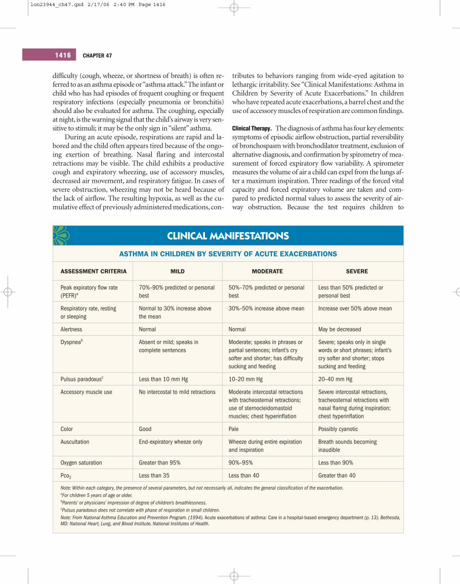
1416 CHAPTER 47
CLINICAL MANIFESTATIONS
ASTHMA IN CHILDREN BY SEVERITY OF ACUTE EXACERBATIONS
ASSESSMENT CRITERIA MILD MODERATE SEVERE
Peak expiratory flow rate (PEFR)a
Respiratory rate, resting or sleeping
Alertness
Dyspneab
Pulsus paradoxusc
Accessory muscle use
Color
Auscultation
Oxygen saturation
Pco2
50%–70% predicted or personalbest
30%–50% increase above mean
Normal
Moderate; speaks in phrases orpartial sentences; infant’s crysofter and shorter; has difficultysucking and feeding
10–20 mm Hg
Moderate intercostal retractionswith tracheosternal retractions;use of sternocleidomastoidmuscles; chest hyperinflation
Pale
Wheeze during entire expirationand inspiration
90%–95%
Less than 40
Less than 50% predicted orpersonal best
Increase over 50% above mean
May be decreased
Severe; speaks only in singlewords or short phrases; infant’scry softer and shorter; stopssucking and feeding
20–40 mm Hg
Severe intercostal retractions,tracheosternal retractions withnasal flaring during inspiration;chest hyperinflation
Possibly cyanotic
Breath sounds becominginaudible
Less than 90%
Greater than 40
Note: Within each category, the presence of several parameters, but not necessarily all, indicates the general classification of the exacerbation.aFor children 5 years of age or older.bParents’ or physicians’ impression of degree of children’s breathlessness.cPulsus paradoxus does not correlate with phase of respiration in small children.Note: From National Asthma Education and Prevention Program. (1994). Acute exacerbations of asthma: Care in a hospital-based emergency department (p. 13). Bethesda,MD: National Heart, Lung, and Blood Institute, National Institutes of Health.
70%–90% predicted or personalbest
Normal to 30% increase abovethe mean
Normal
Absent or mild; speaks incomplete sentences
Less than 10 mm Hg
No intercostal to mild retractions
Good
End-expiratory wheeze only
Greater than 95%
Less than 35
difficulty (cough, wheeze, or shortness of breath) is often re-ferred to as an asthma episode or “asthma attack.”The infant orchild who has had episodes of frequent coughing or frequentrespiratory infections (especially pneumonia or bronchitis)should also be evaluated for asthma. The coughing, especiallyat night, is the warning signal that the child’s airway is very sen-sitive to stimuli; it may be the only sign in “silent” asthma.
During an acute episode, respirations are rapid and la-bored and the child often appears tired because of the ongo-ing exertion of breathing. Nasal flaring and intercostalretractions may be visible. The child exhibits a productivecough and expiratory wheezing, use of accessory muscles,decreased air movement, and respiratory fatigue. In cases ofsevere obstruction, wheezing may not be heard because ofthe lack of airflow. The resulting hypoxia, as well as the cu-mulative effect of previously administered medications, con-
tributes to behaviors ranging from wide-eyed agitation tolethargic irritability. See “Clinical Manifestations: Asthma inChildren by Severity of Acute Exacerbations.” In childrenwho have repeated acute exacerbations,a barrel chest and theuse of accessory muscles of respiration are common findings.
Clinical Therapy. The diagnosis of asthma has four key elements:symptoms of episodic airflow obstruction, partial reversibilityof bronchospasm with bronchodilator treatment, exclusion ofalternative diagnosis, and confirmation by spirometry of mea-surement of forced expiratory flow variability. A spirometermeasures the volume of air a child can expel from the lungs af-ter a maximum inspiration. Three readings of the forced vitalcapacity and forced expiratory volume are taken and com-pared to predicted normal values to assess the severity of air-way obstruction. Because the test requires children to
lon23944_ch47.qxd 2/17/06 2:40 PM Page 1416

The Child with Alterations in Respiratory Function 1417
cooperate and follow instructions, it is usually administered tochildren over 4 or 5 years of age. Skin testing may be used toidentify allergens (asthma triggers).
Medical management includes medications, support ofparents and child, and education. Pharmacologic treatment ismatched to the severity of asthma for long-term control andfor management of acute episodes.See Table 47–12 for nation-ally recommended guidelines for the treatment of childrenover 5 years of age with acute and chronic asthma. Control ofasthma symptoms is the goal, and if control is not achievedwith the regimen prescribed, then the regimen should bechanged to correspond to the next step in asthma severity.Once control of asthma symptoms is achieved, the treatmentplan can be reviewed in 1 to 6 months to determine if a stepback in asthma treatment is appropriate (Hogan & Wilson,2003). See Table 47–13 for medications used to treat asthma.
Some healthcare providers encourage children to use apeak expiratory flow meter to identify when an obstruc-tion is occurring. This device measures the child’s ability topush air forcefully out of the lungs, similar to a spirometer.Medication administration can be based on peak expira-tory flow rate (PEFR) readings and the effectiveness oftreatment confirmed by improved PEFR numbers.
ALBUTEROL
DRUG GUIDE
Administer: For oral: Plain tablets can be crushed and mixed withsmall amounts of food or fluids. Do not crush coated tablets. Side ef-fects are more common with oral form. Inhalation: Shake containerwell. If second inhalation is prescribed, manufacturer recommendsthat 1 minute elapse between doses. Nebulizer: Follow agency pol-icy for operation of the nebulizer apparatus. Follow package insert di-rections for solution dilution. If solution is discolored, do not use. Donot remove nebulization capsules from the original package until justprior to use.
Monitor: Monitor pulse (cardiovascular effects may occur), centralnervous system stimulation (hyperactivity, excitement, nervousness,insomnia), therapeutic response to medication. Maintain hydrationfor weight. Do not use over-the-counter drugs (e.g., cold preparations)without physician approval as many contain drugs that intensify al-buterol action. Perform periodic arterial blood gases, pulmonary func-tion tests, and pulse oximetry.
Patient teaching: Review directions for correct use of medication.Teach the proper use and care of the inhaler. Have child demonstrateinhaler use. Emphasize that inhaler should not be used more fre-quently than prescribed, and to contact physician if albuterol fails toprovide relief. Avoid contact of inhalation drug with eyes. Help thefamily ensure that child has access to medicine during school hoursif needed.
Note: From Bindler, R. M., & Howry, L. B. (2005). Pediatric drugs and nursing im-plications (3rd ed.). Upper Saddle River, NJ: Prentice Hall-Health. Adapted.
Overview of ActionA synthetic sympathomimetic amine and moderately selective beta2-adrenergic agonist that has effect on bronchial, uterine, and vascularsupply to muscles. Produces bronchodilation, decreases airway re-sistance, facilitates mucus drainage, and increases vital capacity.
Routes, Dosage, FrequencyOral■ 2 to 6 years: Initial: 0.1–0.2 mg/kg/day 3 times a day
(maximum dosage 4 mg/dose)■ 6 to 12 years: 2 mg 3 to 4 times per day■ Over 12 years: 2 to 4 mg 3 to 4 times per day; extended-
release tablets: 4 to 8 mg every 12 hours
Inhalation: Oral inhalation for relief of acute bronchospasms or pre-vention of asthma symptoms
■ 6 to 12 years: 1 to 2 inhalations every 4 to 6 hours
Contraindications: Hypersensitivity to drug or its ingredients.
Side Effects: Restlessness, nervousness, tremors, dizziness, weak-ness, headache, hallucination, insomnia, tachycardia, palpitations,hypertension or hypotension, peripheral vasodilation, irritation ofnose and throat, blurred vision, dilated pupils, muscle cramps,hoarseness.
Nursing ImplicationsAssessment: Assess the respiratory status and obtain baseline pulseand peak expiratory flow rate.
NURSING PRACTICE
Markers of good asthma control include the following:
■ Minimal or no chronic symptoms day or night■ Minimal or no exacerbations■ No limitations on activities, no school missed, parents don’t
miss work■ Minimal use of short-acting beta2-agonists (Less than 1 time a
day or less than 1 canister a month)■ Minimal or no adverse effects from medications
Data from National Asthma Education and Prevention Program. (2002). NAEPPExpert Panel Report: Guidelines for the diagnosis and management of asthma.Update on selected topics 2002 (NIH Publication No. 02-5075). Bethesda, MD:National Institutes of Health, NHLBI.
See “Clinical Pathway: Stages of Inpatient Care forChildren with Severe Asthma.”
Most children with acute exacerbations respond to ag-gressive management in the emergency department. Chil-dren who do not respond or who are already being managedat home on corticosteroids have a greater chance of beingadmitted. Some children need mechanical ventilation.
MED
IALIN
KSkill 14–5: Peak Expiratory Flow M
eter
lon23944_ch47.qxd 2/17/06 2:40 PM Page 1417

1418 CHAPTER 47
TABLE 47–12 Asthma Severity Classification and Preferred Clinical Therapy for Children Older than 5 Years of Age
Classification (Steps) Description Medications for Long-Term Control
Step 1: Mild intermittent Brief exacerbations with symptoms no more often than twice a week.
Nighttime symptoms no more than twice a week.
Asymptomatic and normal PEFR between exacerbations.
No emergent visits and no asthma-related absences from school.
PEFR ≥ 80% of predicted with variability < 20%.
Step 2: Mild persistent Exacerbations more than twice a week, but less than once a day.
Nighttime symptoms more than twice a month.
Exacerbations may affect activity and cause absences from school.
PEFR ≥ 80% of predicted with variability of 20%to 30%.
Step 3: Moderate persistent Daily symptoms of coughing and wheezing.Exacerbations at least twice a week that may last for days.
Nighttime symptoms more than once per week.Exacerbations affect activity and several school absences occur.
PEF or FEV1 > 60% but < 80% of predicted with variability > 30%
Step 4: Severe persistent Continuous daytime symptoms, limited physical activity.
Frequent exacerbations.
Frequent nighttime symptoms.
Limited physical activity.
Hospitalizations are frequent with PICU admissions for severe exacerbations.
PEF or FEV1 ≤ 60% of predicted, with variability > 30%
Quick relief Bronchodilator as needed for symptoms. Intensity of treatment will depend on severity of exacerbation.
■ Preferred treatment: Short-acting inhaled �2-agonists by nebulizer, face mask, and space/holding chamber.
■ Alternative treatment: oral �2-agonist.
With viral respiratory infection
■ Bronchodilator every 4–6 hours up to 24 hours (longer with physician counsel); in general, repeat no more than once every 6 weeks.
■ Consider systemic corticosteroids if exacerbation is severe or patient has a history of previous severe exacerbations.
■ Use of short-acting �2-agonists > 2 times a week in intermittent asthma (daily or increasing use in persistent asthma) mayindicate the need to initiate (increase) long-term control therapy.
No daily medications needed.
Severe exacerbation may occur, separated by long periods of normal lungfunction and no symptoms. A course of systemic corticosteroids isrecommended.
Preferred treatment
■ Low-dose inhaled steroid.
Alternate treatment
■ Cromolyn, leukotriene modifier, or nedocromil.
OR
■ Sustained-release theophylline to serum concentration of 5–15 mcg/mL.
Preferred treatment
■ Low- to medium-dose inhaled corticosteroid.
Plus
Long-acting �2-agonists
Alternate treatment
■ Increase inhaled corticosteroids to within medium-dose range
OR
■ Low-dose inhaled steroid and either leukotriene modifier or theophylline.
If needed (particularly in children and adolescents with recurring severeexacerbations)
Preferred treatment
■ Increase inhaled corticosteroids within medium-dose range and addlong-acting inhaled �2-agonists.
Alternative treatment
■ Increase inhaled corticosteroids within medium-dose range and addeither leukotriene modifier or theophylline.
Preferred treatment
■ High-dose inhaled corticosteroids.
Plus
■ Long-acting inhaled �2-agonists.
And if needed,
■ Oral corticosteroids at 2 mg/kg/day (not to exceed 60 mg per day).Repeated efforts should be made to reduce systemic corticosteroidsand maintain control with high-dose inhaled corticosteroids.
MED
IALI
NK
Asth
ma
Guid
elin
es fo
r Chi
ldre
n Un
der 5
Adapted from National Asthma Education and Prevention Program. (2002). Expert Panel Report II: Guidelines for the diagnosis and management of asthma. Update on selectedtopics 2002 (NIH Publication No. 02-5075). Bethesda, MD: National Heart Lung and Blood Institute, National Institutes of Health.
lon23944_ch47.qxd 2/17/06 2:40 PM Page 1418

The Child with Alterations in Respiratory Function 1419
Rescue Medication Action/Indication Nursing Considerations
Beta2-agonists (short-acting)Albuterol, metaproterenol, terbutaline,levalbuterol: inhalation, PO
CorticosteroidsMethylprednisolone: IVPrednisonePrednisolone: PO
AnticholinergicIpratropium: inhalation
TABLE 47–13 Medications Used to Treat Asthma
Relax smooth muscle in airway, increase watercontent in bronchial mucus to promote muciliaryclearance resulting in rapid bronchodilation within5–10 minutes.
Drug of choice for acute therapy (metered-doseinhaler or nebulizer).
Diminish airway inflammation and obstruction,enhance bronchodilating effect of Beta2-agonists.
Used for moderate to severe acute exacerbationswhen single Beta2-agonist dose given in emergencydepartment does not resolve symptoms.
Inhibits bronchoconstriction and decreases mucusproduction.
Provides additive effects to short-acting Beta2-agonists during acute exacerbation.
■ Use this rescue medication before inhaledsteroid, wait 1–2 minutes between puffs, wait 15 minutes to give inhaled steroid. Child shouldhold breath 10 seconds after inspiring. Then rinse mouth and avoid swallowing medication.Use spacer.
■ Some side effects (tachycardia, nervousness,nausea and vomiting, headaches), but these areusually dose related.
■ Repetitive or excessive use can mask increasingairway inflammation and hyperresponsiveness andincrease need for higher dosage to get sameeffect.
■ Use of more than 1 canister a month indicatesinadequate control.
■ Not used as primary treatment.■ Onset of action is 4–6 hours.■ Short-term therapy for 3–10 days until symptoms
resolve or child achieves 80% peak expiratoryflow personal best.
■ Give with food.■ Give daily oral dose in early morning to mimic
normal peak corticosteroid blood level.■ Assess for potential adverse effects of long-term
therapy: decreased growth, unstable blood sugar,immunosuppression.
■ Not for primary treatment.■ Side effects include increased wheezing, cough,
nervousness, dry mouth, tachycardia, dizziness,headache, palpitations.
■ Avoid eye contact.
Controller Medication Action/Indication Nursing Considerations
Beta2-agonists (long acting)SalmeterolFormoterol: inhalation
MethylxanthinesTheophylline: POAminophylline: IV
Mast-Cell InhibitorsCromolyn sodiumNedocromil: aerosol
Relax smooth muscle in airway, used for nocturnalsymptoms and prevention of exercise-inducedbronchospasm.
Relax muscle bundles that constrict airways; dilateairway; provide continuous airway relaxation;sustained release for prevention of nocturnalsymptoms.
Aminophylline may be used for emergency adjuncttherapy in ICU, but use is controversial.
Anti-inflammatory, inhibit early- and late-phaseasthma response to allergens and exercise-inducedbronchospasm; may be used for unavoidable allergenexposure.
■ Should not be used for acute asthma attack.■ Should not be used in place of inhaled
corticosteroids.■ Caution against overdosage as side effects such
as tachycardia, tremor, irritability, insomnia willlast 8–12 hours.
■ Report use of more than 4 puffs a day as this mayindicate need for stepped-up therapy.
■ Tablet should not be crushed or chewed.■ Used for long-term control, so continuous
administration is needed; works best when aspecific amount is maintained in the bloodstream(therapeutic serum level, 10–20 mcg/L).
■ Requires serum level checks and doseadjustment.
■ Side effects include tachycardia, dysrhythmias,restlessness, tremors, seizures, insomnia, hypo-tension, severe headaches, vomiting, and diarrhea.
■ Not used at time of symptom development oracute exacerbation.
■ Must be used up to 4 times a day to be effective.■ Therapeutic response seen in 2 weeks, maximum
benefit may not be seen for 4–6 weeks.■ Adverse reactions include wheezing,
bronchospasm, throat irritation, nasal congestion,anaphylaxis.
(continued)
lon23944_ch47.qxd 2/17/06 2:40 PM Page 1419

1420 CHAPTER 47
Controller Medication Action/Indication Nursing Considerations
CorticosteroidsBeclomethasoneBudesonideFluticasoneTriamcinolone: inhalation
Leukotriene ModifiersMontelukast: POZafirlukast: PO
OtherHyposensitization (allergy shots),subcutaneous
TABLE 47–13 Medications Used to Treat Asthma—continued
Anti-inflammatory, controls seasonal, allergic, andexercise-induced asthma.
Effectively reduces mucosal edema in airways; usuallycombined with other asthma medications for control.
First-line therapy in asthma management.
Reduces inflammation cascade responsible for airwayinflammation.
Improves lung function and diminishes symptoms andneed for rescue medications.
Adjunct to inhaled corticosteroids in moderate tosevere asthma or substitute for inhaledcorticosteroids in mild asthma.
Series of injections that can reduce sensitivity tounavoidable allergens (e.g., environmental organisms—mold, pollen); gradual dose increase over time(buildup) increases the child’s tolerance to allergicsubstances; has been of help in some children.
■ Administer with spacer or holding chamber.■ Rinse mouth following treatment to reduce
chance of thrush and dysphonia.■ Monitor growth.■ Monitor for headache, gastrointestinal upset,
dizziness, infection.■ Use exactly as prescribed.
■ Administer montelukast in evening; may be givenwith or without food.
■ Administer zafirlukast 1 hour before or 2 hoursafter meal.
■ Family needs to report fever, acute asthmaattacks, flulike symptoms, severe headaches, orlethargy.
■ Take as prescribed, do not withdraw abruptly.
■ Use is controversial; some question about actualeffect.
Data from Baker, V. O., Friedman, J., & Schmitt, R. (2002). Asthma management, part II: Pharmacologic management. Journal of School Nursing, 18(5), 257–269; Belcher, D. (2002,November). Breathing easier with pediatric asthma: Pharmacologic management. Advance for Nurse Practitioners, 37–38, 79; Baren, J. M., & Puchalski, A. (2002). Current conceptsin the ED treatment of pediatric asthma. Pediatric Emergency Medicine Reports, 7(10), 105–115.
NURSING MANAGEMENT
NURSING ASSESSMENT AND DIAGNOSISThe nurse usually encounters the child and family in theemergency department or nursing unit. In these settings,acute care has become necessary because the child’s level ofrespiratory compromise cannot be managed at home.
Physiologic AssessmentIdentify the child’s current respiratory status first by assess-ing the ABCs—airway, breathing, and circulation—to makesure the child’s condition is not life threatening. If the child
DEVELOPING CULTURALCOMPETENCE
ASTHMA MANAGEMENTThe number of asthma attacks among children under 18 years variesgreatly by race and ethnicity. Non-Hispanic black children have thehighest prevalence followed by non-Hispanic white children and His-panic children (Centers for Disease Control, 2000).A recent study in-vestigated these disparities and found a difference in preventivemedications used for asthma. Findings revealed that differences inhealth beliefs, fear of steroids, or communication issues rather thanfinancial barriers may play a role in the use of preventive asthma med-ications (Lieu, Lozano, Finkelstein et al., 2002). Learn about the fam-ily’s cultural beliefs and practices. Parents of children from differentcultures may have concerns about daily medication regimens. Someprefer to use folk medicines such as rubbing oils or Vicks preparationson the child’s chest (Sydnor-Greenberg & Dokken, 2000). Individual-izing care through education is a key to successful treatment. Educa-tion should be based on a “thorough assessment of the child’s andfamily’s resources, health care beliefs, access to health care services,and management styles” (Swartz, Cantey-Banasiak, & Meadows-Oliver, 2005, p.78).
ALTERNATIVE ASTHMA TREATMENTSParents of children from different cultures may have concerns aboutdaily medication regimens. Some prefer to use folk medicines suchas rubbing oils or camphor (Vicks VapoRub) preparations on thechild’s chest. Learn about the family’s cultural beliefs and practices(Sydnor-Greenberg & Dokken, 2000). Up to 80% of Hispanic,African-American, and immigrant adolescents attending an inner-city highschool reported the use of complementary therapies for the treat-ment of asthma that included rubs, teas (chamomile, ginger, wildroot, and eucalyptus), prayer, and massage. There was no associationbetween complementary therapy use and ethnicity or immigrant sta-tus. Nearly all adolescents indicated that they would repeat the useof the complementary therapy. It is important to ask about comple-mentary therapy use by children and adolescents with asthma, andhow it is used in conjunction with traditional therapy (Reznik, Ozuah,Franco et al., 2002).
COMPLEMENTARY CARE
lon23944_ch47.qxd 2/17/06 2:40 PM Page 1420

The Child with Alterations in Respiratory Function 1421
CLINICAL PATHWAY
CONTINUOUS NEBULIZER FREQUENT NEBULIZER TRANSITION TO DISCHARGE, NEBULIZER CATEGORY TREATMENTS TREATMENTS TREATMENTS EVERY 4–6 HR
Outcomes
Assessments
Knowledge
STAGES OF INPATIENT CARE FOR CHILDREN WITH SEVERE ASTHMA
Patient will:■ Evidence stable vital signs and be
afebrile.■ Exhibit unlabored respirations and
patent airway.■ Tolerate full diet.■ Tolerate age-appropriate activity
without evidence of respiratorydistress, weakness, or exhaustion.
■ Have moist mucous membranes.■ Verbalize understanding and
demonstrate cooperation withrespiratory therapy.
Family exhibits effective copingmechanisms.
Vital signs every 4 hr and prn.Assess respiratory status every 4 hr
and prn. Monitor carefully forchanges in respiratory rate, skincolor, retractions, and/or flaring.
Discontinue cardiorespiratory monitor.Strict intake and output.Urine specific gravity every 8 hr.Humidification as ordered.Position patient in semi-Fowler’s or
high Fowler’s position.Continue pulse oximetry monitoring
while on oxygen.Wean oxygen, keeping O2 saturation at
greater than or equal to 92%.Provide quiet, restful environment.
Reinforce earlier teaching aboutongoing care.
Reinforce teaching about asthma andtreatments.
Renew detailed teaching with familyand patient about home careincluding medications, respiratorytherapy, activity, trigger agents,avoidance of respiratory infectionsand irritants, early signs andsymptoms of impending acuteepisode, and follow-up care.
Evaluate understanding of teaching.
Patient is afebrile with stable vital signs.Patient exhibits a patent airway and unlabored
respirations with activity.Patient’s respiratory rate and status has returned
to baseline.Patient tolerates activity without evidence of
respiratory distress, weakness, or exhaustion.Patient’s mucous membranes are moist.Family verbalizes/demonstrates home care
instructions, including strategies to reduceexposure to infectious illnesses and respiratoryirritants.
Family exhibits willingness to make lifestylechanges and cope with effects of chronicillness.
Family demonstrates/verbalizes ability to copewith ongoing stressors.
Family verbalizes beginning understanding ofhome care instruction including trigger agents,signs and symptoms of impending acuteepisode, and appropriate actions.
Vital signs every 8 hr and prn.Pulse oximetry reading every 4 hr if patient is on
oxygen.Continue weaning oxygen if O2 saturation greater
than or equal to 92%.Assess respiratory status every 8 hr and prn.
Monitor carefully for changes in respiratoryrate, skin color, retractions, and/or flaring.
Provide quiet, restful environment.
Reinforce earlier teaching about ongoing care.Complete discharge teaching to include diet,
follow-up care, signs and symptoms to report,follow-up physician visit, activity, andmedications: name, purpose, dose, frequency,route, dietary interactions, and side effects.
Provide family/patient with written dischargeinstructions.
Refer unmet teaching needs to outpatientservices.
Evaluate understanding of teaching.Refer knowledge deficits to community resources.
Patient will:■ Evidence stabilization of vital signs.■ Exhibit patent airway; experience
resolution of acute respiratorydistress.
■ Verbalize understanding anddemonstrate cooperation withrespiratory therapy.
■ Have oral intake equal to fluidmaintenance needs.
Family verbalizes feelings aboutillness.
Family displays effective coping.Family understands plan of care for
hospitalization.Family verbalizes beginning
understanding of asthma andongoing care and treatments.
Vital signs every 1–2 hr and prn.Continuous pulse oximetry monitoring.Cardiorespiratory monitoring while on
continuous nebulizer treatments.Potassium level 12 hr after on
continuous nebulizer treatments,then every 12 hr.
Assess respiratory status every 1–2 hrand as necessary.
Monitor carefully for changes inrespiratory rate, skin color,retractions, and/or flaring.
Strict intake and output.Urine specific gravity every 8 hr.Humidification as ordered.Position patient in semi-Fowler’s or
high Fowler’s position.Oxygen as ordered to maintain O2
saturation greater than or equal to 95%.
Provide quiet, restful environment.Assess cognitive/developmental level.
Initiate teaching regarding ongoingcare, including procedures,treatments, and medications.
Initiate patient and family teachingabout asthma, its treatments, triggeragents, avoidance of respiratoryinfections and irritants, and earlysigns and symptoms of infections.
Evaluate understanding of teaching.Provide information at
developmental/cognitive level.
(continued)
lon23944_ch47.qxd 2/17/06 2:40 PM Page 1421

1422 CHAPTER 47
CLINICAL PATHWAY—continued
CONTINUOUS NEBULIZER FREQUENT NEBULIZER TRANSITION TO DISCHARGE, NEBULIZER CATEGORY TREATMENTS TREATMENTS TREATMENTS EVERY 4–6 HR
Psychosocial
Diet
Activity
Medications
Transfer/discharge plans
STAGES OF INPATIENT CARE FOR CHILDREN WITH SEVERE ASTHMA
Provide age-appropriatecommunication and support.
Encourage verbalization of concerns.Encourage family to participate in
care.Reassure patient and family of
ongoing surveillance of patientstatus and responses to therapy.
Age-appropriate diet as tolerated,providing small, frequent, nutritiousfeedings.
Avoid cold fluids and foods.Encourage fluid intake.
Maintain safety precautions.Balance activity with planned rest
periods.Increase activity as tolerated.Encourage age- and status-
appropriate diversional activities.
Oral steroids as ordered.Albuterol nebulizer treatments every
2 hr.Acetaminophen as ordered for
temperature over 38.3°C (101°F).
Continue to review progress towarddischarge goals.
Determine need for home respiratorytherapy and outpatient teaching forbreathing exercises.
Finalize discharge plans.Make appropriate referrals.
Provide age-appropriate communication andsupport.
Encourage verbalization of concerns.Provide ongoing support and encouragement to
family.
Age-appropriate diet as tolerated, providingsmall, frequent, nutritious feedings.
Avoid cold fluids and foods.Encourage fluid intake.
Maintain safety precautions.Balance activity with planned rest periods.Encourage age- and status-appropriate
diversional activities.Activity as tolerated.
Oral steroids as ordered.Albuterol nebulizer treatment every 4–6 hr.Start or restart home therapy.Observe patient for reaction to new medications,
if applicable.Patient sent home with inhaler.
Finalize plans for home care if needed.Make appropriate referrals.Complete discharge teaching.
Orient patient to setting.Provide age-appropriate
communication and support.Encourage verbalization of concerns.Encourage family to participate in
care.Reassure patient and family of
ongoing surveillance of patientstatus and responses to therapy.
Clear liquids as tolerated.Avoid cold fluids and foods.Encourage fluid intake.
Assess safety needs and provideadequate precautions.
Encourage age- and status-appropriate diversional activities.
Balance activity with planned restperiods.
IV fluids only if unable to take oralfluids to equal maintenance.
Oral steroids as ordered.Ipratropium as ordered.Continuous albuterol nebulizer
treatments until respiratory distressdecreases.
Give oral potassium if K level is lessthan 3.5.
Acetaminophen as ordered fortemperature over 38.3°C (101°F).
Continue to review progress towarddischarge goals.
Review discharge plans.
is moving air or talking, assess the quality of breathing. As-sess the respiratory rate. Inspect the chest for retractions toassess the severity of respiratory distress. Auscultate thelungs for the quality of breath sounds and for the presenceor absence of wheezing. Observe the child’s color and assessthe heart rate. Note whether a cough or stridor is present.Obtain oxygen saturation via pulse oximeter. Determine theseverity of symptoms from the “Clinical Manifestations”table on page 1416. Only move on to other symptoms afterfinding no life-threatening respiratory distress.
Assess skin turgor, intake and output, and specificgravity. Because asthma can be a symptom of another ill-ness, perform a head-to-toe assessment to identify otherassociated problems (Tables 47–1 and 47–7). (See Skills9–1 to 9–22.)
Psychosocial AssessmentAssess the child’s anxiety. (See Table 47–9.) In an olderchild whose asthma was previously diagnosed, assesswhether the child thinks this episode could have been
SKILLS
lon23944_ch47.qxd 2/17/06 2:40 PM Page 1422

The Child with Alterations in Respiratory Function 1423
avoided if medication had been taken. Look for clues tohidden stress and self-blaming.
Common nursing diagnoses for the child experiencingan acute asthmatic episode include the following:
■ Ineffective Airway Clearance related to airwaycompromise, copious mucus secretions, and coughing
■ Impaired Gas Exchange related to airway obstruction,possible additional respiratory illness, and poorresponse to medication
■ Risk for Deficient Fluid Volume related to difficulty intaking adequate fluids with respiratory distress
■ Anxiety/Fear (Child or Parents) related to difficultybreathing and change in health status
■ Ineffective Therapeutic Regimen Management (Family)related to inadequate education on daily managementof a chronic disease
PLANNING AND IMPLEMENTATIONPharmacologic and supportive therapies are used to re-verse the airway obstruction and promote respiratoryfunction. Nursing interventions center on maintaining air-way patency, meeting fluid needs, promoting rest and stressreduction for the child and parents, supporting the family’sparticipation in care, and giving the family informationthat lets them manage the child’s disease.
Maintain Airway PatencyIf the child is exhibiting breathing difficulty, give supple-mental oxygen by nasal cannula or face mask. Humidifiedoxygen should be used to prevent drying and thickening ofmucus secretions. Place the child in a sitting (semi-Fowler’s) or upright position to promote and ease respira-tory effort. Evaluate the effectiveness of positioning andoxygen administration by pulse oximeter and by observingfor improved respiratory status. (See Skill 14–1. )
The respiratory distress and need for supplementaloxygen can be stressful for parents and child alike (Figure47–4 ). Encouraging the parents’ presence can be reas-suring for the child. Keep the parents informed of proce-dures and results, and get their input when developing thetreatment plan.
Many medications are given by the aerosol route(Figure 47–5 ). The advantages of aerosol are that themedication acts quickly, enabling the pulmonary bloodvessels to absorb the inhaled medication, systemic effectsare minimized, and the inhaled droplets provide the addedbenefit of moisture. Continuous aerosol treatments maybe used for some children with severe exacerbations. Mon-itor the child for side effects. The frequency of vital signassessment is related to the severity of symptoms. (SeeSkill 11–11. )SKILLS
SKILLS
Meet Fluid NeedsFluid therapy is often necessary to restore and maintain ad-equate fluid balance. Adequate hydration is essential tothin and break up trapped mucous plugs in the narrowedairways. An adequate oral intake may not be possible withthe child’s compromised respiratory status. An intravenousinfusion may be needed, and this route also may be used foradministering medications and providing glucose. Over-hydration must be avoided to prevent pulmonary edema insevere asthma attacks.
As respiratory difficulty diminishes, offer oral fluidsslowly. Iced beverages precipitate bronchospasms in some
Acute exacerbations of asthma may require management in the emergencydepartment. The child is placed in a semisitting position to facilitate respi-ratory effort. Providing support to both the child and parent is an importantpart of nursing care during these acute episodes. This mother is exhaustedafter a sleepless night of caring for her son.
FIGURE 47–4
NURSING PRACTICE
Spacers with valves are often used with metered dose inhalers (MDIs)for children who cannot coordinate inspiration with medication re-lease.The spacer captures the aerosol released by the MDI in a reser-voir for the child to breathe in over a couple of minutes. This providesa benefit similar to nebulizer treatment, but it takes less time and isless expensive (Steinbach, 2000). When teaching the child to use anMDI without a spacer, let the child learn to breathe in slowly withstraws.
lon23944_ch47.qxd 2/17/06 2:40 PM Page 1423

1424 CHAPTER 47
Medications given by aerosol therapy allow children the freedom to playand entertain themselves.
FIGURE 47–5
children with asthma. It is safest to offer the asthmatic childroom-temperature or slightly cooled fluids without ice.Determine the child’s fluid preferences and give choiceswhere possible. Monitor intake and output and assess spe-cific gravity frequently to evaluate the child’s hydration sta-tus. Involving parents in feeding can help gain the child’scooperation in taking oral fluids.
Promote Rest and Stress ReductionThe child who has had an acute asthmatic episode is usu-ally very tired when admitted to the nursing unit. Laboredbreathing and low oxygen status have left the child ex-hausted. Put the child in a quiet, private room if possible,but accessible for frequent monitoring to promote relax-ation and rest. Avoid repeatedly disturbing the child bygrouping tasks.
Support Family ParticipationThe parents may stay with the child, but may be exhaustedafter hours of their child’s respiratory distress. Give par-ents the option of assisting with the child’s treatments,rather than expecting them to do it in addition to com-forting the child. Provide frequent updates about thechild’s condition and encourage the parents to take breaksas needed.
Length of hospitalization depends on the child’s re-sponse to therapy. Any underlying or accompanying healthproblem, such as preexisting lung disease or pneumonia,
can complicate and extend the child’s hospital stay. Com-municate with the family of the hospitalized child at leastonce a day about the child’s condition.
Discharge Planning and Home Care TeachingParents need a thorough understanding of asthma—howto prevent attacks and provide treatment to maintain thechild’s health and avoid unnecessary hospitalization. Whenpossible, educate parents when they are rested but refer thechild to a healthcare provider for more extensive educa-tion. Support of parents and the child should focus onhelping them to understand and cope with the diagnosisand the need for daily management to promote near-normal respiratory function.
Discharge planning for the asthmatic child focuses on in-creasing the family’s knowledge about the disease, medica-tion therapy, and the need for follow-up care according toguidelines of the National Asthma Education and PreventionProgram. The required lifestyle changes may be difficult forthe child and parents. The need to modify the home by re-moving a loved pet, for example, may create stress. When afamily will not give up a pet, discuss the need to bathe the petfrequently to reduce animal dander. The nurse can facilitatediscussion and clarification of ways to prevent asthmaepisodes.Teach the family how to measure and interpret peakexpiratory flow readings (see Skill 11–12 ). Discussrescue medications to manage asthma episodes, as well ascontroller medications for daily management. Reassure thefamily that most children with asthma can lead a normal lifewith some modifications.
Nursing Care in the CommunityNurses provide care to children with asthma in pediatri-cians’ offices, specialty asthma clinics, schools, and summercamps. Once the stress of the acute episode has passed, thecommunity setting is ideal for ongoing coordinated educa-tion about asthma management. Help the parents andchild to understand the diagnosis and the need for dailymanagement to promote near-normal respiratory functionwhile the child continues to grow and develop normally.
Key points to cover in asthma education include thefollowing (Hayes, Djaferis, Gattasso et al., 2004):
■ Explain that asthma is a chronic and progressivecondition, but parents should expect that it can becontrolled.
■ Stress the need to follow the prescribed medicationschedule, and discuss strategies to remember dailymedications (specific time of day or daily event).
■ Discuss possible adverse drug effects.
■ Review proper use of metered dose inhaler, spacer, andpeak flow meter.
SKILLS
MED
IALI
NK
Skill
11–
12
lon23944_ch47.qxd 2/17/06 2:40 PM Page 1424

The Child with Alterations in Respiratory Function 1425
■ Explain that exercise is important for health, and asthmainduced by exercise can be managed with medication.
■ Discuss keeping an asthma symptom diary.
If the child has severe asthma and uses high doses ofaerosol or oral glucocorticoids to control asthma attacks,monitor the child’s growth every 6 months as the diseaseand medications may affect overall growth. Review thefamily’s daily plan for monitoring the child’s respiratorystatus and the parent’s ability to identify the timing andtype of stepped-up care needed to manage worseningsymptoms. Review and reinforce the child’s technique foruse of the metered dose inhaler. The goal is to bring asthmaepisodes under control with stepped-up care before a sig-nificant episode occurs (see Table 47–12).
Make sure the child has a supply of medications atschool or child care as well as at home. Help the childlearn the signs of early respiratory distress and how torequest medications when at school before signs getmore serious. Help the parents communicate withschool personnel regarding the child’s condition, andhave an individual health plan developed so that med-ications are given as needed, even in preparation for ex-
ercise. For a young school-age child, make sure thechild’s teacher can help recognize respiratory distressand reduce the child’s fear of going to the nurse for res-cue medications.
Environmental control is an important part of asthmamanagement. When possible, pets should not be kept in thehome (and never in the child’s bedroom). Active dust mitecontrol should be attempted, but it is challenging as miteslive in carpets, bedding, upholstered furniture, and clothes.To help control dust mites in the child’s bedroom, encasethe pillow and mattress in plastic covers. Initiate cockroachcontrol. Smoke from cigarettes, wood stoves, and fireplacesall have the potential to trigger an asthma attack.
Refer to “Nursing Care Plan: The Child with Asthmain the Community Setting” in Chapter 39 for additionalinformation. ∞EVALUATIONExpected outcomes of nursing care include the following:
■ The child recognizes early asthma symptoms and usesrescue medications, hydration, and relaxationbreathing before severe respiratory distress occurs.
MEDICATION MANAGEMENT IN ASTHMA
Clinical Question (Problem)Many children with asthma have less than optimal medication manage-ment to control symptoms and have increased asthma episodes. Whatinformation about the family’s knowledge and perceptions about man-aging the child with asthma could help nurses collaborate more effec-tively with families to improve asthma management?
EvidenceRecent research on parental beliefs, knowledge, experience of living witha child who has asthma, and attitudes about controller medication wasconducted through interviews with 18 mothers of children and adoles-cents with asthma. Parents indicated that they learned to manage thechild’s asthma through “trial and error.” Even though they learned aboutmedications at one time, they still had gaps in knowledge about med-ication actions. Parents also wished to have health professionals listen tothem regarding their child’s healthcare needs. The parents reported thatdaily medication management was the most difficult aspect of asthmacare, but they saw a good response in the child when used (Peterson-Sweeney, McMullen, Yoos et al., 2003). Another study involving theparents of 109 children with asthma explored their attitudes and under-standing of asthma. Only 27 of the 78 children with persistent asthmahad an appropriate medication regimen, and 17 parents reported usingno anti-inflammatory medication even when the child had moderate tosevere asthma. Some parents believed inhaled steroids should be a last-resort therapy or if steroids were used for a while they would not workwhen needed. These parents also anticipated that their children would
have activity limitations and episodic emergency department visits (Yoos,Kitzman, & McMullen, 2003).
ImplicationsDemonstrating respect for the parent’s knowledge of the child’s healthstatus and response to asthma management is an essential step in de-veloping an effective partnership with the parent. It is also important totalk with parents about their beliefs about daily medications for asthmamanagement, use of inhaled steroids, and their expectations of howasthma will affect their child. Gaining an understanding of the family’sbeliefs can help guide nursing education and collaborative developmentof a plan to manage the child’s asthma.
Critical ThinkingConsider the possible perceptions and beliefs of parents and childrenwith asthma in your practice setting. Develop an education program thatintegrates these beliefs and perceptions to help improve families’ un-derstanding of medication actions, the differences between inhaled andoral corticosteroids, and collaboration with health professionals to im-prove the control of their child’s asthma.
ReferencesPeterson-Sweeney, K., McMullen, A., Yoos, H. L., & Kitzman, H. (2003). Parental per-
ceptions of their child’s asthma: Management and medication use. Journal of Pe-diatric Nursing, 17(3), 118–125.
Yoos, H. L., Kitzman, H., & McMullen, A. (2003). Barriers to anti-inflammatory med-ication use in childhood asthma. Ambulatory Pediatrics, 3(July), 181–190.
EVIDENCE-BASED NURSING
MED
IALIN
KM
edication Administration—
Growth and Development
lon23944_ch47.qxd 2/17/06 2:40 PM Page 1425

1426 CHAPTER 47
Identify parents’ knowledge about the condition:1. Review why asthma occurs and assess parents’ understanding of
the physiologic process. Ask:■ What happens in your child’s lungs during an asthma attack?■ What are the early warning signs of an asthma episode in
your child?■ What are your child’s symptoms and how does he or she
respond to them? Does your child use the peak expiratoryflow meter to evaluate symptoms? Is a diary maintained ofthe child’s symptoms?
2. Identify asthma triggers and assess parents’ understanding of howto prevent, avoid, or minimize their effect in a timely manner. Ask:■ Do you know your child’s personal asthma triggers? Where do
most episodes begin? (Suggest that the parents and childkeep a notebook to track episodes so they can learn moreabout these triggers.)
■ What steps can you take or have you taken to minimize oreliminate your child’s exposure to indoor pollutants (quittingsmoking, environmental control, etc.)?
Set up a schedule for parents and children to learn asthma management:■ Discuss when and where to seek emergency medical help.■ Discuss actions parents can take before seeking medical
assistance.
HOME CARE INSTRUCTIONS FOR THE CHILD WITH ASTHMA
TEACHING HIGHLIGHTS
Review parents’ understanding of medication therapy:■ Provide information about medications: name, type of drug,
dose, method of administration, expected effect, possible sideeffects. Make sure they understand when controller and rescuemedications should be used.
■ Evaluate the child’s technique for the type of inhaler used as thetechniques vary. Review the technique for use of the peak flowmeter. See “Medication Administration—Growth andDevelopment” on the Companion Website for growth anddevelopmental issues regarding the use of inhalers.
Address associated issues:■ Storage and proper transport of medications.■ Financial considerations of medication cost and lifestyle
changes.■ Notification of child’s school or teacher; arrangements for the
child’s use of medications at school.■ Medical identification bracelet or medallion to facilitate
assistance when the child is away from home.
■ The child and family implement a daily treatment planfor asthma and reduce the number of asthma episodesthe child has.
■ The child with a serious asthma episode responds tooxygen, fluids, and medication therapy, avoidinghospital admission.
STATUS ASTHMATICUSStatus asthmaticus is unrelenting, severe respiratory distressand bronchospasm in an asthmatic child, which persists de-spite pharmacologic and supportive interventions. Withoutimmediate intervention the child with status asthmaticusmay progress to respiratory failure and die. The child isplaced in an ICU and may require endotracheal intubationwith assisted ventilation. The section on respiratory failureearlier in the chapter gives additional information on thenurse’s role in providing emergency respiratory care.
CYSTIC FIBROSISCystic fibrosis (CF) is a common inherited autosomal re-cessive disorder of the exocrine glands that results in
physiologic alterations in the respiratory, gastrointestinal,integumentary, musculoskeletal, and reproductive sys-tems. The incidence of CF varies by race—1:3300 inwhites, 1:17,000 in blacks, 1:8000 in Hispanics, and1:32,000 in Asian Americans (McMullen & Bryson, 2004).Approximately 20,000 children and 10,000 adults have CFin the United States (Cystic Fibrosis Foundation, 2004)(Figure 47–6 ). The median life span for individualswith cystic fibrosis is 35 years (Cystic Fibrosis Founda-tion, 2005).
ETIOLOGY AND PATHOPHYSIOLOGYA gene isolated on the long arm of chromosome 7 directsthe function of the CF transmembrane conductance regu-lator (CFTR). With a defective CFTR, the exocrine and ep-ithelial cells have defective chloride-ion transport anddecreased water flows across cell membranes. This resultsin an abnormal accumulation of viscous, dehydrated mu-cus that affects the respiratory, gastrointestinal, and geni-tourinary systems. Inflammation and lung changes arepresent as early as 4 weeks of age. Ultimately, all body or-gans with mucous ducts become obstructed and damaged(McMullen & Bryson, 2004).
lon23944_ch47.qxd 2/17/06 2:40 PM Page 1426

The Child with Alterations in Respiratory Function 1427
Cystic fibrosis is an inherited autosomal recessive disorder of the exocrineglands, so it is not uncommon to see siblings with it such as this brotherand sister.
FIGURE 47–6
Because of the blocked pancreatic ducts and resultingpancreatic damage, the natural enzymes necessary to digestfats and proteins are not secreted, and malabsorption re-sults in the majority of children by 1 year of age. In somechildren, the pancreas may stop producing insulin and thebody may fail to use insulin normally, resulting in the de-velopment of diabetes mellitus.
The lungs are always filled with mucus, which the res-piratory cilia cannot clear. This causes air to becometrapped in the small airways, resulting in atelectasis (pul-monary collapse). Secondary respiratory infections occurbecause secretions provide an environment conducive tobacterial growth. Respiratory failure is the leading cause ofmortality.
Nearly all males who have CF are sterile because ofblockage or absence of the vas deferens. Females have diffi-culty conceiving because of chronic illness, and increasedmucus secretions in the reproductive tract interfere withthe passage of sperm (McMullen & Bryson, 2004).
Metabolic function is altered as a result of the imbal-ances created by excessive electrolyte loss through perspi-ration, saliva, and mucus secretion. The children are at riskfor dehydration secondary to electrolyte imbalance. The“salty taste” of the skin is the result of sodium chloride thatmakes its way through skin pores to the skin surface.
CLINICAL MANIFESTATIONSThe primary symptom of CF is the production of thick,sticky mucus. One of the earliest signs is meconium ileus,a small bowel obstruction in the newborn’s first 48 hoursof life. Stools of the child with cystic fibrosis characteristi-cally are frothy (bulky and large quantity), smell foul, con-tain fat (are greasy), and float. Constipation is commonand intestinal obstruction may occur in older children.Rectal prolapse, resulting from the large, bulky, difficult-to-pass stools, may occur.
Other signs and symptoms include a chronic moist,productive cough and frequent respiratory infections.Frontal headaches, facial tenderness, and purulent nasaldischarge are signs of a chronic sinus infection. Nasalpolyps are found in 10% of children with CF (McMullen &Bryson, 2004). Most children have difficulty maintainingand gaining weight despite a voracious appetite because ofmalabsoption and frequent infections. Infants and chil-dren may have a delayed bone age, short stature, and de-layed onset of puberty. Clubbing of the tips of the fingersand toes occurs as the disease progresses (Figure 47–7 ).
CLINICAL THERAPYCF is usually diagnosed in infancy or early childhood withone of three major presentations: newborn meconiumileus, malabsorption or failure to thrive, or chronic recur-rent respiratory infections. Some children with a milderform of the disease, however, may reach the teen or youngadult years before symptoms appear. The mean age at di-agnosis is 4.8 years (Farrell, Kosorok, Rock et al., 2001).
NURSING PRACTICE
Recent advances in localization of the CF gene have led to suc-cessful techniques for prenatal diagnosis and carrier testing. Thereare nearly 1000 mutations of the CFTR gene that can cause CF(Orenstein, Winnie, & Altman, 2002). Genetic testing to identify themajority of CF gene alterations is available to adults with a positivefamily history, partners of people with CF, couples planning a preg-nancy in a high-risk population, and couples seeking prenatal test-ing. Rare CF gene alterations are not always detected by genetictesting (Cystic Fibrosis Foundation, 2004).
Digital clubbing.
FIGURE 47–7
lon23944_ch47.qxd 2/17/06 2:40 PM Page 1427

1428 CHAPTER 47
This 6-month-old girl is being evaluated for cystic fibrosis using the sweat test.
FIGURE 47–8
Clinical Therapy Rationale
Respiratory TherapyExercise and physical fitness Promotes maintenance of lung function
Chest physiotherapy for all lung segments (bilateral percussion or In association with coughing and breathing techniques secretions vibration while the patient is in a position to promote sputum drainage) move to bronchi from lung areas
Immunizations Prevention of viral and some bacterial infections
Chest tube drainage of air leaks Resolves pneumothorax
Thorocoscopy to sew over ruptured alveoli Repairs area of recurrent pneumothorax and prevents future episode insame location
Lung transplantation Reversal of respiratory failure
Gastrointestinal Tract TherapyAcid suppression preparation Gastroesophageal reflux worsens lung function; enteric coating of
enzyme supplements is affected by high acid content in duodenum
Hyperosmolar enemas, Isotonic fluid lavage of the intestines orally or Enema relieves meconium ileus in most infants; fluid lavage reducesby nasogastric tube distal intestinal obstruction
NutritionWell-balanced diet with 120–150% of RDA recommended calories and Promotes essential nutrient balance for health, growth, and weight 200% of RDA recommended protein and moderate fat maintenance; nutritional counseling to support high-caloric intake;
cultural-socioeconomic issues important
Pancreatic enzyme supplements Assists in digestion of nutrients and decreasing fat and bulk
TABLE 47–14 Clinical Therapy for Cystic Fibrosis
Newborn screening using dried blood samples for im-munoreactive trypsinogen is mandated in some states, andgenetic testing of the child’s DNA is performed to confirma positive test (Parad & Comeau, 2003). A sweat chloridetest by pilocarpine iontophoresis is used to test childrenwith classic symptoms or a positive family history (Mc-Mullen & Bryson, 2004). A sweat chloride concentration of50 to 60 mEq/L is suspicious. If the chloride concentra-tion is greater than 60 mEq/L, it is diagnostic with othersigns. Two tests are performed to confirm the diagnosis(Figure 47–8 ). A spirometer is used on children olderthan 6 years to monitor pulmonary function. Sputum cul-tures are obtained to identify infectious organisms and an-tibiotic sensitivities.
Clinical therapy focuses on maintaining respiratoryfunction, managing infection, promoting optimal nutri-tion and exercise, and preventing gastrointestinal blockage(Table 47–14). See Table 47–15 for medications used totreat CF. Newly diagnosed children will have no or minimalsymptoms and near-normal lung function if aggressivelytreated. Pulmonary function declines 2% to 4% per yeareven with aggressive treatment (Varlotta, 1998).
Improvements in medical management and optimalnutrition have prolonged the lives of children with CF.However, new complications such as CF-related diabetesmust be carefully coordinated along with the progression ofthe disease. Lung transplantation is occasionally performed,
and approximately 50% of cases survive for the first 5 years(McMullen & Bryson, 2004). CF is ultimately terminal,however, because of the progressive multisystem changesand the difficulty of long-term infection management.M
ED
IALI
NK
Nurs
ing
Care
Pla
n: Th
e Ch
ild w
ith C
ystic
Fib
rosi
s
lon23944_ch47.qxd 2/17/06 2:40 PM Page 1428

The Child with Alterations in Respiratory Function 1429
Medications Actions
Aerosol bronchodilators
Aerosol DNAse
Corticosteroids and high-dose ibuprofen on alternate days
Antibiotics (oral, IV, and inhalation)
Pancreatic enzyme supplements (Cotazym-S,Pancrease, Viokase)
Multivitamins and vitamin E in water-soluble form; vitamins A, D, and K given when deficient; iron supplementation
Ursodeoxycholate
Lactulose
TABLE 47–15 Medications Used to Treat Cystic Fibrosis
Opens large and small airways; use before chest physiotherapy and with symptoms; few studiesexist to demonstrate their effectiveness.
Loosens, liquefies, and thins pulmonary secretions; decreases risk of developing pulmonaryinfections requiring parenteral treatment in some patients (McMullen & Bryson, 2004).
Anti-inflammatory agents: reduces inflammatory response to infection; alternate day use todecrease side effects of steroids; decreases progression of lung damage in preadolescents withmild disease.
Treats infections. Higher doses than normal and prolonged courses may be needed. Antibioticselection should be based on culture sensitivities. Intermittent administration of tobramycin byinhalation improves pulmonary function.
Assists in digestion of nutrients decreasing fat and bulk; given prior to food ingestion, takenwith meals and snacks.
Cystic fibrosis interferes with vitamin production; supplements are required in water-solubleform for better absorption (vitamins A, D, E, and K are naturally fat soluble); iron deficiencyresults from malabsorption syndrome.
May slow progression of hepatic lesion in CF. Given when patient has elevated liver enzymes orevidence of portal hypertension.
May abort early distal intestinal obstruction syndrome and prevent recurrences.
NURSING MANAGEMENTCare of the child with previously diagnosed CF is the focusof the following discussion.
NURSING ASSESSMENT AND DIAGNOSIS
Physiologic AssessmentPhysical assessment of the child focuses on adequacy ofrespiratory function. The child with CF usually is admittedwith symptoms of an upper respiratory infection. Obtain aset of baseline vital signs. Assess the child’s respiratory sta-tus. Auscultate the chest for breath sounds, crackles, andwheezes. Inquire about the frequency and character of thechild’s cough and characteristics of sputum as changes maybe related to a new infection. Note any cyanosis or clubbingof the extremities.
Evaluate the child’s growth, plotting the weight andheight on a growth curve. Determine whether the child ismaintaining an appropriate growth pattern or is malnour-ished. Inquire about the child’s appetite and dietary intake.How are nutritional supplements, pancreatic enzymes, andvitamins used?
Assess the child’s stooling pattern. Identify whether thechild has problems with abdominal pain or bloating, and
whether these problems can be related to eating, stooling,or other activities. Palpate the abdomen for liver size, fecalmasses, and evidence of pain.
Psychosocial AssessmentThe emotional stress of this chronic disease may not bereadily apparent on admission, particularly if the child’ssymptoms are mild and not imminently life threatening.Ongoing observation of the child’s and parents’ behaviorhelps direct nursing interventions throughout hospitaliza-tion (see Table 47–9). Parents may feel guilt as carriers ofthe disease. Siblings may also show signs of difficulty indealing with the illness. Link the family to support groups.School-age children and adolescents often are embarrassedat being viewed as different from playmates and peers. Askhow the child and adolescent feel about the need for a spe-cial diet, medications, and limitations.
Ask parents how the child’s illness has affected day-to-day functioning, any potential conflicts with family activi-ties, and how they have adapted to the child’s plan of care.Ask what the parents have told the child and siblings aboutthe disease. What questions have the child and siblingsasked about CF, and how have parents answered them? Hasthe child ever asked about his or her life expectancy? If not,what would parents say if asked?
lon23944_ch47.qxd 2/17/06 2:40 PM Page 1429

1430 CHAPTER 47
Developmental AssessmentGrowth and development may be altered by the chronicnature of the disease. Children with cystic fibrosis may begrowth retarded. Compare the child’s height and weight toage norms and observe the adolescent for the appearanceof secondary sex characteristics, which are often delayed.
Common nursing diagnoses for the child with cystic fi-brosis include the following:
■ Ineffective Airway Clearance related to thick mucus inlungs
■ Risk for Infection related to the presence of mucoussecretions conducive to bacterial growth
■ Imbalanced Nutrition: Less than Body Requirementsrelated to inability to digest nutrients
■ Parental Role Conflict related to interruptions in familylife due to the home care regimen and child’s frequentexacerbations
PLANNING AND IMPLEMENTATIONNursing management involves supporting the child andfamily initially, when the diagnosis is made, during subse-quent hospitalizations, and during visits to specialty andprimary healthcare providers. The nurse’s role begins withimplementing specific medical therapies and providingnursing care to meet the child’s physiologic and psy-
chosocial needs. Respiratory therapy, medications, anddiet must be coordinated to promote optimal body func-tion. Psychosocial support and reinforcement of thechild’s daily care needs are important in preparation forhome care.
Children with cystic fibrosis require periodic hospital-ization when a severe infection occurs or for a pulmonaryand nutritional “tune-up.” The child is usually placed in asingle room to reduce the spread of infectious organismswith standard precautions. Children with CF are not co-roomed to reduce the risk for transfer of the infectious or-ganisms Pseudomonas and Burkholderia cepacia (CysticFibrosis Foundation,2000).Respect the parents’experiencesas the child’s primary care provider and include them in thechild’s routine care as much as possible. However, parentsmay view the hospital stay as a break from the rigorous dailypulmonary routine at home and need support in taking ad-vantage of some “down” time. While the family is often pro-ficient at providing physical care to the child, the nurseshould take the opportunity to review basic and new infor-mation about respiratory care, medications, and nutrition.
Provide Respiratory TherapyChest physiotherapy is usually performed one to threetimes per day before meals to clear secretions from thelungs, as coughing may stimulate vomiting (Figure 47–9
). Parents and other family members can learn to help
Postural drainage can be achieved by clapping with a cupped hand on the chest wall over the segment to be drained to create vibrations that are trans-mitted to the bronchi to dislodge secretions. A, If the obstruction is in the posterior apical segment of the lung, the nurse can do this with the child sit-ting up. B, If the obstruction is in the left posterior segment, the child should be lying on the right side. Several other positions can be used dependingon the location of the obstruction. C, A high-frequency chest wall oscillation vest is another option for chest physiotherapy that the child can indepen-dently manage.
FIGURE 47–9
A B C
lon23944_ch47.qxd 2/17/06 2:40 PM Page 1430

The Child with Alterations in Respiratory Function 1431
with these necessary treatments. Pulmonary care may in-volve aerosol treatments and antibiotics when indicated(see Table 47–11). (See Skill 14–25.)
Administer Medications and Meet Nutritional NeedsAntibiotics for acute exacerbation are provided by oral, in-halation, and intravenous routes. Because children with CFhave an increased clearance of most antibiotics, they needhigher doses and long treatment courses. Renal functionneeds to be monitored and serum antibiotic levels may betaken to ensure therapeutic dosing.
Digestive problems can be eased with pancreatic en-zymes and dietary modification. Pancreatic enzyme sup-plements come in powder sprinkles and capsule form andare taken orally with all meals and large snacks. The amountneeded is individualized based on the child’s nutritionalneeds and digestive response to these supplements. Fami-lies need to learn which foods if any to avoid that contributeto a child’s gastrointestinal problems. The goal is to achievenear-normal, well-formed stools and adequate weight gain.
Fat-soluble vitamins (A, D, E, and K) are not com-pletely absorbed from food; therefore, they must be takenin water-soluble form. Multivitamins taken twice daily usu-ally are sufficient to prevent deficiency. The diet should bewell balanced, with an emphasis on high caloric value. Fatsand salt are both necessary in the diet. Balanced with pan-creatic enzyme supplements, moderate fat intake adds animportant source of extra fuel. Respiratory complicationscause additional energy expenditure, and some children re-quire supplemental nasogastric or gastrostomy feedings togain and maintain weight.
Provide Psychosocial SupportHelp the parents and child learn what they must do to main-tain health after discharge. Emotional support is essentialbecause the diagnosis of this disorder creates anxiety andfear in both the parents and the child. They need assistancewith emotional and psychosocial issues relating to disci-pline, body image (stooling and odor), frequent rehospital-ization, the potential fatal nature of the illness, the child’sfeeling of being different from friends, and overall financial,social, and family concerns. Because the disorder is inher-ited, families may have more than one child with cystic fi-brosis. Parents may have unspoken feelings of anger andguilt, blaming themselves for their children’s condition.
Discharge Planning and Home Care TeachingReview the family’s potential need for financial assistancedue to the out-of-pocket costs for medications, supplies,equipment, and medical follow-up. Home care of the childwith CF is expensive and can be draining on the family’s fi-nances. If the family requires financial assistance, referthem to the appropriate social services.
SKILLS
CF affects all family members and disrupts activities ofdaily living for everyone. It is important to refer families tofamily counseling and group therapy with families of otherchildren with CF, if indicated. The Cystic Fibrosis Founda-tion is a source for family information. See the CompanionWebsite for links.
Nursing Care in the CommunityNurses may encounter the child with cystic fibrosis in anyof the following settings: clinics specializing in the disease,pediatricians’ offices, and schools. The primary goal is tokeep the disease under control by promoting optimal nu-trition and assisting the family to reduce the incidence ofinfection. Nurses may also provide home care to the childwith cystic fibrosis following hospitalization for an acuteexacerbation or provide hospice care.Assessment. Perform the physical assessment as described forthe hospitalized child. Obtain oxygen saturation and spirom-etry readings if changes in respiratory status are suspected.Observe the child’s physical appearance, noting overall bodyproportions and any changes characteristic of long-term CF.Assess hearing acuity on a regular basis if the antibiotic to-bramycin is used as it has been associated with hearing loss.
NURSING PRACTICE
Parents often have a difficult time getting the child with cystic fibrosisto eat the extra calories needed for optimal nutrition, setting the stagefor a potential mealtime battleground. To be successful, parents needguidance about managing mealtime behaviors in addition to guidelinesfor preparing nutritional calorie-dense foods. Increase calorie intake byadding fats and high-calorie snacks between meals and before bed. Ex-tra intervention may be needed when the child’s weight is 85% to 90%of ideal weight for height. Children with adequate nutrition have a longerlife expectancy. See the Companion Website for links.
Review chest physiotherapy techniques with the par-ents, which should be performed as often as two or threetimes a day. Alternate bronchial hygiene therapy tech-niques such as a vest may be more easily accepted by thefamily. See “Bronchial Hygiene Therapy” on the Compan-ion Website. Encourage the child to exercise and participatein physical activities as this helps improve lung function,endurance, and airway clearance.
Managing the child’s nutritional needs is importantand takes time and energy. Parents need to learn how toprepare high-calorie foods, provide needed enzymes be-fore meals, what vitamins need to be given daily, and whatfoods should be avoided or eliminated because of thechild’s digestive problems. Make a referral to a nutritionisteither before or at the time of discharge.
MED
IALIN
KSkill 14–25: Perform
ing Chest Physiotherapy/Postural DrainageM
ED
IALIN
KBronchial Hygiene Therapy
lon23944_ch47.qxd 2/17/06 2:40 PM Page 1431

1432 CHAPTER 47
Inquire about the family’s and child’s emotional andpsychosocial responses to managing the illness. These is-sues are very important when the child is going throughmajor developmental stages.Management. Review the child’s use of bronchodilators andairway clearance techniques. To prevent a change in pul-monary status from progressing, short-term changes in caremay be recommended. These may include intravenous andaerosol medications and antibiotics, an increase in the num-ber of times chest physiotherapy is performed daily, andchanges in dietary management. Help the family select thebest time to fit the additional treatment into their schedule.
Malnutrition is a major problem for children with CF.Par-ents often need to plan meals and snacks for the young child toensure that adequate calories are consumed.Arrange for a con-sultation with a nutritionist if the family would benefit fromnew strategies to help meet the child’s nutritional needs.
Children with CF lose more than normal amounts ofsalt in their sweat. This loss can become intensified duringhot weather, strenuous exercise, and fever. Parents shouldallow the child to add extra salt to food and should permitsome salty snacks (pretzels with salt, pickles, carbonatedsoda). During periods of increased sweating, the childshould be encouraged to drink more fluids and increasesalt intake. Teach parents to recognize early symptoms ofsalt depletion, including fatigue, weakness, abdominalpain, and vomiting, and to contact the child’s healthcareprovider if these symptoms occur.
Adolescents with CF need special assistance in copingwith their disorder, especially as survival into adulthood iscommon. Help them identify normal adolescent changesversus those related to CF. Adolescents must learn how tocope with the difference they know exists between them-selves and peers. Provide information about potential infer-tility along with guidelines for safe sexual practices to reducethe risk for sexually transmitted infections. Females with CFmay be able to conceive and should be offered contraception.
Gradual assumption of responsibility for daily diseasemanagement is necessary. Adherence with the daily diseasemanagement may be a problem during adolescence. Individ-ualized planning to achieve their daily care regimen while en-abling them to interact with peers and participate in schoolactivities may be most helpful. Link adolescents to services toassist with planning appropriate educational and occupa-tional goals for their future. Palliative care planning shouldbe initiated as the disease progresses to respiratory failure.
EVALUATIONExpected outcomes of nursing care include the following:
■ The child and family develop proficiency in providingthe daily pulmonary care and reducing the incidence ofrespiratory infections.
■ The child and family develop a schedule and routinefor daily pulmonary care that fits into family andschool activities.
■ The child consumes adequate calories and pancreaticenzymes to support growth and to stay withindesirable weight ranges.
INJURIES OF THE RESPIRATORYSYSTEM
Airway compromise after an unintentional injury can causedeath if not managed quickly and effectively. Children arevulnerable to changes in respiratory function after injury. Achild’s airway can become easily obstructed because of itssmall size. The airway may be obstructed by the tongue,small amounts of blood, mucus, or foreign debris, as well asswelling in the respiratory tract or adjacent neck tissue. Ifthe child’s neck is flexed or hyperextended, the soft laryn-geal cartilage may also compress and obstruct the airway.
SMOKE-INHALATION INJURYExposure to fire conditions sets up dramatic responses inthe respiratory tract of children. In every age group, in-halation injury increases the child’s chance of death by20%, and it also increases the likelihood that the child willdevelop pneumonia (Kim, 2001).
The severity of the smoke-inhalation injury is influ-enced by the type of material burned and whether the childwas found in an open or closed space. The composition ofmaterials determines how easily they ignite, how fast theyburn, and how much heat they release. These factors influ-ence the production of smoke and toxic gases. Smoke, aproduct of the burning process that is composed of gasesand particles, is generated in varying volumes and density.The type and concentration of toxic gases, which are usu-ally invisible, affect the severity of pulmonary damage. Theduration of exposure to the smoke produced and any toxicgases contribute significantly to the child’s prognosis.
Exposure to extreme heat, common in house fires, leadsto surface injury and upper airway damage.The upper airwaynormally removes heat from inhaled gases, sparing the lowerairway from thermal damage. However, this action results inmarked edema, placing the small child at particular risk forairway obstruction. Edema develops rapidly over a few hoursand may lead to acute respiratory distress syndrome.
Carbon monoxide (CO) is a clear, colorless, odorlessgas present in all fire conditions as the fire consumes oxy-gen. The CO molecule binds more firmly to hemoglobinthan does oxygen. As a result, it replaces oxygen in circula-tion and rapidly produces hypoxia in the child. The longer
lon23944_ch47.qxd 2/17/06 2:40 PM Page 1432

The Child with Alterations in Respiratory Function 1433
the exposure to CO, the greater the hypoxia. The brain re-ceives inadequate oxygen, resulting in confusion. This ac-counts for the inability of fire victims to escape asconfusion progresses to loss of consciousness. The processcan be rapidly reversed, however, by timely administrationof 100% oxygen (Schweich & Zempsky, 1999).
Damage to the lower airway most often results fromchemicals or toxic gas inhalation. Soot is carried deep intothe lungs, where it combines with water in the lungs to de-posit acid-producing chemicals on the lung tissue. Theseacids burn the tissue, causing loss of cilia, loss of surfactant,and edema. Tissue destruction, edema, and disruption of gasexchange produce the initial insult to the lungs and potentialairway obstruction. Days later, the damaged tissue sloughsoff, obstructing the airways. Because the cilia that normallyhelp remove debris have been destroyed, the lungs become abreeding ground for microorganisms. Pneumonia becomesa major health concern. The damaged alveoli heal by scar tis-sue formation. This can greatly reduce future lung function.
Burns of the face and neck, singed nasal hairs, sootaround the mouth or nose, and hoarseness with stridor orvoice change all indicate inhalation injury, even when thechild initially has no respiratory distress. Edema developsrapidly over a few hours and may lead to airway obstruc-tion with signs such as tachypnea, stridor, coughing, andwheezing. Respiratory distress develops and can lead torespiratory failure.
NURSING MANAGEMENTMost children who survive smoke-inhalation injury are ad-mitted for close observation, airway management, andventilatory support, if indicated. Initial treatment is 100%humidified oxygen administered through a nonrebreathermask. Respiratory assessment and pulmonary therapy areusually required to reestablish adequate oxygenation andrespiratory function. If respiratory distress develops, ag-gressive airway management with an endotracheal tube,mechanical ventilation, and monitoring are usually pro-vided in an intensive care unit. Care is provided as de-scribed for the child with respiratory failure.
BLUNT CHEST TRAUMABlunt trauma is a common injury in children, especiallyassociated with motor vehicle crashes (Pieper, 2000).Chest injuries may not be obvious and can be extremelydifficult to evaluate.
Most children who die after sustaining severe blunttrauma were hypoxic because of poor airway and ventilatorycontrol. A child’s elastic, pliable chest wall and thin abdomi-nal muscles provide minimal protection to underlying or-gans. This elasticity often prevents rib fractures, but the
energy from blunt trauma is transferred directly from an ex-ternal force to the internal organs, often causing a pulmonarycontusion or pneumothorax.A rib fracture in children under12 years old indicates trauma of significant force.
PULMONARY CONTUSIONA pulmonary contusion is defined as bruising damage tothe tissues of the lung. This causes bleeding into the alveoli,which may lead to capillary rupture in the air sacs. Pul-monary edema develops in the lower airways as blood andfluid from damaged tissues accumulate. Lower airway ob-struction and atelectasis may result in impaired gas ex-change, acute respiratory distress, and respiratory failure(Hazinski, 1999).
Pulmonary contusion occurs in the majority of chil-dren with nonpenetrating chest trauma. Initially the childmay appear asymptomatic. Respiratory distress, along withfever, wheezing, hemoptysis, and crackles, often developsover several hours. Careful observation is required duringthe first 12 hours after the injury to detect decreased per-fusion related to ventilatory impairment.
Nursing ManagementNursing care centers on providing necessary physiologicsupport, such as oxygen therapy, positioning, positive pres-sure ventilation, oxygen, and comfort measures. The child’slevel of consciousness is an excellent indicator of respira-tory function. Agitation and lethargy can signal increasinghypoxia. When monitoring the status of a child who has apulmonary contusion, do not rely on the child’s color as anindicator of adequate oxygenation. Cyanosis in children isoften a late indicator of respiratory distress. Observe for he-moptysis (fresh blood in the emesis), dyspnea, decreasedbreath sounds, wheezes, crackles, and a transient tempera-ture elevation.
Inspect the thorax for symmetric chest wall movementand equal presence of breath sounds in both lungs. The childmay initially appear well but requires careful and thoroughmonitoring to detect signs of deterioration. Children withsignificant injuries are cared for in the ICU. Some childrenrequire ventilator support as the pulmonary tissues heal.
PNEUMOTHORAXA pneumothorax occurs when air enters the pleural spacebecause of tears in the tracheobronchial tree, the esopha-gus, or the chest wall. If blood collects in the pleural space,it is called a hemothorax, and if blood and air collect, it iscalled a pneumohemothorax. A pneumothorax is one ofthe more common thoracic injuries in pediatric traumapatients.
There are three types of pneumothorax: open, closed,and tension.An open pneumothorax, sometimes referred to
lon23944_ch47.qxd 2/17/06 2:40 PM Page 1433

1434 CHAPTER 47
Parenial pleura(outer lining)
Air in pleuralspace
Visceral pleura(inner lining)
Partiallycollapsedlung
PneumothoraxPneumothorax
A, A pneumothorax is air in the pleural space that causes a lung to collapse. Whether the air results from an open injury or from bursting of alve-oli due to a blunt injury, it is important to focus on airway management and maintain lung inflation. B, Tension pneumothorax, note the collapsedlung on the patient’s right side and the deviation of the child’s heart and trachea to the right side of the chest. Courtesy of Dorothy Bulas, M.D.,Professor of Radiology and Pediatrics, Children’s National Medical Center, Washington, DC.
A B
as a sucking chest wound, results from any penetrating in-jury that exposes the pleural space to atmospheric pressure,thereby collapsing the lung. A sucking sound may be heardas the air moves through the opening on the chest wall.
A closed pneumothorax is sometimes caused by bluntchest trauma with no evidence of rib fracture (see “Patho-physiology Illustrated: Pneumothorax”). The chest may becompressed against a closed glottis, causing a sudden increasein pressure within the thoracic cavity. The child sponta-neously holds his or her breath when the thorax is struck, ac-counting for the involuntary closing of the glottis. Thepressure increase is transferred to the alveoli, causing them toburst. A single burst alveolus may be able to seal itself off, butwith the destruction of many alveoli the lung collapses.Breath sounds are decreased or absent on the injured side,and the child is in respiratory distress. A thoracostomy is per-formed and a chest tube inserted (see Skill 14–15.) Aclosed drainage system is attached to help remove the air andreinflate the lung by reestablishing negative pressure.
A tension pneumothorax is a life-threatening emer-gency that results when the air leaks into the chest during
SKILLS
inspiration but cannot escape during expiration. Internalpressure continues to build, compressing the chest con-tents and collapsing the lung. Venous return to the heartis impaired as the trachea, heart, vena cava, and esopha-gus are compressed toward the unaffected lung when themediastinum shifts, leading to decreased cardiac output.Signs of tension pneumothorax include increasing respi-ratory distress, decreased breath sounds, and paradoxicalbreathing.
Nursing ManagementNursing management focuses on airway management andmaintaining lung inflation. The child arrives on the nurs-ing unit with a chest tube and drainage system in place.Continued close observation for respiratory distress is es-sential. Carefully monitor vital signs. Complications in-clude hemothorax (if the thoracostomy and chest tube areimproperly placed), lung tissue injury, and scarring frompoor tube placement (especially if the tube is placed toonear the breast in girls).
lon23944_ch47.qxd 2/17/06 2:40 PM Page 1434

The Child with Alterations in Respiratory Function 1435
Critical Concept ReviewLEARNING OBJECTIVES CONCEPTS
Describe unique characteristics of thepediatric respiratory system anatomy andphysiology and apply that information to thecare of children with respiratory conditions.
List the different respiratory conditions andinjuries that can cause respiratory distress ininfants and children.
Assess the child’s respiratory signs andsymptoms to distinguish between respiratorydistress and respiratory failure and describethe appropriate nursing care for each level ofseverity.
1. A child’s airway is shorter and narrower than an adult’s:■ Creates increased potential for obstruction.
2. Trachea is higher and at a different angle:■ Causes increased risk for right mainstem obstruction.
3. Newborns are obligatory nose breathers:■ Do not open mouth if nose is obstructed.
4. Newborn doesn’t have enough smooth muscle bundles to help trap airway invaders:■ Increases possibility of upper respiratory infection.
5. Until age 6 the child uses the diaphragm for breathing:■ Observe the abdomen to count respirations.
Respiratory conditions and injuries that can cause respiratory distress include:1. Foreign body aspiration.2. Obstructive sleep apnea.3. Croup syndrome.4. Epiglottitis.5. Viral and bacterial respiratory infections.6. Asthma.7. Smoke inhalation.8. Blunt chest trauma.9. Pulmonary contusion.
10. Pneumothorax.
Signs of respiratory distress:■ Tachypnea and tachycardia■ Retractions■ Nasal flaring■ Inspiratory stridor■ Pallor or mottled color■ Labored breathing (dyspnea)Signs of impending respiratory failure include:■ Irritability, anxiety, mood changes■ Lethargy or decreased responsiveness■ Cyanosis■ Wheezing■ Nasal flaring■ Retractions and accessory muscle use■ GruntingNursing care includes:1. Elevate head of the bed.2. Monitor vital signs and level of responsiveness frequently for any changes.3. Monitor oxygen saturation.4. Give oxygen as ordered.5. Keep emergency equipment at the bedside to assist ventilations if necessary.6. Keep child from crying, if possible.
(continued)
lon23944_ch47.qxd 2/17/06 2:40 PM Page 1435

1436 CHAPTER 47
CRITICAL THINKING IN ACTION
View the Critical Thinking in Action video in Chapter 47 of the CD-ROM. Then, answer the questions that follow.
Adam and his mother havecome to speak with you todiscuss the plan of care for hisasthma. Adam, who is 7 yearsold, has a history of episodicwheezing and nebulizertreatments, but was never washospitalized for asthma untillast week. He was treated in theemergency room and kept 2days. His parents were
educated about how to manage his mild persistent asthma.His mother has brought a doctor’s order along with Adam’s Albuterol
MDI, his peak flow meter, spacer, and his asthma action plan. She tells
you that he is also completing a dose of oral steroids and was placed onSingulair to help prevent the asthma. You discuss the peak flow meter, theguidelines, and what type of action to take as needed. Based on hisheight of 48 inches, his peak flow meter green zone is 160–128, hisyellow zone is 128–80, and his red zone is 80 or below. If he is havingproblems, he is to take his Albuterol MDI with spacer, two puffs every 4–6hours as needed.
1. What are some of the side effects associated with Adam’sAlbuterol MDI?
2. What is the benefit to using a spacer on Adam’s Albuterol MDI?3. What are the signs of respiratory distress to observe for with Adam?4. Describe the pathophysiology with asthma and why Adam had an
asthma attack.
MEDIALINK www.prenhall.com/london
■ NCLEX-RN® Review, case studies, and otherinteractive resources for this chapter can befound on the Companion Website athttp://www.prenhall.com/london. Click on“Chapter 47” and select the activities for thischapter.
■ For animations, more NCLEX-RN® Reviewquestions, and an audio glossary, access theaccompanying CD-ROM in this textbook.
Develop a nursing care plan for the childwith a chronic respiratory condition.
1. Instruct parents in the use of daily medications.2. Instruct parents in respiratory therapy techniques.3. Instruct parents about specific nutritional needs of the child.4. Review the potential need for financial assistance.5. Encourage child to exercise and to participate in physical activities to the level of child’s
respiratory condition.6. Link family to appropriate support groups.7. Ensure that child gets immunizations and has other health promotion needs addressed.
LEARNING OBJECTIVES CONCEPTS
Develop a nursing care plan for a child withcommon acute respiratory conditions.
1. Maintain airway patency.2. Frequently assess vital signs.3. Allow child to assume position of comfort.4. Meet fluid needs.5. Promote rest and stress reduction for the child and parents.6. Support the family’s participation in care.7. Give the family members information that lets them learn to manage the child’s disease.
lon23944_ch47.qxd 2/17/06 2:40 PM Page 1436

The Child with Alterations in Respiratory Function 1437
REFERENCES
Agency for Healthcare Research and Quality.(2003). Management of bronchiolitis in infants andchildren, summary (Evidence Report/TechnologyAssessment: Number 69, AHRQ PublicationNumber 03-E009). Rockville, MD: Author. RetrievedMarch 29, 2004, from http://www.ahrq.gov/clinic/epcsums/broncsom.htm
Akinbami, L. J., & Schoendorf, K. C. (2002).Trends in childhood asthma: Prevalence, health careutilization, and mortality. Pediatrics, 110(2), 315–322.
American Academies of Pediatrics. (2003). Redbook: 2003 Report of the Committee on InfectiousDiseases (26th ed.). Elk Grove Village, IL: Author.
American Academy of Pediatrics Committee onChild Abuse and Neglect. (2001). Distinguishingsudden infant death syndrome from child abuse fa-talities. Pediatrics, 107(2), 437–441.
American Academy of Pediatrics Committee onFetus and Newborn. (2003). Apnea, sudden infantdeath syndrome, and home monitoring. Pediatrics,111(4), 914–917.
American Academy of Pediatrics Committee onInfectious Diseases and Committee on Fetus andNewborn. (2003). Revised indications for the use ofPalivizumab and Respiratory Syncytial VirusImmune Globulin intravenous for the preventionof Respiratory Syncytial Virus infections. Pediatrics,112(6), 1442–1446.
American Academy of Pediatrics Section onPediatric Pulmonology, Subcommittee onObstructive Sleep Apnea Syndrome. (2002).Clinical practice guideline: Diagnosis and manage-ment of childhood obstructive sleep apnea syn-drome. Pediatrics, 109(4), 704–712.
American Thoracic Society and Centers forDisease Control and Prevention. (2000). Diagnosticstandards and classification of tuberculosis inadults and children. American Journal of Respiratoryand Critical Care Medicine, 161, 1376–1395.
Anbar, R. D. (2001, February). Self-hypnosis formanagement of chronic dyspnea in pediatric pa-tients. Pediatrics, 107(2), E21.
Baker, V. O., Friedman, J., & Schmitt, R. (2002).Asthma management, part I: An overview of theproblem and current trends. Journal of SchoolNursing, 18(3), 128–137.
Baren, J. M., & Puchalski, A. (2002). Current con-cepts in the ED treatment of pediatric asthma.Pediatric Emergency Medicine Reports, 7(10), 105–115.
Belcastro, M. R. (2004). Bronchopulmonarydysplasia: A new look at an old problem. Newbornand Infant Nursing Reviews, 4(2), 121–125.
Belcher, D. (2002, November). Breathing easierwith pediatric asthma: Pharmacologic manage-ment. Advance for Nurse Practitioners, 37–38, 79.
Bindler, R. M., & Howry, L. B. (2005). Pediatricdrugs and nursing implications (3rd ed.). UpperSaddle River, NJ: Prentice Hall-Health.
Capper-Michel, B. (2004). Bronchopulmonarydysplasia. In P. J. Allen & J. A. Vessey, Primary care ofthe child with a chronic condition (4th ed.,pp. 282–298). St. Louis, MO: Mosby.
Centers for Disease Control and Prevention.(2000). Measuring childhood asthma prevalencebefore and after the 1997 redesign of the National
Health Interview Survey. Morbidity and MortalityWeekly Report, 49(40), 908–911.
Centers for Disease Control and Prevention.(2004). Trends in tuberculosis—United States,1998–2003. Morbidity and Mortality Weekly Report,53(10), 209–214.
Centers for Disease Control and Prevention,National Center for Health Statistics. (2001,October). New estimates for asthma tracked.Retrieved January 23, 2003, fromwww.cdc.gov/nchs/releases/01facts/asthma.htm.
Cooper, A. C., Banasiak, N. C., & Allen, P. J.(2003). Management and prevention strategies forrespiratory syncytial virus (RSV) bronchiolitis ininfants and young children: A review of evidence-based practice interventions. Pediatric Nursing,29(6), 452–456.
Cote, A., Gerez, T., Brouillette, R. T., &Laplante, S. (2000). Circumstances leading to achange to prone sleeping in sudden infant deathsyndrome victims. Pediatrics, 106(6), 1–5.
Cystic Fibrosis Foundation. (2000). B. cepaciapolicy. Retrieved March 17, 2004, from http://www.cff.org/living_with_cf/B_cepacia_policy.cfm
Cystic Fibrosis Foundation. (2004). Genetic car-rier testing for CF. Retrieved March 10, 2004, fromhttp://www.cff.org
Cystic Fibrosis Foundation. (2005). Median age ofsurvival for people with CF increased to 35 years in2004. Retrieved July 8, 2005, from http://www.cff.org
Davies, F., & Gupta, R. (2002). Apparent lifethreatening events in infants presenting to anemergency department. Emergency MedicineJournal, 19, 11–16.
Farrell, P. M., Kosorok, M. R., Rock, M. J.,Laxova, A., Zeng, L., Lai, H. C., et al. (2001). Earlydiagnosis of cystic fibrosis through neonatal screen-ing prevents severe malnutrition and improves longterm growth. Pediatrics, 107(1), 1–13.
Field, T., Henteleff, T., Hernandez-Reif, M.,Martinez, E., Mavanda, K., Kuhn, C., et al. (1998).Children with asthma have improved pulmonaryfunctions after massage therapy. Journal ofPediatrics, 132(5), 854–858.
Foley, S. M. (2002). Infant asthma: Genetic pre-disposition and environmental influences. Newbornand Infant Nursing Reviews, 2(4), 200–206.
Froh, D. L. (2002). Alterations in pulmonaryfunction in children. In K. L. McCance &S. E. Huether (Eds.), Pathophysiology: The biologicbasis for disease in adults and children (4th ed.,pp. 1145–1169). St. Louis, MO: Mosby.
Gadomski, A. (2002). Bronchiolitis dilemma: Ahappy wheezer and his unhappy parent.Contemporary Pediatrics, 19(11), 40–59.
Gilliland, F. D., Yu-Fen, L., & Peters, J. M. (2001).Effects of maternal smoking during pregnancy andenvironmental tobacco smoke on asthmatic andwheezing in children. American Journal ofRespiratory and Critical Care Medicine, 163, 429–436.
Grant, M. J., & Curley, M. A. Q. (2001).Pulmonary critical care problems. InM. A. Q. Curley & P. A. Moloney-Harmon, Criticalcare nursing of infants and children (2nd ed.,pp. 655–694). Philadelphia: WB Saunders.
Hayes, E., Djaferis, M., Gattasso, S., Hosmer, T.,& Williamson, K. (2004). Documenting to improvepediatric asthma outcomes. Advance for NursePractitioners, 12(9), 51–56.
Hazinski, M. F. (1999). Manual of pediatric criti-cal care. St. Louis, MO: Mosby.
Health Resources and Services Administration,Maternal and Child Health Bureau. (2002). Childhealth USA 2002. Rockville, MD: U.S. Departmentof Health and Human Services.
Hogan, M. B., & Wilson, N. W. (2003). Asthma inthe school-aged child. Pediatric Annals, 32(1), 20–25.
Institute of Medicine. (2000). Clearing the air:Asthma and indoor air exposures. Washington, DC:National Academy of Sciences.
Isaacson, G., & Isaacson, D. M. (2003). Pediatricepiglottitis caused by group G beta-hemolyticstreptococcus. Pediatric Infectious Disease Journal,22(9), 846–847.
Kato, I., Franco, P., Groswasser, J., Scaillet, S.,Kelmanson, I., & Kahn, A. (2003). Incompletearousal processes in infants who were victims ofsudden death. American Journal of Respiratory andCritical Care Medicine, 168(1), 1298–1303.
Kemper, K. J., & Lester, M. R. (1999). Alternativeasthma therapies: An evidence-based review.Contemporary Pediatrics, 16, 162–195.
Kieckhefer, G., & Ratcliffe, M. (2004). Asthma.In P. L. Jackson & J. A. Vessey (Eds.), Primary care ofthe child with a chronic condition (4th ed.,pp. 174–197). St. Louis, MO: Mosby.
Kim, Y. (2001). Smoke inhalation injury in chil-dren. Office and Emergency Pediatrics, 14(1), 27–30.
Kohen, D. P., & Wynne, E. (1997). Applying hyp-nosis in a preschool family asthma education pro-gram: Uses of storytelling, imagery and relaxation.American Journal of Clinical Hypnosis, 39, 169–181.
Krous, H. F. (2004). Sudden infant death syn-drome and unclassified sudden infant deaths: A de-finitional and diagnostic approach. Pediatrics,114(1), 234–238.
Leung, A. K. C., Kellner, J. D., & Johnson, D. W.(2004). Viral croup: A current perspective. Journalof Pediatric Health Care, 18(6), 297–301.
Lieu, T. A., Lozano, P., Finkelstein, J. A.,Chi, F. W., Jensvold, N. G., et al. (2002). Racial/ethnic variations in asthma status and managementpractices among children in managed Medicaid.Pediatrics, 109(5), 857–865.
Mannino, D. M., Homa, D. M., & Redd, S. C.(2002). Involuntary smoking and asthma severity inchildren: Data from the Third National Health andNutrition Examination Survey. Chest, 122(2),409–415.
Margolis, P., & Gadomski, A. (1998). Does thisinfant have pneumonia? Journal of the AmericanMedical Association, 279(4), 308–313.
Markowitz, G. (2000, April). Don’t be sidelined.Managing exercise-induced asthma in children.Advanced Nurse Practitioner, 8(4), 77–80.
McMullen, A. H., & Bryson, E. A. (2004). Cysticfibrosis. In P. J. Allen & J. A. Vessey, Primary care ofthe child with a chronic condition (4th ed.,pp. 404–425). St. Louis, MO: Mosby.
lon23944_ch47.qxd 2/17/06 2:40 PM Page 1437

1438 CHAPTER 47
Milgrom, H., & Taussig, L. M. (1999,September). Keeping children with exercise-induced asthma active. Pediatrics, 104(3), e38.
Moon, R. Y., Patel, K. M., & Shaefer, S. J. M.(2000). Sudden infant death syndrome in child caresettings. Pediatrics, 106(2), 295–300.
Morisky, D. E., Malotte, C. K., Ebin, V.,Davidson, P., Cabrera, D., et al. (2001). Behavioralinterventions for the control of tuberculosis in ado-lescents. Public Health Reports, 116(6), 568–574.
National Asthma Education and PreventionProgram. (2002). Expert Panel Report II: Guidelinesfor the diagnosis and management of asthma. Updateon selected topics 2002 (NIH Publication No. 02-5075). Bethesda, MD: National Heart Lung andBlood Institute, National Institutes of Health.
National Center for Health Statistics. (2004).Summary health statistics for U.S. children: NationalHealth Interview Survey, 2002, 10(221); 87(PHS)-1549.Retrieved February 4, 2005, from www.cdcc.gov/nchs
National Respiratory and Enteric VirusSurveillance System. (2000). Respiratory syncytialvirus activity—U.S., 1999–2000 season. Morbidityand Mortality Weekly Report, 49(48), 1091–1093.
Orenstein, D. M., Winnie, G. B., & Altman, H.(2002). Cystic fibrosis: A 2002 update. Journal ofPediatrics, 140(2), 156–164.
Parad, R. B., & Comeau, A. M. (2003).Newborn screening for cystic fibrosis. PediatricAnnals, 32(8), 528–535.
Parnigrahy, A., Filiano, J., Sleeper, L. A.,Mandell, F., Valdes-Dapena, M., et al. (2000).Decreased serotonergic receptor binding in rhom-bic lip-derived regions of the medulla oblongata inthe sudden infant death syndrome. Journal ofNeuropathology and Experimental Neurology,59(5), 377–384.
Perkin, R. M., & Swift, J. D. (2002). Infectiouscauses of upper airway obstruction in children.Pediatric Emergency Medicine Reports, 7(11), 117–128.
Peterson-Sweeney, K., McMullen, A., Yoos, H. L.,& Kitzman, H. (2003). Parental perceptions of theirchild’s asthma: Management and medication use.Journal of Pediatric Nursing, 17(3), 118–125.
Pieper, P. (2000). Pediatric trauma. InB. V. Wise, C. McKenna, G. Garvin, & B. J. Harmon(Eds.), Nursing care of the general pediatric surgicalpatient (pp. 459–479). Gaithersburg, MD: Aspen.
Reznik, M., Ozuah, P. O., Franco, K.,Cohen, R., & Motlow, F. (2002, October). Use ofcomplementary therapy by adolescents withasthma. Archives of Pediatric and AdolescentMedicine, 156, 1042–1044.
Schulte, A., Musolf, J., Meurer, J. R., Cohn,D. H., & Kelly, K. J. (2004). Pediatric asthma casemanagement: A review of evidence and an experi-mental study design. Journal of Pediatric Nursing,19(4), 304–310.
Schweich, P. J., & Zempsky, W. T. (1999).Emergent issues. In J. A. McMillan, C. D. DeAngelis,R. D. Feigin, & J. B. Warshaw, Oski’s pediatrics:Principles and practice (3rd ed., pp. 584–585).Philadelphia: Lippincott Williams & Wilkins.
Solomon, G., Humphreys, E. H., & Miller, M. D.(2004). Asthma and the environment: Connectingthe dots. Contemporary Pediatrics, 21(8), 73–81.
Steinbach, S. F. (2000). Four controversies in pe-diatric asthma care. Contemporary Pediatrics,17(10), 150–172.
Stowe, C. D., & Jacobs, R. F. (1999). Treatmentof tuberculosis infection and disease in children:The North American perspective. Pediatric Drugs,1(4), 299–312.
Stricker, P. R. (2000). Swimming: A case-basedapproach to exercise-induced asthma and rotatorcuff tendonitis. Pediatric Annals, 29(3), 166–170.
Stroud, R. H., & Friedman, N. R. (2001). An up-date on inflammatory disorders of the pediatric air-way: Epiglottitis, croup, and tracheitis. AmericanJournal of Otolaryngology, 22(4), 268–275.
Swartz, M., Cantey-Banasiak, N., & Meadows-Oliver, N. (2005). Barriers to effective pediatricasthma care. Journal of Pediatric Health Care,19(2), 71–79.
Sydnor-Greenberg, N., & Dokken, D. (2000).Communicating information at diagnosis: Helpingfamilies and children manage asthma. Journal ofChild and Family Nursing, 3(4), 290–295.
Theobald, K., Botwinski, C., Albanna, S., &McWilliam, P. (2000). Apnea of prematurity:Diagnosis, implications for care, and pharmaco-logic management. Neonatal Network, 19(6), 17–24.
van Veldhoven, N. H., Vermeer, A.,Bogaard, J. M., Hessels, M. G., Wijnroks, L.,Colland, V. T., et al. (2001, August). Children withasthma and physical exercise: Effects of an exerciseprogramme. Clinical Rehabilitation, 15(4), 360–370.
Varlotta, L. (1998). Management and care of thenewly diagnosed patient with cystic fibrosis. CurrentOpinion in Pulmonary Medicine, 4, 311–318.
Vedanthan, P. K., Kesavalu, L. K., Murthy, K. C.,Duvall, K., Hall, M. J., Baker, S., et al. (1998).Clinical study of yoga techniques in university stu-dents with asthma: A controlled study. Allergy andAsthma Proceedings, 19(1), 3–9.
Webster, H., & Huether, S. E. (1998). Alterationsin pulmonary function in children. InK. L. McCance & S. E. Huether (Eds.),Pathophysiology: The biologic basis for disease inadults and children (3rd ed., pp. 1201–1220). St.Louis, MO: Mosby.
Wright, R. B., Pomerantz, W. J., & Luria, J. W.(2002). New approaches to respiratory infections inchildren. Emergency Medicine Clinics of NorthAmerica, 20(1), 93–114.
Yoos, H. L., Kitzman, H., & McMullen, A.(2003, July). Barriers to anti-inflammatory medica-tion use in childhood asthma. AmbulatoryPediatrics, 3, 181–190.
lon23944_ch47.qxd 2/17/06 2:40 PM Page 1438