vascular mechanisms of terminal shock
TRANSCRIPT
VASCULAR MECHANISMS OF TERMINAL SHOCK*
I R V I N E H . P A G E , M . D .
From the Research Division of the Cleveland Clinic Foundation
Cleveland, Ohio
Experience during the war has shown conclusively that treatment of early shock with plasma and blood is satisfactory and demands no more improvement than the perfection of technics. These agents are not as effective, however, in the late, so-called "irreversible" or "ter-minal" phase of shock. The pressing problem therefore is the study of the nature and treatment of terminal shock.
In its essence, the problem is one of vascular failure; the approach adopted by our groups, first in Indianapolis and later in Cleveland, lay in a study of the reactions of the whole vascular system. These reactions determine the volume and rate of tissue blood flow. They are basic in this study because the degree of tissue perfusion determines the survival of tissues and the survival of vital tissues determines the survival of the shocked patient.
First it is necessary to know something of the relation between the rate of tissue blood flow and the level of arterial pressure.1 Fortunately, as this study was being undertaken, a patient was seen whose condition all but settled this relationship. At admission, his blood pressure while reclining was 38—0 mm. mercury by auscultation and approximately 30 mm. mercury by intra-arterial puncture. In spite of this, he had few complaints, talked well and clearly, his skin was warm, and there were no signs of shock. Other than extreme hypotension, the only distinct abnormality was failure of urinary secretion. Remarkably, cardiac out-put was doubled. He failed to show signs of shock because he was able to maintain tissue blood flow by increasing greatly the volume of blood ejected under low pressure into vessels which were not excessively con-stricted. The abnormality lay basically in a great decrease of peripheral resistance due to the suicidal ingestion of arsenic trioxide. Infusion of angiotonin and fluids seemed in part responsible for recovery so far as hypotension was concerned. Clearly, the level of arterial pressure is not the major determinant of tissue perfusion nor is hypotension coextensive with shock. Rather, the caliber of the arterial blood vessels is a major issue.
* First presented in lecture form at the meeting of the Surgical Air Consultants, Patterson Field, Dayton, Ohio, July 28, 1944.
1
other uses require permission. on November 29, 2021. For personal use only. Allwww.ccjm.orgDownloaded from

I R V I N E H . P A G E
C A L I B E R O F T H E B L O O D V E S S E L S
The size of arterioles and veins in shocked animals was then ascer-tained. We and others had observed, although without systematic record, that the arterioles in the eyegrounds of patients in shock seemed constricted. But it was not known whether vasodilatation or vasocon-striction predominated in animals brought into shock by such varied stimuli as burns, limb tourniquets, bleeding, and intestinal manipula-tion. Indeed, there was at one time a body of opinion in favor of vaso-dilatation.
The usual methods of microscopic examination of vessels were not applicable in this study because trauma causes locally important changes in circulation.
To solve this problem, working with Dr. Richard Abell, we used the methods of blood vessel observation developed by Clark. Mica windows were placed in rabbits' ears and tissue allowed to grow between them. The blood vessels could then be studied under the higher powers of the microscope without any local tissue irritation. Transparent plastic molds were made for observations on intestinal and mesenteric vessels. These could be inserted a day or more before the observations were made.
As the study began, it was soon clear that vasoconstriction of the new vessels in the ear and of normal mesenteric and intestinal vessels is a constant accompaniment of shock induced by a variety of means.2'3,4
Moderate vasodilatation appears only about an hour before death. An intense vasoconstriction develops in the renal circulation of dogs during the onset of shock and persists even after blood volume is fully restored by transfusion.5 In part, this renal vasoconstriction underlies the appear-ance of crush syndrome or post-traumatic anuria.6 The spleen of dogs was greatly constricted early in shock, and if the hypotension is pro-longed it undergoes a further slow constriction uninfluenced by rein-fusion of the withdrawn blood. Thus, direct and indirect evidence establishes the presence of severe vasoconstriction in shock and its persistence until shortly before death.7,8
MECHANISM OF VASOCONSTRICTION
The mechanism of this vasoconstriction was then studied. There can be little doubt that during and for a short time after the initiation of shock the vasoconstriction is neurogenic and due to vasomotor reflexes. But, as this brief period of nervous irritation passes off, an ultra-filtrable substance appears in the plasma which, when injected into the vessels of an isolated, perfused rabbit 's ear, causes severe and prolonged vasoconstriction.9 There is therefore good reason to believe that the
2
other uses require permission. on November 29, 2021. For personal use only. Allwww.ccjm.orgDownloaded from
VASCULAR MECHANISMS OF TERMINAL S H O C K
persistent, non-neurogenic vasoconstriction of shock is due to the appearance of this substance in the blood. Evidence indicates that it is neither of renal nor adrenal origin but arises from the sites of tissue injury.10
REACTIVITY OF VESSELS
We established in 1943 that the pressor response to injection of angiotonin was slightly enhanced in the early phases of shock, but that it weakened as shock deepened and was finally lost at a point when death from sudden vascular collapse was imminent.11 Extending this study12 to a variety of pressor (adrenalin, tyramine) and depressor sub-stances (histamine, acetylcholine), it became evident that in burn shock three phases of reactivity could be clearly defined: (1) the injury phase, a brief one during the period of burning in which arterial pressure rises and the phenomena are those of acute nervous excitation; (2) the tran-sitional phase during which arterial pressure falls moderately and the response to pressor drugs is often, but not always, enhanced; and (3) the terminal phase during which the responses, both pressor and depressor, are reduced and finally fail. Gardiometric studies showed that this re-fractoriness arises not only in peripheral vessels, but in the heart as well.
Shock due to bleeding does not exhibit the phase in which enhance-ment of the response to adrenalin occurs. Partial refractoriness appears early and may persist for several hours until it becomes complete, even with strong concentrations of adrenalin. Usually restoration of the blood only partially restores this response.
Clearly then, the terminal phase of shock may be partially character-ized as a state of vascular and myocardial refractoriness to chemical stimulation. It seems the vascular tree is robbed of its ability to respond rapidly and accurately to the demands placed upon it. In this vulnerable condition stimuli which of themselves seem insignificant—such as the removal of 50 or 100 cc. of blood from a large dog—may immediately precipitate vascular collapse and death.
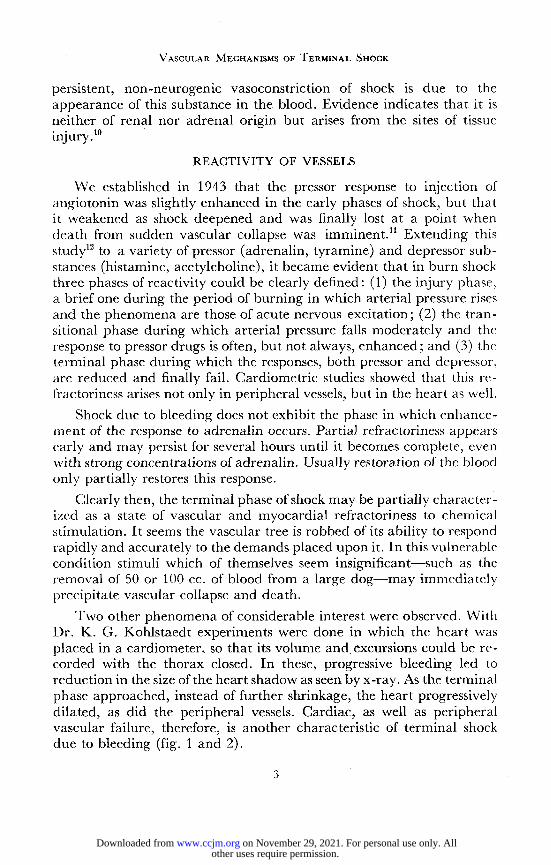
Two other phenomena of considerable interest were observed. With Dr. K. G. Kohlstaedt experiments were done m which the heart was placed in a cardiometer, so that its volume and excursions could be re-corded with the thorax closed. In these, progressive bleeding led to reduction in the size of the heart shadow as seen by x-ray. As the terminal phase approached, instead of further shrinkage, the heart progressively dilated, as did the peripheral vessels. Cardiac, as well as peripheral vascular failure, therefore, is another characteristic of terminal shock due to bleeding (fig. 1 and 2).
3
other uses require permission. on November 29, 2021. For personal use only. Allwww.ccjm.orgDownloaded from

I R V I N E H. P A G E
UlnUTCi I'Ki. 1—Schematic diagram of the effect of hemorrhage and prolonged hypotension on circulatory dynamics. A, Cardiometer record of stroke volume in prehemorrhagic period. B, Point of greatest reduction of diastolic and systolic volume. C, I), and E, 3.5 cc. of blood injected intra-arterially. VP, Intra-thoracic venous pressure in millimeters of water. AP, Arterial pressure in millimeters of mercury. Shaded area denotes total amount of blood re-moved (scale at right side). Scale at upper right, indicates stroke volume units of cardiac dilatation. (Courtesy of SURGERY 16:430, 1944)
a b c
Flu. 2—X-ray photographs taken before and during prolonged post-hemorrhagic hypo-tension. (a), Photograph of roentgenogram taken during prehemorrhagic period, (b), Photo-graph taken during hypotensive period when cardiometer indicated greatest reduction in cardiac size, (c), Photograph taken just before treatment began. Cardiometer indicated an increase of 3.5 stroke volume units in systolic volume. (Courtesy of SURGERY 16:430,1944)
4
other uses require permission. on November 29, 2021. For personal use only. Allwww.ccjm.orgDownloaded from

V A S C U L A R M E C H A N I S M S O F T E R M I N A L S H O C K
The second phenomenon concerns the rate at which blood flows into a shocked animal when the arterial pressure is maintained by con-necting a larger artery to a reservoir of blood under the desired pressure. Blood will flow in or out according to the variations of the animal's blood pressure and, presumably, the capacity of the blood vessels and reservoirs. Such an animal may live for several hours at a low level of arterial pressure in spite of persistent vasoconstriction. But the approach of the terminal phase, which, as we have seen, is heralded by vascular and cardiac dilation, may also be gauged from a sudden inflow of blood from the reservoir.
Chambers, Zweifach, Lowenstein, Schorr, and their associates have arrived at somewhat similar conclusions concerning the state of the very smallest arterioles (the metarterioles) in shock, although from an en-tirely different approach. The metarterioles of the exteriorized meso-appendix of rats are measured under the microscope. Sensitivity to adrenalin contained in the fluid bathing the tissue is estimated roughly by the degree of constriction which occurs. Early in hemorrhagic shock the response is greatly heightened, while late, refractoriness appears along with what they term loss of vasomotion. As described above, we have not found after hemorrhage a phase of enhanced pressor response to adrenalin.
THERAPEUTICS OF TERMINAL SHOCK
The experimental data make it clear that once vascular and cardiac refractoriness appears in shock, vascular collapse is imminent, and at present such collapse is irremediable. But it generally takes a long and variable time for this state to develop. A method which would rapidly restore arterial pressure so that, in spite of vasoconstriction, the per-fusion of tissues would be adequate for survival—and here the cardiac and cerebral beds should be considered—would have much to recom-mend it.13
Such a method was suggested to us by Seeley and the experimental evidence obtained in work with Dr. K. G. Kohlstaedt and, more recently with Dr. Otto Glasser.14 The method consists in the rapid administra-tion of blood into a large (radial or femoral) artery under positive pres-sure. For example, if the patient's blood pressure be taken as 30 mm. mercury and the duration of shock not such that total vascular irrespon-siveness would be present, a number 18 needle is inserted into the artery with the point toward the heart, and blood is administered freely from a reservoir set at 50 mm. mercury. When equilibrium is reached at this pressure so that inflow ceases, the positive pressure is increased to 70 mm. mercury and so on by increments until the final equilibrium of
5
other uses require permission. on November 29, 2021. For personal use only. Allwww.ccjm.orgDownloaded from

IRVINE H . PAGE
arterial and reservoir pressure is reached at a pressure of approximately 100 mm. mercury. This point should be reached in a few minutes.
If it were believed that vascular refractoriness appeared because of a long period of deep shock, it sometimes seemed desirable to give a small amount (200 mg.) of 2-amino heptane into the infusion tubing just as the blood started to flow in. The drug is used because it tends to constrict the heart and, as has been shown, cardiac dilatation is a serious danger in this stage of shock. The drug has no use apart from the co-incident infusion of blood or plasma, and repeated doses are increasingly ineffective. It has not been shown to have value in the treatment of terminal shock either alone or in conjunction with intravenous trans-fusion.
The intra-arterial infusion method has several advantages. These seem to be: (1) the delivery of blood into the aorta perfuses the coronary vessels, relieving myocardial ischemia, and since myocardial ischemia probably underlies the cardiac dilatation and insufficiency, places the heart rapidly in a state in which it can pump the infused blood to other areas; (2) when, before the infusion, the patient may have been apneic, at its start he will take a deep breath, as if the arterial filling had rapidly extended to the vital medullary centers; (3) in contrast with intravenous
m m Hi mo 90 80 70 60 50 40 30 !0 10 0
minutes elapsed
3-1/2-
FIG. 3—Example of the effect of intra-arterial infusion in a patient in deep shock with un-diagnosed abdominal bleeding. Arterial pressures are recorded on a kymograph. A, Initial pressure. B, During intra-arterial infusion. C, Infusion discontinued and pressure fell off rapidly. D, Reinstituted. E, Infusion stopped. F, Same sequence repeated. Clearly blood was leaking rapidly from the vascular tree. (Kohlstaedt and Page)
6
other uses require permission. on November 29, 2021. For personal use only. Allwww.ccjm.orgDownloaded from

VASCULAR MECHANISMS OF T E R M I N A L S H O C K
transfusion, blood pressure is rapidly restored, and the volume of blood infused is determined, not by time-consuming analyses or by guess, but by the actual capacity of the vascular tree; and (4) a subsidiary advan-tage lies in the fact that latent bleeding (spleen, liver, kidney), which causes arterial pressure to fall off rapidly, is detectable within minutes rather than hours (fig. 3).
Possibly the most important advantage of the method lies in the emphasis it places on the need for speed in the treatment of severe shock. Untold lives would be saved if physicians thought of the treat-ment of shock in terms of minutes rather than hours.
REFERENCES
1. Page, I. H., Taylor, R. D. and Kohlstaedt, K. G.: Case of extreme hypotension fol-lowing acute arsenic poisoning with adequate blood supply to the tissues. Am. J. M. Sc. 205: 730-734 (May) 1943.
2. Page, I. H. and Abell, R. G.: State of the vessels of mesentery in shock produced by constricting limbs and behavior of the vessels following hemorrhage. J. Exper. Med. 77:215-232 (March) 1943.
3. Abell, R. G. and Page, I. H.: Study of smaller blood vessels in burned dogs and cats. Surg., Gynec. & Obst. 77:348-353 (Oct.) 1943.
4. Page, I. H. and Abell, R. G.: Effects of acute hemorrhage and of subsequent infusion upon the blood vessels and blood flow as seen in the mesenteries of anesthetized dogs. Am. J. Physiol. 143:182-190 (Feb.) 1945.
5. Corcoran, A. C. and Page, I. H.: Effects of hypotension due to hemorrhage and of blood transfusion on renal function in dogs. J. Exper. Med. 78:205-224 (Sept.) 1943.
6. Corcoran, A. C. and Page, I. H.: Post-traumatic renal injury. Arch. Surg. 51:93-101 (Sept.) 1945.
7. Taylor, R. D. and Page, I. H.: Mechanism of erythremia; erythremia resulting from traumatic shock in dogs and from injection of epinephrine into human beings and dogs. Arch. Surg. 47:59-68 (July) 1943.
8. Lewis, R. N., Werle, J. M. and Wiggers, C. J. : Behavior of the spleen in hemorrhagic hypotension and shock. Am. J. Physiol. 138:205-211 (Jan.) 1943.
9. Page, I. H.: Occurrence of vasoconstrictor substance in blood during shock induced by trauma, hemorrhage and burns. Am. J. Physiol. 139:386-398 (July) 1943.
10. Corcoran, A. C., Taylor, R. D. and Page, I. H.: Immediate effects on renal function of onset of shock due to partially occluding limb tourniquets. Ann. Surg. 118:871-886 (Nov.) 1943.
11. Page, I. H.: Hypotension and loss of pressor response to angiotonin as result of trauma to central nervous system and severe hemorrhage. J. Exper. Med. 78:41-58 (July) 1943.
12. Page, I. H.: Cardiovascular changes resulting from severe scalds. Am. J. Physiol. 142:366-378 (Oct.) 1944.
13. Kohlstaedt, K. G. and Page, I. H.: Hemorrhagic hypotension and its treatment by intra-arterial and intravenous infusion of blood. Arch. Surg. 47:178-191 (Aug.) 1943.
14. Kohlstaedt, K. G. and Page, I. H.: Terminal hemorrhagic shock; circulating dynamics, recognition and treatment. Surgery 16:430-465 (Sept.) 1944.
7
other uses require permission. on November 29, 2021. For personal use only. Allwww.ccjm.orgDownloaded from