university of groningen magnetic nanoparticles for the
TRANSCRIPT

University of Groningen
Magnetic Nanoparticles for the Control of Infectious BiofilmsQuan, Kecheng
DOI:10.33612/diss.170829667
IMPORTANT NOTE: You are advised to consult the publisher's version (publisher's PDF) if you wish to cite fromit. Please check the document version below.
Document VersionPublisher's PDF, also known as Version of record
Publication date:2021
Link to publication in University of Groningen/UMCG research database
Citation for published version (APA):Quan, K. (2021). Magnetic Nanoparticles for the Control of Infectious Biofilms. University of Groningen.https://doi.org/10.33612/diss.170829667
CopyrightOther than for strictly personal use, it is not permitted to download or to forward/distribute the text or part of it without the consent of theauthor(s) and/or copyright holder(s), unless the work is under an open content license (like Creative Commons).
The publication may also be distributed here under the terms of Article 25fa of the Dutch Copyright Act, indicated by the “Taverne” license.More information can be found on the University of Groningen website: https://www.rug.nl/library/open-access/self-archiving-pure/taverne-amendment.
Take-down policyIf you believe that this document breaches copyright please contact us providing details, and we will remove access to the work immediatelyand investigate your claim.
Downloaded from the University of Groningen/UMCG research database (Pure): http://www.rug.nl/research/portal. For technical reasons thenumber of authors shown on this cover page is limited to 10 maximum.
Download date: 30-11-2021
CHAPTER 2
Homogeneous Distribution of Magnetic,
Antimicrobial-Carrying Nanoparticles through an
Infectious Biofilm Enhances Biofilm-Killing Efficacy
Kecheng Quan, Zexin Zhang, Yijin Ren, Henk J. Busscher, Henny C. van der Mei,
Brandon W. Peterson.
ACS Biomaterials Science & Engineering 2020, 6: 205-212.
Reproduced with permission from America Chemical Society.
Chapter 2
58

Chapter 2
59
Abstract
Magnetic, antimicrobial-carrying nanoparticles provide a promising, new and direly
needed antimicrobial strategy against infectious bacterial biofilms. Penetration and
accumulation of antimicrobials over the thickness of a biofilm is a conditio sine qua
non for effective killing of biofilm inhabitants. Simplified schematics on magnetic-
targeting, always picture homogeneous distribution of magnetic, antimicrobial-carrying
nanoparticles over the thickness of biofilms, but this is not easy to achieve. Here,
gentamicin-carrying magnetic nanoparticles (MNPs-G) were synthesized through
gentamicin-conjugation with iron-oxide nanoparticles and used to demonstrate the
importance of their homogeneous distribution over the thickness of a biofilm.
Diameters of MNPs-G were around 60 nm, well below the limit for reticulo-endothelial
rejection. MNPs-G killed most ESKAPE-panel pathogens, including Escherichia coli,
equally as well as gentamicin in solution. MNPs-G distribution in a Staphylococcus
aureus biofilm was dependent on magnetic-field exposure time and most homogeneous
after 5 min magnetic-field exposure. Exposure of biofilms to MNPs-G with 5 min
magnetic-field exposure, not only yielded homogeneous distribution of MNPs-G, but
concurrently better staphylococcal killing at all depths than MNPs, gentamicin in
solution, and MNPs-G, or after other magnet-field exposure times. In summary,
homogeneous distribution of gentamicin-carrying magnetic nanoparticles over the
thickness of a staphylococcal biofilm was essential for killing biofilm inhabitants and
required optimizing of the magnetic-field exposure time. This conclusion is important
for further successful development of magnetic, antimicrobial-carrying nanoparticles
towards clinical application.

Chapter 2
60
2.1 Introduction
Development of new infection-control strategies is becoming more and more urgent.
Antimicrobial-resistant bacterial infections have been predicted to become the number
one cause of death in the year 2050, exceeding the number of deaths caused by cancer
[1]. New infection-control strategies should not only evade rapidly arising bacterial
antimicrobial-resistance mechanisms [2], but preferentially also self-target infectious
biofilms and achieve homogeneous distribution of an antimicrobial over the entire
thickness of a biofilm. However, such a homogeneous distribution is hard to achieve.
Antimicrobials even when applied to antimicrobial-susceptible strains, solely kill those
bacteria that reside on the outer part of a biofilm [3]. As a result, many infections are
re-current, which increases the chances upon development of antimicrobial-resistance
[4]. Hopes are high that nanotechnology will contribute to the development of new
infection-control strategies [5], for which self-targeting, pH-adaptive antimicrobial
micelles, liposomes or polymersomes, antimicrobial carbon dots and dendrimers,
photothermal and magnetic antimicrobial nanoparticles have all been considered
“promising”.
Magnetic targeting of drugs is considered promising as a new infection-control
strategy and in tumor treatment, as it allows to establish high drug concentrations at the
target site [6,7]. Magnetic targeting of drugs requires two trivial components: drug-
carrying magnetic nanoparticles and a magnet targeting system. Magnetic targeting of
micron-sized infections is arguably more difficult, especially in vivo, than the targeting
of much larger tumors. It is frequently assumed [8-12], that magnetic, antimicrobial
nanoparticles fully penetrate and distribute homogeneously over the thickness of a
biofilm under the influence of a magnetic-field (see Fig. 1). Usually however,
particularly in vivo, this is not easy and even targeting of magnetic, antimicrobial
nanoparticles to a biofilm site, as opposed to homogeneous distribution over the
thickness of a biofilm, requires sophisticated instrumentation [13].
Chapter 2
61
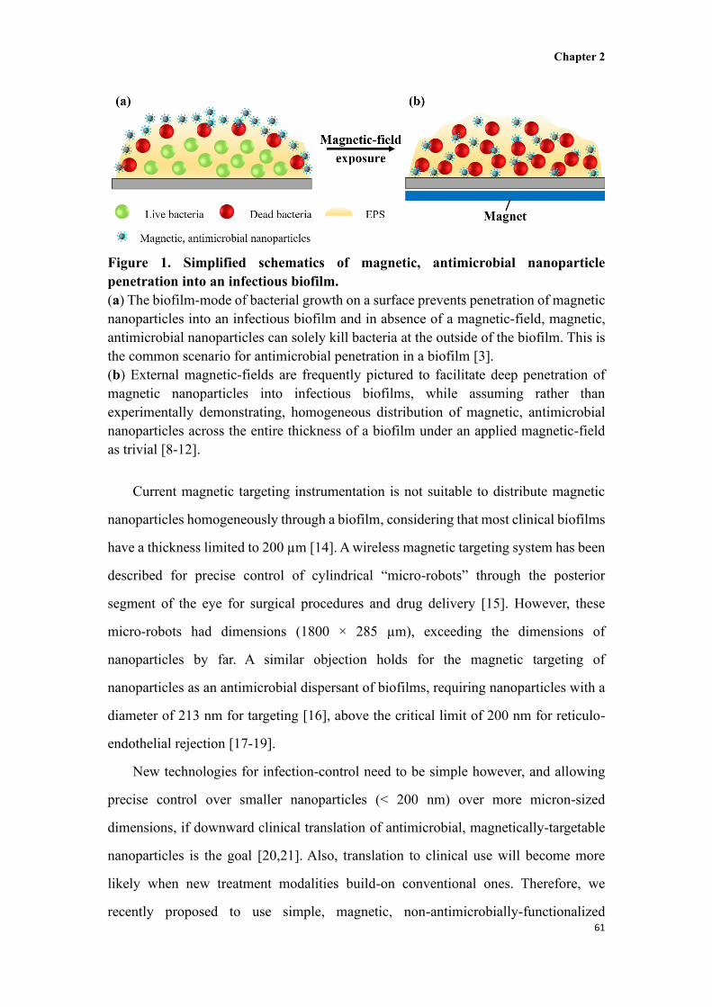
Figure 1. Simplified schematics of magnetic, antimicrobial nanoparticle
penetration into an infectious biofilm.
(a) The biofilm-mode of bacterial growth on a surface prevents penetration of magnetic
nanoparticles into an infectious biofilm and in absence of a magnetic-field, magnetic,
antimicrobial nanoparticles can solely kill bacteria at the outside of the biofilm. This is
the common scenario for antimicrobial penetration in a biofilm [3].
(b) External magnetic-fields are frequently pictured to facilitate deep penetration of
magnetic nanoparticles into infectious biofilms, while assuming rather than
experimentally demonstrating, homogeneous distribution of magnetic, antimicrobial
nanoparticles across the entire thickness of a biofilm under an applied magnetic-field
as trivial [8-12].
Current magnetic targeting instrumentation is not suitable to distribute magnetic
nanoparticles homogeneously through a biofilm, considering that most clinical biofilms
have a thickness limited to 200 µm [14]. A wireless magnetic targeting system has been
described for precise control of cylindrical “micro-robots” through the posterior
segment of the eye for surgical procedures and drug delivery [15]. However, these
micro-robots had dimensions (1800 × 285 µm), exceeding the dimensions of
nanoparticles by far. A similar objection holds for the magnetic targeting of
nanoparticles as an antimicrobial dispersant of biofilms, requiring nanoparticles with a
diameter of 213 nm for targeting [16], above the critical limit of 200 nm for reticulo-
endothelial rejection [17-19].
New technologies for infection-control need to be simple however, and allowing
precise control over smaller nanoparticles (< 200 nm) over more micron-sized
dimensions, if downward clinical translation of antimicrobial, magnetically-targetable
nanoparticles is the goal [20,21]. Also, translation to clinical use will become more
likely when new treatment modalities build-on conventional ones. Therefore, we
recently proposed to use simple, magnetic, non-antimicrobially-functionalized

Chapter 2
62
nanoparticles to create artificial water channels in infectious biofilms by magnetically-
induced movement of nanoparticles to make biofilms more penetrable and susceptible
to conventional antibiotic treatment [22]. Artificial channel digging does not require
any accumulation nor precise control or homogeneous distribution of nanoparticles
inside the biofilm.
Here, we created magnetic, antimicrobial-carrying nanoparticles, with the aim of
demonstrating the difficulty in achieving homogeneous distribution of magnetic,
antimicrobial nanoparticles and bacterial killing across the thickness of a biofilm. A
simple methodology to achieve homogeneous distribution of magnetic, gentamicin-
carrying nanoparticles across the thickness of an infectious biofilm growing on a
biomaterial surface was developed and demonstrated to be accompanied by enhanced
killing of biofilm inhabitants. Biomaterial-associated infections are a special class of
recalcitrant infections, caused by bacteria forming an infectious biofilm on biomaterials
implants and devices, such as total hip or knee arthroplasties, heart valves, vascular
grafts and many other types of implants and devices [20,23].
2.2 Results and Discussions
First, gentamicin (G), a commonly used aminoglycoside with a wide spectrum of
antibacterial activity and particularly suitable for local application [24][32] was
conjugated through its amino groups to the carboxyl groups on the surface of an iron-
oxide, magnetic nanoparticle (MNP) using a peptide-coupling (Fig. 2a) [25]. Effective
conjugation of G to MNPs was demonstrated from the presence of characteristic G- and
peptide-coupling bands in Fourier transform infrared (FTIR) spectra (Fig. 2b), i.e. the
bands at 1400 cm-1, 1575 cm-1 and 1650 cm-1 due to the stretching of N-H, C-N and
C=O of the peptide-coupling and the band at 1030 cm-1 attributed to C-O-C stretching
of G. Zeta potentials of MNPs were highly negative (-38.8 ± 2.1 mV; see Fig. 2c) due
to their carboxyl-rich surface and became positive (8.8 ± 0.2 mV) after G-conjugation
[26] as a result of amino-groups in gentamicin. Magnetic properties of MNPs-G (43.4
emu g-1; see Fig. 2d) were only slightly lower than of MNPs (46.8 emu g-1).
Thermogravimetric and elemental analysis indicated that G-conjugation in MNPs-G

Chapter 2
63
amounted 24 – 25% by mass (Fig. 2e and 2f, respectively). The diameter of MNPs-G
as obtained using Transmission Electron Microscopy (TEM) was around 60 nm (Fig.
2g).
Figure 2. Preparation and characterization of magnetic, gentamicin-carrying
nanoparticles (MNPs-G).
(a) Synthesis of MNPs-G. The carboxyl (-COOH) group of the carboxyl-functionalized
MNP is conjugated with the one of the amino (-NH2) groups on gentamicin through a
peptide-coupling, using 1-(3-dimethylaminopropyl)-3-ethylcarbodiimide
hydrochloride (EDC) and N-hydroxy succinimide (NHS) as catalysts. The reaction
occurs at room temperature.
(b) Fourier transform infrared (FTIR) spectra of G, MNPs and MNPs-G.
(c) Zeta potentials of MNP before and after conjugation of gentamicin in water (pH 7.0).
(d) Magnetic hysteresis loops at 300 K for MNPs before and after conjugation of
gentamicin, measured by Vibrating Sample Magnetometry. Insert in the lower right
corner shows the magnetic behavior of MNPs-G under an applied external magnetic-
field.
(e) Thermogravimetric analysis of G, MNPs and MNPs-G. The % weight loss over the

Chapter 2
64
temperature range between 210oC and 490oC was applied to calculate the weight % of
gentamicin in MNPs-G.
(f) Elemental analysis of G, MNPs and MNPs-G. Nitrogen (N) is absent in MNPs, while
present in G and MNPs-G, from which it can be concluded that the weight increase of
MNPs-G compared to MNPs (panel e) is due to G. Data are expressed as means ±
standard deviations over three separate measurements.
(g) TEM micrograph of the MNPs-G as synthesized in this study.
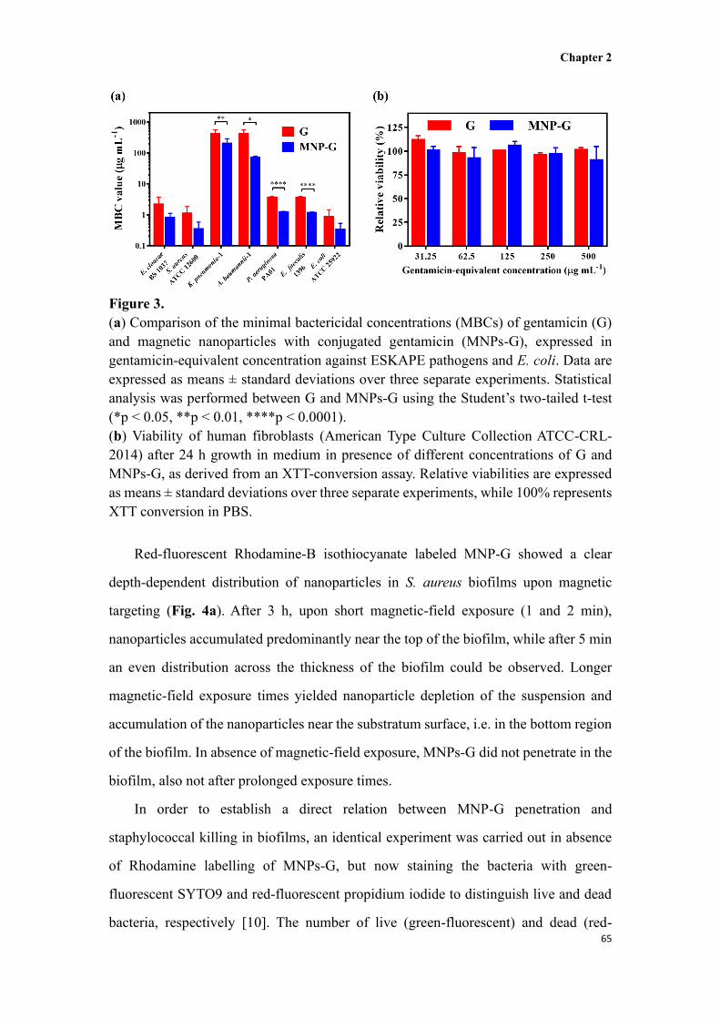
MNPs-G had a broad antibacterial activity against a variety of pathogen members
from the so-called ESKAPE-panel [27], Enterobacter cloacae BS 1037,
Staphylococcus aureus ATCC 12600, Klebsiella pneumonia-1, Acinetobacter
baumannii-1, Pseudomonas aeruginosa PA01 and Enterococcus faecalis 1396 with the
exception of K. pneumonia and A. baumannii (Fig. 3a). In addition to ESKAPE-panel
pathogens, MNPs-G were also anti-bacterially active against Escherichia coli ATCC
25922. In general, MBCs of MNPs-G were slightly, but significantly lower than of
gentamicin. This is likely because electrostatic double-layer attraction between
negatively-charged bacteria [28] and positively charged MNPs-G demonstrates that
conjugation did not negatively impact the antibacterial properties of gentamicin.
Growth of mouse fibroblasts with MNPs-G did not negatively impact the metabolic
activity of the cells (Fig. 3b), while bare magnetic iron-oxide nanoparticles were fully
biocompatible with these mammalian cells, as shown previously [29]. This is in line
with the known biocompatibility of iron-oxide nanoparticles to mammalian cells
[17,30]. Moreover, iron-oxide nanoparticles are known to be removed from the body
through phagocytosis [31].
Chapter 2
65
Figure 3.
(a) Comparison of the minimal bactericidal concentrations (MBCs) of gentamicin (G)
and magnetic nanoparticles with conjugated gentamicin (MNPs-G), expressed in
gentamicin-equivalent concentration against ESKAPE pathogens and E. coli. Data are
expressed as means ± standard deviations over three separate experiments. Statistical
analysis was performed between G and MNPs-G using the Student’s two-tailed t-test
(*p < 0.05, **p < 0.01, ****p < 0.0001).
(b) Viability of human fibroblasts (American Type Culture Collection ATCC-CRL-
2014) after 24 h growth in medium in presence of different concentrations of G and
MNPs-G, as derived from an XTT-conversion assay. Relative viabilities are expressed
as means ± standard deviations over three separate experiments, while 100% represents
XTT conversion in PBS.
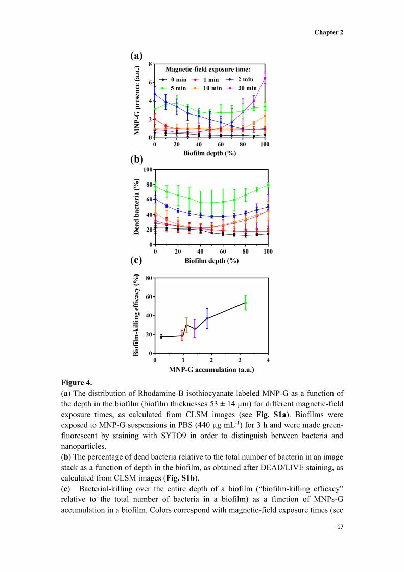
Red-fluorescent Rhodamine-B isothiocyanate labeled MNP-G showed a clear
depth-dependent distribution of nanoparticles in S. aureus biofilms upon magnetic
targeting (Fig. 4a). After 3 h, upon short magnetic-field exposure (1 and 2 min),
nanoparticles accumulated predominantly near the top of the biofilm, while after 5 min
an even distribution across the thickness of the biofilm could be observed. Longer
magnetic-field exposure times yielded nanoparticle depletion of the suspension and
accumulation of the nanoparticles near the substratum surface, i.e. in the bottom region
of the biofilm. In absence of magnetic-field exposure, MNPs-G did not penetrate in the
biofilm, also not after prolonged exposure times.
In order to establish a direct relation between MNP-G penetration and
staphylococcal killing in biofilms, an identical experiment was carried out in absence
of Rhodamine labelling of MNPs-G, but now staining the bacteria with green-
fluorescent SYTO9 and red-fluorescent propidium iodide to distinguish live and dead
bacteria, respectively [10]. The number of live (green-fluorescent) and dead (red-

Chapter 2
66
fluorescent) bacteria was determined from the CLSM images, using ImageJ to quantify
the number of green- and red-fluorescent bacteria. The distribution of dead bacteria
across the thickness of a biofilm roughly followed the same distribution pattern as of
MNPs-G (compare Fig. 4a and 4b). Shorter magnetic-field exposure times yielded
more dead bacteria near the top of the biofilm, while longer exposure times also caused
bacterial death in the bottom of the biofilm. However, the total number of MNPs-G
accumulated in a biofilm related well with the number of dead staphylococci over the
entire thickness of a biofilm (Fig. 4c). This attests to the importance of penetration and
homogeneous accumulation of antimicrobial-carrying nanoparticles for killing bacteria
in a biofilm-mode of growth. Fig. 4a and 4b yield the conclusion that deep killing of
biofilm inhabitants requires good penetration of antimicrobial, magnetic nanoparticles
in the biofilm (Fig. 4b) and that overall killing is highest when antimicrobial
nanoparticles distribute homogeneously across the thickness of a biofilm (Fig. 4a and
4b). For the MNPs and magnet set-up used here, optimal magnet-exposure time thus
equals 5 min.
Chapter 2
67
Figure 4.
(a) The distribution of Rhodamine-B isothiocyanate labeled MNP-G as a function of
the depth in the biofilm (biofilm thicknesses 53 ± 14 µm) for different magnetic-field
exposure times, as calculated from CLSM images (see Fig. S1a). Biofilms were
exposed to MNP-G suspensions in PBS (440 µg mL-1) for 3 h and were made green-
fluorescent by staining with SYTO9 in order to distinguish between bacteria and
nanoparticles.
(b) The percentage of dead bacteria relative to the total number of bacteria in an image
stack as a function of depth in the biofilm, as obtained after DEAD/LIVE staining, as
calculated from CLSM images (Fig. S1b).
(c) Bacterial-killing over the entire depth of a biofilm (“biofilm-killing efficacy”
relative to the total number of bacteria in a biofilm) as a function of MNPs-G
accumulation in a biofilm. Colors correspond with magnetic-field exposure times (see
Chapter 2
68
panel a).
Data are expressed as means ± standard deviations over three separate experiments.
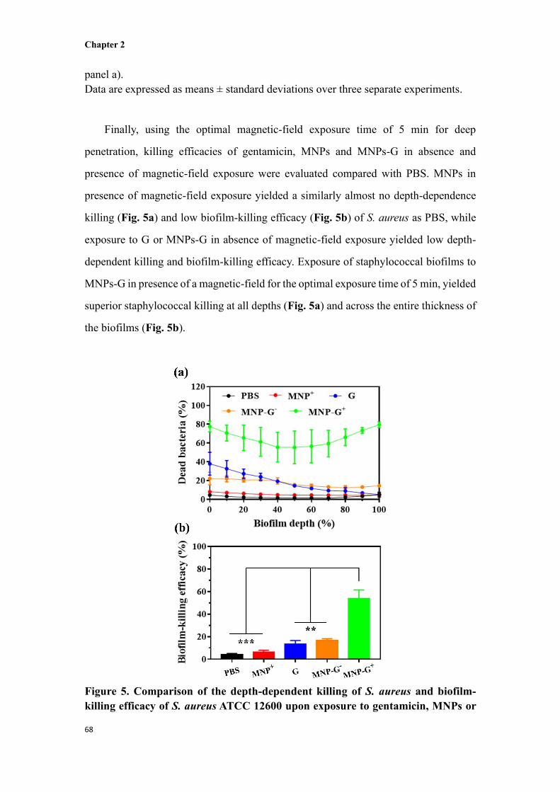
Finally, using the optimal magnetic-field exposure time of 5 min for deep
penetration, killing efficacies of gentamicin, MNPs and MNPs-G in absence and
presence of magnetic-field exposure were evaluated compared with PBS. MNPs in
presence of magnetic-field exposure yielded a similarly almost no depth-dependence
killing (Fig. 5a) and low biofilm-killing efficacy (Fig. 5b) of S. aureus as PBS, while
exposure to G or MNPs-G in absence of magnetic-field exposure yielded low depth-
dependent killing and biofilm-killing efficacy. Exposure of staphylococcal biofilms to
MNPs-G in presence of a magnetic-field for the optimal exposure time of 5 min, yielded
superior staphylococcal killing at all depths (Fig. 5a) and across the entire thickness of
the biofilms (Fig. 5b).
Figure 5. Comparison of the depth-dependent killing of S. aureus and biofilm-
killing efficacy of S. aureus ATCC 12600 upon exposure to gentamicin, MNPs or
Chapter 2
69
MNPs-G in absence and presence of optimized 5 min magnetic-field exposure. PBS
was included as a control. Total exposure time to the antimicrobials was 180 min.
(a) The percentage of dead bacteria relative to the total number of bacteria in an image
stack as a function of depth in the biofilm, as obtained after DEAD/LIVE staining, as
calculated from CLSM images (Fig. S2). Superscripts ‘-’ and ‘+’ denote absence and
presence of magnet-field exposure, respectively.
(b) Bacterial-killing over the entire depth of a staphylococcal biofilm (“biofilm-killing
efficacy” relative to the total number of bacteria in a biofilm).
Data are expressed as means ± standard deviations over three separate experiments.
Statistical analysis was performed using the Student’s two-tailed t-test (**p < 0.01,
***p < 0.001).
2.3 Conclusion
In conclusion, this work shows that it should not be a priori assumed that magnetic-
field exposure yields a homogeneous distribution of magnetic nanoparticles over the
entire thickness of a biofilm as is the commonly assumed scenario in the current
literature [8-12]. Too short magnetic-field exposure yields accumulation of magnetic
nanoparticle near the top of a biofilm, while too long exposure times create more
accumulation near the bottom. Under the conditions applied in this work, homogeneous
distribution of magnetic nanoparticles could be achieved using an intermediate
magnetic-field exposure time of 5 min, but different culturing platforms, including
clinical biofilms, may yield different optimal exposure times. Homogeneous
distribution of magnetic, gentamicin-carrying nanoparticles achieved after the optimal
magnetic-field exposure time, yielded better depth-dependent staphylococcal killing
and biofilm-killing efficacy (over an entire biofilm) than other magnetic-field exposure
times. Moreover, a homogeneous distribution of magnetic, gentamicin-carrying
nanoparticles yielded better killing than gentamicin or magnetic, gentamicin-carrying
nanoparticles in absence of magnetic-field exposure. Thus homogeneous distribution
of magnetic, antimicrobial-carrying nanoparticles is a conditio sine qua non for optimal
killing. Clinical translation of the use of magnetic, antimicrobial-carrying nanoparticles
is not trivial, but easiest to achieve for biomaterial-associated infections, in which the
bottom of a biofilm is well defined, as it concurs with the surface of the implant or
device (i.e. demonstrated in the current study). In other types of infections, like e.g.
Chapter 2
70
organ infections, a magnetic field might be placed at multiple angles towards an
infection site to achieve homogeneous distribution of magnetic, antimicrobial-carrying
MNPs in the infectious biofilm, which we demonstrate here is absolutely needed to kill
biofilm inhabitants over the depth of a biofilm.
2.4 Materials and methods
2.4.1 Materials
Gentamicin, 3,4-dihydroxyhydrocinnamic acid (DHCA), 1-(3-dimethylaminopropyl)-
3-ethylcarbodiimide hydrochloride (EDC), N-hydroxysuccinimide (NHS), 1-
octadecene, oleic acid, sodium oleate and iron (III) chloride (FeCl3·6H2O) were
purchased from Aldrich. Tetrahydrofuran (THF), ethanol and hexane were purchased
from Sinopharm Chemical Reagent Co. (China). All chemicals were used as received.
2.4.2 Preparation and characterizations of magnetic, gentamicin-carrying
nanoparticles (MNPs-G)
Carboxyl-functionalized, iron-oxide magnetic nanoparticles (MNPs, 10 mg mL-1, 1 mL,
prepared as described in Supporting Information) were dispersed in 10 mL
demineralized water (pH 4.0, adjusted by diluted hydrochloric acid) in a 50 mL round
bottom flask. After adding 1 mL EDC (0.1 M) and 1 mL NHS (0.1 M), the mixture was
stirred for 12 h at room temperature (RT). Then, the pH of the mixture was adjusted to
9.0 by adding 0.4 mL NaOH (0.05 M) and 1 mL gentamicin (0.1 M) was added,
followed by stirring for another 12 h at RT. The black particles obtained were
magnetically separated and washed with demineralized water for 3 times in order to
remove unreacted gentamicin molecules. Finally, MNPs-G were dispersed in phosphate
buffered saline (PBS, 5 mM K2HPO4, 5 mM KH2PO4, 150 mM NaCl, pH 7.4) using
sonication (Transonic TP 690, ELMA, Germany, 160 W, 35 kHz) at RT for 30 min.
The size and shape of MNPs-G were determined using transmission electron
microscopy (TEM, G-120, Hitachi, Japan). The conjugation of G to MNP was
characterized by Fourier transform infrared spectroscopy (FTIR, Nicolet-20DXB, US).
Spectra were taken over a wavenumber range from 500 to 4000 cm-1 at a resolution of

Chapter 2
71
2.0 cm-1. All spectra represent averages from 16 interferograms. Zeta potentials were
measured in demineralized water using a Malvern NanoSizer ZS2000 (UK). Zeta
potentials were measured in triplicate on separately-prepared batches of MNPs-G.
Magnetic properties of MNPs-G were measured at RT using a vibrating sample
magnetometer (Model 7410, Lake Shore, USA). The mass content of G in MNPs-G
was analyzed using a combination of thermogravimetric (TA2100 USA, heating rate
10°C min-1) and elemental analysis (Vario EL cube, Germany).
2.4.3 Bacterial strains, growth conditions and harvesting
Enterobacter cloacae BS 1037, Staphylococcus aureus ATCC 12600, Klebsiella
pneumonia-1, Acinetobacter baumannii-1, Pseudomonas aeruginosa PA01,
Enterococcus faecalis 1396 and Escherichia coli ATCC 25922 were grown from stock
solutions (7% DMSO, kept at -80°C) on blood agar plates at 37°C for 24 h. For pre-
cultures, a single bacterial colony was transferred into 10 mL tryptone soy broth (TSB,
OXOID, Basingstoke, UK) and incubated 24 h at 37°C. For main cultures, the pre-
culture was transferred into 200 mL TSB and incubated for 16 h at 37°C. Then, bacteria
were harvested by centrifugation (5000 g, 5 min, 10 °C) followed by washing twice in
sterile PBS. The bacterial suspension was sonicated (Vibra cell model 375, Sonics and
Material Inc., Danbury, CT, USA) 3 times each for 10 s with 30 s intervals between
each cycle on ice to obtain a suspension with single bacteria. The bacterial
concentrations of the suspension were adjusted values appropriate for later use, as
determined in a Bürker-Türk counting chamber.
2.4.4 Minimal bactericidal concentration (MBC)
To determine the MBCs of different strains, 100 µL of G, MNPs and MNPs-G (1 mg
mL-1) in PBS were put in a 96 wells plate and 100 µL of TSB was added. Then, solutions
were mixed and two-fold serially diluted. Next, 10 µL bacterial suspension (1 × 10⁵
bacteria mL-1) was added to mixed solutions in the 96 wells plates. After 24 h incubation
at 37°C, 10 µL was taken out of a well and placed on an agar plate and incubated for
24 h at 37°C. The lowest concentration at which no visible colonies were formed, was

Chapter 2
72
taken as the MBC value. The experiment was repeated twice with separate bacterial
cultures.
2.4.5 MNP-G distribution in S. aureus biofilms
For biofilm formation, we selected S. aureus ATCC 12600 for further experiments, as
it is a prominent pathogen in many types of infection [27,32]. A staphylococcal
suspension (1× 10⁹ bacteria mL-1, 2 mL) was put into a sterile polystyrene 12-well plate
for 2 h at RT in order to allow bacterial adhesion. Thereafter, the suspension was
removed, and the well was washed 3 times with sterile PBS, filled with fresh TSB and
incubated for 24 h at 37°C.
In order to visualize depth-dependent distribution of MNPs-G after penetration and
accumulation in staphylococcal biofilms, MNPs-G were first labelled with red-
fluorescent Rhodamine-B isothiocyanate (Sigma-Aldrich, USA). To this end, 10 mg
MNP-G and 1 mg Rhodamine-B isothiocyanate were mixed in 10 mL PBS and stirred
for 8 h in the dark at RT. Then, the suspension was dialyzed for 48 h in demineralized
water to remove unreacted Rhodamine-B isothiocyanate, while refreshing the water
every 12 h. After dialysis, the Rhodamine-B isothiocyanate labeled MNPs-G were
magnetically separated and re-suspended in PBS for later use. Next, 24 h S. aureus
biofilms were washed once with sterile PBS, exposed to 2 mL of red-fluorescent MNP-
G (440 µg mL-1) under a magnetic-field created by a NdFeB magnet (1 mm thickness
and 10 mm in diameter with 1.17-1.21 Tesla residual magnetism) for different times (0,
1, 2, 5, 10 and 30 min) at 37°C. After magnetic-field exposure, biofilms were placed in
the incubator again. The total exposure time of the biofilms to MNPs-G amounted 180
min, including magnetic-field exposure and incubation. After incubation, the
nanoparticle suspension was removed and bacteria in the biofilms were stained with
green-fluorescent SYTO9 (Thermo Fisher Scientific, Waltham, Massachusetts, USA)
for 15 min at RT in the dark. Finally, biofilms were washed once with PBS and
subsequently imaged by CLSM (Leica TCS SP2 Leica, Wetzlar, Germany) with an
HCX APO L40×/0.80 W U-V-1 objective. An argon ion laser at 488 nm and a green
HeNe laser at 543 nm were used to excite the SYTO9 and Rhodamine-B isothiocyanate

Chapter 2
73
and fluorescence was collected at 500-540 nm (SYTO9) and 583-688 nm (Rhodamine-
B isothiocyanate). CLSM images were acquired using Leica software, version 2.0.
The presence of Rhodamine-B isothiocyanate labeled MNPs-G in each image stack
of a biofilm or in an entire biofilm (“MNPs-G accumulation”) was calculated as the
ratio of red-fluorescent over green-fluorescent pixels, either in an image stack or in an
entire biofilm. The distribution of MNPs-G across the thickness of staphylococcal
biofilms was measured in triplicate, using separate bacterial cultures.
2.4.6 S. aureus killing in biofilms
24 h S. aureus biofilms were exposed to MNPs-G (no Rhodamine-B isothiocyanate
labeling), but afterwards stained with green-fluorescent SYTO9 and red-fluorescent
propidium iodide (Thermo Fisher Scientific, Waltham, Massachusetts, USA) for 15 min
at RT in the dark to label live and dead bacteria, respectively in the biofilm. Biofilms
were subsequently imaged by CLSM and depth-dependent staphylococcal killing or
killing over the entire thickness of a biofilm (“biofilm killing efficacy”) was calculated
using ImageJ as the percentage red-fluorescent over the sum of red- and green-
fluorescent pixels, either in an image stack or in an entire biofilm, respectively.
In order to compare the biofilm-killing efficacy of MNPs-G after optimized
magnetic-field exposure, 24 h staphylococcal biofilms were exposed to PBS, G (110 µg
mL-1 in PBS, equivalent concentration as in MNPs-G), MNPs-G or MNPs (440 µg mL-
1, in PBS) with magnetic-field exposure, and MNP-G (440 µg mL-1 in PBS) with and
without magnetic-field for 5 min, while total exposure time to antimicrobials was 180
min, including magnetic-field exposure and incubation. The experiment was repeated
in triplicate with separate staphylococcal cultures.
2.4.7 Effects of MNPs-G on mammalian cells
The cytotoxicity of MNP-G and G were evaluated according to a previous method [22].
Briefly, human fibroblasts (American Type Culture Collection ATCC-CRL-2014) were
cultured in 96 well plates (5 ×10³ cells per well), filled with 100 μL cell growth medium
(Dulbecco's modification of Eagle's medium (DMEM, ThermoFisher Scientific)
Chapter 2
74
supplemented with 10% Fetal Bovine Serum (FBS; Invitrogen)). Subsequently, 100 μL
MNPs-G or G in cellular growth medium (concentration range 31.25 to 500 µg mL-1 in
G-weight equivalents) was added and incubated for 24 h in 5% CO2 at 37°C. After 24
h, 50 μL XTT ((2,3-Bis-(2-methoxy-4-nitro-5-sulfophenyl)-2H-tetrazolium-5-
carboxanilide salt), AppliChem) reagent solution combined with activation solution
(PMS, (n-methyl dibenzopyrazine methyl sulfate), Sigma-Aldrich) was added. After
another 4 h at 37°C, absorbances A485 nm were measured using a spectrophotometer
(Shimadzu, Japan). According to the manufacturer’s instructions, A690 nm was measured
and subtracted as a reference control. The viability of the fibroblasts after material
exposure was calculated relative to the one of cells exposed to PBS in absence of
material according to
Relative viability (%)= Amaterial 485 nm- Amaterial 690 nm
APBS 485 nm- APBS 690 nm×100% (1)
Statistics: All comparisons of MBCs and biofilm-killing efficacies between the
different treatments were performed with a two-tailed Student t-test, accepting
significance at p < 0.05.
Acknowledgements
This work was financially supported by National Key Research and Development
Program of China (2016YFC1100402), the National Natural Science Foundation of
China (21334004, 11574222 and 21522404), and UMCG, Groningen, The Netherlands.
References
[1] G. Humphreys, F. Fleck, Bull. World Health Organ. 94 (2016) 638-639.
[2] M. Simoes, L. C. Simoes, M. J. Vieira, LWT-Food Sci. Technol. 43 (2010) 573-583.
[3] D. Davies, Nat. Rev. Drug Discovery 2 (2003) 114-122.
[4] C. Fuente-Nunez, F. Reffuveille, L. Fernandez, R. E. W. Hancock, Curr. Opin. Microbiol. 16
(2013) 580-589.
[5] Y. Liu, L. Shi, H. C. Van der Mei, P. C. Jutte, Y. Ren, H. J. Busscher, Chem. Soc. Rev. 48 (2019)
428-446.
[6] M. J. Hajipour, K. M. Fromm, A. Ashkarran, D. Aberasturi, L. Larramendi, T. Rojo, V.
Serpooshan, W. J. Parak, M. Mahmoudi, Trends Biotechnol. 30 (2012) 499-511.
Chapter 2
75
[7] K. Ulbrich, K. Hola, V. Subr, A. Bakandritsos, J. Tucek, R. Zboril, Chem. Rev. 116 (2016) 5338-
5431.
[8] G. Subbiahdoss, S. Sharifi, D. W. Grijpma, S. Laurent, H. C. Van der Mei, M. Mahmoudi, H. J.
Busscher, Acta Biomater. 8 (2012) 2047-2055.
[9] B. M. Geilich, L. Gelfat, S. Sridhar, A. L. Van de Ven, T. J. Webster, Biomaterials 119 (2017) 78-
85.
[10] X. Wang, A. Deng, W. Cao, Q. Li, L. Wang, J. Zhou, B. Hu, X. Xing, J. Mater. Sci. 53 (2018)
6433-6449.
[11] X. Wang, J. Wu, P. Li, L. Wang, J. Zhou, G. Zhang, X. Li, B. Hu, X. Xing, ACS Appl. Mater.
Interfaces 10 (2018) 34905-34915.
[12] C. Zhang, C. Du, J. Liao, Y. Gu, Y. Gong, J. Pei, H. Gu, D. Yin, L. Gao, Y. Pan, Biomater. Sci. 7
(2019) 2833-2840.
[13] B. Shapiro, S. Kulkarni, A. Nacev, A. Sarwar, D. Preciado, D. A. Depireux, Annu. Rev. Biomed.
Eng. 16 (2014) 455-481.
[14] T. Bjarnsholt, M. Alhede, M. Alhede, S. R. Eickhardt-Sorensen, C. Moser, M. Kuhl, P. Jensen,
N. Hoiby, Trends Microbiol. 21 (2013) 466-474.
[15] F. Ullrich, C. Bergeles, J. Pokki, O. Ergeneman, S. Erni, G. Chatzipirpiridis, S. Pane, C. Framme,
B. J. Nelson, Invest. Ophthalmol. Visual Sci. 54 (2013) 2853-2863.
[16] G. Hwang, A. J. Paula, E. E. Hunter, Y. Liu, A. Babeer, B. Karabucak, K. Stebe, V. Kumar, E.
Steager, H. Koo, Science Robotics 4 (2019) eaaw2388.
[17] A. K. Gupta, M. Gupta, Biomaterials 26 (2005) 3995-4021.
[18] S. M. Moghimi, A. C. Hunter, J. C. Murray, Pharmacol. Rev. 53 (2001) 283-318.
[19] R. A. Petros, J. M. DeSimone, Nat. Rev. Drug Discovery 9 (2010) 615-627.
[20] H. J. Busscher, H. C. van der Mei, G. Subbiahdoss, P. C. Jutte, J. J. A. M. Van den Dungen, S. A.
J. Zaat, M. J. Schultz, D. W. Grainger, Sci. Transl. Med. 4 (2012) 153rv10.
[21] H. J. Busscher, V. Alt, H. C. van der Mei, P. H. Fagette, W. Zimmerli, T. F. Moriarty, J. Parvizi,
G. Schmidmaier, M. J. Raschke, T. Gehrke, R. Bayston, L. M. Baddour, L. C. Winterton, R. O.
Darouiche, D. W. Grainger, ACS Biomater. Sci. Eng. 5 (2018) 402-406.
[22] K. Quan, Z. Zhang, H. Chen, X. Ren, Y. Ren, B. W. Peterson, H. C. van der Mei, H. J. Busscher,
Small 15 (2019) 1902313.
[23] C. R. Arciola, D. Campoccia, L. Montanaro, Nat. Rev. Microbiol. 16 (2018) 397-409.
[24] M. Lucke, G. Schmidmaier, S. Sadoni, B. Wildemann, R. Schiller, N. P. Haas, M. Raschke, Bone
32 (2003) 521-531.
[25] E. E. M. G. Loomans, J. V. Wiltenburg, M. Koets, A. V. Amerongen, J. Agric. Food Chem. 51
(2003) 587-593.
[26] N. Pothayee, N. Pothayee, N. Jain, N. Hu, S. Balasubramaniam, L. M. Johnson, R. M. Davis, N.
Sriranganathan, J. S. Riffle, Chem. Mater. 24 (2012) 2056-2063.
[27] J. N. Pendleton, S. P. Gorman, B. F. Gilmore, Expert Rev. Anti-Infect. Ther. 11 (2013) 297-308.
[28] M. E. Bayer, J. L. Sloyer, J. Gen. Microbiol. 136 (1990) 867-874.
[29] R. Dai, Y. Hang, Q. Liu, S. Zhang, L. Wang, Y. Pan, H. Chen, J. Mater. Chem. B 7 (2019) 4161-
4168.
[30] M. Mahmoudi, A. Simchi, M. Imani, M. A. Shokrgozar, A. S. Milani, U. O. Hafeli, P. Stroeve,
Colloids Surf. B Biointerfaces 75 (2010) 300-309.
[31] Q. Yu, X. Xiong, L. Zhao, T-T. Xu, H. Bi, R. Fu, Q-H. Wang, Curr. Med. Sci. 38 (2018) 1096-

Chapter 2
76
Chapter 2 73 1102.
[32] F. D. Lowy, N. Engl. J. Med. 339 (1998) 520-532.

Chapter 2
77
Supporting Information
Synthesis of magnetic, carboxyl-functionalized iron oxide nanoparticles
Iron oleate synthesis
Iron oleate was synthesized according to a previously published method [1].
Specifically, 10.8 g iron (III) chloride (FeCl3·6H2O, 40 mmol) and 36.5 g sodium oleate
(120 mmol) were dissolved in a mixture of 80 mL ethanol, 60 mL demineralized water
and 140 mL hexane. The mixed solution was heated to 70oC and subsequently refluxed
for 4 h at 70oC under continuously stirring. After cooling the mixture to room
temperature (RT), the upper brown organic liquid was separated and washed 3 times
with demineralized water using a separatory funnel. Then, hexane was evaporated by a
vacuum rotary evaporator. Finally, the liquid was put overnight into a vacuum oven.
Synthesis of oleic acid, iron oxide particles
3.6 g (4 mmol) of the iron oleate synthesized and 0.57 g of oleic acid (20 mmol) were
dissolved in 20 g of 1-octadecene at RT. The mixture was heated to 320oC at a heating
rate of 10oC per 3 min under nitrogen flow and refluxed at 320oC for 30 min. When the
mixture was cooled to RT, 50 mL of ethanol was added to precipitate the nanoparticles
and nanoparticles were separated by centrifugation (5000 g, 10 min, RT). Finally, the
precipitated nanoparticles were dried in a vacuum oven overnight.
Carboxyl-functionalized magnetic nanoparticles (MNPs)
Iron oxide nanoparticles were made hydrophilic using a previously published method
[2]. Briefly, 50 mg 3,4-dihydroxyhydrocinnamic acid (DHCA) was dissolved in 6 mL
tetrahydrofuran (THF) in a 25 mL round bottom flask and the mixture was heated to
50oC under nitrogen flow and 20 mg of the iron oxide nanoparticles synthesized
suspended in 1 mL THF under sonication for 30 min. Then, the nanoparticle suspension
was added dropwise into the round bottom flask and the mixture was stirred for 3 h at
50oC under a nitrogen flow. After the mixture was cooled down to RT, 500 µL NaOH
(0.5 M) was added and carboxyl-functionalized, hydrophilic MNPs were separated by

Chapter 2
78
centrifugation (3000 g, 5 min) and dried in vacuum oven over night at RT. Finally, the
MNPs were re-suspended in demineralized water for further use.

Chapter 2
79
Figure S1.
(a) Representative 3D-CLSM images of S. aureus ATCC 12600 biofilms exposed to
Rhodamine-B isothiocyanate labeled MNPs-G (440 µg mL-1 in PBS) after different
magnetic-field exposure times. Note that single, one-directional magnetic field
exposure does not cause damage the biofilm structure. Green and red represent bacteria
(SYTO9 stained) and MNPs-G, respectively. All scale bars represent 50 µm.
(b) Representative 3D-CLSM images of S. aureus ATCC 12600 biofilms exposed to
MNP-G (440 µg mL-1 in PBS) after different magnetic-field exposure times. Note that
single, one-directional magnetic field exposure does not cause damage the biofilm
structure. Green and red represent live (SYTO9 stained) and dead (propidium iodide
stained) staphylococci, respectively. All scale bars represent 50 µm.
Chapter 2
80
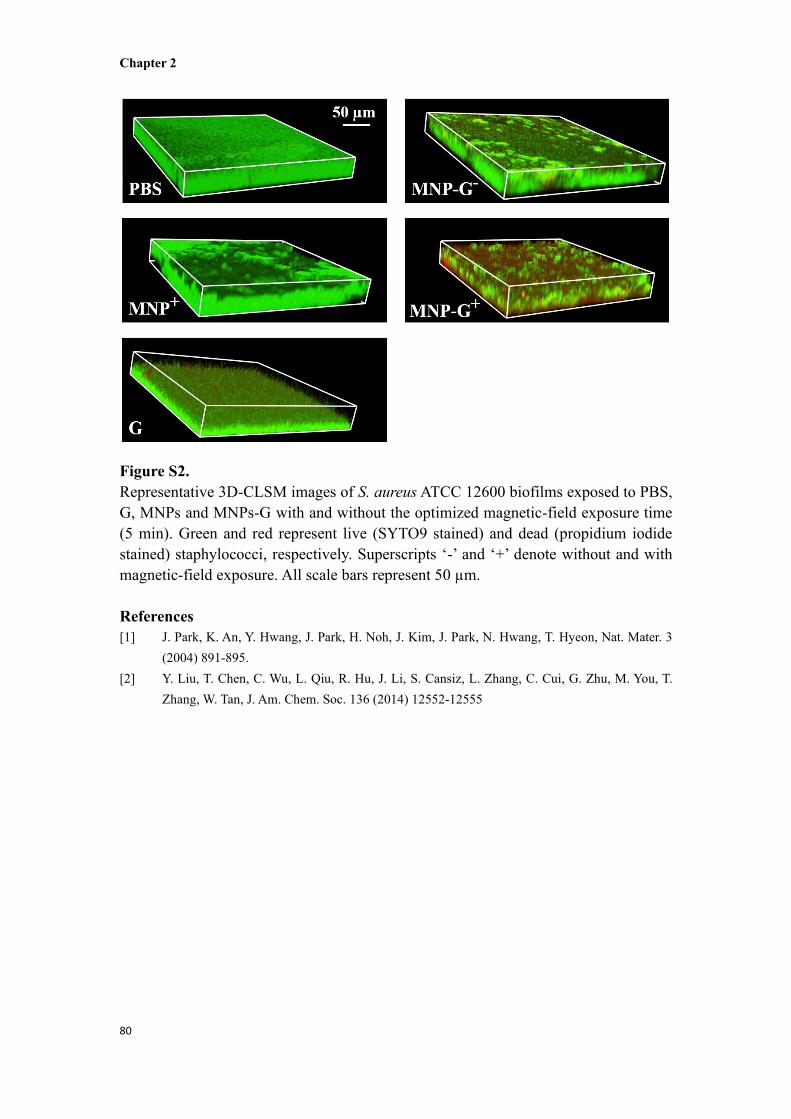
Figure S2.
Representative 3D-CLSM images of S. aureus ATCC 12600 biofilms exposed to PBS,
G, MNPs and MNPs-G with and without the optimized magnetic-field exposure time
(5 min). Green and red represent live (SYTO9 stained) and dead (propidium iodide
stained) staphylococci, respectively. Superscripts ‘-’ and ‘+’ denote without and with
magnetic-field exposure. All scale bars represent 50 µm.
References
[1] J. Park, K. An, Y. Hwang, J. Park, H. Noh, J. Kim, J. Park, N. Hwang, T. Hyeon, Nat. Mater. 3
(2004) 891-895.
[2] Y. Liu, T. Chen, C. Wu, L. Qiu, R. Hu, J. Li, S. Cansiz, L. Zhang, C. Cui, G. Zhu, M. You, T.
Zhang, W. Tan, J. Am. Chem. Soc. 136 (2014) 12552-12555