applications of magnetic nanoparticles in medicine
TRANSCRIPT

227
PRHSJ Vol. 28 No. 3September, 2009
Magnetic Fluid HyperthermiaRinaldi C, et al.
Applications of Magnetic Nanoparticles in Medicine: Magnetic Fluid Hyperthermia
Magda LatoRRe, Ph d; CaRLoS RiNaLdi, Ph d
department of Chemical engineering, University of Puerto Rico, Mayagüez
address correspondence to: Prof. Carlos Rinaldi, Po Box 9046, Mayagüez, PR 00681. tel: 787-832-4040 ext 3585 • Fax: 787-265-3818 • email: [email protected]
Nanoparticle systems are an intense subject of research for various biomedical applications. Colloidal suspensions of magnetic nanoparticles are of special interest, particularly in bioimaging, and more recently, in Magnetic Fluid Hyperthermia (MFH). MFH promises to be a viable alternative in the treatment of localized cancerous tumors. The treatment consists of locally injecting magnetic nanoparticles in fluid suspension into the tumor site and exposing the site to an oscillating magnetic field, where nanoparticles
dissipate energy in the form of heat, causing a localized rise in temperature and tumor cell death. Here we will review methods of magnetic nanoparticle synthesis, and the role of the nanoparticle surface coating in achieving colloidal stability, minimizing toxicity, and targeting. Finally, we review in vitro and in vivo MFH experiments, and clinical studies in the treatment of glioblastoma multiforme and prostate cancer.
Key words: Nanoparticles, Iron oxide, Magnetic fluid hyperthermia
1. Nanotechnology in Medicinethe term nanotechnology applies to the creation,
manipulation, and application of materials at the molecular and atomic scale, that is 1-100 nm. the fundamental properties of many materials at the nanoscale are uniquely different than those of bulk materials (1). these differences are mainly due to the vastly increased ratio of surface area to volume. this in turn results in an increased number of surface atoms, quantum electromagnetic interactions, increased surface tension, and size confinement effects. For example, quantum effects become significant for structures under 50 nm, resulting in unusual optoelectronic and magnetic properties of nanostructured materials compared to bulk materials (2). Nanomaterials have become an intense subject of research in fields such as optimizing the use and harnessing of energy, waste management, electronics, information and communication, and medicine (3).
Because the nanoscale lies at the intersection between the largest biological molecules and the smallest manmade devices, nanomaterials are expected to have a major impact in the medical field. Already, there are a few examples of commercially available nanomedicines that are approved by the Fda. abraxanetM, an albumin-
bound form of paclitaxel with a mean particle size of approximately 130 nanometers, is used to treat breast cancer. doxil® is used for the treatment of refractory ovarian cancer and aidS-related Kaposi’s sarcoma and it consists of lipid nanoparticles with a polyethylene glycol (Peg) coating. this coating helps evade the potential impact of the immune system and provides a means for delivery of drugs to disease-specific areas of the body. other examples are FeridextM and ResovisttM, iron oxide-nanoparticle based Magnetic Resonance imaging (MRi) contrast agents (4).
1.1. Biomedical Applications of Nanoparticles.
Biomedical applications of nanoparticles can be divided into two broad categories: diagnostics and therapeutics. Nanotechnology promises to optimize technologies used for diagnostics as well as the development of numerous types of therapies, with potential uses in biosensing (5), imaging (6), drug delivery (7), and in cancer treatment (4). For general reviews on the development of nanoparticles for clinical and biomedical applications see (8-10).
the sensing of biological agents and diseases is an important goal for biomedical diagnosis (11). the unique physicochemical properties of nanoparticles make these systems promising candidates for sensing applications. as an example, Mirkin and colleagues have devised a system consisting of oligonucleotide- labeled nanoparticles (12). Upon recognition of a target dNa sequence, usually associated with some pathology, the oligonucleotide-nanoparticle complexes aggregate changing the color of the solution. this technique allows for recognition of
04 Rinaldi.indd 227 8/4/2009 10:17:20 aM

PRHSJ Vol. 28 No. 3September, 2009
228
Magnetic Fluid HyperthermiaRinaldi C, et al.
target dNa without need of polymerase chain reaction (PCR), which is the method currently employed for screening of genetic diseases.
in the field of bioimaging, there are two types of nanoparticles that have played important roles: quantum dots are used for optical imaging (13) and magnetic nanoparticles are used for magnetic resonance imaging (MRi) (6). Quantum dots are characterized by stable, narrow fluorescence emissions, an advantage over organic dyes which suffer from rapid photo-bleaching. Magnetic nanoparticles on the other hand serve as MRi contrast agents which can be targeted to tissues with insufficient contrast for MRi.
Nanotechnology has also helped advance the design of novel drug delivery systems. Researchers exploit nanoparticle surface chemistry by encapsulating the drug within a polymeric layer that will allow release under desired conditions. as an example, Hong, et al. prepared cationic gold nanoparticles attached to a hydrophobic drug analog (14). the cationic surface of the nanoparticle allows it to penetrate into the cell. drug release was triggered by the high intracellular concentrations of glutathione (gSH) relative to extracellular gSH concentrations.
1.2. Nanoparticle/Cell Interactionsthe biodistribution of nanoparticles is primarily
governed by their ability to negotiate biological barriers, such as the endothelial and epithelial barriers found in vessel walls, the placenta, and the intestines. additional barriers include the cell membrane and cell organelles, enzymatic degradation, uptake by phagocytic cells, abnormal blood flow, abnormal hydrostatic pressure at target sites, and molecular and ionic efflux pumps that expel drugs form target cells (15).
in order for many of the biomedical applications mentioned above to be successful, the nanoparticle’s physicochemical properties must be tailored to promote specific interactions between the cell and the nanoparticle. For example, for some of the imaging, sensing, and sorting applications it is required that nanoparticles selectively attach to a targeted cell type. on the other hand, for some therapeutic applications it is necessary that the nanoparticle be internalized into the targeted cell type, or even localized to a specific intracellular compartment (16). A major obstacle in the refinement of these tools is the lack of fundamental insight on the interaction of nanoparticles with biological systems. in order to achieve a desired effect, nanoparticles need to interact in precise ways with specific cell types, and a major setback has been a lack of research focused on studying such interactions. it is to be expected that the interaction between nanoparticles and cells should be governed by physicochemical properties
such as particle size, shape, and surface chemistry (e.g., surface charge, hydrophillicity, chemical functional groups, etc.) (17). there is therefore a need for detailed studies that take into account how these physicochemical properties affect nanoparticle/cell interactions.
an essential component of a biocompatible nanoparticle is the shell and surface functional groups. Most targeting efforts have been directed towards attaching ligands that are selectively recognized by receptors that are expressed in the cells of interest. in antibody-directed cell targeting, antibodies against cell surface markers are attached to the nanoparticles. they allow precise selection of cells bearing an antigenic determinant (18, 19). However, antibodies are costly and even though the targeted cell population is recognized with high specificity, the fraction of targeted cells interacting with the antibody- decorated nanoparticle is relatively low. therefore, other approaches besides antibody-directed cell targeting should be advantageous for applications where high fractions of nanoparticles interacting with cells are needed. there is much interest in studying nanoparticle/cell interactions where the nanoparticle coating is not specific, i.e., nanoparticles coated with various polymers or polysaccharides (16). There are various reports where “unspecific” nanoparticle coatings show preferential specifity for certain cell types (20-22). elucidation of molecular mechanisms involved in nanoparticle uptake with non-specific polymeric coatings is pivotal for further studies aimed at regulation of molecular mechanisms of nanoparticle internalization. this will allow researchers to exploit these mechanisms to enhance total nanoparticle uptake and to manipulate intracellular sorting and trafficking of these nanoparticles for specific intracellular targeting.
1.3. Colloidal StabilityFor nanoparticles to be useful in biomedical applications
they must be colloidally stable in biological media. Colloidal stability refers to the capacity of a nanoparticle solution to resist agglomeration and subsequent precipitation. Colloidal nanoparticle systems for biomedical applications should exhibit low toxicity, resist sterilization techniques such as autoclaving, and possess a long shelf life (7).
Bare nanoparticles are inherently unstable under physiological conditions, thus they are usually coated with biocompatible polymers that improve stability. Colloidal stability will depend upon repulsive and attractive forces that exist between particles. Nanoparticles are attracted towards each other due to so-called dispersive van der Waals forces, which are very strong at close distances. in the case of magnetic nanoparticles, there is also an attractive magnetic interaction which is effective over
04 Rinaldi.indd 228 8/4/2009 10:17:20 aM

229
PRHSJ Vol. 28 No. 3September, 2009
Magnetic Fluid HyperthermiaRinaldi C, et al.
much longer range. attraction between nanoparticles may also be the result of the presence of other solutes, such as high molecular weight molecules, in the medium in what is called depletion flocculation. On the other hand, common repulsive interactions include electrostatic repulsion of like-charged surfaces and steric and osmotic repulsion between surfaces coated with polymers. in many cases the attractive interactions are inevitable and the nanoparticle surface must be engineered to introduce sufficient repulsion to avoid aggregation and the undesirable changes in properties which ensue.
in order to be effective in biological applications, nanoparticles should remain colloidally stable when encountering physiological ionic strengths and the range of pH found in biological systems, and should resist absorption of proteins present in the bloodstream. For example, in oral drug delivery systems, the nanoparticle must withstand the acidic stomach environment until reaching its target, but release the drug in the more neutral pH of the intestines, where the drug can be more readily absorbed into the bloodstream (23). Figure 1 illustrates the colloidal stability of polyethylene glycol (Peg) -coated magnetite nanoparticles across a wide range of pH and ionic strengths. Monodisperse magnetite nanoparticles were synthesized and then modified with Peg using a silane functionalized Peg obtained by reacting 3-aminopropyl triethoxysilane with carboxylic acid methoxy Peg. Colloidal stability was studied by determining particle size in aqueous solutions with increasing pH and salt concentrations (24).
2. Magnetic NanoparticlesMagnetic nanoparticles are the subject of intense
research focusing on their synthesis, characterization,
and functionalization. the growing interest in magnetic nanoparticles stems from the capability to induce particle motion and rotation using an external magnetic field, coupled with their small size, ease with which their surfaces may be functionalized with surfactants and polymers, and, in the case of iron oxides, their biocompatibility. they are attractive in various novel applications including: a) MRi contrast enhancement agents (25-27), b) magnetically targeted drug delivery (25-26), c) magnetic cell sorting schemes (28), d) nano-/bio-sensors (5, 29), and e) Magnetic Fluid Hyperthermia (30). Magnetite and maghemite are the most ubiquitous class of magnetic nanoparticles being studied, but other materials such as iron alloyed with cobalt, nickel, and platinum are being investigated.
in the field of diagnostics, Magnetic Resonance imaging (MRi), based on the nuclear magnetic resonance phenomenon, provides the possibility of detecting early malignant tumors with the assistance of appropriate contrast agents. Researchers continue to develop novel magnetic materials to achieve this aim. While signal intensity of the contrast agent is a crucial feature for the detection of small tumors (31), the coating of the contrast agent must also be taken into consideration when engineering these nanoparticles. the surface coating will have important effects on nanoparticle half-life before excretion from the system, targeting specificity, and amount of nanoparticle internalization into cells (32). MRi detection of atherosclerotic plaques using magnetic nanoparticles is currently being investigated. Patients with substantial carotid narrowing caused by atheroscletotic plaques are at increased risk for major stroke. Magnetic nanoparticles show promise in identifying vulnerable plaque inflammation in vivo in humans, in which areas
8642
pH
a) b)
D (n
m)
0
20
40
60
10 0.30.20.10.0
[NaCl]
0
20
40
60
0.4
V
D (n
m)
v
Figure 1. diameter of Peg–silane coated magnetite nanoparticles as a function of a) pH and b) ionic strength. With permission from Barrera, et al. (24).
04 Rinaldi.indd 229 8/4/2009 10:17:21 aM

PRHSJ Vol. 28 No. 3September, 2009
230
Magnetic Fluid HyperthermiaRinaldi C, et al.
of focal signal loss on MR images have been shown to correspond to the accumulation of iron particles in plaque macrophages (33).
in the field of therapeutics, the use of magnetic nanoparticles for the treatment of cancer and other diseases is still at the proof-of-concept stage. there are currently many nanoparticle-based methods being investigated for the purposes of specifically targeting drugs and other molecules to diseased cells and tissues. one example is how magnetic cell sorting techniques exploit interactions between specific cells and nanoparticles specifically engineered to target these cells. these techniques have proven to be effective in such applications as embryonic stem cell purification (34) and in the enrichment of plasma cells from murine bone marrow (35).
one of the most recent and promising applications for the use of magnetic nanoparticles is the magnetically actuated delivery of drugs (36-40). targeting is typically achieved, as mentioned previously, by tailoring the nanoparticle surface to recognize the cell of interest. Various researchers have successfully shown this can be achieved. Magnetically actuated drug delivery has the features of most other nanoparticle-based drug delivery systems, but it has the advantage that the release of the drug or molecule of interest can be controlled by a magnetic field. This application takes advantage of the temperature increase generated by the magnetic nanoparticles in the presence of an oscillating magnetic field. This temperature increase is then utilized to stimulate a thermoresponsive polymer which is surface grafted onto the nanoparticle. another advantage of such a system is that the magnetic field can be applied after the particles reach the desired tissue, as determined by Magnetic Resonance imaging. With proper design one can envision these particles as both therapeutic and diagnostic agents, so-called theranostics.
the rest of this review is focused on the application of magnetic nanoparticles in Magnetic Fluid Hyperthermia (MFH). MFH consists of targeting magnetic nanoparticles to cancerous tumors and applying an oscillating magnetic field to the tumor region. This will cause the magnetic particles to dissipate energy in the form of heat. the associated rise in temperature causes cancerous cells to undergo apoptotic cell death. Various groups have shown this treatment to be successful in in vitro and in vivo experiments, which will be discussed below.
2.2. Synthesis MethodsSuperparamagnetic iron oxide Nanoparticles (SPioN’s)
are typically monocrystalline and composed of magnetite (Fe3o4) or maghemite (γ-Fe2o4). iron oxide nanoparticles vary in their size and types of surface coating, factors which
significantly affect their blood half-life, biodistribution, and extent of uptake. In vivo, large iron nanoparticles tend to have a short blood half-life and are quickly removed by macrophages of the liver and spleen (26).
the synthesis method utilized to produce SPioNs determines the size and polydispersity of the particle population (10, 41). a commonly used method for magnetite synthesis is the coprecipitation of iron salts in aqueous media at room temperature under basic, inert conditions (42). the size, shape, and composition of the particles produced are highly dependant on reaction temperature, pH, and ionic strength (43). this method has the advantages of being facile, producing large amounts of particles in a single batch, and access to a substantial literature on surface modification of the particles. As an example, our group has produced superparamagnetic magnetite nanoparticles through the co-precipitation method (44). an aqueous solution of 0.36 M ferric chloride hexahydrate is mixed with an aqueous solution of with a solution of 0.18 ferrous chloride. this mixture is taken to pH 8.0 in the presence of a nitrogen stream and vigorously stirred for 1 hour. Magnetite nanoparticles are then precipitated from the aqueous solution. this relatively straightforward method results in the formation of large amounts of magnetic core clusters of about 36 nm composed of single particles around 10 with the drawback that the clusters generated are very polydisperse. Difficult control of aggregation and particle size distribution are the disadvantages of the co-precipitation method.
attractive alternatives to co-precipitation are the thermal decomposition methods reported in recent years (45-50). in the method due to Park, et al. (47) one prepares an iron oleate precursor which is then decomposed into an iron oxide at high temperature in an organic solvent. the resulting nanoparticles have narrow size distributions but are unfortunately coated with a hydrophobic layer of oleic acid. in order to obtain stable aqueous dispersions of these particles in water, oa on the surface of the particles is exchanged for another ligand (51), which not only stabilizes the particle in suspension but can also serve to covalently attach other molecules to the surface of the particle (24).
Figure 2 shows teM images of magnetite nanopar-ticles synthesized through the coprecipitation and thermodecomposition methods. Clearly the former method produces particles with a wider size distribution, but more importantly, the latter method produces particles that are separated from each other, i.e. that are singly dispersed in solution. Retaining this state of individual dispersion while modifying the particle surface to make the particles hydrophilic is an important step toward applying these very uniform particles in medicine and biology.
04 Rinaldi.indd 230 8/4/2009 10:17:22 aM

231
PRHSJ Vol. 28 No. 3September, 2009
Magnetic Fluid HyperthermiaRinaldi C, et al.
2.3. Nanoparticle CoatingsWhile manipulation of the magnetic
core is a very important challenge in the engineering of SPioNs, additional factors need to be addressed. Colloidal stability, cytotoxicity, and target specificity of the nanopaticle system need to be considered (9, 41, 52). these challenges have motivated efforts to modify the surface of nanoparticles to improve their colloidal stability by introducing coatings that provide steric and/or electrostatic repulsive interactions. in addition to providing stability, the nanoparticle coating must also be non-toxic. Various groups have reported on the effect of nanoparticle coating on cellular toxicity. goodman and colleagues demonstrated that cationic nanoparticles were moderately toxic, whereas anionic nanoparticles were nontoxic (53). they found that nanoparticles functionalized with quaternary ammonium had mild effects on cell viability in CoS-2 and red blood cells, while carboxy-functionalized nanoparticles did not. Pisanic, et al. found that magnetic nanoparticles coated with dimercaptosuccinic acid (dMSa) were toxic to neurons in a dose-dependent manner (54). on the other hand Wilhelm and colleagues have shown that dMSa coated nanoparticles are non-toxic to HeLa cells or RaW macrophages (20). these examples serve to illustrate the importance and complexity of choosing an appropriate surface coating for the desired application.
Poly-ethylene glycol (Peg) (55), poly-vinyl alcohol (PVa) (56), and the polysaccharides chitosan (57), dextran (58), and carboxymethyl dextran (CMdx) (59) are commonly used polymers for coating magnetic nanoparticles. the stabilizing coating can also be used as a platform to anchor other molecules that give the original nanoparticle an extra degree of functionalization or specificity. Proteins and antibodies can be attached to the nanoparticle coating and these may be recognized by specific receptors in targeted cell types (60).
the commercially available liver contrast agent FeridextM is composed of magnetic nanoparticles coated with cross-linked dextran. Commercially available carboxymethyl dextran coated nanoparticles (Magneticfluids, Berlin, germany) were shown by Schwalbe and colleagues to efficiently separate tumor cells from leukocyte/cell suspensions (21). Bhattari, et al. have demonstrated that N-hexanoyl chitosan-coated magnetite nanoparticles are efficiently taken up by RAW macrophages, showing potential use in applications such as MRi and cellular labeling (57). Yoo and colleagues created magnetic nanoparticles for use as liver MRi
contrast agents by successfully targeting rat hepatocytes in vivo (61). Nanoparticles were coated with polyvin ylbenzyl-o-B-d-galactopyranosyl-d-gluconamide (PVLa) containing galactose moieties. the galactose group is recognizable to the hepatic asialoglycoprotein receptors.
3. Magnetic Fluid Hyperthermia
Current cancer treatments generally fall into a few general categories: chemotherapy, radiotherapy, and tumor extirpation. although these approaches have saved countless lives, they are not always enough to eradicate the disease. in addition, chemo- and radiotherapy produce such debilitating side effects that the patient’s quality of life is so poor that they often refuse further treatment.
There is a need for localized, efficient treatments that allow the patient a better quality of life. efforts are being made in locally treating tumors with high temperatures. the idea behind this approach is that due to poor oxygenation, tumor cells are more susceptible to damage from heat. Healthy cells, but not cancer cells, can survive temperatures of up to 42°C. according to the National Cancer institute, hyperthermia treatment kills cancerous cells by elevating their temperatures to the therapeutic temperature range of 42-45°C. this approach can destroy tumors with minimal damage to healthy tissues and, therefore, limit negative side effects. there are various methods used to apply hyperthermia cancer treatment. Laser therapy uses high-intensity light to shrink and destroy tumors (62). in microwave therapy, body tissue is exposed to high temperatures to damage and kill cancer cells or to make cancer cells more sensitive to the effects of radiation and certain anticancer drugs (63). However, these treatments are also limited by the ability of laser and microwave energy to penetrate body tissues.
Magnetic fluid hyperthermia (MFH) cancer treatment involves injecting a fluid containing magnetic
A B
Figure 2. teM images of magnetite nanoparticles pre-pared by a) coprecipitation and b) thermal decomposition.
04 Rinaldi.indd 231 8/4/2009 10:17:23 aM

PRHSJ Vol. 28 No. 3September, 2009
232
Magnetic Fluid HyperthermiaRinaldi C, et al.
nanoparticles directly into tumors. When placed in an alternating magnetic field, the nanoparticles dissipate heat and destroy the tumors (Figure 3). this minimally invasive procedure, unlike the alternatives of laser and microwave hyperthermia, prevents unnecessary heating in healthy tissues because only the magnetic nanoparticles absorb the magnetic field energy (64). Cancerous cells typically have diameters of 10 to 100 micrometers and have been shown to absorb magnetic particles. one way of targeting magnetic nanoparticles to tumors is through passive targeting by the enhanced Permeation and Retention (ePR). effect, originally described by Maeda and colleagues (65). the defective vasculature surrounding tumors have increased endothelial fenestrations and defective architecture, resulting in preferential lodging of injected nanoparticles into the tumor area (Figure 4). another approach to targeting magnetic nanoparticles to the tumor site is through active targeting. this consists of attaching specific ligands to the nanoparticle surface that recognize specific receptors in the cancerous cells. Both these targeting mechanisms increase the effectiveness of hyperthermia by delivering therapeutic heat directly to cancerous cells. Nanoparticles can also effectively cross the blood-brain barrier (66), an essential step in treating brain tumors.
efforts are being made in order to optimize the magnetic properties of nanoparticles for use in MFH.
doping the magnetic core with other metals can change properties such as specific absorption rate (SAR) and Curie temperature. SaR can be viewed as the heating capacity of a specific material (67). By developing materials that have higher heating capacity one can reduce the dosage required for MFH, in this way reducing potential toxicity and side effects. one alternative is using thermally blocked materials, such as cobalt ferrite, which have higher SaR compared to magnetite (Figure 5).
on the other hand, an important challenge in realizing MFH as a viable cancer treatment is monitoring and controlling the temperature during treatment. Left unchecked, magnetic nanoparticles embedded in a tumor and under the action of a magnetic field may dissipate enough energy to raise the local temperature well above the target of 42-45°C. this underscores the need to either monitor temperature and control the magnetic field conditions or develop magnetic materials which somehow stop dissipating heat once the target temperature range has been reached. the Curie point of a ferromagnetic material is the temperature above which it loses its characteristic ferromagnetic properties. at temperatures below the Curie point the magnetic moments are partially aligned within magnetic domains in ferromagnetic materials. as the temperature is increased towards the Curie point, the alignment within each domain decreases. above the Curie point, there are no magnetized domains of aligned moments
(68). if a material were designed so that its Curie temperature is just above the target temperature range in MFH such a material would stop dissipating heat at the target temperature. Various candidate materials have been suggested, such as manganese/zinc ferrites, iron/platinum alloys, and more recently, substituted manganese oxides (69-70).
3.1. In vitro studiesin order for an emerging
biomedical technology to be accepted, an understanding of what happens at the cellular level is needed to safely assess possible safety issues and side- effects associated with the technology. In vitro studies are extremely useful
Figure 3. Schematic representation of Magnetic Fluid Hyperthermia. a suspension of magnetic nanoparticles is introduced into a tumor. When placed in an alternating magnetic field, the nanoparticles dissipate energy in the form of heat, causing a rise in temperature that leads to cell death.
magnetic nanoparticles
45°C
37°C
Oscillating magnetic field
tumor cell death causedby increase in temperature
04 Rinaldi.indd 232 8/4/2009 10:17:25 aM

233
PRHSJ Vol. 28 No. 3September, 2009
Magnetic Fluid HyperthermiaRinaldi C, et al.
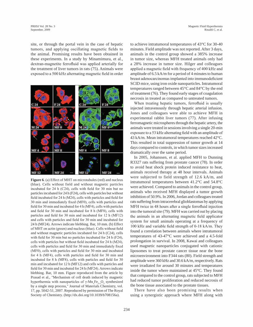
for these purposes. in 1996, Jordan and colleagues found that dextran-coated magnetite nanoparticles were non-toxic to human colonic adenocarcinoma cells at concentrations of up to 5mg/mL. they demonstrated that these cells can internalize up to 1.0 pg of magnetic material per cell (71). in subsequent publications Jordan and colleagues went on to show time-dependent uptake of magnetic nanoparticles into tumor cells and were among the first to point out that the magnetite nanoparticle surface coating affects cellular internalization kinetics (72). they also demonstrated cell death by MFH at temperatures between 43-45°C. Prasad, et al. showed that inducing MFH in HeLa cells with manganese-doped iron oxides caused apoptosis and disrupted the actin and tubulin cytoskeleton (Figure 6) (73). Bergey, et al. labeled magnetite nanoparticles with luteinizing hormone releasing hormone (LHRH) and were able to selectively lyse cells that overexpressed the LHRH receptor when MFH was applied (74). Yan, et al. conducted MFH experiments with maghemite nanoparticles on human hepatocarcinoma (SMMC-7721) cells (30). they showed that 60 minutes of magnetic field application was capable of controlling cell proliferation and increasing chromatin condensation, features of apoptotic cell death. the decrease of cell proliferation and increase in apoptosis correlated with nanoparticle
concentration. From these and other in vitro studies, the basic science behind MFH is being elucidated.
3.2. In vivo studiesto date, various groups have studied the principles
of Magnetic Fluid Hyperthermia (MFH) in rodents and rabbits. the typical methods of application consist of injecting a superparamagetic ferrofluid into the tumor
Figure 4. Passive versus active targeting of tumors. Nanoparticles can be passively delivered to tumor site by the enhanced Permeation and Retention (ePR) effect. defective tumor vascularization and increased vascular fenestrations permit lodging of the nanoparticles into the tumor. active targeting is achieved by attaching a ligand that can be recognized by a surface receptor in the cell of interest.
Active Targeting
Passive Targeting through EPR
Nanoparticle with unspecific coating
Nanoparticle with ligand attached for active recognition through cellular receptors
Figure 5. temperature increase as a function of time for magnetic nanoparticles suspended in heptane at 2.5 %w-ferrite/v upon the application of a magnetic field (6.6 ka/m and 233 kHz). Circles represent magnetite-oleic acid nanoparticles. triangles illustrate cobalt ferrite-oleic acid nanoparticles.
100
Tem
per
ature
(C
)
Time (min)
90
80
70
60
50
40
30
20
0 2 4 6 8 10 12 14
04 Rinaldi.indd 233 8/4/2009 10:17:27 aM
PRHSJ Vol. 28 No. 3September, 2009
234
Magnetic Fluid HyperthermiaRinaldi C, et al.
Figure 6. (a) effect of MHt on microtubules (red) and nucleus (blue). Cells without field and without magnetic particles incubated for 24 h (C24), cells with field for 30 min but no particles incubated for 24 h (F24), cells with particles but without field incubated for 24 h (M24), cells with particles and field for 30 min and immediately fixed (MF0), cells with particles and field for 30 min and incubated for 4 h (MF4), cells with particles and field for 30 min and incubated for 8 h (MF8), cells with particles and field for 30 min and incubated for 12 h (MF12) and cells with particles and field for 30 min and incubated for 24 h (MF24). arrows indicate blebbing. Bar, 10 mm. (b) effect of MHT on actin (green) and nucleus (blue). Cells without field and without magnetic particles incubated for 24 h (C24), cells with field for 30 min but no particles incubated for 24 h (F24), cells with particles but without field incubated for 24 h (M24), cells with particles and field for 30 min and immediately fixed (MF0), cells with particles and field for 30 min and incubated for 4 h (MF4), cells with particles and field for 30 min and incubated for 8 h (MF8), cells with particles and field for 30 min and incubated for 12 h (MF12) and cells with particles and field for 30 min and incubated for 24 h (MF24). Arrows indicate blebbing. Bar, 10 mm. Figure reproduced from the article by Prasad et al., "Mechanism of cell death induced by magnetic hyperthermia with nanoparticles of γ-MnxFe2-xo3 synthesized by a single step process," Journal of Materials Chemistry, vol. 17, pp. 5042-51, 2007. Reproduced by permission of the Royal Society of Chemistry. (http://dx.doi.org/10.1039/b708156a).
site, or through the portal vein in the case of hepatic tumors, and applying oscillating magnetic fields to the animal. Promising results have been obtained in these experiments. in a study by Minamimura, et al., dextran-magnetite ferrofluid was applied arterially for the treatment of liver tumors in rats (75). animals were exposed to a 500 kHz alternating magnetic field in order
to achieve intratumoral temperatures of 43°C for 30-40 minutes. Field amplitude was not reported. after 3 days, animals in the control group showed a 385% increase in tumor size, whereas MFH treated animals only had a 28% increase in tumor size. Hilger and colleagues applied a magnetic field with frequency of 400 kHz and amplitude of 6.5 ka/m for a period of 4 minutes to human breast adenocarcinomas implanted into immunodeficient SCid mice, using iron oxide nanoparticles. intratumoral temperatures ranged between 45°C and 84°C by the end of treatment (76). they found early stages of coagulation necrosis in treated as compared to untreated tumors.
When treating hepatic tumors, ferrofluid is usually injected intravenously through hepatic arterial infusion. Jones and colleagues were able to achieve MFH in experimental rabbit liver tumors (77). after infusing ferromagnetic microspheres through the hepatic artery, the animals were treated in sessions involving a single 20-min exposure to a 53 kHz alternating field with an amplitude of 43 ka/m. Mean intratumoral temperatures reached 42°C. this resulted in total suppression of tumor growth at 14 days compared to controls, in which tumor sizes increased dramatically over the same period.
in 2005, Johanssen, et al. applied MFH to dunning R3327 rats suffering from prostate cancer (78). in order to avoid heat shock protein induced resistance to heat, animals received therapy at 48 hour intervals. animals were subjected to field strength of 12.6 ka/m, and intratumoral temperatures between 41.2°C and 54.8°C were achieved. Compared to animals in the control group, animals who received MFH displayed a tumor growth inhibition of 50.9%. in 2006, Jordan and colleagues treated rats suffering from intracerebral glioblastomas by applying MFH twice in 48 hours after a single ferrofluid injection into the tumoral site (79). MFH was carried out by placing the animals in an alternating magnetic field applicator system for small animals operating at a frequency of 100 kHz and variable field strength of 0-18 kA/m. They found a correlation between animals where intratumoral temperatures of 43-47°C were achieved and a 4.5-fold prolongation in survival. in 2008, Kawai and colleagues used magnetic nanoparticles conjugated with cationic liposomes to treat prostate cancer tissue near the bone microenvironment into F344 rats (80). Field strength and amplitude were 360 kHz and 30.6 ka/m, respectively. Rats were irradiated for around 30 minutes and temperatures inside the tumor where maintained at 45°C. they found that compared to the control group, rats subjected to MFH had reduced tumor proliferation and reduced necrosis of the bone tissue associated to the prostate tissues.
there have also been promising results when using a synergistic approach where MFH along with
M 24
MF 4
MF 12
M 24
MF 4
MF 12
MF 0
MF 8
MF 24
C 24 C 24F 24 F 24
MF 0
MF 8
MF 24
04 Rinaldi.indd 234 8/4/2009 10:17:28 aM

235
PRHSJ Vol. 28 No. 3September, 2009
Magnetic Fluid HyperthermiaRinaldi C, et al.
chemotherapeutic drugs or other molecules are utilized for the treatment of tumors. the high temperatures make the cancerous cells more susceptible to the agent designed to destroy the tumor cell. one example is the work of ito and colleagues, who utilized tumor necrosis factor (tNF) alpha gene therapy along with MFH to successfully inhibit the growth of tumors in mice (81). tNF alpha is a factor that starts a signaling cascade that leads to cell death. tNF alpha expression was driven by the heat- and stress-inducible promoter, gadd 153, with MFH using magnetite cationic liposomes (MCLs) being responsible for the rise in temperature. Magnetic field and amplitude were not reported. in tumor bearing athymic mice, MCLs induced cell death throughout much of the tumor area on heating under an alternating magnetic field. This heat stress also resulted in a 3-fold increase in tNF-alpha gene expression driven by the gadd 153 promoter as compared with that of non-heated tumor. over a 30-day period, the combined treatment strongly arrested tumor growth in nude mice, results being more dramatic than with MFH alone.
3.3. Pre-Clinical Studies
although positive results have been reported in vitro and in vivo using MFH as treatment for cancer in various animal models, MFH has not yet been established in the clinical setting. this is most likely due to limitations in current techniques, where problems are encountered in selectively targeting the tumor and in homogenously and controllably distributing the heat within tumor tissues. to date, all SPioNs used in the pre-clinical setting are composed of the iron oxides magnetite (Fe3o4) and maghemite (γ-Fe2o3). this is due to the fact that these materials have been proven to show low toxicity and their metabolic pathway is known.
MagForce Nanotechnologies ag, a german company, has designed an alternating magnetic field applicator (MFH 300FTM). This magnetic field applicator generates alternating fields of 100kHz at variable field strengths of 0-18 ka/m. it can be used to treat malignancies at almost every location of the human body (82).
The first clinical feasibility study was carried out in March 2003, on 14 terminally ill patients suffering from glioblastoma multiforme (83). all patients received intratumoral injections and received 4-10 MFH sessions of 1-hour duration. intratumoral temperatures reached were between 42.4 - 45.5°C. treatment was well tolerated, and patient follow-up is ongoing. in 2008 a follow-up report was published about three patients that had died during the study due to progression of the disease (84). When brain autopsies were performed, the group found the installed magnetic nanoparticles were dispersed or distributed as aggregates within geographic
tumor necroses, restricted in distribution to the sites of instillation.
in 2005, Johanssen and colleagues started a feasibility trial in ten patients suffering from recurrent prostate cancer. the ferrofluid was injected transperineally. the patients received six 60-minute MFH treatments at weekly intervals, at field strengths of 4 to 5 kA/m. temperatures between 38.8°C and 43.4°C where achieved in 90% of the prostates. in a follow-up study performed 17.5 months earlier, nanoparticle deposits were still detectable in the prostate, and no systemic toxicity was observed (64).
4. Challenges and Conclusionsan important factor responsible for the slow adoption of
hyperthermia as an available resource for physicians and patients is the limited targeting capabilities of currently available nanoparticles. this limitation hinders the ability to concentrate the nanoparticles within the targeted tumor site, negatively affecting the homogeneous distribution of heat. in order to overcome this constraint, the direct injection of the nanoparticles into the tumor sites has been presented as a viable solution for targeted delivery of superparamagnetic nanoparticles. However, more information needs to be uncovered in order to maximize nanoparticle concentration and energy dissipation within and in the vicinity of cancer tumors.
Magnetic Fluid Hyperthermia seems to be a viable approach in the treatment of localized tumors. tumor size regression has been achieved in in vivo experiments in animal models such as rodents and rabbits. Human feasibility studies are under way, treatment seems to be well tolerated, and to date there is no toxicity associated with the treatments given. it remains to be seen if results in human patients will be as promising as those achieved in animal studies.
Resumen Los sistemas de nanopartículas están siendo
intensamente investigados para varias aplicaciones biomédicas. Suspensiones coloidales de nanopartículas magnéticas son de interés especial, particularmente como una herramienta para la capturación de bioimágenes, y más recientemente, por su utilidad en Hipertermia causada por Fluidos Magnéticos, o “MFH” (Magnetic Fluid Hyperthermia) por sus siglas en inglés. MFH promete ser una alternativa viable en el tratamiento de tumores cancerosos localizados. el tratamiento consiste en inyectar una suspensión fluida de nanopartículas magnéticas en el lugar del tumor, y luego exponer el
04 Rinaldi.indd 235 8/4/2009 10:17:28 aM

PRHSJ Vol. 28 No. 3September, 2009
236
Magnetic Fluid HyperthermiaRinaldi C, et al.
area del tumor a un campo magnético oscilante. esto causa que las nanopartículas disipen energía en forma de calor, causando un aumento localizado en temperatura y eventualmente muerte celular. aquí presentamos una revisión de métodos de síntesis de partículas magnéticas, el papel que juega la cubierta de la nanopartícula en alcanzar estabilidad coloidal, minimizar toxicidad, y su reconocimiento por parte de la célula cancerosa. Finalmente, discutiremos experimentos in vivo e in vitro de MFH, y estudios clínicos en el tratamiento de pacientes con glioblastoma multiforme y cáncer de la próstata.
Acknowledgements
ML is supported by a postdoctoral fellowship from the Puerto Rico institute for Functional Nanomaterials, NSF ePSCoR (oia-0701525). CR is supported through a grant from the United States National Science Foundation (NSF) Nanoscale interdisciplinary Research teams program (CBet-0609117). the authors have received funding for this work from the United States National Science Foundation. The authors have no conflict of interest to disclose.
References
1. Roduner e. Size matters: Why nanomaterials are different. Chem Soc Rev 2006;35:583-592.
2. Roco M. Nanoparticles and nanotechnology research. J Nanopar-ticle Res 1999;1:1-6.
3. Roco MC. Nanoscale Science and engineering: Unifying and transforming tools. aiChe Journal 2004;50:890-7.
4. alexis F, Rhee JW, Richie JP, et al. New frontiers in nanotechnol-ogy for cancer treatment. Urol oncol 2008;26:74-85.
5. Meyer MH, Stehr M, Bhuju S, et al. Magnetic biosensor for the de-tection of Yersinia pestis. J Microbiol Methods 2007;68:218-24.
6. Kirsch Je. Basic principles of magnetic resonance contrast agents. top Magn Reson imaging 1991;3:1-18.
7. de Jong W, Borm P. drug delivery and Nanoparticles: applica-tions and Hazards. int J Nanomedicine 2008;3:133-149.
8. Nie S, Xing Y, Kim gJ, et al. Nanotechnology applications in can-cer. annu Rev Biomed eng 2007;9:257-288.
9. de M, ghosh P, Rotello V. applications of nanoparticles in Biol-ogy. adv Mat 2008;20:1-17.
10. duran Jd, arias JL, gallardo V, delgado aV. Magnetic colloids as drug vehicles. J Pharm Sci 2008;97:2948-2983.
11. diamond d. overview. in: Wiley N, editor. Principles of Chemical and Biological Sensors; 1998: p. 1-18.
12. Park SJ, taton ta, Mirkin Ca. array-based electrical detection of dNa with nanoparticle probes. Science 2002;295:1503-1506.
13. Wang S, Jarrett BR, Kauzlarich SM, et al. Core/shell quantum dots with high relaxivity and photoluminescence for multimodality im-aging. J am Chem Soc 2007;129:3848-3856.
14. Hong R, Fischer N, goodman C, et al. Control of Protein Structure and Function through Surface Recognition by tailored Nanopar-ticle Scaffolds. J am Chem Soc 2004;126:739-743.
15. Sanhai WR, Sakamoto JH, Canady R, et al. Seven challenges for nanomedicine. Nat Nanotechnol 2008;3:242-244.
16. Bareford LM, Swaan PW. endocytic mechanisms for targeted drug delivery. adv drug deliv Rev 2007;59:748-758.
17. Hu L, Mao Z, gao C. Colloidal particles for cellular uptake and delivery. J Mater Chem 2009;19:3108-3115.
18. Miltenyi S, Muller W, Weichel W, et al. High gradient magnetic cell separation with MaCS. Cytometry 1990;11:231-238.
19. abts H, emmerich M, Miltenyi S, et al. Cd20 positive human B lymphocytes separated with the magnetic cell sorter (MaCS) can be induced to proliferation and antibody secretion in vitro. J im-munol Methods 1989;125:19-28.
20. Wilhelm C, Billotey C, Roger J, et al. intracellular uptake of anion-ic superparamagnetic nanoparticles as a function of their surface coating. Biomaterials 2003;24:1001-1011.
21. Schwalbe M, Jörkeu C, Buske N, et al. Selective reduction of the interaction of magnetic nanoparticles with leukocytes and tumor cells by human plasma. J Mag Mag Mat 2005;293:433-437.
22. Lorenz MR, Holzapfel V, Musyanovych a, et al. Uptake of func-tionalized, fluorescent-labeled polymeric particles in different cell lines and stem cells. Biomaterials 2006;27:2820-2828.
23. Peppas N, Huang Y, torres-Lugo M, et al. Physicochemical foun-dations and structural design of hydrogels in medicine and biology. ann Rev Biomed eng 2000;2:9-30.
24. Barrera C, Herrera a, Rinaldi C. Colloidal dispersions of mono-disperse magnetite nanoparticles modified with poly(ethylene gly-col). J Colloid interface Sci 2009;329:107-113.
25. Roger J, Pons J, Massart R, et al. Some biomedical applications of ferrofluids. Euro Phys J - Applied Physics 1999;5:321-325.
26. Ramchand CN, Pande P, Kopcansky P, et al. application of mag-netic fluids in medicine and biotechnology. Ind J of Pure Applied Physics 2001;39:683-686.
27. Na HB, Song iC, Hyeon t. inorganic nanoparticles for MRi con-trast agents. adv Mat 2009;21:2133-2148.
28. Berger M, Castelino J, Huang R, Shah M, austin RH. design of a microfabricated magnetic cell separator. electrophoresis 2001; 22:3883-3892.
29. Baselt dR, Lee gU, Natesan M, Metzger SW, Sheehan Pe, Colton RJ. a biosensor based on magnetoresistance technology. Biosens Bioelect 1998;13:731-739.
30. Yan S, Zhang d, gu N, Zheng J, ding a, Wang Z, Xing B, Ma M, Zhang Y. therapeutic effect of Fe2o3 nanoparticles com-bined with magnetic fluid hyperthermia on cultured liver cancer cells and xenograft liver cancers. J Nanosci Nanotechnol 2005;5: 1185-1192.
31. Bonnemain B. Superparamagnetic agents in magnetic resonance imaging: physicochemical characteristics and clinical applica-tions. a review. J drug target 1998;6:167-174.
32. de Haen C. Conception of the first magnetic resonance imag-ing contrast agents: a brief history. top Magn Reson imaging 2001;12:221-230.
33. Yuan C, Kerwin WS. MRi of atherosclerosis. J Magn Reson imag-ing 2004;19:710-719.
34. David R, Groebner M, Franz WM. Magnetic cell sorting purifica-tion of differentiated embryonic stem cells stably expressing trun-cated human Cd4 as surface marker. Stem Cells 2005;23:477-482.
35. Minges Wols HA, Witte PL. Plasma cell purification from mu-rine bone marrow using a two-step isolation approach. J immunol Methods 2008;329:219-224.
36. derfus a, von Maltzahn g, Harris t, et al. Remotely triggered Re-lease from Magnetic Nanoparticles. adv Mat 2007;19:3932-3936.
37. Kaiser A, Gelbrich T, Schmidt AM. Thermosensitive magnetic flu-ids. J Phys-Condensed Matter 2006;18:S2563-S2580.
38. Schmidt aM. thermoresponsive magnetic colloids. Colloid Poly-mer Sci 2007;285:953-966.
04 Rinaldi.indd 236 8/4/2009 10:17:28 aM

237
PRHSJ Vol. 28 No. 3September, 2009
Magnetic Fluid HyperthermiaRinaldi C, et al.
39. Zhang J, Misra RdK. Magnetic drug-targeting carrier encapsu-lated with thermosensitive smart polymer: Core-shell nanoparticle carrier and drug release response. acta Biomat 2007;3:838-850.
40. Zhang JL, Srivastava RS, Misra RdK. Core-shell magnetite nanoparticles surface encapsulated with smart stimuli-responsive polymer: Synthesis, characterization, and LCSt of viable drug-targeting delivery system. Langmuir 2007;23:6342-6351.
41. gupta aK, gupta M. Synthesis and surface engineering of iron oxide nanoparticles for biomedical applications. Biomaterials 2005;26:3995-4021.
42. Lefebure S, dubois e, Cabuil V, et al. Monodisperse magnetic nanoparticles: Preparation and dispersion in water and oils. J Mat Res,1998;13:2975-2981.
43. Lu aH, Salabas eL, Schuth F. Magnetic nanoparticles: synthesis, protection, functionalization, and application. angew Chem int ed engl 2007;46:1222-1244.
44. Herrera a, Barrera C, Rinaldi C. Synthesis and functionalization of magnetite nanoparticles with aminopropylsilane and carboxym-ethyldextran. J Mat Chem 2008;18:3650-3654.
45. Hyeon t, Chung Y, Park J, et al. Synthesis of highly crystalline and monodisperse cobalt ferrite nanocrystals. J Phys Chem B 2002;106:6831-6833.
46. Hyeon t, Lee SS, Park J, et al. Synthesis of highly crystalline and monodisperse maghemite nanocrystallites without a size-selection process. aCS J 2001;123:12798-12801.
47. Park J, an K, Hwang Y, et al. Ultra-large-scale syntheses of mono-disperse nanocrystals. Nat Mater 2004;3:891-895.
48. Rockenberger J, Scher eC, alivisatos aP. a new nonhydrolytic single-precursor approach to surfactant-capped nanocrystals of transition metal oxides. aCS J 1999;121:11595-11596.
49. Sun SH, Murray CB. Synthesis of monodisperse cobalt nanocrys-tals and their assembly into magnetic superlattices (invited). J app Phys 1999;85:4325-4330.
50. Sun SH, Zeng H. Size-controlled synthesis of magnetite nanoparti-cies. aCS J 2002;124:8204-8205.
51. de Palma R, Peeters S, Van Bael M, et al. Silane Ligand exchange to Make Hydrophobic Superparamagnetic Nanoparticles Water-dispersible. Chem Mat 2007;19:1821-1831.
52. Villanueva a, Canete M, Roca a, Calero M, Veintemillas-Ver-daguer S, Serna CJ, Morales Mdel P, Miranda R. The influence of surface functionalizationon the enhanced internalization of mag-netic nanoparticles in cancer cells. Nanotechnology 2009;20:1-9.
53. goodman CM, McCusker Cd, Yilmaz t, et al. toxicity of gold nanoparticles functionalized with cationic and anionic side chains. Bioconjug Chem 2004;15:897-900.
54. Pisanic tR, 2nd, Blackwell Jd, Shubayev Vi, et al. Nanotoxicity of iron oxide nanoparticle internalization in growing neurons. Bio-materials 2007;28:2572-81.
55. Zhang Y, Zhang J. Surface modification of monodisperse magne-tite nanoparticles for improved intracellular uptake to breast can-cer cells. J Colloid interface Sci 2005;283:352-357.
56. Kim JS, Yoon tJ, Yu KN, et al. Cellular uptake of magnetic nano-particle is mediated through energy-dependent endocytosis in a549 cells. J Vet Sci 2006;7:321-326.
57. Bhattarai SR, Kc RB, Kim SY et al. N-hexanoyl chitosan stabi-lized magnetic nanoparticles: implicationfor cellular labeling and magnetic resonance imaging. J Nanobiotech 2008;6:1-9.
58. trehin R, Figueiredo JL, Pittet MJ, et al. Fluorescent nanoparticle uptake for brain tumor visualization. Neoplasia 2006;8:302-311.
59. Clement JH, Schwalbe M, Buske N, et al. differential interaction of magnetic nanoparticles with tumor cells and peripheral blood cells. J Cancer Res Clin oncol 2006;132:287-292.
60. Phanapavudhikul P, Shen S, Ng WK, et al. Formulation of Fe3o4/acrylate co-polymer nanocomposites as potential drug carriers. drug deliv 2008;15:177-183.
61. Yoo MK, Kim iY, Kim eM, et al. Superparamagnetic iron oxide nanoparticles coated with galactose-carrying polymer for hepato-cyte targeting. J Biomed Biotechnol 2007;2007:94740-94748.
62. Chen WR, adams RL, Carubelli R, et al. Laser-photosensitizer assisted immunotherapy: a novel modality for cancer treatment. Cancer Lett 1997;115:25-30.
63. Lin JC, Wang J. interstitial microwave antennas for thermal thera-py. int J Hyperthermia 1987;3:37-47.
64. Johannsen M, gneveckow U, thiesen B, et al. thermotherapy of prostate cancer using magnetic nanoparticles: feasibility, im-aging, and three-dimensional temperature distribution. eur Urol 2007;52:1653-1661.
65. Maeda H, Wu J, Sawa t, et al. tumor vascular permeability and the ePR effect in macromolecular therapeutics: a review. J Control Release 2000;65:271-284.
66. Peira e, Marzola P, Podio V, et al. in vitro and in vivo study of solid lipid nanoparticles loaded with superparamagnetic iron ox-ide. J drug target 2003;11:19-24.
67. Jordan a, Rheinländer t, Waldöfner N, et al. increase of the spe-cific absorption rate (SAR) by magnetic fractionation of magnetic fluids. J Nanoparticle Res 2003;5:597-600.
68. todaka t, Kishino t, enokisono N. Low Curie temperature ma-terial for induction heating self-temperature controlling system. J Mag Mag Mat 2008;320:702-707.
69. Kaman o, Pollert e, Veverka P, et al. Silica encapsulated man-ganese perovskite nanoparticles for magnetically induced hy-perthermia without the risk of overheating. Nanotechnology 2009;20:275610.
70. Melnikov oV, gorbenko oY, Markelova MN, et al. ag-doped manganite nanoparticles: New materials for temperature-controlled medical hyperthermia. J Biomed Mater Res a 2009;90:317-325.
71. Jordan a, Wust P, Scholz R, tesche B, Fähling H, Mitrovics t, Vogl t, Cervós-Navarro J, Felix R. Cellular uptake of mag-netic fluid particles and their effets on human adenocarcinoma cells exposed to AC fields in vitro. Int J Hyperthermia 1996;12: 705-722.
72. Jordan a, Scholz R, Wust P, et al. endocytosis of dextran and si-lan-coated magnetite nanoparticles and the effect of intracellular hyperthermia on human mammary carcinoma cells in vitro. J Mag Mag Mat 1999;194:185-196.
73. Prasad N, Rathinasamy K, Panda d, et al. Mechanism of cell death induced by magnetic hyperthermia with nanoparticles of gamma-MnxFe2-xo3 synthesized by a single step process. J Mat Chem 2007;17:5042-5051.
74. Bergey e, Levy L, Wang X, et al. dC Magnetic Field induced Magnetocytolysis of Cancer Cells targeted by LH-RH Magnetic Nanoparticles in vitro. Biomed Microdevices 2002;4:293-299.
75. Minamimura t, Sato H, Kasaoka S, Saito t, ishizawa S, takemori S, tazawa K, tsukada K. tumor regression by inductive hyper-thermia combined with hepatic embolization using dextran mag-netite-incorporated microspheres in rats. int J oncol 2000;16: 1153-1158.
76. Hilger i, Hiergeist R, Hergt R, Winnefeld K, Schubert H, Kaiser Wa. thermal ablation of tumors using magnetic nanoparticles: an in vivo feasibility study. invest Radiol 2002;37:580-586.
77. Jones S, Winter J, gray B. treatment of experimental rabbit liver tumours by selectively targeted hyperthermia. int J Hyperthermia 2002;18:117-128.
78. Johannsen M, Thiesen B, Jordan A, et al. Magnetic fluid hyper-thermia (MFH)reduces prostate cancer growth in the orthotopic dunning R3327 rat model. Prostate 2005;64:283-292.
79. Jordan a, Scholz R, Maier-Hauff K, et al. the effect of thermo-therapy using magnetic nanoparticles on rat malignant glioma. J Neurooncol 2006;78:7-14.
04 Rinaldi.indd 237 8/4/2009 10:17:28 aM

PRHSJ Vol. 28 No. 3September, 2009
238
Magnetic Fluid HyperthermiaRinaldi C, et al.
80. Kawai N, Futakuchi M, Yoshida t, et al. effect of heat therapy us-ing magnetic nanoparticles conjugated with cationic liposomes on prostate tumor in bone. Prostate 2008;68:784-792.
81. ito a, Shinkai M, Hakamada K, et al. Radiation-inducible tNF-alpha gene expression under stress-inducible promoter gadd 153 for cancer therapy. J Biosci Bioeng 2001;92:598-601.
82. thiesen B, Jordan a. Clinical applications of magnetic nanopar-ticles for hyperthermia. int J Hyperthermia 2008;24:467-474.
83. Maier-Hauff K, Rothe R, Scholz R, et al. intracranial thermother-apy using magnetic nanoparticles combined with external beam radiotherapy: results of a feasibility study on patients with glio-blastoma multiforme. J Neurooncol 2007;81:53-60.
84. van Landeghem FK, Maier-Hauff K, Jordan a, Hoffmann Kt, gneveckow U, Scholz R, thiesen B, Brück W, von deimling a. Post-mortem studies in glioblastoma patients treated with thermo-therapy using magnetic nanoparticles. Biomaterials 2008: in print.
04 Rinaldi.indd 238 8/4/2009 10:17:28 aM