preparation of functionalized magnetic nanoparticles
TRANSCRIPT

RSC Advances
PAPER
Ope
n A
cces
s A
rtic
le. P
ublis
hed
on 0
1 M
ay 2
019.
Dow
nloa
ded
on 3
/15/
2022
6:4
8:12
PM
. T
his
artic
le is
lice
nsed
und
er a
Cre
ativ
e C
omm
ons
Attr
ibut
ion-
Non
Com
mer
cial
3.0
Unp
orte
d L
icen
ce.
View Article OnlineView Journal | View Issue
Preparation of fu
aCentro de Investigacions Cientıcas Avanz
Facultade de Ciencias, Universidade da C
[email protected]; carlos.jimenez@udbDepartment of Microbiology and Parasitolo
de Santiago de Compostela, Campus Sur, ScInstitute of Functional Surfaces, School o
Leeds, Leeds LS2 2JT, UKdBioResource Systems Research Group, Schoo
Leeds LS2 9JT, UKeFaculty of Mathematics and Physical Scie
University of Leeds, Leeds LS2 9JT, UKfDepartamento de Ingenierıa Quımica, Unive
Nubia, Manizales, Colombia
† Electronic supplementary informationmagnetization hysteresis loops, thermogNPs, SEM images, TEM images and EDXinteraction assay results for iron and irDOI: 10.1039/c8ra10440a
Cite this: RSC Adv., 2019, 9, 13533
Received 20th December 2018Accepted 22nd April 2019
DOI: 10.1039/c8ra10440a
rsc.li/rsc-advances
This journal is © The Royal Society of C
nctionalized magneticnanoparticles conjugated with feroxamine andtheir evaluation for pathogen detection†
Diana Martınez-Matamoros, a Socorro Castro-Garcıa, a Miguel Balado, b
Adriana Matamoros-Veloza, c Miller Alonso Camargo-Valero, df
Oscar Cespedes,e Jaime Rodrıguez, *a Manuel L. Lemos b
and Carlos Jimenez *a
This work reports the preparation of a conjugate between amino-functionalized silica magnetite and the
siderophore feroxamine. The morphology and properties of the conjugate and intermediate magnetic
nanoparticles (MNPs) were examined by powder X-ray diffraction (XRD), Fourier Transform Infrared
spectroscopy (FT-IR), Raman spectroscopy, X-ray photoelectron spectroscopy (XPS), magnetization
studies, zeta potential measurements, Transmission Electron Microscopy (TEM) and Energy Dispersive X-
ray (EDX) mapping. Furthermore, this study investigated the interaction between the functionalized
magnetic NPs and Yersinia enterocolitica wild type (WC-A) using Scanning Electron Microscopy (SEM)
and TEM images. In addition, the interaction between MNPs and a Y. enterocolitica mutant strain lacking
feroxamine receptor FoxA, was also used to study the binding specificity. The results showed that the
capture and isolation of Y. enterocolitica by the MNPs took place in all cases. Moreover, the specific
interaction between the MNP conjugate and bacteria did not increase after blocking the free amine
groups with t-butoxycarbonyl (Boc) and carboxylic acid (COOH) functional groups. Electrostatic surface
interactions instead of molecular recognition between MNP conjugate and feroxamine receptor seem to
rule the attachment of bacteria to the conjugate.
Introduction
A growing interest in magnetic nanoparticles (MNP) based onmagnetite (Fe3O4) has been observed over the last 10 years inanalytical sensing and nanomedicine due to its strongmagneticproperties and biocompatibility.1 The magnetic eld of MNPplays a key role in the capture and bio-separation of analytes.
adas (CICA), Departamento de Quımica,
oruna, 15071 A Coruna, Spain. E-mail:
c.es
gy, Institute of Aquaculture, Universidade
antiago de Compostela 15782, Spain
f Mechanical Engineering, University of
l of Civil Engineering, University of Leeds,
nces, School of Physics and Astronomy,
rsidad Nacional de Colombia, Campus La
(ESI) available: p-XRD, FT-IR spectra,ravimetric analysis (TGA) of blockedmaps and Y. enterocolitica WC-A-MNPon deciency growth conditions. See
hemistry 2019
The functionalization of MNP's surface allows the developmentof multiple applications such as magnetic hyperthermia,magnetic resonance imaging (MRI), target drug delivery anddetection of bacteria, since MNP are capable of capturingbacteria using specic recognition. In fact, MNP are capable ofinteracting with biological entities such as proteins and bacte-rial membranes, among others, can be manipulated by anexternal magnetic eld, and are easy to synthesize.2
The development of rapid, sensitive and reliable methods forthe detection and identication of infectious microorganisms isone of the main concerns in food and health industries.Nowadays, this interest has become more important with theemerge of virulent strains of common pathogenic bacteria andthe need of limiting the spread of related contagious diseases.The traditional methods based on cell culturing are usually veryslow and time-consuming processes. Numerous rapid andsensitive methods for microbial detection have been developed(e.g., immunoassays, enzyme-linked immunosorbent assays(ELISA) and polymerase chain reaction methodologies (PCR)).3
However, they are not effective in complex systems whenbacteria are present in low concentrations. An emergingresearch area based on the magnetic, electronic, photonic, andoptical properties and functionalization of MNP has beingadopted to develop alternative methods based on the isolation
RSC Adv., 2019, 9, 13533–13542 | 13533

RSC Advances Paper
Ope
n A
cces
s A
rtic
le. P
ublis
hed
on 0
1 M
ay 2
019.
Dow
nloa
ded
on 3
/15/
2022
6:4
8:12
PM
. T
his
artic
le is
lice
nsed
und
er a
Cre
ativ
e C
omm
ons
Attr
ibut
ion-
Non
Com
mer
cial
3.0
Unp
orte
d L
icen
ce.
View Article Online
of pathogenic bacteria using nanomaterials for biologicalidentication.4
The specicity of MNP is based on the chemical recognitionof pathogenic bacteria through the conjugation of MNP withantibodies, aptamers, bioproteins, carbohydrates and bacte-riophages.1 Moreover, MNP can be also coupled to siderophoresthat are recognized by specic membrane receptors of micro-organisms.5 Siderophores are small organic molecules recog-nised for playing a role in the mechanisms controlling Fe3+
uptake by bacteria.6,7 So far, three different approaches havebeen documented for the detection of microbial pathogensusing siderophore scaffolds. The rst one utilizes an immobi-lized siderophore to capture human pathogens, in whicha siderophore conjugate is attached to gold-plated glass chipsthrough bovine serum albumin (BSA).8,9 Other example isa modied, articial siderophore complex attached to thesurface of an Au electrode and placed on quartz crystal micro-balance (QCM) chips.10,11 A recent work using this approach,documented the use of a siderophore-based active bacterialremoval integrated in a localized surface plasmon resonance(LSPR) sensing platform.12 The second approach employsa siderophore attached to functionalized quantum dots (QDs)for the bacterial interaction with a specic receptor.13 The thirdapproach uses functionalized agarose columns bound toa specic bacterial siderophore for the capture of siderophore-binding proteins.14 However, the conjugation of siderophoresand magnetic nanoparticles to isolate and capture pathogenicbacteria has not been studied yet.
Herein, we report the rst synthesis of a conjugate betweenamino-functionalized silica magnetite and the siderophoreferoxamine, the blocking of free amine groups on the surface ofamino-functionalized silica magnetite and the conjugate with t-butoxycarbonyl (Boc) and carboxylic acid (–COOH) functionalgroups and its evaluation for the capture of wild type (WC-A)and a mutant lacking feroxamine receptor FoxA (FoxA WC-A12-8) Y. enterocolitica strains.
Experimental
All starting materials, reagents and solvents were obtained fromcommercial suppliers and used without further purication.Argon gas was used to avoid the presence of moisture andoxygen in sensitive reactions. Size exclusion chromatographywas performed on Sephadex™ LH-20 resins. LREIMS andHRESIMS were measured on Applied Biosystems QSTAR Elite.
Synthesis
Synthesis of Fe3O4 magnetic nanoparticles (MNP).15 A solu-tion of 0.5 g of iron(III) acetylacetonate (Fe(acac)3) in 10 mL ofbenzyl alcohol was sonicated for 2 min, transferred to a heatingblock and le to react at 180 �C for 72 h. Aer that time, theresulting mixture was allowed to cool down before the precipi-tates were decanted by centrifugation (5000 rpm for 30 min),while the supernatant was discarded. The solids were rinsedthree times with 96% ethanol, sonicated and recovered usinga magnet.
13534 | RSC Adv., 2019, 9, 13533–13542
Synthesis of SiO2 coating of MNP (MNP@SiO2).16 80 mL ofisopropanol, 4 mL of ammonia (21%), 7.5 mL of distilled waterand 0.56 mL of tetraethyl orthosilicate (TEOS) were carefullyadded in this order to 2 g of MNP. The mixture was heated at40 �C for 2 h with continuous stirring and then sonicated for1 h. Aer that time, the MNP were removed from the solutionusing a magnet and re-dispersed in 30 mL of isopropanol. Thiscoating procedure was repeated a second time. Finally, the SiO2
coated MNP were rinsed with ethanol and separated from thedispersion using a magnet.
Synthesis of amino-functionalized silica magnetite(MNP@SiO2@NH2).17 A modied procedure described by Chenet al.17 was used for the functionalization of MNP@SiO2. Forthat, 500 mg of MNP@SiO2 were rinsed and sonicated threetimes with 3 mL of dimethylformamide (DMF). Then, theparticles were re-suspended in 9 mL of DMF and 9 mL of 3-aminopropyltriethoxysilane (APTES). The resulting mixture wasthen shaken at 60 �C for 12 h. Finally, the functionalizedparticles (MNP@SiO2@NH2) were separated with a magnet andsonicated with 96% ethanol, three times.
Synthesis of feroxamine (2).18 100 mg (0.15 mmol) of defer-oxamine mesylate (1) salt and 53.0 mg (0.15 mmol) of Fe(acac)3were dissolved in 5 mL of distilled water and le stirring over-night. The resulting product was washed three times with 20mLof EtOAc and then, the solvent was removed under vacuumusing a rotavapor. The aqueous phase was freeze-dried to obtainferoxamine as a red solid (94.4 mg, 78% yield). (+)-HR-ESIMSm/z 614.2751 [M + H]+ (calculated for C25H45FeN6O8: 614.2729).
Synthesis of N-succinyl feroxamine (3).19 350 mg (3.50 mmol)of succinic anhydride were added to a solution of 100 mg (0.17mmol) of feroxamine in 5 mL of pyridine. The resulting mixturewas stirred at room temperature for 16 h. Aer that time, theexcess of pyridine was eliminated in a rotavapor under vacuum.The red solid product was puried by size exclusion chroma-tography using methanol as eluent to separate 93.1 mg of N-succinyl feroxamine (3) as a dark red solid. (+)-HR-ESIMS m/z736.2700 [M + Na]+; (calculated for C29H49FeN6O11Na:736.2706).
Synthesis of MNP@SiO2@NH@Fa (4). 30 mg of dryMNP@SiO2@NH2 were rinsed twice with DMF and sonicatedfor 30 minutes. A solution of N-succinyl feroxamine (3; 200 mg,0.30 mmol), benzotriazole-1-yl-oxy-tris-(dimethylamino)-phosphonium hexauorophosphate (BOP, 173 mg, 0.45mmol), 1-hydroxybenzotriazole (HOBt, 46 mg, 0.39 mmol) andN,N-diisopropylethylamine (DIPEA, 128.8 mg, 1.21 mmol) in10 mL of DMF was added dropwise to a suspension of 30 mg ofMNP@SiO2@NH2 in 3 mL of DMF under sonication in dry andoxygen free conditions using an argon gas atmosphere.20 Themixture was le stirring at room temperature overnight. Finally,the resulting conjugate (MNP@SiO2@NH@Fa, 4) was separatedfrom the suspension with a magnet and the separated solid wasrinsed and sonicated 5 times with 10 mL of ethanol. The solidwas vacuum dried for 24 h.
Synthesis of MNP@SiO2@NHBoc and MNP@SiO2@-NHBoc@Fa (5). 28.5 mg of MNP@SiO2@NH2 was rinsed withdry DMF and sonicated twice for 5 minutes under an argon gas
This journal is © The Royal Society of Chemistry 2019

Paper RSC Advances
Ope
n A
cces
s A
rtic
le. P
ublis
hed
on 0
1 M
ay 2
019.
Dow
nloa
ded
on 3
/15/
2022
6:4
8:12
PM
. T
his
artic
le is
lice
nsed
und
er a
Cre
ativ
e C
omm
ons
Attr
ibut
ion-
Non
Com
mer
cial
3.0
Unp
orte
d L
icen
ce.
View Article Online
atmosphere and then suspended in 10 mL of dry DMF. Aerthat, 200 mg of Boc2O (di-tert-butyl dicarbonate) were dissolvedin dry DMF and mixed with the nanoparticles. The reactionmixture was sonicated for 30 min and then stirred at roomtemperature in an orbital shaker at 200 rpm for 24 h. Finally, thesolids were separated using a magnet and rinsed with 10 mL ofethanol and sonicated ve times. The solids were then vacuumdried for 24 h to yield 27.5 mg of MNP@SiO2@NHBoc. Thesame procedure was repeated for 13.6 mg of MNP@SiO2@-NH@Fa (4) to obtain 13.8 mg of MNP@SiO2@NHBoc@Fa (5).
Synthesis of MNP@SiO2@NHCOOH and MNP@SiO2@-NHCOOH@Fa (6). 25 mg of MNP@SiO2@NH2 were rst rinsedwith dry pyridine and sonicated twice for 5 minutes under anargon atmosphere and then suspended in 10 mL of dry pyridine.Aer this, 200 mg of succinic anhydride were added to thenanoparticles. The reaction mixture was sonicated 30 min andthen stirred at room temperature in an orbital shaker at 200 rpmfor 24 h. Finally, the solids were separated using amagnet, rinsedand sonicated ve times using 10 mL of ethanol and vacuumdried for 24 h to yield 21.3 mg of MNP@SiO2@NHCOOH. Thesame procedure was repeated for 9.3 mg of MNP@SiO2@NH@Fa(4) to obtain 8.7 mg of MNP@SiO2@NHCOOH@Fa (6).
Characterization
Powder X-ray diffraction (XRD). XRD analyses of samplescontaining MNP were performed using a Bruker D8 diffrac-tometer (CuKa) with a scan range between 2 and 70 2q at 0.052q min�1. The MNP containing samples were re-dispersed inethanol and mounted onto a poly(methyl methacrylate) spec-imen holder for analysis. Peak identication was performed byusing X'Pert High Score Plus soware by comparing thecollected diffraction data with the International Centre forDiffraction Data database.
FT-IR and Raman spectroscopy. FT-IR analyses were carriedout on powdered nanoparticle samples using an A2-TechnologyMicroLab Portable mid-IR spectrometer equipped with a dia-mond internal reection (DATR). For the analysis, the back-ground was collected without deposition of the sample andthen a sample was re-dispersed in ethanol and placed onto thediamond window of the instrument. Individual spectra (4096)were acquired between 650 to 4000 cm�1 at a resolution of1 cm�1 and then co-added and processed using Origin 8 (Ori-ginLab, Northampton, MA, USA). Raman analyses were carriedout in a LabRAM HR 800 Horiba Scientic spectrometer, witha 633 nm laser using a 10% power (�1 mW) and a diffractiongrating of 600 ln per mm. Each spectrum included tenmeasurements of 300 seconds (total measuring time 3000seconds).
X-ray photoelectron spectroscopy (XPS). Analysis of thesamples was performed using a Thermo Scientic K-AlphaESCA instrument equipped with aluminum Ka mono-chromatized radiation at 1486.6 eV X-ray source. Due to the noconductor nature of samples, it was necessary to use an electronood gun to minimize surface charging. Neutralization of thesurface charge was performed by using both a low energy oodgun (electrons in the range 0 to 14 eV) and a low energy Argon
This journal is © The Royal Society of Chemistry 2019
ions gun. The XPS measurements were carried out usingmonochromatic Al-Ka radiation (hn ¼ 1486.6 eV). Photoelec-trons were collected from a take-off angle of 90� relative to thesample surface. The measurement was done in a ConstantAnalyser Energy mode (CAE) with a 100 eV pass energy forsurvey spectra and 20 eV pass energy for high resolution spectra.Charge referencing was done by setting the lower bindingenergy C1s photo peak at 285.0 eV C1s hydrocarbon peak.Surface elemental composition was determined using thestandard Scoeld photoemission cross sections. Data analysisand quantication were performed using the Avantage sowareversion 5 from the manufacturer Thermo Scientic.
Magnetic characterization. Magnetization was measuredusing an Oxford Instruments VSM with a magnetic eld of 1 Tand a sensitivity of 10 micro-emu.
Zeta potential analysis. Zeta potential measurements wereperformed with a NanoBrook 90 Plus from Brookhaven Instru-ments. Samples were prepared with ultra-pure water andanalyzed immediately aer sonication.
Thermogravimetric analysis (TGA). Thermogravimetricanalyses were carried out using a differential scanning calo-rimeter STA 449 F3 Jupiter (Netzsch), equipped with a SiC oven.The samples were analyzed in a nitrogen gas atmosphere by anincrement of the temperature of 5 �C min�1 until 900 �C.Weight loss of each sample was obtained by measurements atdifferent temperatures.
Transmission electron microscopy (TEM) and energydispersive X-ray (EDX) mapping. Bright eld images and mapswere acquired at room temperature using a Tecnai TF20 FEG-TEM with an operating voltage of 200 keV tted with a highangle annular dark eld (HAADF) detector and a Gatan OriusSC600 CCD camera. EDX maps were collected at roomtemperature using a FEI Titan G2 S/TEM with an operatingvoltage of 200 keV, a beam current of 0.1 nA, a convergenceangle of 18 mrad and a HAADF inner angle of 54 mrad.
Bacterial capture study with Y. enterocolitica strains
Yersinia enterocolitica WC-A and FoxA WC-A 12-8 were donatedby Professor Klaus Hantke (University of Tubingen, Germany).Trypticase Soy Broth (TSB), Trypticase Soy Agar (TSA), Ringerssolution and PBS buffer were prepared with distilled water (DW)for biological assays.
Trypticase soy broth (TSB) cultures of Y. enterocolitica (wildtype and mutant strains) were incubated up to an OD600
between 0.5 and 0.8 in iron decient conditions by adding 2,20-bipyridyl up to 100 mM. Then, 100 mL of a 1 mgmL�1 solution ofbare, MNP@SiO2, MNP@SiO2@NH2 or MNP@SiO2@NH@Fa(4) was added to 1 mL of 1 : 100 dilution of each Y. enterocoliticastrain (equivalent to ca. 6 � 106 bacterial cells per mL) inPhosphate Buffer Saline (PBS) pH 7.4 and incubated for 1 h. TheMNP/bacteria aggregates were separated with a magnet and thesupernatant was carefully discarded. The remaining aggregateswere rinsed twice with PBS and re-suspended again in freshPBS. Serial ten-fold dilutions of this suspension were plated onTrypticase Soy Agar (TSA) and incubated at 37 �C for 24 h. Aer
RSC Adv., 2019, 9, 13533–13542 | 13535

RSC Advances Paper
Ope
n A
cces
s A
rtic
le. P
ublis
hed
on 0
1 M
ay 2
019.
Dow
nloa
ded
on 3
/15/
2022
6:4
8:12
PM
. T
his
artic
le is
lice
nsed
und
er a
Cre
ativ
e C
omm
ons
Attr
ibut
ion-
Non
Com
mer
cial
3.0
Unp
orte
d L
icen
ce.
View Article Online
this time, the colony forming units (CFU) captured with theMNP conjugate were counted.
Evaluation of bacteria–nanoparticle interaction
Scanning electron microscopy (SEM). SEM images were ob-tained using a FEI Quanta 650 FEGESEM environmental SEMwith an Oxford Instruments INCA 350 EDX system/80 mm X-MaxSDD detector, EBSD and KE Centaurus EBSD system. Imageanalysis was performed in ImageJ soware.21 Y. enterocoliticaWC-A was grown in 10 mL of TSB until a OD600 ¼ 0.5, then thebacteria in solution was diluted 1 : 10 and mixed with 1 mL ofa suspension of MNP@SiO2@NH@Fa (4) in PBS. The bacteriawere allowed to interact with the nanoparticles at room temper-ature for 1 h, then the solids (bacteria–nanoparticles) wereseparated from the suspension with a magnet, and rinsed twicewith 1 mL of PBS. The captured bacteria were mixed with 2.5%glutaraldehyde in 0.1M phosphate buffer and allowed to react for2 h. Aer that time, the solids were rinsed twice with 0.1 Mphosphate buffer for 30 min. Post-xed samples were mixed with1% osmium tetroxide in 0.1 M phosphate buffer overnight. Aerthat time, the solids were dehydrated using an ascending acetoneseries (20–40–60–80–100%) for 30 min, each run. Aer this, thesamples were dried with a Polaron E3000 critical point dryingapparatus using liquid carbon dioxide as the transition uid toafford enough solid to bemounted on 13mm-diameter pin stubsusing double sided adhesive tape. Finally, these samples werecoated with platinum to a thickness of 5 nm using a Cressington208HR high resolution sputter coating unit.
Transmission electron microscopy (TEM) and EDX maps.Bacteria–nanoparticle interaction was performed as describedin the SEM analysis. The solids were xed in 2.5% glutaralde-hyde and 0.1 M phosphate buffer for 2 h, and rinsed twice (for30 min each) with a 0.1 M phosphate buffer. 1% osmiumtetroxide in 0.1 M phosphate buffer was added to the post xedsample and le overnight. Aer that time, samples were dehy-drated using an ascending acetone series (20–40–60–80–100%),for 30 min each run. Then, the sample was treated twice withpropylene oxide for 20 min each time. A 50 : 50 propyleneoxide–araldite solution was added to the sample and le over-night, then 25 : 75 and le for several hours, and nally 100%araldite was added and le for 8 h. The resulting preparationwas transferred to embedding moulds with fresh araldite andpolymerase overnight at 60 �C. Ultra-thin sections (silver–gold80–100 nanometers) were picked up on 3.05 mm grids andstained with saturated uranyl acetate (120 min).
Results and discussionSurface modication and characterization
The preparation of the conjugate between feroxamine andfunctionalized silica-coated magnetite nanoparticles throughthe formation of an amide bond is shown in Fig. 1. First,magnetite (Fe3O4, MNP) was synthetized using iron(III) acetyla-cetonate (Fe(acac)3) and benzyl alcohol,15,22 and then, its surfacefunctionalization was accomplished via a ligand additionmechanism.
13536 | RSC Adv., 2019, 9, 13533–13542
The silica-coated magnetite (MNP@SiO2) was then func-tionalized with 3-aminopropyltriethoxysilane (APTES) usinga sol–gel method17 that provides abundant NH2 terminal func-tional groups on the coated particle surface. Silane chemistrywas employed for the surface modication of bare Fe3O4 (MNP).The coating with SiO2 using tetraethoxysilane (TEOS)16 providedan adequate scaffold to create tailored variation in the surfacefunctional groups such as amine groups as well as to facilitatethe dispersion of the nanoparticles in water, and the posteriorfunctionalization would result to be more uniform. In parallel,commercial deferoxamine mesylate salt (1) was complexed withiron(III) using aqueous Fe(acac)3 to produce ferroxaminecomplex (2) that was then treated with succinic anhydride toform the corresponding N-succinyl feroxamine (3).18 Thecoupling between the amine functionalized silica coated MNP(MNP@SiO2@NH2) and N-succinyl feroxamine (3) using BOPand HOBt20 nally formed the desired MNP@SiO2@NH@Faconjugate (4) through the formation of a covalent amide bond.Further functionalization of MNP@SiO2@NH2 andMNP@SiO2@NH@Fa (4) via nucleophilic reaction of –NH2 withBoc2O and succinic anhydride allowed us the introduction ofBoc and carboxylic acid groups, respectively, in their free aminegroups to obtain the nanoparticles MNP@SiO2@NHBoc andMNP@SiO2@NHCOOH and the conjugates MNP@SiO2@-NHBoc@Fa (5) and MNP@SiO2@NHCOOH@Fa (6).
The nal MNP@SiO2@NH@Fa conjugate (4) and the MNPintermediate solids were characterized by different methodsincluding powder XRD, Raman Spectroscopy, FTIR, XPS,magnetization studies, Z potential measurements, TEM andEDX mapping.
XRD analysis conrmed the crystalline structure of oursynthetic magnetite (MNP) by comparing with the diffractionpeaks of a standard magnetite JCPDS le 00-003-0863 (Fig. S1†).Raman analyses of the MNP@SiO2@NH@Fa (4) conjugate andintermediates allowed us to conrm the silica coating andfunctionalization of the bare MNP (Fig. 2). The peaks present inall Raman spectra at 305.8, 537.2 and 665.6 cm�1 correspond toFe–O vibrations.23 The appearance of a shoulder on the peak at713.5 cm�1 in all spectra of silica coated MNP relates to Si–O–Sivibrations.24 TheMNP@SiO2@NH2 spectrum shows two intensepeaks at 1001.5 and 1027.4 cm�1 also associated to the presenceof SiO2. The presence of two intense and well-dened peaks at1578.6 and 1597.9 cm�1 in the MNP@SiO2@NH2 spectrumconrmed the formation of Si–C bonds. Moreover, a shoulderobserved at 703.0 cm�1 conrmed the presence of APTES(Fig. 2C).25,26 The two intense peaks in the MNP@SiO2@NH2
spectrum at �1570 and 1590 cm�1 related to Si–C bondsbecome a single broader peak centred at �1580 cm�1 in theRaman spectrum of the MNP@SiO2@NH@Fa (4) conjugate dueto the now presence of amide groups (�1630–1680 cm�1).26
Fig. 3 shows the FTIR spectra of MNP, MNP@SiO2,
MNP@SiO2@NH2 and MNP@SiO2@NH@Fa (4). The beginningof a band within the spectral range of the analysis at 600 cm�1
in all the FTIR spectra relates to the Fe–O vibrations. The FTIRspectrum of MNP@SiO2 showed an intense and broad band at1050 cm�1 corresponding to the Si–O–Si stretching vibrationconrming the silica coating, and it is was also present in the
This journal is © The Royal Society of Chemistry 2019

Fig. 1 Synthesis of conjugates MNP@SiO2@NH@Fa (4), MNP@SiO2@NHBoc@Fa (5) and MNP@SiO2@NHCOOH@Fa (6).
Paper RSC Advances
Ope
n A
cces
s A
rtic
le. P
ublis
hed
on 0
1 M
ay 2
019.
Dow
nloa
ded
on 3
/15/
2022
6:4
8:12
PM
. T
his
artic
le is
lice
nsed
und
er a
Cre
ativ
e C
omm
ons
Attr
ibut
ion-
Non
Com
mer
cial
3.0
Unp
orte
d L
icen
ce.
View Article Online
MNP@SiO2@NH2 and MNP@SiO2@NH@Fa (4) spectra. Thebroad band between 830 and 1275 cm�1 in the FTIR spectrumof MNP@SiO2 is attributed to the Si–O bond, and the bandbecomes more intense in the FTIR spectrum of MNP@SiO2@-NH2 as a result of the functionalization of MNP@SiO2 withAPTES and it is probably due to the Si–C bond expected between1175 and 1250 cm�1. Finally, the FTIR spectrum ofMNP@SiO2@NH@Fa (4) shows bands at 2995 cm�1 (C–Hstretching bonds), 1640 cm�1 (O]C amide vibration) and
Fig. 2 Raman spectra of bare iron oxide (Fe3O4) MNP (A), MNP@SiO2 (B),iron oxide phases, likely formed from the transformation of magnetite b
This journal is © The Royal Society of Chemistry 2019
1577 cm�1 (O]C–N hydroxamic acid vibration) that conrmedthe presence of feroxamine conjugated with the nanoparticles.27
FTIR spectra for MNP@SiO2@NHBoc@Fa (5) andMNP@SiO2@NHCOOH@Fa (6) are shown in Fig. S2.†
Magnetization studies aer coating and functionalizationtreatments were performed using hysteresis loop tests. Theparticles exhibit a superparamagnetic behavior, with onlya little remanence and coercivity, which suggests the presenceof a long-range magnetic dipole–dipole interaction among the
MNP@SiO2@NH2 (C) and MNP@SiO2@NH@Fa (D). (*) APTES, (**) othery the laser power.
RSC Adv., 2019, 9, 13533–13542 | 13537
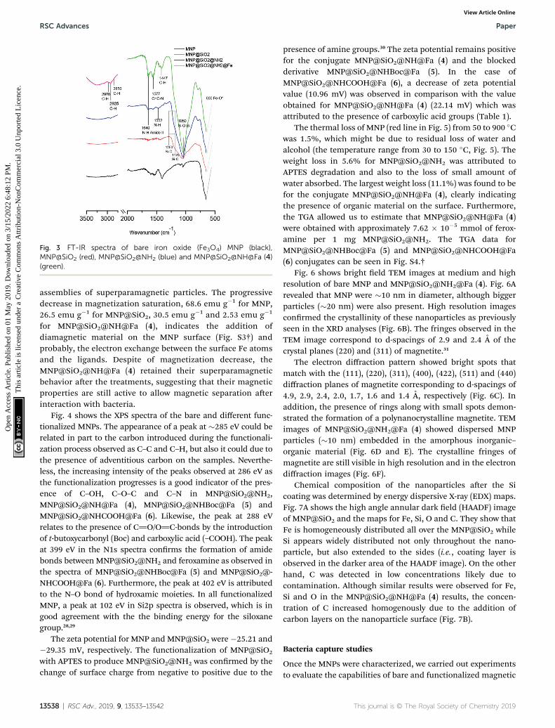
Fig. 3 FT-IR spectra of bare iron oxide (Fe3O4) MNP (black),MNP@SiO2 (red), MNP@SiO2@NH2 (blue) and MNP@SiO2@NH@Fa (4)(green).
RSC Advances Paper
Ope
n A
cces
s A
rtic
le. P
ublis
hed
on 0
1 M
ay 2
019.
Dow
nloa
ded
on 3
/15/
2022
6:4
8:12
PM
. T
his
artic
le is
lice
nsed
und
er a
Cre
ativ
e C
omm
ons
Attr
ibut
ion-
Non
Com
mer
cial
3.0
Unp
orte
d L
icen
ce.
View Article Online
assemblies of superparamagnetic particles. The progressivedecrease in magnetization saturation, 68.6 emu g�1 for MNP,26.5 emu g�1 for MNP@SiO2, 30.5 emu g�1 and 2.53 emu g�1
for MNP@SiO2@NH@Fa (4), indicates the addition ofdiamagnetic material on the MNP surface (Fig. S3†) andprobably, the electron exchange between the surface Fe atomsand the ligands. Despite of magnetization decrease, theMNP@SiO2@NH@Fa (4) retained their superparamagneticbehavior aer the treatments, suggesting that their magneticproperties are still active to allow magnetic separation aerinteraction with bacteria.
Fig. 4 shows the XPS spectra of the bare and different func-tionalized MNPs. The appearance of a peak at �285 eV could berelated in part to the carbon introduced during the functionali-zation process observed as C–C and C–H, but also it could due tothe presence of adventitious carbon on the samples. Neverthe-less, the increasing intensity of the peaks observed at 286 eV asthe functionalization progresses is a good indicator of the pres-ence of C–OH, C–O–C and C–N in MNP@SiO2@NH2,MNP@SiO2@NH@Fa (4), MNP@SiO2@NHBoc@Fa (5) andMNP@SiO2@NHCOOH@Fa (6). Likewise, the peak at 288 eVrelates to the presence of C]O/O]C-bonds by the introductionof t-butoxycarbonyl (Boc) and carboxylic acid (–COOH). The peakat 399 eV in the N1s spectra conrms the formation of amidebonds between MNP@SiO2@NH2 and feroxamine as observed inthe spectra of MNP@SiO2@NHBoc@Fa (5) and MNP@SiO2@-NHCOOH@Fa (6). Furthermore, the peak at 402 eV is attributedto the N–O bond of hydroxamic moieties. In all functionalizedMNP, a peak at 102 eV in Si2p spectra is observed, which is ingood agreement with the the binding energy for the siloxanegroup.28,29
The zeta potential for MNP and MNP@SiO2 were�25.21 and�29.35 mV, respectively. The functionalization of MNP@SiO2
with APTES to produce MNP@SiO2@NH2 was conrmed by thechange of surface charge from negative to positive due to the
13538 | RSC Adv., 2019, 9, 13533–13542
presence of amine groups.30 The zeta potential remains positivefor the conjugate MNP@SiO2@NH@Fa (4) and the blockedderivative MNP@SiO2@NHBoc@Fa (5). In the case ofMNP@SiO2@NHCOOH@Fa (6), a decrease of zeta potentialvalue (10.96 mV) was observed in comparison with the valueobtained for MNP@SiO2@NH@Fa (4) (22.14 mV) which wasattributed to the presence of carboxylic acid groups (Table 1).
The thermal loss of MNP (red line in Fig. 5) from 50 to 900 �Cwas 1.5%, which might be due to residual loss of water andalcohol (the temperature range from 30 to 150 �C, Fig. 5). Theweight loss in 5.6% for MNP@SiO2@NH2 was attributed toAPTES degradation and also to the loss of small amount ofwater absorbed. The largest weight loss (11.1%) was found to befor the conjugate MNP@SiO2@NH@Fa (4), clearly indicatingthe presence of organic material on the surface. Furthermore,the TGA allowed us to estimate that MNP@SiO2@NH@Fa (4)were obtained with approximately 7.62 � 10�5 mmol of ferox-amine per 1 mg MNP@SiO2@NH2. The TGA data forMNP@SiO2@NHBoc@Fa (5) and MNP@SiO2@NHCOOH@Fa(6) conjugates can be seen in Fig. S4.†
Fig. 6 shows bright eld TEM images at medium and highresolution of bare MNP and MNP@SiO2@NH2@Fa (4). Fig. 6Arevealed that MNP were �10 nm in diameter, although biggerparticles (�20 nm) were also present. High resolution imagesconrmed the crystallinity of these nanoparticles as previouslyseen in the XRD analyses (Fig. 6B). The fringes observed in theTEM image correspond to d-spacings of 2.9 and 2.4 A of thecrystal planes (220) and (311) of magnetite.31
The electron diffraction pattern showed bright spots thatmatch with the (111), (220), (311), (400), (422), (511) and (440)diffraction planes of magnetite corresponding to d-spacings of4.9, 2.9, 2.4, 2.0, 1.7, 1.6 and 1.4 A, respectively (Fig. 6C). Inaddition, the presence of rings along with small spots demon-strated the formation of a polynanocrystalline magnetite. TEMimages of MNP@SiO2@NH2@Fa (4) showed dispersed MNPparticles (�10 nm) embedded in the amorphous inorganic–organic material (Fig. 6D and E). The crystalline fringes ofmagnetite are still visible in high resolution and in the electrondiffraction images (Fig. 6F).
Chemical composition of the nanoparticles aer the Sicoating was determined by energy dispersive X-ray (EDX) maps.Fig. 7A shows the high angle annular dark eld (HAADF) imageof MNP@SiO2 and the maps for Fe, Si, O and C. They show thatFe is homogeneously distributed all over the MNP@SiO2 whileSi appears widely distributed not only throughout the nano-particle, but also extended to the sides (i.e., coating layer isobserved in the darker area of the HAADF image). On the otherhand, C was detected in low concentrations likely due tocontamination. Although similar results were observed for Fe,Si and O in the MNP@SiO2@NH@Fa (4) results, the concen-tration of C increased homogenously due to the addition ofcarbon layers on the nanoparticle surface (Fig. 7B).
Bacteria capture studies
Once the MNPs were characterized, we carried out experimentsto evaluate the capabilities of bare and functionalized magnetic
This journal is © The Royal Society of Chemistry 2019

Fig. 4 XPS narrow spectra of MNP, MNP@SiO2@NH2, MNP@SiO2@NH@Fa (4) MNP@SiO2@NHBoc@Fa (5) and MNP@SiO2@NHCOOH@Fa (6).
Table 1 Zeta potential measurements
Sample Z potential
MNP �25.21MNP@SiO2 �29.35MNP@SiO2@NH2 17.03MNP@SiO2@NH@Fa (4) 22.14MNP@SiO2@NHBoc@Fa (5) 19.16MNP@SiO2@NHCOOH@Fa (6) 10.96
Fig. 5 Thermogravimetric analysis of MNP (red), MNP@SiO2@NH2
(green), and MNP@SiO2@NH@Fa (pink).
Fig. 6 Bright field TEM images and electron diffraction of bareMNP (A,B and C), and of MNP@SiO2@NH@Fa (4) (D, E and F). Images atmedium and high resolution.
This journal is © The Royal Society of Chemistry 2019
Paper RSC Advances
Ope
n A
cces
s A
rtic
le. P
ublis
hed
on 0
1 M
ay 2
019.
Dow
nloa
ded
on 3
/15/
2022
6:4
8:12
PM
. T
his
artic
le is
lice
nsed
und
er a
Cre
ativ
e C
omm
ons
Attr
ibut
ion-
Non
Com
mer
cial
3.0
Unp
orte
d L
icen
ce.
View Article Online
nanoparticles to capture wild type (WC-A) and a mutant lackingferoxamine receptor FoxA (FoxA WC-A 12-8) Y. enterocoliticastrains.
Bare MNP and functionalized MNPs were incubated in a PBSsolution containing each Y. enterocolitica strain. The aggregateswere then separated from the bacteria suspension by usinga magnet. Aer rinsing the separated aggregates two times withPBS, they were re-suspended in PBS, to prepare serial dilutionsthat were plated for colony counting.
The results obtained from colony counting are shown inFig. 8. Both Y. enterocolitica strains evaluated did not showa signicant binding specicity for the functionalized MNP inrelation to bare MNP. The lack of binding specicity is likely
RSC Adv., 2019, 9, 13533–13542 | 13539

Fig. 7 EDX maps of MNP@SiO2: HAADF image and the corresponding Fe, Si, O and C maps of (A) MNP@SiO2 and (B) MNP@SiO2@NH@Fa (4).
Fig. 9 CFU of Y. enterocolitica WC-A (wild type) captured per 100 mgof magnetic nanoparticles (A) MNP@SiO2@NH2, MNP@SiO2@NHBoc,MNP@SiO2@NH@Fa (4), MNP@SiO2@NHBoc@Fa (5), (B)MNP@SiO2@NH2, MNP@SiO2@NHCOOH, MNP@SiO2@NH@Fa (4),MNP@SiO2@NHCOOH@Fa (6).
RSC Advances Paper
Ope
n A
cces
s A
rtic
le. P
ublis
hed
on 0
1 M
ay 2
019.
Dow
nloa
ded
on 3
/15/
2022
6:4
8:12
PM
. T
his
artic
le is
lice
nsed
und
er a
Cre
ativ
e C
omm
ons
Attr
ibut
ion-
Non
Com
mer
cial
3.0
Unp
orte
d L
icen
ce.
View Article Online
caused by surface interactions between nanoparticles andbacteria. Most bacteria have a net negative surface charge,particularly during the early stationary phase of cell growth,32,33
that makes them to preferentially interact with positivelycharged surfaces such as MNP@SiO2@NH2, due to the presenceof free amine groups through the protonation in physiologicsolution. Therefore, our results are in good agreement withprevious works reporting bacteria adsorption through freeamine groups of functionalized MNP.30,34,35 The bacteriaadsorption achieved withMNP@SiO2 particles can be attributedto mutually hydrophobic interaction.36
In order to reduce the non-specic binding behavior due tothe electrostatic interactions between the free amine function-alized nanoparticles and bacteria, we made attempts to blockthe surface of the particles with two different groups, one ofthem of neutral nature (Boc) and a second group with polarcharacter (COOH). Boc groups were introduced ontoMNP@SiO2@NH2 and MNP@SiO2@NH@Fa (4) by using(Boc)2O to give MNP@SiO2@NHBoc and conjugateMNP@SiO2@NHBoc@Fa (5), respectively. Carboxylic acidgroups were also introduced onto the same conjugate by usingsuccinic anhydride to give the corresponding MNP@SiO2@-NHCOOH and MNP@SiO2@NHCOOH@Fa (6) similar to thosereported by Gunawan and coworkers.34
When testing the bacteria capture with these new conju-gates, the colony counting did not show any signicant changes
Fig. 8 CFU of Y. enterocolitica captured per 100 mg of magneticnanoparticles: bare, MNP@SiO2, MNP@SiO2@NH2 and MNP@SiO2@-NH@Fa (4). (A) WC-A (wild type) (B) FoxA WC-A 12-8 (mutant lackingferoxamine receptor FoxA).
13540 | RSC Adv., 2019, 9, 13533–13542
for the adsorption of Y. enterocolitica WC-A (wild type strain)indicating that the molecular recognition of the siderophoreagain was not observed (Fig. 9A and B). Thus, these modica-tions were not enough to attenuate the electrostatic interactionsbetween bacteria and the modied nanoparticles as conrmedwith the low decrease value of zeta potential (Table 1). Similarresults were obtained when the experiments were repeated withand without iron deciency growth conditions (Fig. S8†).
Fig. 10A shows the attachment of the nano-sized conjugateMNP@SiO2@NH@Fa (4) to the surface of Y. enterocoliticaWC-A.
Fig. 10 (A) SEM images of Y. enterocolitica WC-A interacting withMNP@SiO2@NH@Fa (4). (B) TEM images of Y. enterocolitica WC-Ainteracting with MNP@SiO2@NH@Fa (4), (B1) attachment of nano-particles to the surface of a single bacteria, (B2) detail of the attach-ment on the bacterial membrane.
This journal is © The Royal Society of Chemistry 2019

Paper RSC Advances
Ope
n A
cces
s A
rtic
le. P
ublis
hed
on 0
1 M
ay 2
019.
Dow
nloa
ded
on 3
/15/
2022
6:4
8:12
PM
. T
his
artic
le is
lice
nsed
und
er a
Cre
ativ
e C
omm
ons
Attr
ibut
ion-
Non
Com
mer
cial
3.0
Unp
orte
d L
icen
ce.
View Article Online
The corresponding thin-sectioned samples measured by TEM(Fig. 10B1 and B2) conrmed the capability of the modiednanoparticles to attach to the bacterial membrane. Additionalimages and EDX maps are shown in Fig. S7 of the ESI.†
Conclusions
In this study, we describe the preparation of the conjugateMNP@SiO2@NH@Fa (4) using surface modied magneticnanoparticles and deferoxamine iron(III) complex (feroxamine)and its structural characterization using several techniques.The interaction of MNP@SiO2@NH@Fa (4) with Y. enter-ocolitica WC-A and FoxA WC-A 12-8 showed no signicantdifference in the number of colonies captured in relation tobare, MNP@SiO2, and MNP@SiO2@NH2. The lack of bindingspecicity was attributed to the presence of electrostatic forcessuch as the positive charged free amine groups present inMNP@SiO2@NH2 and the low concentration of siderophoremembrane receptor in bacteria. These results suggest that theelectrostatic and other surface interactions are dominant overthose due to the molecular recognition between MNP conjugateand feroxamine receptor. The effect of free amine groups andthe change of charge on the surface were evaluated with Boc andCOOH groups in MNP@SiO2@NHBoc@Fa (5) andMNP@SiO2@NHCOOH@Fa (6), respectively. Unfortunately,these new conjugates did not improve bacteria capture. Furtherefforts are needed to explore other blocking materials in orderto remove or decrease non-specic binding of magnetic nano-particles surface to bacteria. While the reported siderophore-based methods for detection of microbial pathogens allow thedetection of the target bacteria, the development of the presentstrategy would also allow bacteria isolation from a complexmixture of microorganisms for their posterior identication.
Conflicts of interest
There are no conicts to declare.
Acknowledgements
The authors gratefully acknowledge Professor Klaus Hantke(University of Tubingen, Germany) for kindly supply the Yersiniaenterocolitica strains used in this work. This work was supportedby grants AGL2015-63740-C2-2-R and AGL2015-63740-C2-1-R(AEI/FEDER, EU) from the State Agency for Research (AEI) ofSpain, both co-funded by the FEDER Programme from theEuropean Union.
Notes and references
1 Y. Pan, X. Du, F. Zhao and B. Xu, Chem. Soc. Rev., 2012, 41,2912–2942.
2 R. A. Bohara, N. D. Thorat and S. H. Pawar, RSC Adv., 2016, 6,43989–44012.
3 O. Lazcka, F. J. Del Campo and F. X. Munoz, Biosens.Bioelectron., 2007, 22, 1205–1217.
This journal is © The Royal Society of Chemistry 2019
4 T. Mocan, C. T. Matea, T. Pop, O. Mosteanu, A. D. Buzoianu,C. Puia, C. Iancu and L. Mocan, J. Nanobiotechnol., 2017, 15,25.
5 T. Zheng and E. M. Nolan, Metallomics, 2012, 4, 866–880.6 R. C. Hider and X. Kong, Nat. Prod. Rep., 2010, 27, 637–657.7 M. Sandy and A. Butler, Chem. Rev., 2010, 109, 4580–4595.8 D. D. Doorneweerd, W. A. Henne, R. G. Reifenberger andP. S. Low, Langmuir, 2010, 26, 15424–15429.
9 Y. Kim, D. P. Lyvers, A. Wei, R. G. Reifenberger and P. S. Low,Lab Chip, 2012, 12, 971–976.
10 T. Inomata, H. Eguchi, Y. Funahashi, T. Ozawa andH. Masuda, Langmuir, 2012, 28, 1611–1617.
11 T. Inomata, H. Tanabashi, Y. Funahashi, T. Ozawa andH. Masuda, Dalton Trans., 2013, 42, 16043–16048.
12 J. Hu, M. Ghosh, M. J. Miller and P. W. Bohn, Anal. Methods,2019, 11, 296–302.
13 S. Wu, Z. Zhang, X. Wang, M. Zhang, J. Peng, Z. Xie andD. Pang, J. Phys. Chem. C, 2009, 113, 9169–9174.
14 N. Bugdahn, F. Peuckert, A. G. Albrecht, M. Miethke,M. A. Marahiel and M. Oberthur, Angew. Chem., Int. Ed.,2010, 49, 10210–10213.
15 N. Pinna, S. Grancharov, P. Beato, P. Bonville, M. Antoniettiand M. Niederberger, Chem. Mater., 2005, 17, 3044–3049.
16 Y. S. Li, J. S. Church, A. L. Woodhead and F. Moussa,Spectrochim. Acta, Part A, 2010, 76, 484–489.
17 J. P. Chen, P. C. Yang, Y. H. Ma, S. J. Tu and Y. J. Lu, Int. J.Nanomed., 2012, 7, 5137–5149.
18 D. Goswami, M. T. Machini, D. M. Silvestre, C. S. Nomuraand B. P. Esposito, Bioconjugate Chem., 2014, 25, 2067–2080.
19 J. D. Herscheid, a Hoekstra and C. M. Vos, Eur. J. Nucl. Med.,1984, 9, 508–510.
20 K. El-Boubbou, C. Gruden and X. Huang, J. Am. Chem. Soc.,2007, 129, 13392–13393.
21 M. D. Abramoff, P. J. Magalhaes and S. J. Ram, BiophotonicsInt., 2004, 11, 36–42.
22 J. Yanez-Vilar, M. Sanchez-Andujar, S. Castro-Garcıa, J. Mira,J. Rivas and M. A. Senarıs-Rodrıguez, Bol. Soc. Esp. Ceram.Vidrio, 2010, 49, 81–88.
23 O. N. Shebanova and P. Lazor, J. Solid State Chem., 2003, 174,424–430.
24 P. Gonzalez, J. Serra, S. Liste, S. Chiussi, B. Leon andM. Perez-Amor, J. Non. Cryst. Solids, 2003, 320, 92–99.
25 M. Veres, M. Koos, S. Toth, M. Fule, I. Pocsik, A. Toth,M. Mohai and I. Bertoti, Diam. Relat. Mater., 2005, 14,1051–1056.
26 Y. You, T. Yu, J. Kasim, H. Song, X. Fan, Z. Ni, L. Cao,H. Jiang, D. Shen, J. Kuo and Z. Shen, Appl. Phys. Lett.,2008, 93, 103111–103113.
27 O. Cozar, N. Leopold, C. Jelic, V. Chis, L. David, A. Mocanuand M. Tomoaia-Cotisel, J. Mol. Struct., 2006, 788, 1–6.
28 N. Graf, E. Yegen, T. Gross, A. Lippitz, W. Weigel, S. Krakert,A. Terfort and W. E. S. Unger, Surf. Sci., 2009, 603, 2849–2860.
29 W. Michaeli, C. J. Blomeld, R. D. Short, F. R. Jones andM. R. Alexander, Appl. Surf. Sci., 2002, 137, 179–183.
30 W. Fang, C. Han, H. Zhang, W. Wei, R. Liu and Y. Shen, RSCAdv., 2016, 6, 67875–67882.
RSC Adv., 2019, 9, 13533–13542 | 13541

RSC Advances Paper
Ope
n A
cces
s A
rtic
le. P
ublis
hed
on 0
1 M
ay 2
019.
Dow
nloa
ded
on 3
/15/
2022
6:4
8:12
PM
. T
his
artic
le is
lice
nsed
und
er a
Cre
ativ
e C
omm
ons
Attr
ibut
ion-
Non
Com
mer
cial
3.0
Unp
orte
d L
icen
ce.
View Article Online
31 X. Teng and H. Yang, J. Mater. Chem., 2004, 14, 774–779.32 H. Hayashi, H. Seiki, S. Tsuneda, A. Hirata and H. Sasaki, J.
Colloid Interface Sci., 2003, 264, 565–568.33 N. P. Boks, H. J. Busscher, H. C. Van Der Mei and W. Norde,
Langmuir, 2008, 24, 12990–12994.
13542 | RSC Adv., 2019, 9, 13533–13542
34 A. E. Liana, C. P. Marquis, C. Gunawan, J. J. Gooding andR. Amal, Colloids Surf., B, 2017, 151, 47–57.
35 S. Zhan, Y. Yang, Z. Shen, J. Shan, Y. Li, S. Yang and D. Zhu, J.Hazard. Mater., 2014, 274, 115–123.
36 H. H. Tuson and D. B. Weibel, So Matter, 2013, 9, 4368–4380.
This journal is © The Royal Society of Chemistry 2019