title vacuum ultraviolet-induced surface modification of
TRANSCRIPT
RIGHT:
URL:
CITATION:
AUTHOR(S):
ISSUE DATE:
TITLE:
Vacuum ultraviolet-induced surfacemodification of cyclo-olefinpolymer substrates forphotochemical activation bonding
KIM, Young-Jong; TANIGUCHI, Yoshinao; MURASE,Kuniaki; TAGUCHI, Yoshihiro; SUGIMURA, Hiroyuki
KIM, Young-Jong ...[et al]. Vacuum ultraviolet-induced surface modification of cyclo-olefinpolymer substrates for photochemical activation bonding. Applied Surface Science 2009,255(6): 3648-3654
2009-01-01
http://hdl.handle.net/2433/97891
Copyright © 2008 Elsevier B.V.; この論文は出版社版でありません。引用の際には出版社版をご確認ご利用ください。; This is not thepublished version. Please cite only the published version.
1
Vacuum Ultraviolet-Induced Surface Modification of Cyclo-olefin
Polymer substrates for Photochemical Activation Bonding
Young-Jong KIM, Yoshinao TANIGUCHI*, Kuniaki MURASE,
Yoshihiro TAGUCHI*, and Hiroyuki SUGIMURA*
Department of Materials Science and Engineering, Kyoto University,
Yoshida-hommachi, Sakyo-ku, Kyoto 606-8501, Japan
*Present address: ALPS ELECTRIC, Business Development HQ,
Production Engineering Development Center 136-1, Kitahara, Tsukanome,
Furukawa, Osaki-City, Miyagi-Pref, 989-6225, Japan
*E-mail address : hiroyuki.sugimura@ materials.mbox.media.kyoto-u.ac.jp
Abstract
The surfaces of cyclo-olefin polymer (COP) were treated using vacuum
ultraviolet (VUV) light at 172 nm in wavelength in order to improve its
wettability and adhesion properties. The effect of the VUV-light treatment
with variable conditions was studied, and then the condition was
A Self-archived copy inKyoto University Research Information Repository
https://repository.kulib.kyoto-u.ac.jp
2
optimized. The chemical and physical properties of the sample was
examined by using water contact angle measurements, X-ray
photoelectron spectroscopy, fourier transform infrared spectroscopy, and
atomic force microscopy. The VUV treatment of an COP sample was
carried out by placing the sample in air and then irradiating the sample
surface with a Xe-excimer lamp. At that time, The surface terminal
groups were oxidized into the functional groups (detected as C–O, C=O,
and COO components) readily on COP surfaces. The introduction of
oxygen functional groups formed hydrophilic COP surfaces. We
investigated how the oxygen functional groups are formed on the COP
surface from the viewpoint of different distances between the lamp and the
sample. The dependence of oxygenation extent on different experimental
parameters of VUV irradiation distance or treatment duration is measured
by atomic ratio O1s / C1s of XPS spectra and [C=O]/[C–H] area ratio of
FTIR-ATR spectra. It was shown that the surface property of COP
samples, such as hydrophilicity and functionalization, were improved
A Self-archived copy inKyoto University Research Information Repository
https://repository.kulib.kyoto-u.ac.jp
3
remarkably after VUV treatment with suitable conditions. A much higher
concentration of oxygen functional groups was observed for distance 5
mm than for the corresponding distance 30 mm at the same extent of
VUV-light treatment. VUV-light treatment also has a more obvious effect
on the short irradiation distance. In both cases, however, the concentration
of oxygen functional groups changes remarkably. It was concluded that
VUV treatment is a promising technique to modify COP surface property.
KEYWORDS: surface modification, excimer lamp, vacuum ultraviolet
(VUV), oxygen functional groups
1. Introduction
Today, cyclo-olefin polymer (COP) resins are used in a variety of
applications according to their various properties and low costs. COP
properties such as excellent transparency, high heat resistance, low water
absorbency, stable and guaranteed refractive index, and low birefringence
are fully utilized in applications, especially for camera lenses/prisms,
A Self-archived copy inKyoto University Research Information Repository
https://repository.kulib.kyoto-u.ac.jp
4
lenses for cameras incorporated into mobile phones, pick-up lenses, and
Fq lenses for laser beam printers. Also, the COP is used in such
applications as medical vials, syringes, optical lab test cells, and syringes
pre-filled with pharmaceutical content. The COP is especially suitable for
use in medical devices that undergo autoclave sterilization at 121 ˚C. The
market for COPs is growing every year due to their excellent properties,
primarily in optical applications.1
Extensive work to develop practical and economical method for
surface modification of COP has been carried out by many workers. For
improvement of adhesivity, dyeability, and wettability, surface
photografting modification in gas-phase or liquid-phase has attracted wide
attention.2-7 Especially, adhesion is a critical design feature of many
commercially available products. In order to obtain better adhesion
property, it is necessary to improve the adhesive chemical bond at the
interface. This is possible by increasing the distribution of polar
functional groups at the surface. An appropriate surface treatment is
A Self-archived copy inKyoto University Research Information Repository
https://repository.kulib.kyoto-u.ac.jp
5
required for this purpose.
Polymeric materials, especially in continuous format, i.e. films,
have been largely treated through different oxidative process, such as
corona discharge,8-11 plasma etching,12,13 ultraviolet radiation,14,15 chemical
solutions,16-18 etc. The resulting changes in surface composition,
morphology, and surface energy can improve adhesive bonding,
wettability, biocompatibility, and many other surface-related
properties. Although it is desirable to give new functions to polymer
surfaces, it is also important to modify polymer material surfaces without
affecting their bulk characteristics, such as mechanical, thermal and other
intrinsic properties. ultraviolet radiation is a good method in this respect
because ultraviolet radiation interacts with the polymer surface and not
with its bulk. Also, ultraviolet radiation is a very efficient, economical and
potentially practical oxidative process for modifying polymer
surface. Particularly, it is widely known that the energy of photons
becomes stronger as its wavelength becomes shorter. This enables it to
A Self-archived copy inKyoto University Research Information Repository
https://repository.kulib.kyoto-u.ac.jp
6
cause chemical reactions unattainable by a conventional UV light and
allows acceleration of the reaction speed. In addition, short-wavelength
[vacuum ultraviolet (VUV), λ<200 nm] radiation is believed to play an
important role in the near-surface chemistry of plasma-treated
polymers.19,20 One possible advantage of VUV photochemistry over its
plasma counterparts may be that a more specific surface chemistry is
achieved using monochromatic radiation because of more specific and
selective (photo) chemistry both on the solid surface and in the gas
phase.21
For this study, we investigated whether VUV-light treatment
modifies the COP surface to produce a hydrophilic surface, whether the
chemical components on the COP surfaces are modified by VUV-light
treatment, and whether the rates of the introduction of functional groups
on COP surfaces by VUV-light treatment differ or not, according to the
influence of experimental parameters such as VUV irradiation distance
and treatment duration on the extent of surface photochemical
A Self-archived copy inKyoto University Research Information Repository
https://repository.kulib.kyoto-u.ac.jp
7
modification. The chemical analysis of polymer surfaces has provided a
fruitful realm for the application of X-ray photoelectron spectroscopy
(XPS). XPS is now well-known for the unique qualitative and quantitative
surface information derived from core and valence level spectra. However,
when utilized alone, XPS has some major weaknesses. We have been
active in the extension of surface analysis of polymers by fourier
transform infrared spectroscopy (FTIR). Especially, using XPS and FTIR,
analyzed the generation of hydroxyl, carbonyl, and carboxyl groups on the
surface of COP samples treated with VUV-light, and it was shown that
both XPS and FTIR complemented each other and were useful in
analyzing the changes in chemical composition due to surface oxidation.
2. Experimental
2.1. Materials
The sample used in this study was a transparent cyclo-olefin
A Self-archived copy inKyoto University Research Information Repository
https://repository.kulib.kyoto-u.ac.jp
8
polymer (COP) supplied by Alps Electric (Furukawa, Osaki-City,
Miyagi-Pref, Japan) with a thickness of 1 mm. Samples were prepared for
the VUV-light treatment and, front this, samples of different dimensions
were cut for different measurements. Detailed properties of the COP have
already been reported.1
2.2. VUV-light treatment
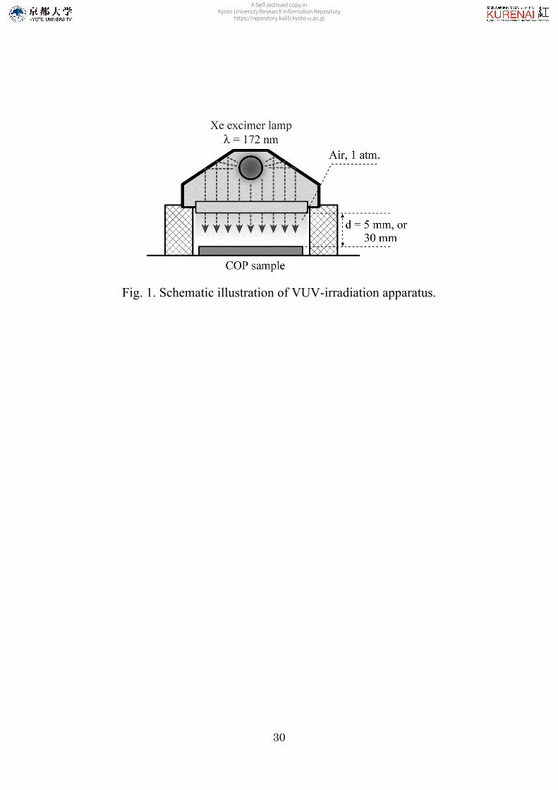
A schematic of the VUV-irradiation apparatus is shown in Figure.
1. We used an excimer lamp as a source of VUV light at a wavelength of
172 nm (Ushio., UER20-172V; intensity at the lamp window, 10 mW
cm-2) as a light source. This lamp consists of two (inner and outer) quartz
glass tubes. The metal electrode is mounted within the inner tube, while
the metal mesh electrode is mounted outside of the outer tube. The quartz
glass tubes are filled with a discharge gas. The COP samples were placed
on the sample stage. In our VUV-light-exposure system, the chamber was
filled with ambient air, and the distance between the lamp window and the
A Self-archived copy inKyoto University Research Information Repository
https://repository.kulib.kyoto-u.ac.jp
9
sample surface was fixed at 5 and 30 mm. Since oxygen molecules
strongly absorb VUV light at 172 nm, the VUV light is attenuated when
propagating through an air layer with a certain thickness. The optical
absorption coefficient of the VUV light at wavelength 172 nm in ambient
air with an oxygen partial pressure 0.2 atm was reported to be in the range
of 10-15 cm-1 atm-1,22 indicating that the transmittance the light through a
10 mm-thick air layer is calculated to be in the range of 5 - 13%, and we
also observed measured the value was about 10%. Therefore, the
transmittance for 5 mm is estimated to be less than 50%, which means that
the light intensity at the COP surface is less than 5 mW cm-2. At a
distance of 5 mm, the sample surface was directly irradiated with the VUV
light emitted from the lamp window, although there was a absorption loss
with about 50%. Simultaneously, the sample surface was exposed to
active oxygen species generated in the space just on the sample
surface. Both the reactions, that is, VUV-excitation of the COP surface
and the oxidation of the COP surface with the active oxygen, were
A Self-archived copy inKyoto University Research Information Repository
https://repository.kulib.kyoto-u.ac.jp
10
considered to proceed on the sample surface. In contrast, the
transmittance for 30 mm is estimated to be less than 0.1%, which means
that the light intensity at the ODS-SAM surface is less than 0.010 mW
cm-2. In other words, at the distance of 30 mm, VUV light was absorbed
almost completely by atmospheric oxygen molecules, yielding active
oxygen species such as ozone and atomic oxygen, hence no substantial
amount of VUV-light reached the sample surface. The direct irradiation of
COP surfaces with VUV photons is not expected in the system of an air
layer of 30 mm, and only the VUV-light-generated active oxygen can
participate in the surface modification of the COP.
2.3. Chemical and physical properties analysis
After VUV-light treatment, photochemical effects of VUV-light
treatments on the COP surfaces were investigated using a combination of
analytical techniques. The static water contact angles of the sample
surfaces were measured with a contact angle meter (Kyowa Interface
A Self-archived copy inKyoto University Research Information Repository
https://repository.kulib.kyoto-u.ac.jp
11
Science, CA-X) in an atmospheric environment; here, we fixed the size of
water droplets at about 1.5 mm in diameter. At least four different
measurements on the sample surfaces were obtained and the average
values for water contact angles were calculated. The chemical bonding
states of each sample were examined by X-ray photoelectron spectroscopy
(XPS; Kratos Analytical, ESCA-3400) using a Mg Kα X-ray source with
10 mA in emission current and 10 kV in accelerating voltage. The
background pressure in the analytical chamber was 8.0 × 10⁻⁷ Pa. The
X-ray spot diameter was 6 mm. The binding energy scales were
referenced to 285.0 eV as determined by the locations of the maximum
peaks on the C 1s spectra of hydrocarbon (CHx), associated with
adventitious contamination. The C 1s and O 1s spectra were decomposed
by fitting a Gaussian-Lorentzian mixture function (mixture rate, 20 :
80). Surface modification process of COP samples were also monitored
by quantitative fourier transform infrared spectroscopy (FTIR; Digilab
Japan Co., Ltd, Excalibur FTS-3000). We used a single reflection ATR
A Self-archived copy inKyoto University Research Information Repository
https://repository.kulib.kyoto-u.ac.jp
12
(attenuated total reflection) mode for measurement of the samples. The
ATR IR spectra were obtained with 65˚ of incident angle, and
hemispherical Ge ATR crystal with diameter of 2.5 cm (internal reflection
element, from Harrick Scientific). IR Spectra were measured in a dry
atmosphere of a sample compartment purged with nitrogen and were
referenced to background spectra determined under the same
conditions. All spectra were measured at a resolution of 4 cm-1 and with
1024 times of scan cycles. The morphology and surface roughness of the
samples were measured by an atomic force microscopy (AFM; SII
Nanotechnology, SPA-300HV + SPI-3800N) in tapping mode at a scan
rate of 0.5 Hz, using silicon cantilever probe (Seiko Instruments Inc.,
SI-DF20, force constant of 15 N m-1). From the analysis of the images,
the root-mean-squared roughness (RRMS) for the topographic profiles
measured on 5×5 µm2 images was evaluated.
3. Results and Discussion
A Self-archived copy inKyoto University Research Information Repository
https://repository.kulib.kyoto-u.ac.jp
13
3.1. Water contact angle on COP sample surfaces treated with the direct
and remote VUV-light
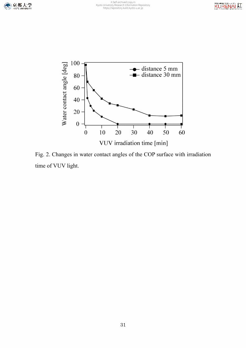
We investigated whether VUV-light treatment modifies COP
surfaces to make the COP surface hydrophilic. The changes of water
contact angles on the COP surface treated with VUV-light for irradiation
distance 5 and 30 mm are shown in Figure 2, respectively. As can be
observed, the COP surface treated with VUV-light for irradiation distance
5 mm became hydrophilic more rapidly than that for irradiation distance
30 mm. In case of distance 5 mm, water contact angles show large
decrease in the modifying hydrophilic surfaces with increasing VUV
irradiation time but level off from 20 min. After 20 min, the water contact
angle settled at zero. In contrast, in case of distance 30 mm, it gradually
decreased but level off from 40 min. Finally, the water contact angle
remained at approximately 14˚. Thus, for VUV irradiation time of 5 min
the water contact angle loss is around 78 and 44% for distance 5 and 30
A Self-archived copy inKyoto University Research Information Repository
https://repository.kulib.kyoto-u.ac.jp
14
mm, respectively. Results showed that, on the COP surface, the surface
contact angle made by VUV-light treatment decreased to modify
hydrophilically. It was expected that the COP surfaces treated by
VUV-light treatment were introduced by the oxygen functional groups.
3.2. Chemical composition of direct and remote VUV-light treated COP
sample surface
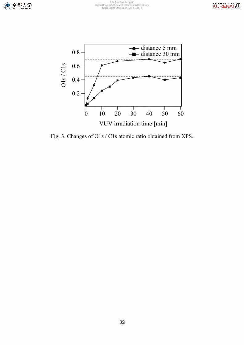
This VUV-irradiated surface chemical conversion of COP was
studied in more detail using XPS. By means of XPS measurement, we
investigated the dependency of surface oxygenation of COP on different
experimental conditions, such as VUV irradiation distance and treatment
duration. We can use the atomic ratio of intensity of O1s to that of C1s
(O1s/C1s) as a measure of the surface oxygenation extent. The increase in
the extent of surface oxidation with VUV irradiation time is shown in
Figure 3. The initially faster oxidation in case of distance 5 mm shows
that the atomic ratio of O1s/C1s can reach a level which changes the
A Self-archived copy inKyoto University Research Information Repository
https://repository.kulib.kyoto-u.ac.jp
15
surface composition distinctly in a short time (on the order of one
minute). The most reasonable explanation for these results is the
assumption that active oxygen species oxide intact COP only with a low
rate. Obviously, a photolytic activation is needed for the increase of COP
oxidation. In our previous paper,23,24 we investigated the vacuum
ultraviolet (VUV) photodegradation of alkyl monolayers in the presence
of atmospheric oxygen molecules. Here, VUV light of 172 nm in
wavelength excites atmospheric oxygen molecules, resulting in the
generation of ozone molecules as well as oxygen atoms in singlet and
triplet states [O(1D) and O(3P), respectively], as described in the
following equation.25
O2 + hv → O(1D) + O(3P)
Since these active oxygen species, particularly O(1D), have
strong oxidative reactivity. In case of distance 5 mm, the sample surface
was directly irradiated with VUV light while the sample surface was
exposed to active oxygen species. Therefore, in addition to the oxidation
A Self-archived copy inKyoto University Research Information Repository
https://repository.kulib.kyoto-u.ac.jp
16
of the sample surface with the active oxygen, a VUV excitation of the
sample surface might be induced. The overall oxidation is expected to be
autoaccelerating. And this point is very important for commercial
practice. From Figure 3, an optimum experimental condition for
irradiation distance 5 and 30 mm can be found. This is that the VUV
irradiation time is 40 min, respectively.
In addition to providing the total heteroatom concentration on the
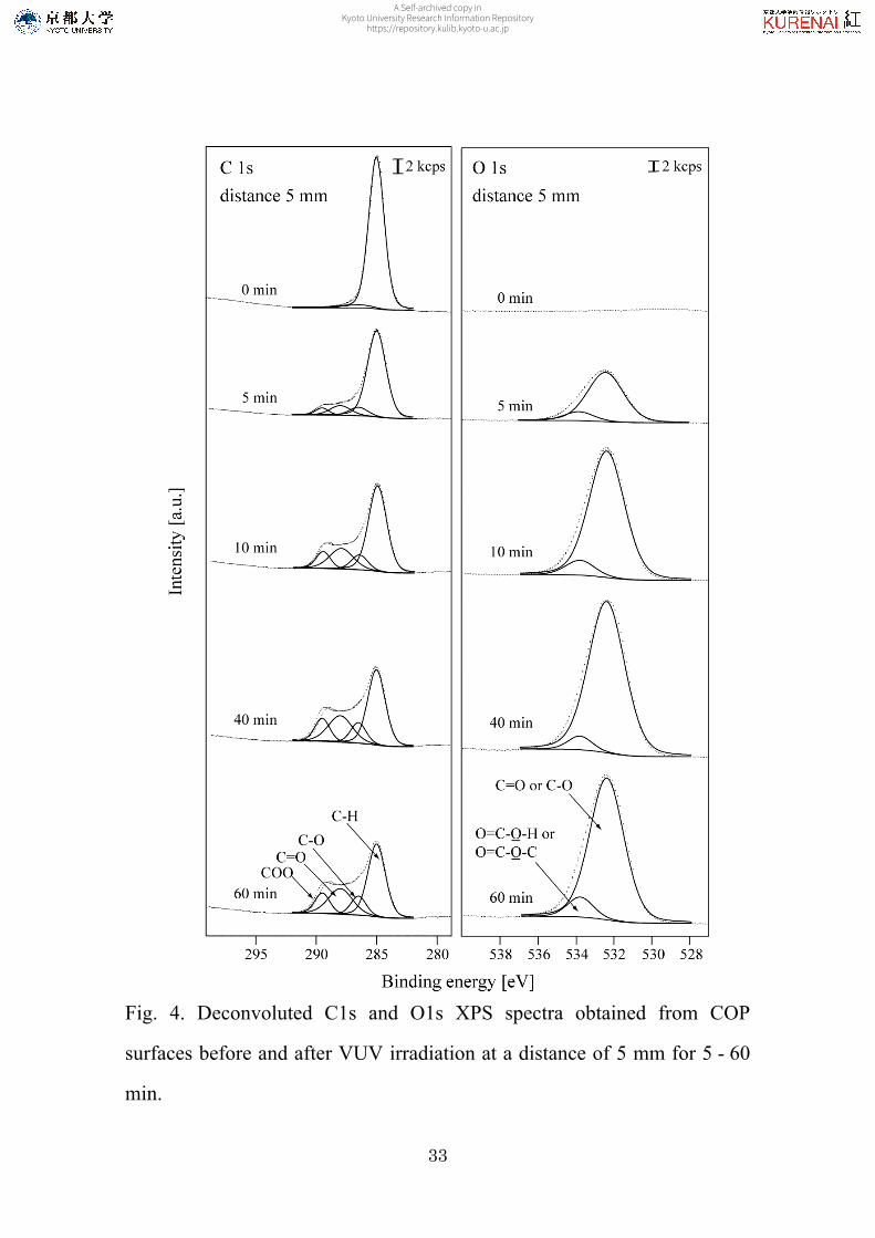
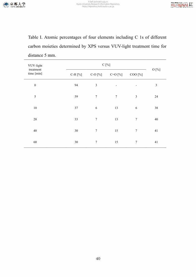
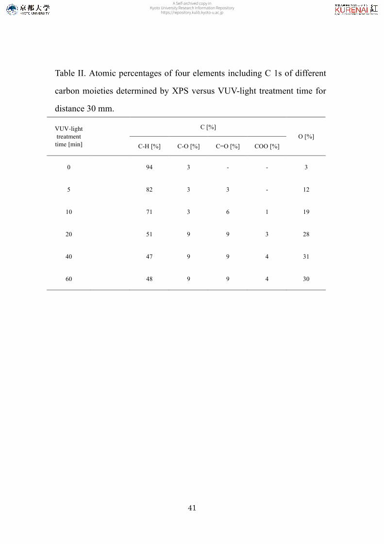
surfaces of VUV-treated COP presented above, XPS was also used to
characterize their chemical bonding states. Remarkable differences
between distance 5 mm and distance 30 mm were found not only in the
rate of oxygen incorporation and the final oxygen concentration but also in
the evolution of the oxygen functional groups. Figures 4 and 5 shows the
C1s and O1s XPS spectra of COP samples prior to and after VUV
irradiation for 5, 10, 40, and 60 min. In Figures 4 and 5, the spectra for
the untreated sample are also shown figure for comparison with the
VUV-light treatment results. The tail on the high binding energy side of
A Self-archived copy inKyoto University Research Information Repository
https://repository.kulib.kyoto-u.ac.jp
17
the main C1s peak for COP shows that oxygen incorporation into the COP
surfaces gave rise to a variety of functional groups. These C1s peaks can
be decomposed into four main components at 285.0 (C–H groups), 286.5
(C–O groups), 288.0 (C=O groups), and 289.5 eV (COO). The O1s peaks
can be decomposed into two peaks with binding energies of 532.4 and
533.8 eV, respectively. The peaks are assigned as follows: O1s 532.4 eV
(C=O or C–O groups) and 533.8 eV (O=C–O–H or O=C–O–C
groups). From XPS analysis, it can be seen that after VUV-light treatment,
there appeared oxygen functional groups such as carbonyl, ether, and
carboxyl in COP surface. A much higher concentration of oxygen
functional groups was observed for distance 5 mm than for the
corresponding distance 30 mm at the same extent of VUV-light
treatment. In case of distance 30 mm, prolonged VUV-light treatment
(>10 min) can result in the appearance of the COO
components. VUV-light treatment also has a more obvious effect on the
short irradiation distance. By VUV irradiation for 5, 10, and 40 min, the
A Self-archived copy inKyoto University Research Information Repository
https://repository.kulib.kyoto-u.ac.jp

18
amount of carbon on the sample decreased, whereas the amount of oxygen
on the sample increased. XPS analysis as a function of the VUV-light
treatment duration also indicates a progressive functionalization. The C1s
atomic percentage data summarized in Table I and II shows progressive
increases in the surface densities of oxygen functional groups (detected as
C–O, C=O, and COO components) with VUV-light irradiation until 40
min as confirmed by Figure 3. However the differences between the
samples with VUV irradiation times of 40 and 60 min are not evident. It
is remarkable that the functionalization of COP by VUV-light treatment
remains almost constant for VUV irradiation times higher than 40
min. The increase of amount of oxygen functional groups are in
agreement with the reduction of the water contact angles showed by
Figure 2. These results indicate that surface modification with VUV-light
treatment is effective in enhancing their functionalization.
For the surface chemical-analysis obtained by XPS, we also
employed a supplemental measurement by using FTIR. The FTIR-ATR
A Self-archived copy inKyoto University Research Information Repository
https://repository.kulib.kyoto-u.ac.jp
19
spectra of untreated and treated COP are presented in Figure 6 and 7,
respectively. Given that the sampling depth of the present FTIR-ATR
technique (0.13 µm and below) is large compared with the estimated depth
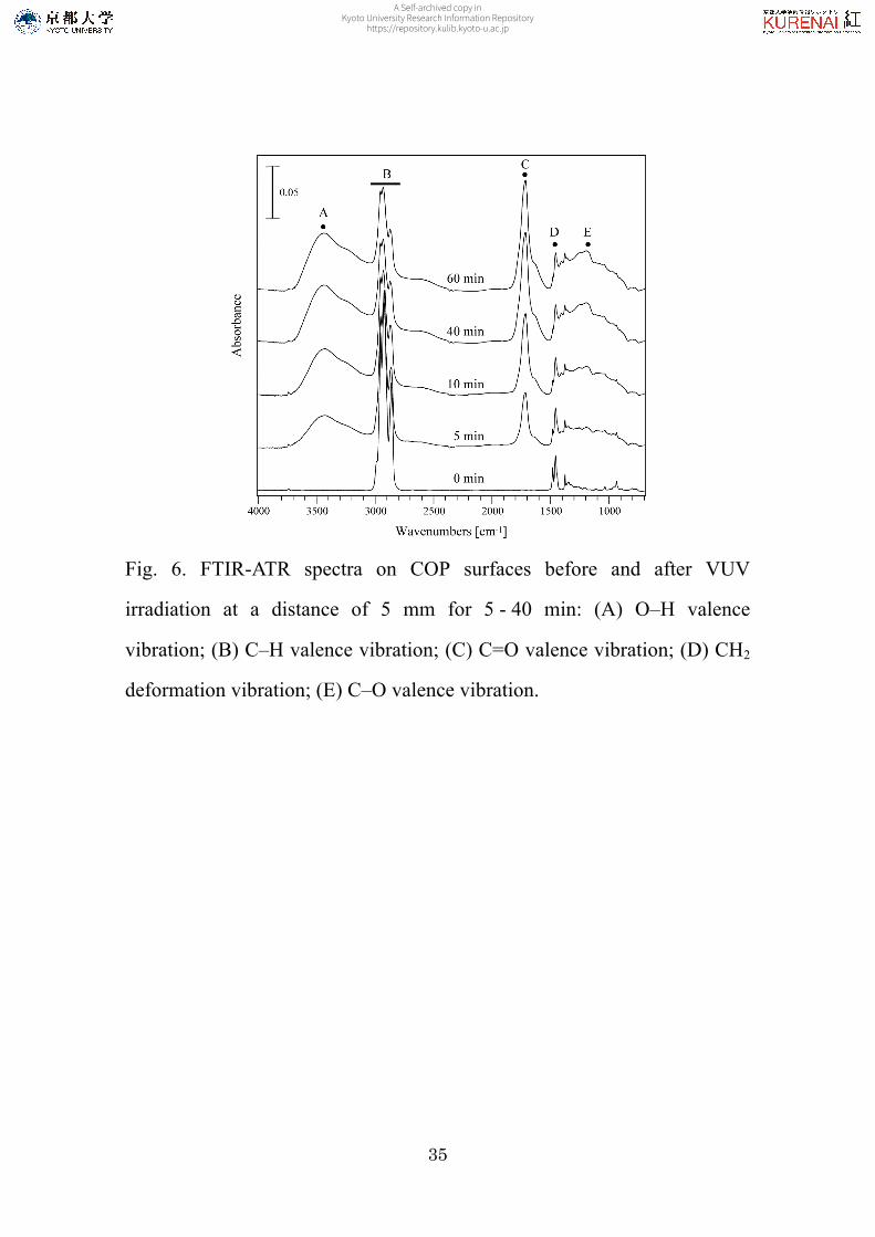
of VUV-light treatment effects. To highlight the chemical changes in the
surface, the untreated sample are shown. As we can see, the VUV-light
treatment introduces three new IR bands such as O–H, C=O, and C–O
valence vibration. A very broad between 3750 and 3050 cm-1, centered at
about 3450 cm-1, can be attributed to the O–H stretch in alcohols and
phenols. A relatively sharp band between 1897 and 1519 cm-1 has a
doublet structure, subpeaks being centered at 1716 and 1624 cm-1,
respectively. This feature can be assigned to C=O stretch in aliphatic
ketones (1725-1705 cm-1), C=C and C=O in unsaturated ketones
(1705-1665 cm-1), C=O stretch in primary (1680-1519 cm-1). A broad
band, between 1290 and 1180 cm-1, can be assigned to C–O–C
antisymetric stretch in esters. A broad band, between 1000 and 900 cm-1
with a peak at 940 cm-1, can be assigned to CH out-of-plane deformation
A Self-archived copy inKyoto University Research Information Repository
https://repository.kulib.kyoto-u.ac.jp
20
(1000-950 cm-1), CH2 out-of-plane wagging (950-900 cm-1) in vinyl
(–CH=CH2), and CH out-of-plane deformation (980-955 cm-1) in vinylene
(–CH=CH–) with a high level of confidence. As above mentioned, Figure
6 and 7 shows the increase of the C=C double bonds concentration in a
sample surface. This can be explained as follows: C=C double bonds are
formed by the abstraction of hydrogen, known as the dominant mechanism
during VUV irradiation of hydrocarbon polymers.26-28 Figure 6 and 7
shows that the vibrational bands that are significantly perturbed as a
function of VUV irradiation time are the carbonyl band vibrations at about
1897-1519 cm-1. It should be pointed out that no spectrum is free from
carbonyl band vibrations except for non-treated sample, but the intensity
of this band increased relatively from one sample to another as a function
of VUV irradiation time. Here, in the FTIR analysis, we mean by the
word intensity the height of the peak and not the area. The carbonyl band
area is measured relatively to the hydrocarbon (C–H) band area, and the
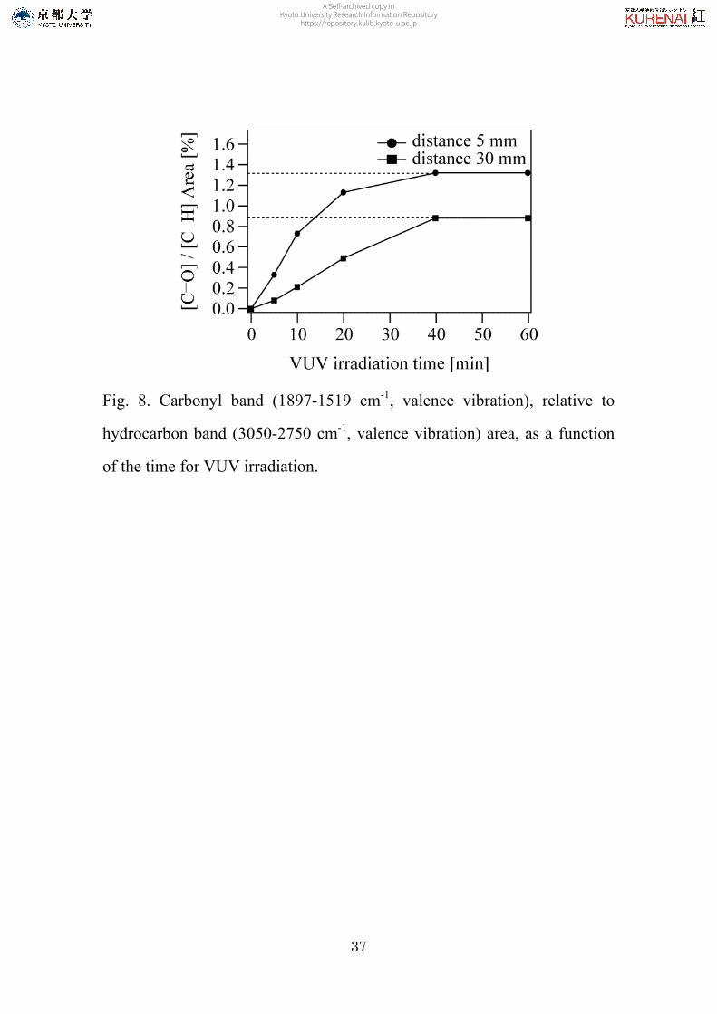
result is shown in Figure 8, as a function of VUV irradiation time. In this
A Self-archived copy inKyoto University Research Information Repository
https://repository.kulib.kyoto-u.ac.jp
21
figure, a substantial increase in the [C=O]/[C–H] area ratio until 40 min
can be observed. This fact corroborates the activation/functionalization
effect of VUV-light treatment on the COP surface. However the
differences between the samples with VUV irradiation times of 40 and 60
min are not evident. It is remarkable that the functionalization of COP by
VUV-light treatment remains almost constant for VUV irradiation times
higher than 40 min. As discussed above (XPS results) with O1s/C1s
atomic ratio, Figure 3 showed an outstanding increase in the O1s/C1s
atomic ratio at the time of VUV light until 40 min. An exact agreement
between XPS and FTIR analysis can be expected though they have
different sampling depths. The large increase in the [C=O]/[C–H] area
ratio thus means that the non-treated sample has been oxidized largely,
while the outermost layers have lost part of their oxygen-containing
moieties, as evidenced by the XPS O1s/C1s atomic ratio.
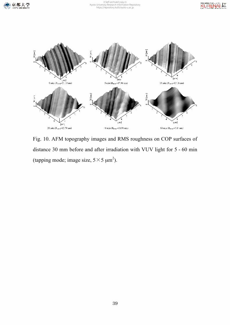
3.3. Topography of direct and remote VUV-light treated COP sample
A Self-archived copy inKyoto University Research Information Repository
https://repository.kulib.kyoto-u.ac.jp
22
surface
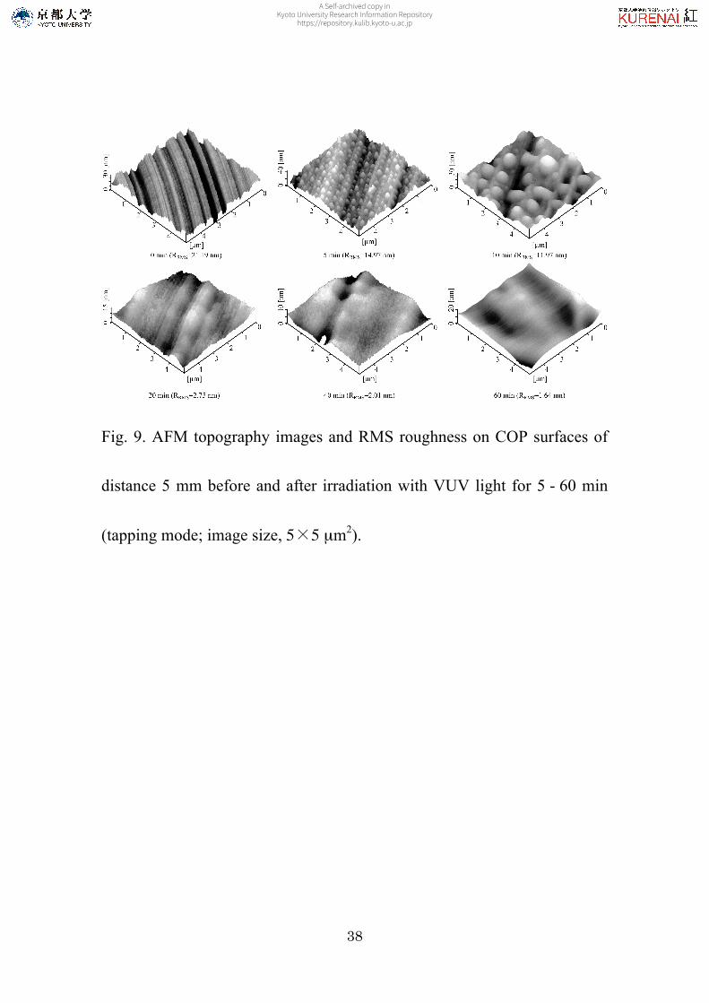
The surface structure of the VUV-light-treated COP was
monitored using AFM. AFM is a more adapted technique for the
characterization of VUV treated surface, allowing to abtain a 3D
representation of the surface topography and roughness
measurements. Figure 9 and 10 shows topography images of COP
samples prior to and after VUV irradiation time for 5, 10, 20, 40, and 60
min. As can be observed the VUV-light treatment shows an increase in
smooth degree of the sample surface with an increase in the VUV
irradiation time. That is, the VUV-light treatment on the sample causes
changes in the surface topography. In case of distance 5 mm, both the
reactions, that is, VUV-excitation of the COP surface and the oxidation of
the COP surface with the active oxygen species, promotes surface
topography changes greatly; opposite to this, in case of distance 30 mm,
only by oxidation of the COP surface with the active oxygen species make
difficult significant changes on the surface topography. In case of distance
A Self-archived copy inKyoto University Research Information Repository
https://repository.kulib.kyoto-u.ac.jp
23
5 mm, particularly, the surface topography of treated COP for 5, 10 min
shows structures like bubbles; however, for longer VUV irradiation times,
there was a smooth structure without bubbles appears. This indicates the
photon energy effects of 172 nm light with photons of 7.2 eV. In both
cases, however, with increasing VUV irradiation time, the surface
roughness gradually decreases. Changes in surface topography are
observed, and they do contribute in a significant way to improve
wettability as confirmed by Figure 2. In addition, this result suggests that
surface oxidation has occurred uniformly. Consequently, the photons and
active oxygen species would be the important parameters for the
mechanism of surface modification of COP.
4. Conclusions
In this study, COP surfaces were irradiated using an excimer lamp
as a source of VUV light at a wavelength of 172 nm. There appeared
oxygen functional groups such as ether, ketone, and carboxyl groups in the
A Self-archived copy inKyoto University Research Information Repository
https://repository.kulib.kyoto-u.ac.jp

24
COP surface after VUV-light treatment. These groups formed hydrophilic
polymer surfaces. The effects of the introduction of functional groups on
COP surfaces by VUV-light treatment differed among experimental
parameters such as VUV irradiation distance and treatment duration. A
much higher concentration of oxygen functional groups was observed for
distance 5 mm than for the corresponding distance 30 mm at the same
extent of VUV-light treatment. VUV-light treatment also has a more
obvious effect on the short irradiation distance. These differences in
effects engendered differences in their chemical structures. The atomic
ratio O1s / C1s of XPS spectra and the [C=O]/[C–H] area ratio of
FTIR-ATR spectra can be taken as a measure of the degree of surface
oxidation in VUV process. From above analysis, an optimum
experimental condition for irradiation distance 5 and 30 mm can be
found. This is that the VUV irradiation time is 40 min, respectively. In
particular for both cases, which appears to be quite stable against active
oxygen, the photochemical activation seems to be crucial. It has also been
A Self-archived copy inKyoto University Research Information Repository
https://repository.kulib.kyoto-u.ac.jp
25
shown that the reaction path can be influenced by the selection of
VUV-light treatment conditions. Furthermore, from present investigation
it can be seen that VUV-light treatment technology for COP surface
modification appears to offer a number of advantages: 1) the equipment is
simple and cheap; 2) the equipment can be safely and easily operated; 3)
no chemical reagents are required; 4) there are no residual polluting
byproducts and tedious after treatment. Therefore, it is expected that
VUV-light treatment is a promising technology to modify COP surface
properties in industry field.
Acknowledgements
This work was supported by KAKENHI (Grants-in-Aid for Scientific
Research) and Kyoto University Global COE Program, "International
Center for Integrated Research and Advanced Education in Materials
Science," from the Japan Society for the Promotion of Science (JSPS), and
by the Sasakawa Scientific Research Grant from The Japan Science
A Self-archived copy inKyoto University Research Information Repository
https://repository.kulib.kyoto-u.ac.jp

26
Society.
A Self-archived copy inKyoto University Research Information Repository
https://repository.kulib.kyoto-u.ac.jp
27
References
1. Yamazaki, M. J. Mol. Catal. A: Chem. 2004, 213, 81.
2. Tazuke, S.; Kimura, H. Makromol. Chem. 1978, 179, 2603.
3. Allmer, K.; Hult, A.; Ranby, B. J. Polym. Sci.: Part A: Polym. Chem.
1988, 26, 2099.
4. Yamada, K.; Tsutaya, H.; Tatekawa, S.; Hirata, M. J. Appl. Polym.
Sci. 1992, 46, 1065.
5. Feng, Z.; Icherenska, M.; Ranby, B. Die Angewandte Makromol.
Chem. 1992, 199, 33.
6. Hamilton, L. M.; Green, A.; Edge, S.; Badyal, J. P. S.; Feast, W. J.;
Pacynko, W. F. J. Appl. Polym. Sci. 1994, 52, 413.
7. Mingbo, H.; Xingzhou, H. Polym. Degrad. Stab. 1987, 18, 321.
8. Owens, D. K. J. Appl. Polym. Sci. 1975, 19, 3315.
9. Carly, J. F.; Kitze, P. T. Polym. Eng. Sci. 1980, 20, 330.
10. Iwata, H.; Kishida, A.; Suzuki, M.; Hata, Y.; Ikada, Y. J. Polym. Sci.
Polym. Chem. Ed. 1988, 26, 3309.
A Self-archived copy inKyoto University Research Information Repository
https://repository.kulib.kyoto-u.ac.jp
28
11. Iwata, H.; Kishida, A.; Suzuki, M.; Hata, Y.; Ikada, Y. J. Polym. Sci.
Polym. Chem. Technol. 1978, 21, 483.
12. Yasuda, H.; Marsh, H. C.; Brandt, S.; Reilley, C. N. J. Polym. Sci.
Polym. Chem. Ed. 1977, 15, 991.
13. Schonhorn, H.; Hansen, R. H. J. Appl. Polym. Sci. 1967, 11, 1461.
14. Hudis, M.; Prescott, L. E. J. Polym. Sci. 1972, B10, 179.
15. Hudis, M. J. Appl. Polym. Sci. 1972, 16, 2379.
16. Snogren, R. C. Adhes. Age 1969, 12(7), 26.
17. DeLollis, N. J.; Montoya, O. Adhes. Age 1963, 6(1), 32.
18. Nelson, E. R.; Kilduff, T. J.; Benderly, A. A. Ind. Eng. Chem. 1958,
50, 329.
19. Fozza, A. C.; Klemberg-Sapieha, J. E.; Wertheimer, M. R. Plasmas
Polym. 1999, 4, 183.
20. Fozza, A. C.; Moisan, M.; Wertheimer, M. R. J. Appl. Phys. 2000,
88, 20.
21. Esrom, H.; Kogelschatz, U. Thin Solid Films 1992, 218, 231.
A Self-archived copy inKyoto University Research Information Repository
https://repository.kulib.kyoto-u.ac.jp
29
22. Watanabe, K.; Inn, E. C. Y.; Zelikoff, M. J. Chem. Phys. 1953, 21,
1026.
23. Hong, L.; Sugimura, H.; Furukawa, T.; Takai, O. Langmuir 2003, 19,
1966.
24. Sugimura, H.; Hong, L.; Lee, K. H. Jpn. J. Appl. Phys. 2005, 44,
5185.
25. Roland, R. P.; Bolle, M.; Anderson, R. W. Chem. Mater. 2001, 13,
2493.
26. Hong, J.; Truica-Marasescu, F.; Martinu, L.; Wertheimer, M. R.
Plasmas Polym. 2002, 7, 245.
27. Wilken, R.; Holländer, A.; Behnisch, J. Plasmas Polym. 2002, 7,
185.
28. Holländer, A.; Wilken, R.; Behnisch, J. Surf. Coat. Technol. 1999,
116-119, 788.
A Self-archived copy inKyoto University Research Information Repository
https://repository.kulib.kyoto-u.ac.jp
30
Fig. 1. Schematic illustration of VUV-irradiation apparatus.
A Self-archived copy inKyoto University Research Information Repository
https://repository.kulib.kyoto-u.ac.jp
31
Fig. 2. Changes in water contact angles of the COP surface with irradiation
time of VUV light.
A Self-archived copy inKyoto University Research Information Repository
https://repository.kulib.kyoto-u.ac.jp
32
Fig. 3. Changes of O1s / C1s atomic ratio obtained from XPS.
A Self-archived copy inKyoto University Research Information Repository
https://repository.kulib.kyoto-u.ac.jp
33
Fig. 4. Deconvoluted C1s and O1s XPS spectra obtained from COP
surfaces before and after VUV irradiation at a distance of 5 mm for 5 - 60
min.
A Self-archived copy inKyoto University Research Information Repository
https://repository.kulib.kyoto-u.ac.jp
34
Fig. 5. Deconvoluted C1s and O1s XPS spectra obtained from COP
surfaces before and after VUV irradiation at a distance of 30 mm for 5 - 60
min.
A Self-archived copy inKyoto University Research Information Repository
https://repository.kulib.kyoto-u.ac.jp
35
Fig. 6. FTIR-ATR spectra on COP surfaces before and after VUV
irradiation at a distance of 5 mm for 5 - 40 min: (A) O–H valence
vibration; (B) C–H valence vibration; (C) C=O valence vibration; (D) CH2
deformation vibration; (E) C–O valence vibration.
A Self-archived copy inKyoto University Research Information Repository
https://repository.kulib.kyoto-u.ac.jp
36
Fig. 7. FTIR-ATR spectra on COP surfaces before and after VUV
irradiation at a distance of 30 mm for 5 - 40 min: (A) O–H valence
vibration; (B) C–H valence vibration; (C) C=O valence vibration; (D) CH2
deformation vibration; (E) C–O valence vibration.
A Self-archived copy inKyoto University Research Information Repository
https://repository.kulib.kyoto-u.ac.jp
37
Fig. 8. Carbonyl band (1897-1519 cm-1, valence vibration), relative to
hydrocarbon band (3050-2750 cm-1, valence vibration) area, as a function
of the time for VUV irradiation.
A Self-archived copy inKyoto University Research Information Repository
https://repository.kulib.kyoto-u.ac.jp
38
Fig. 9. AFM topography images and RMS roughness on COP surfaces of
distance 5 mm before and after irradiation with VUV light for 5 - 60 min
(tapping mode; image size, 5×5 µm2).
A Self-archived copy inKyoto University Research Information Repository
https://repository.kulib.kyoto-u.ac.jp
39
Fig. 10. AFM topography images and RMS roughness on COP surfaces of
distance 30 mm before and after irradiation with VUV light for 5 - 60 min
(tapping mode; image size, 5×5 µm2).
A Self-archived copy inKyoto University Research Information Repository
https://repository.kulib.kyoto-u.ac.jp
40
Table I. Atomic percentages of four elements including C 1s of different
carbon moieties determined by XPS versus VUV-light treatment time for
distance 5 mm.
C [%] VUV-light treatment time [min]
C-H [%] C-O [%] C=O [%] COO [%]
O [%]
0 94 3 - - 3
5 59 7 7 3 24
10 37 6 13 6 38
20 33 7 13 7 40
40 30 7 15 7 41
60 30 7 15 7 41
A Self-archived copy inKyoto University Research Information Repository
https://repository.kulib.kyoto-u.ac.jp
41
Table II. Atomic percentages of four elements including C 1s of different
carbon moieties determined by XPS versus VUV-light treatment time for
distance 30 mm.
C [%] VUV-light treatment time [min]
C-H [%] C-O [%] C=O [%] COO [%]
O [%]
0 94 3 - - 3
5 82 3 3 - 12
10 71 3 6 1 19
20 51 9 9 3 28
40 47 9 9 4 31
60 48 9 9 4 30
A Self-archived copy inKyoto University Research Information Repository
https://repository.kulib.kyoto-u.ac.jp