title: dermographic, clinical and histopathological prole of … · of classical lp, 6 cases of...
TRANSCRIPT
Introduction: Unique, common in�ammatory disorder affecting skin, mucous membranes, nails and hair with prototypic “lichenoid” papules showing characteristic 4 P' are – 1)purple 2)polygonal 3)pruritic
14)papule . Lichen Planus(LP) has worldwide distribution, incidence 2varying from 0.22% to 1% depending upon geographic location .
3LP represents 0.38% of all dermatology outpatients in India.
Histopathologically LP is characterized by massive band like in�ltration of lymphocytes and histiocytes impinging on
4,5dermoepidermal junction (DEJ). The rete ridges appear �attened. Epidermal melanocytes are absent/decrease in number with
6pigmentary incontinence within dermal melanophages.
7,8There are studies about clinical features of LP in Indian patients but the histopathological feature have not been studied in detail. A combination of clinical data along with histopathological correlation,help in arriving at a more speci�c diagnosis.
Material and methods:This is a cross-sectional descriptive study which was conducted over period from January 2013 to January 2015. Local Ethics Committee permission was taken for this study. All clinically suspected cases of lichen planus were evaluated and subjected to histopathological examination.
Inclusion criteria:Patients with clinical diagnosis of lichen planus irrespective of age and gender were included and patients willing to participate in the study were included.
Exclusion criteria:Patients not willing to be the part of study or undergo the required investigation were excluded. And patients having only mucosal lesions were excluded.
Methods of collection of data:Patient's clinical history like age, sex, duration, site, number of lesion, signi�cant personal history, family history, history of
associated diseases and any drug intake was taken and entered in Performa. After detailed local and systemic examination, site of biopsy was selected. Consent was taken. Punch biopsy technique was used for biopsy. Each biopsy tissue was sent to pathology department and stained with hematoxylin and eosin (H&E) stain for histopathological examination. These histopathological features were correlated with clinical features to arrive at an accurate diagnosis. I have done this study during my residency period.
Results: The study group comprised of clinical diagnosed 87 cases of lichen planus.
TABLE 1 : ACCORDING TO TYPE OF LICHEN PLANUS
Out of 87 cases of lichen planus most common was classical LP with 44(50.57%), followed by hypertrophic LP with 9(10.34%); LP pigmentosus were 8(9.19%); linear LP and eruptive LP were 6 (6.89%)each; atrophic LP were 4(4.59%); LP pilaris, follicular LP and actinic LP were 3(3.44%) each; and bullous LP was 1(1.14%).
Original Research Paper Dermatology
Title: Dermographic, clinical and histopathological pro�le of cutaneous lichen planus
Dr. Rikeeta S. Deshmukh
Senior resident, ESIC model hospital, Ahmedabad
ISSN - 2250-1991 | IF : 5.215 | IC Value : 79.96Volume : 6 | Issue : 1 | January - 2017
KEYWORDS Lichen planus, clinical features, histopathological characteristics, band like lymphocytic in�ltrate
AB
STR
AC
T
Objective: To study clinical and histopathological characteristics of lichen planusMethod: This is a cross-sectional descriptive study conducted over period from January 2013 to January 2015. All clinically suspected cases of cutaneous lichen planus were evaluated and subjected to histopathological examination.Results: The study group comprised of clinical diagnosed 87 cases of lichen planus(LP). Out of 87 cases of lichen planus, most common was classical LP with 44(50.57%), followed by hypertrophic LP with 9(10.34%); LP pigmentosus were 8(9.19%); linear LP and eruptive LP were 6 (6.89%)each; atrophic LP were 4; LP pilaris, follicular LP and actinic LP were 3(3.44%) each; and bullous LP comprised of 1 patient. Among the 87 cases maximum 24 patients(27.58%) were in the age group 31 to 40 years. In this study male to female ratio was 0.89. Lower limb was the most common involved body part with 65 cases; followed by 49cases of upper limb involvement; minimum involvement of face/scalp with 11cases.Out of 87 cases biopsy was done in 62 patients. The lymphohistiocytic in�ltrate in the upper dermis was band-like in 55/62 (88.71%) cases and basal cell vacuolation in 54 cases (87.09%); hyperkeratosis was found in 55cases (88.70%), 38 (61.29%) had wedge shaped hypergranulosis. Saw tooth rete ridges were identi�ed in 21(33.87%) cases. Out of the 62 cases taken for biopsy, 55 cases (88.70%) were con�rmed on histology. 3 cases (4.83%) were diagnosed as other than LP and diagnosis of 4 cases (6.45%) were inconclusive.
Dr. Raju G. Chaudhary
Professor and head of the department, V.S. hospital, Ahmedabad
66 | PARIPEX - INDIAN JOURNAL OF RESEARCH
Types of LP No. of patients Percentage (%)
LP 44 50.57
Hypertrophic LP 9 10.34
Atrophic LP 4 4.59
Linear LP 6 6.89
LP pigmentosus 8 9.19
LP pilaris 3 3.44
Follicular LP 3 3.44
Actinic LP 3 3.44
Bullous LP 1 1.14
Eruptive LP 6 6.89
Total 87 100
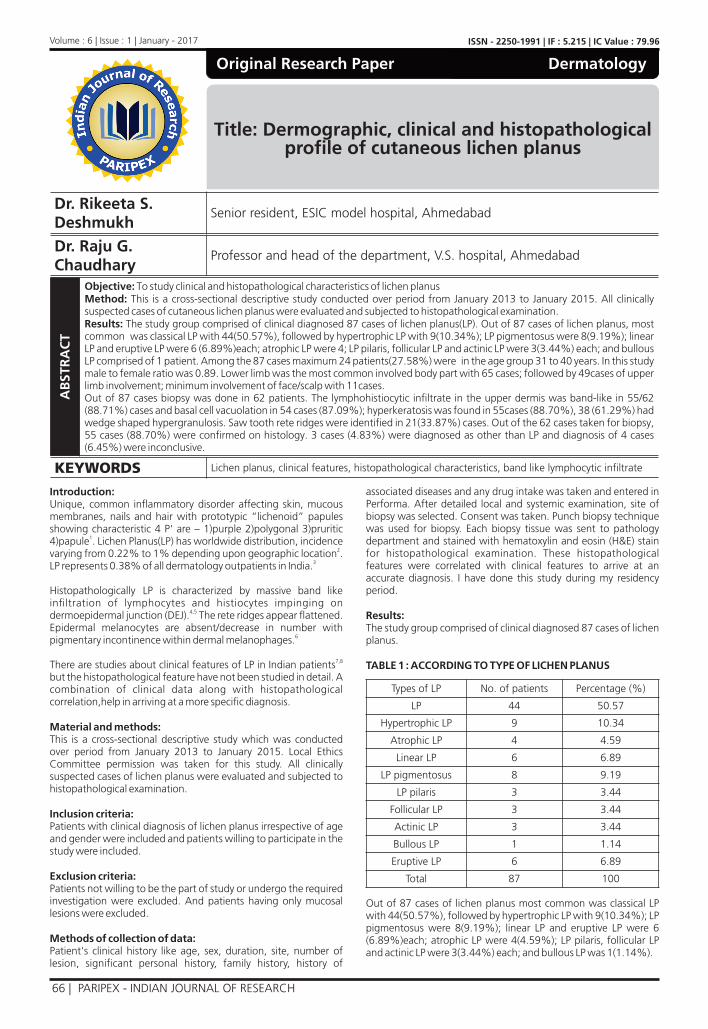
TABLE 2 : ACCORDING TO AGE AND SEX
Among the 87 cases maximum 24 patients(27.58%) were in the age group 31 to 40 years, comprising of 10 cases of classical lichen planus.4 cases of LP pigmentosus; 2cases of atrophic LP and linear LP each , 3 cases of hypertrophic LP; 1 case of eruptive LP, actinic LP and follicular LP each.
The next common age group was 0-20 years comprising 21(24.13%) patients followed by 21-30 years age group having 16 (18.39%), then 41-50 age group with 13 patients (14.94%) followed by 8 patients (9.19%) of 51-60years age group and 3patients(3.44%) of 60-70 years age group and least common being 70 years and above age group with only 2 patients (2.29%).
In my study male to female ratio was 41/46 that is 0.89.
TABLE 3 : AGE DISTRIBUTION OF THE INDIVIDUAL CASES
In the present study, maximum number of cases occurred in the middle aged group of 21 to 50 years in which there were 25 cases of classical LP, 6 cases of hypertrohic LP, 5 cases of LP pigmentosus, 4 eruptive LP.3 actinic LP, linear LP; 2 cases each of follicular LP, LP pilaris atrophic LP.
In the age group of above 50 years, 7 cases were of classical LP;2 cases of hypertrophic LP. 1 case of LP pigmentosus, LP pilaris, follicular LP, atrophic LP
The minimum number of cases occurred with 0-10year age group with only 2 cases of LP
TABLE 4: ACCORDING TO DURATION OF DISEASE
TABLE 5: CLINICAL SYMPTOMS
Moderate to severe degree of itching was present in almost all the patients. The itching was more severe in patients with generalized variety than in localized form except those with localized hypertrophic type which was extremely pruritic.
TABLE 6: ANATOMIC DISTRIBUTION OF DIFFERENT TYPES OF LICHEN PLANUS
Lower limb was the most common involved body part with 65 cases; followed by 49 cases of upper limb involvement; trunk 26 cases; mucosa 19 cases and minimum involvement of face/scalp with 11cases.Five patients had nail involvement in the form of longitudinal ridges, pitting and pterygium.
TABLE 7: ACCORDING TO ASSOCIATION WITH OTHER DISEASE
Associated conditions were Diabetes, Hypertension, Thyroid disorder and minimum association of 1 case each in Vitiligo and Alopecia. None of them had psoriasis.
Out of total 87 patients biopsy was done in 62 patients.
TABLE 8 : HISTOPATHOLOGICAL FINDING OF LICHEN PLANUS
Hyperkeratosis was found in 55cases (88.70%). Irregular acanthosis was seen in 46/62 (74.19%) cases. Saw tooth rete ridges and dome shaped papillae were identi�ed in 21/87 (33.87%) cases. 38/62 (61.29%) had wedge shaped hypergranulosis. Parakeratosis was found in 11 cases (17.74%). Liquefaction degeneration was found in 54 cases (87.09%). The least common histopathological �nding was spongiosis in 2 patients followed by max joseph space in 3 cases. Civatte bodies or necrotic keratinocytes were present in 14/62 (22.58%) of cases in the lower epidermis and especially in the papillary dermis. They had a homogeneous, eosinophilic appearance and fair number of them also contained pyknotic or fragmented nuclei. The in�ltrate in the upper dermis was bandlike in 55/62 (88.71%) at DEJ. In�ltrate was composed almost entirely of lymphocytes intermingled with few histiocytes. Pigment incontinence is a result of damage to the basal cells and was seen in 33/62 (53.22%)
ISSN - 2250-1991 | IF : 5.215 | IC Value : 79.96Volume : 6 | Issue : 1 | January - 2017
Age Group LP Hypertrophic LP
Atrophic LP
Linear LP
LP Pigmentosus
LP pilari
s
Follicular LP
Actinic LP
Bullous LP
Eruptive LP
Birth - 10 Yr 2 - - - - - - - - -11 - 20 Yr 9 1 1 3 2 - - - 1 221 - 50 Yr 25 6 2 3 5 2 2 3 - 4
> 50 Yr 7 2 1 - 1 1 1 - - -
Duration of disease No of cases
≤ 1 month 20
1-6 months 37
6-12 months 5
>1 year 25
Symptoms No of cases
No symptoms 11
Itching 75
Burning 13
Pain 2
Type of LP Face/Scalp
Trunk/abdomen
Upper limb
Lower limb
Mucosa(oral/genital)
LP 3 11 28 36 12Hypertrophic LP - 1 4 9 3
Atrophic LP - 1 1 4 -Linear LP - 2 1 3 1
LP pigmentosus 4 5 5 2 1LP pilaris 1 - - - -
Follicular LP - 1 2 3 -Actinic LP 2 1 2 1 1Bullous LP - 1 1 1 -Eruptive LP 1 3 5 6 1
Systemic No of cases Cutaneous No of casesDiabetes 2 Vitiligo 1
Hypertension 2 Psoriasis -Thyroid disorders 2 Alopecia 1
Epidermal DermalFinding No of
casesPercentage(%)
Finding No of cases
Percentage(%)
Hyperkeratosis 55 88.70 Bandlike lymphocytic in�ltration
55 88.71
Parakeratosis 11 17.74 Perivascular in�ltration
18 29.03
Acanthosis 46 74.19 Pigment incontinence
33 53.22
Hypergranulosis 38 61.29Saw tooth rete ridges 21 33.87Basal cell vacuolation 54 87.09
Spongiosis 2 3.22Colloid bodies 14 22.58
Atrophy 8 12.90Papillomatosis 5 8.06
Max joseph space 3 4.83
PARIPEX - INDIAN JOURNAL OF RESEARCH | 67
cases.
TABLE 9: CLINICAL AND HISTOPATHOLOGICAL CORRELA-TION OF LICHEN PLANUS
Out of the 62 cases taken for biopsy, 55 cases (88.70%) were diagnosed as LP and were con�rmed on histology.
Three (4.83%) patients had negative correlat ion on histopathology. One case of clinically diagnosed hypertrophic LP had histopathological �ndings of verrucous hyperplasia with dermatophytes, one case of LP was histopathlogically diagnosed as lichen nitidus, and one actinic LP was diagnosed as pigmented nevus on histopathology.
Histopathology was inconclusive in 4 (6.45%) patients and didn't show characteristic �nding like BCV or band like lympho-histiocytic in�ltration.
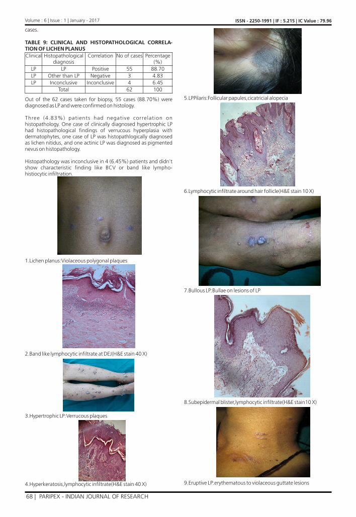
1.Lichen planus:Violaceous polygonal plaques
2.Band like lymphocytic in�ltrate at DEJ(H&E stain 40 X)
3.Hypertrophic LP:Verrucous plaques
4.Hyperkeratosis,lymphocytic in�ltrate(H&E stain 40 X)
5.LPPilaris:Follicular papules,cicatricial alopecia
6.Lymphocytic in�ltrate around hair follicle(H&E stain 10 X)
7.Bullous LP:Bullae on lesions of LP
8.Subepidermal blister,lymphocytic in�ltrate(H&E stain10 X)
9.Eruptive LP:erythematous to violaceous guttate lesions
ISSN - 2250-1991 | IF : 5.215 | IC Value : 79.96Volume : 6 | Issue : 1 | January - 2017
Clinical Histopathological diagnosis
Correlation No of cases Percentage (%)
LP LP Positive 55 88.70LP Other than LP Negative 3 4.83LP Inconclusive Inconclusive 4 6.45
Total 62 100
68 | PARIPEX - INDIAN JOURNAL OF RESEARCH
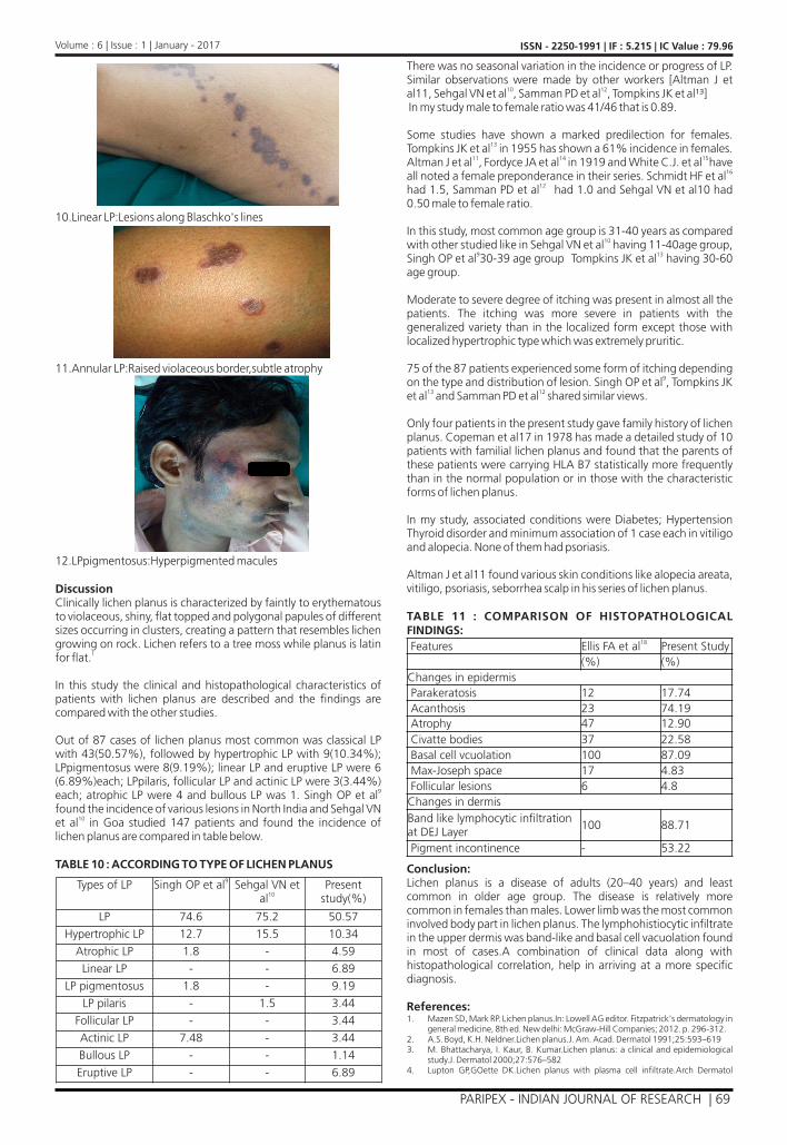
10.Linear LP:Lesions along Blaschko's lines
11.Annular LP:Raised violaceous border,subtle atrophy
12.LPpigmentosus:Hyperpigmented macules
DiscussionClinically lichen planus is characterized by faintly to erythematous to violaceous, shiny, �at topped and polygonal papules of different sizes occurring in clusters, creating a pattern that resembles lichen growing on rock. Lichen refers to a tree moss while planus is latin
1for �at.
In this study the clinical and histopathological characteristics of patients with lichen planus are described and the �ndings are compared with the other studies.
Out of 87 cases of lichen planus most common was classical LP with 43(50.57%), followed by hypertrophic LP with 9(10.34%); LPpigmentosus were 8(9.19%); linear LP and eruptive LP were 6 (6.89%)each; LPpilaris, follicular LP and actinic LP were 3(3.44%)
9each; atrophic LP were 4 and bullous LP was 1. Singh OP et al found the incidence of various lesions in North India and Sehgal VN
10et al in Goa studied 147 patients and found the incidence of lichen planus are compared in table below.
TABLE 10 : ACCORDING TO TYPE OF LICHEN PLANUS
There was no seasonal variation in the incidence or progress of LP. Similar observations were made by other workers [Altman J et
10 12al11, Sehgal VN et al , Samman PD et al , Tompkins JK et al��] In my study male to female ratio was 41/46 that is 0.89.
Some studies have shown a marked predilection for females. 13Tompkins JK et al in 1955 has shown a 61% incidence in females.
11 14 15 Altman J et al , Fordyce JA et al in 1919 and White C.J. et al have 16all noted a female preponderance in their series. Schmidt HF et al
12had 1.5, Samman PD et al had 1.0 and Sehgal VN et al10 had 0.50 male to female ratio.
In this study, most common age group is 31-40 years as compared 10with other studied like in Sehgal VN et al having 11-40age group,
9 13Singh OP et al 30-39 age group Tompkins JK et al having 30-60 age group.
Moderate to severe degree of itching was present in almost all the patients. The itching was more severe in patients with the generalized variety than in the localized form except those with localized hypertrophic type which was extremely pruritic.
75 of the 87 patients experienced some form of itching depending 9on the type and distribution of lesion. Singh OP et al , Tompkins JK
13 12et al and Samman PD et al shared similar views.
Only four patients in the present study gave family history of lichen planus. Copeman et al17 in 1978 has made a detailed study of 10 patients with familial lichen planus and found that the parents of these patients were carrying HLA B7 statistically more frequently than in the normal population or in those with the characteristic forms of lichen planus.
In my study, associated conditions were Diabetes; Hypertension Thyroid disorder and minimum association of 1 case each in vitiligo and alopecia. None of them had psoriasis.
Altman J et al11 found various skin conditions like alopecia areata, vitiligo, psoriasis, seborrhea scalp in his series of lichen planus.
TABLE 11 : COMPARISON OF HISTOPATHOLOGICAL FINDINGS:
Conclusion:Lichen planus is a disease of adults (20–40 years) and least common in older age group. The disease is relatively more common in females than males. Lower limb was the most common involved body part in lichen planus. The lymphohistiocytic in�ltrate in the upper dermis was band-like and basal cell vacuolation found in most of cases.A combination of clinical data along with histopathological correlation, help in arriving at a more speci�c diagnosis.
References:Mazen SD, Mark RP. Lichen planus.In: Lowell AG editor. Fitzpatrick's dermatology in general medicine, 8th ed. New delhi: McGraw-Hill Companies; 2012. p. 296-312.A.S. Boyd, K.H. Neldner.Lichen planus.J. Am. Acad. Dermatol 1991;25:593–619M. Bhattacharya, I. Kaur, B. Kumar.Lichen planus: a clinical and epidemiological study.J. Dermatol 2000;27:576–582Lupton GP,GOette DK.Lichen planus with plasma cell in�ltrate.Arch Dermatol
ISSN - 2250-1991 | IF : 5.215 | IC Value : 79.96Volume : 6 | Issue : 1 | January - 2017
Types of LP 9Singh OP et al Sehgal VN et 10al
Present study(%)
LP 74.6 75.2 50.57
Hypertrophic LP 12.7 15.5 10.34
Atrophic LP 1.8 - 4.59
Linear LP - - 6.89
LP pigmentosus 1.8 - 9.19
LP pilaris - 1.5 3.44
Follicular LP - - 3.44
Actinic LP 7.48 - 3.44
Bullous LP - - 1.14
Eruptive LP - - 6.89
Features 18Ellis FA et al Present Study(%) (%)
Changes in epidermis Parakeratosis 12 17.74 Acanthosis 23 74.19 Atrophy 47 12.90 Civatte bodies 37 22.58 Basal cell vcuolation 100 87.09 Max-Joseph space 17 4.83 Follicular lesions 6 4.8Changes in dermisBand like lymphocytic in�ltration at DEJ Layer
100 88.71
Pigment incontinence - 53.22
1.
2.3.
4.
PARIPEX - INDIAN JOURNAL OF RESEARCH | 69

1981;117:124-5.Roustan G,hospital M ,Vileges C et al.Lichen planus with predominant plasma cell in�ltrate.Am J Dermatol 1994;16:311-4.Black MM,Wilson Jones E.The role of the epidermis in the histopathogenesis of lichen planus.Arch Dermatol 1972;105:81-6.Singh OP, Kanwar AI. Lichen planus in India - an appraisal of 441 cases, Int J Dermatol 1976; 15: 752-756. Kachwa D, Kachawa V, Kalla G, et al. A clinicoaetiological pro�le of 375 cases of lichen planus, Indian J Dermatol Venerol Leprol 1995; 61:276379. SINGH OP,KANWAR AJ.Lichen planus in India.An appraisal of 441 cases.Int.J Dermat,1976;15(10):752-756SEHGAL VN AD REGE V.Lichen planus;An appraisal of 147 cases,Ind J Ddermat ,1974;40(3):104-107.ALTMAN JUCES AND PERRY HAROLDO.The variation and course of lichen planus.Arch Dermatol,1961:84:179-191.SAMMAN p .D .L i chen p lanus ; s ta t i s t i ca l s tudy o f 41 cases .Arch Defmatol,1961,71:515.TOMPKINS JAMES K,Lichen planus-A statistical study of 41 cases.Arch Dermatol.1955:71:515-21.FORDYCE JA,MACKEE GM.Clinical types of lichen planus.J of cut dis,1919:37:671.WHITE CJ.Lichen planus-critical analysis of 64 cases.J cut dis,1919,37:671.SCHMIDT.H.Frequency,duration and localization of lichen planus,A study based on 181 patients.Acta Derm Venerol:1961:41:164.COPEMAN PWM.TANRSH,TIMLIN DAND SAMMAN P.D.Familial lichen planus,another disease or a distinct people.Br J Dermatol,1978;98;573-577.FRANCIS A ELLIS,Histopathology of lichen planus based on analysis of 100 biopsy specimen.J Invest dermatol.1967:48:143-148.
5.
6.
7.
8.
9.
10.
11.
12.
13.
14.15.16.
17.
18.
70 | PARIPEX - INDIAN JOURNAL OF RESEARCH
ISSN - 2250-1991 | IF : 5.215 | IC Value : 79.96Volume : 6 | Issue : 1 | January - 2017