david burkholder, md - mayo clinic · • may be helpful if lp is delayed relative to antibiotic...
TRANSCRIPT
©2015 MFMER | 3543652-1
David Burkholder, MD Assistant Professor of Neurology
Faculty photo will be placed here

©2015 MFMER | 3543652-2
Neurologic Emergencies
Wednesday-Saturday, October 19-22, 2016 Sawgrass Marriott Hotel • Ponte Vedra Beach, Florida
Mayo School of Continuous Professional Development
2nd Annual Inpatient Medicine for NPs & PAs:
Hospital Care from Admission to Discharge

©2016 MFMER | 3543652-3
Disclosures Financial
None
Off-label use Use of medications for status epilepticus other
than lorazepam IV, midazolam IM, and phenytoin/fosphenytoin

©2016 MFMER | 3543652-4
Objectives • At the conclusion of this lecture, the audience
should be able to describe and enact: • Appropriate management for intracerebral
hemorrhage • Management for status epilepticus • Clinical signs that suggest need for
ventilatory support for neuromuscular respiratory failure
• Evaluation and empiric treatment for bacterial and HSV meningoencephalitis

©2016 MFMER | 3543652-5
Case • 48 year old woman found down and poorly
responsive at home in pool of vomit • Reported to PCP 2 days prior reporting neck
pain and headache • Febrile (38.6 C), tachycardic • Nuchal rigidity on examination, severely
encephalopathic

©2016 MFMER | 3543652-6
Case • 69 year old man, with headache and
progressive encephalopathic over 1-2 days • Today, had a convulsion (seizure) that prompted
ED evaluation • Febrile (38.6 C), without nuchal rigidity; clearly
encephalopathic and doesn’t follow commands, but has mildly reduced movement of his right side

©2016 MFMER | 3543652-7
These clinical presentations are most consistent with: A. Stroke
B. CNS infection
C. Status epilepticus
D. Systemic infection with delirium
E. Malingering

©2016 MFMER | 3543652-8
These clinical presentations are most consistent with: A. Stroke
B. CNS infection
C. Status epilepticus
D. Systemic infection with delirium
E. Malingering

©2016 MFMER | 3543652-9
What is the most appropriate next step? A. STAT CT scan prior to LP,
then initiate antimicrobial therapy
B. Imaging not necessary; STAT LP, then initiate antimicrobial therapy
C. STAT CT prior to LP, but don’t wait to initiate antimicrobial therapy
D. Imaging not necessary; STAT LP, but don’t wait to initiate antimicrobial therapy

©2016 MFMER | 3543652-10
What is the most appropriate next step? A. STAT CT scan prior to LP,
then initiate antimicrobial therapy
B. Imaging not necessary; STAT LP, then initiate antimicrobial therapy
C. STAT CT prior to LP, but don’t wait to initiate antimicrobial therapy
D. Imaging not necessary; STAT LP, but don’t wait to initiate antimicrobial therapy

©2016 MFMER | 3543652-11
Bacterial meningitis • Decreasing incidence in
US over time
• Average age • 15 months (1986) • 25 years (1998)
• First Hib vaccine introduced in US in 1985
• 20,000 cases (1987) • 255 cases (1998)
Thigpen MC et al. N Engl J Med 2011;364:2016-2025.

©2016 MFMER | 3543652-12
Bacterial meningitis: Symptoms • Classic triad: Fever, headache, neck stiffness
• Subacute over hours to <2 days • Only about half of patients with all 3 signs
• Other symptoms • Vomiting • Encephalopathy (“altered mental status”), ranging
from irritability to coma • Light sensitivity
• Kernig’s sign (K = knee)
• Brudzinski’s sign
Netter’s Neurology, 2nd ed., 2012

©2016 MFMER | 3543652-13
Bacterial meningitis: Risk factors • Ends of age spectrum (infants and elderly) • Chronic disease (DM, renal, hepatic) • Immunocompromise or suppression • Alcoholism • Pregnancy (Group B strep, Listeria) • Crowded living arrangement (N. meningitidis)
Brouwer MC, et al. Clin Microbiol Rev 2010;23(3):467.

©2016 MFMER | 3543652-14
Bacterial meningitis: Diagnosis • Basic labs – CBC, electrolytes, coags • Blood cultures
• Up to 50% positive in common meningitis-causing bacteria
• May be helpful if LP is delayed relative to antibiotic use
• Imaging? • Before LP only in certain cases if worried about
intracranial pressure • Immunocompromised, history of CNS disease,
seizure within 1 week, papilledema, decreased consciousness, focal neurologic deficit
Tunkel AR, et al. Clin Infect Dis 2004 Nov 1. 39(9):1267-84

©2016 MFMER | 3543652-15
Bacterial meningitis: Diagnosis • Lumbar puncture, including gram stain and
culture
Meningitis etiology
Opening pressure
Cell count Glucose Protein
Normal <25 cm H20 0-5 60% serum <40 mg/dL
Bacterial Elevated 1000+, neutrophilic
Very decreased
Very elevated
Viral Normal to mildly elevated
<500, lymphocytic
Normal to mildly decreased
Normal to mildly elevated

©2016 MFMER | 3543652-16
Bacterial meningitis: Treatment (age 2 and up) • For all:
• Vancomycin 15-20 mg/kg IV q8-12 hours • Ceftriaxone 2 g IV q12 hours • Plus: empiric viral coverage…yet to come!
• For age >50 years or immune impaired, add to above:
• Ampicillin 2 g IV q4 hour
• DO NOT WITHOLD TREATMENT TO WAIT FOR TESTS!
van de Beek D, et al. Lancet 2012;380:1693.

©2016 MFMER | 3543652-17
Dexamethasone and S. pneumo • S. pneumoniae may be more effectively treated
if dexamethasone given before (or with) initial antibiotic therapy
• Must decide to give before knowing diagnosis! • Dexamethasone 10 mg IV x 1 prior to abx, then
10 mg q6 hours for 4 days • Discontinue if CSF returns inconsistent with
bacterial meningitis
van de Beek D, et al. Lancet 2012;380:1693.

©2016 MFMER | 3543652-18
Viral meningitis • Majority treated with supportive care • Most common exception: Herpes simplex virus
(HSV)
Singh TD, et al. J Neurol 2016;263(2):277.
©2016 MFMER | 3543652-19
HSV encephalitis • May not be associated with meningismus • Frequently encephalopathic, seizures • MRI shows characteristic T2 hyperintensity in
one or both temporal lobes • CSF: HSV PCR (may be false negative if <48
hrs) • Treatment with acyclovir 10 mg/kg IV q8 hours

©2016 MFMER | 3543652-20
Approach to suspected CNS infection

©2016 MFMER | 3543652-21
Approach to suspected CNS infection • Labs and treatment performed simultaneously
• Don’t forget blood cultures!
• LP as soon as possible, combining bacterial and viral studies
• Treat empirically for both bacterial meningitis and HSV until CSF findings confirm etiology
• Tailor treatment as testing comes in
Basic CSF studies: - Cell count and diff - Glucose - Protein - Gram stain and culture - HSV PCR
Basic treatment: - Vancomycin 15-20 mg/kg IV q8-12 - Ceftriaxone 2 g IV q12 - Acyclovir 10 mg/kg IV q8 - +/- Ampicillin 2 g IV q4 (if risk factors) - +/- Dexamethasone 10 mg IV (prior to first abx dose if suspect S. pneumoniae)

©2016 MFMER | 3543652-22
Case • 56 year old man presents with acute right-sided
weakness • History of hypertension and atrial fibrillation on
warfarin • BP 180/110 mmHg, pulse 85, O2 sat 96% • On exam, has dense right hemiparesis.
Language is impaired • INR = 5

©2016 MFMER | 3543652-23
What is the most appropriate next step? A. Reversal with IV vitamin K
alone
B. Reversal with fresh frozen plasma (FFP) alone
C. Reversal with both IV vitamin K and FFP
D. Aggressive blood pressure reduction (SBP <140 mmHg) and reversal with IV vitamin K alone
E. Aggressive blood pressure reduction (SBP <140) and reversal with both IV vitamin K and FFP

©2016 MFMER | 3543652-24
What is the most appropriate next step? A. Reversal with IV vitamin K
alone
B. Reversal with fresh frozen plasma (FFP) alone
C. Reversal with both IV vitamin K and FFP
D. Aggressive blood pressure reduction (SBP <140 mmHg) and reversal with IV vitamin K alone
E. Aggressive blood pressure reduction (SBP <140) and reversal with both IV vitamin K and FFP

©2016 MFMER | 3543652-25
Intracranial hemorrahge • Many varieties
• Epidural (surgical emergency) • Subdural • Subarachnoid • Intracerebral
• Will focus on intracerebral since care is often (but not always) non-operative
• Highlights of subarachnoid hemorrhage (SAH)

©2016 MFMER | 3543652-26
Intracerebral hemorrhage • Presentation similar to
stroke
• May have associated headache, worsening mental status
• Imaging key to diagnosis
• Risk factors: • Hypertension • Age • Anticoagulation/antithr
ombotic use
Ariesen MJ, et al. Stroke 2003;34(8):2060. Sturgeon JD, et al. Stroke 2007;38(10):2718.

©2016 MFMER | 3543652-27
Intracerebral hemorrhage Goal of early intervention is to prevent expansion!
Emiru T, et al. Clin Appl Thromb Hemost 2013;19:652-62.

©2016 MFMER | 3543652-28
Intracerebral hemorrhage • If anticoagulated, REVERSE
• Vitamin K 5-10 mg IV if warfarin – DOES NOT ACT QUICKLY
• Fresh frozen plasma or prothrombin complex concentrates
• Ensure INR stays normalized (repeat!) • Direct thrombin inhibitors and factor Xa
inhibitors • Dabigatran (DTI) – idarucizumab • Currently investigating options for Xa
inhibitors
Hemphill JC, et al. Stroke 2015;46.

©2016 MFMER | 3543652-29
Intracerebral hemorrhage • Blood pressure control
• Previously, SBP <180 mmHg
• INTERACT2 trial – trend towards primary outcome, no difference in safety
• Now recommend lowering SBP <140 mmHg in patients without contraindication
Anderson CS, et al. New Engl J Med 2013; 368(25):2355. Hemphill JC, et al. Stroke 2015;46.

©2016 MFMER | 3543652-30
What about surgery? • Cerebellar hemorrhage
• Not much room to expand • Brainstem compression, hydrocephalus,
decompensation may occur early • Neurosurgeon may decompress at first signs of
deterioration
• Cerebral hemorrhage • Not usually recommended • May intervene if decompensating, comatose, or
large hematoma
Hemphill JC, et al. Stroke 2015;46.

©2016 MFMER | 3543652-31
Cerebellar hemorrhage anatomy

©2016 MFMER | 3543652-32
Subarachnoid hemorrhage: Highlights • Early intervention strategies similar
• Reverse anticoagulation • Control blood pressure (parameters not well defined)
• Angiography to evaluate for aneurysm • If present and able, secure aneurysm (coiling or
clipping) • Later complications
• Delayed cerebral ischemia (vasospasm) • Cerebral salt wasting

©2016 MFMER | 3543652-33
Restarting anticoagulation • No guidelines regarding
timing of restart
• Appears to be safe (and beneficial) in the appropriate context
• Studies confounded • Small numbers • Non-controlled • Primarily warfarin
(NOACs small numbers)
Nielsen PB, et al. Circulation 2015;132(6):517.

©2016 MFMER | 3543652-34
Case • 65 year old woman with history of left
hemisphere stroke 6 months prior presents to the ED
• Found unresponsive convulsing on floor, duration unknown at time of discovery
• EMS activated, lorazepam 2 mg IV given en route, brought to ED
• On examination, remains unresponsive with continuous rhythmic clonic jerks of the face and extremities, eye deviation to the right

©2016 MFMER | 3543652-35
What is the most appropriate next step? A. Lorazepam 2-4 mg IV
B. Fosphenytoin 20 PE/kg IV
C. Lorazepam 2-4 mg IV now, and also ordering fosphenytoin
D. Anesthetic induction and intubation
E. Hide in the corner, maybe no one will notice

©2016 MFMER | 3543652-36
What is the most appropriate next step? A. Lorazepam 2-4 mg IV
B. Fosphenytoin 20 PE/kg IV
C. Lorazepam 2-4 mg IV now, and also ordering fosphenytoin
D. Anesthetic induction and intubation
E. Hide in the corner, maybe no one will notice

©2016 MFMER | 3543652-37
Status Epilepticus
Falco-Walter JJ and Bleck T. J Clin Med 2016;5(5):49.

©2016 MFMER | 3543652-38
Status Epilepticus • Common hospital entity • High mortality rates
• Convulsive: up to 27% at 30 days • Non-convulsive: up to 65% at 30 days • Refractory: up to 61% at “discharge”
• Associated morbidity • Non-refractory: 39% return to functional baseline • Refractory: 15% independent (still moderately
disabled)
Brophy GM, et al. Neurocrit Care 2012;17:3-23

©2016 MFMER | 3543652-39
Initial management • Adrenaline runs high • Remember to stay calm • First issue: treat the seizures • Second issue: address the source
• Treating seizures difficult if etiology not adequately addressed
• Concurrent with symptomatic treatment

©2016 MFMER | 3543652-40
Hirsch LJ and Gaspard N. Continuum 2013;19(3):767-794.
Addressing the source

©2016 MFMER | 3543652-41
Addressing the source • History • CT head (acutely) • MRI brain (later on) • Labs
• CBC • Glucose • Complete metabolic panel • Magnesium • Calcium • Phosphorus • Lumbar puncture • Urine drug screen • Blood cultures • Urinalysis and cultures • AED levels (if on treatment)
• Administer dextrose and thiamine • Just like ACLS, remember your
ABCs: stabilizing the patient is priority

©2016 MFMER | 3543652-42
Treatment reality
Lorazepam 2 mg
Fos/phenytoin 1 g Levetiracetam 1 g Valproate 1 g
Other IV agent
Other IV agent
Lorazepam 2 mg
Lorazepam 2 mg Lorazepam 2 mg
©2016 MFMER | 3543652-43
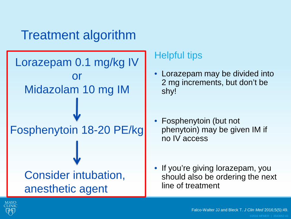
Treatment algorithm Helpful tips
• Lorazepam may be divided into 2 mg increments, but don’t be shy!
• Fosphenytoin (but not phenytoin) may be given IM if no IV access
• If you’re giving lorazepam, you should also be ordering the next line of treatment
Lorazepam 0.1 mg/kg IV or
Midazolam 10 mg IM
Fosphenytoin 18-20 PE/kg
Consider intubation, anesthetic agent
Falco-Walter JJ and Bleck T. J Clin Med 2016;5(5):49.

©2016 MFMER | 3543652-44
Alternatives to fosphenytoin/phenytoin • Why avoid the only FDA-approved second line agent?
• If cardiac arrhythmia or hypotension is a problem in an unstable patient
• Very significant hepatic disease
• _________ – 20-40 mg/kg IV • Avoid in hepatic disease
• _____________ – 2.5-4.5 grams IV • Requires dose adjustment for renal disease
• Lacosamide – 400 mg IV • Avoid in arrhythmia
(But what evidence do we have for these?)
Falco-Walter JJ and Bleck T. J Clin Med 2016;5(5):49.

©2016 MFMER | 3543652-45

©2016 MFMER | 3543652-46
Established status epilepticus treatment trial (ESETT)
• Multicenter double-blind RCT • Goal 795 patients • Intention to treat • Enrolled if seizure >5 min after adequate benzodiazepine
• 3 treatment arms, all infused over 10 minutes • Fosphenytoin 20 PE/kg • _____________ 60 mg/kg • Valproate 40 mg/kg
• At 60 minutes post intervention: • Seizure • Mental status • Adverse events
• Chart review for admission status, requirement for intubation, and seizure recurrence during hospitalization
Bleck T, et al. Epilepsia 2013;54:89.

©2016 MFMER | 3543652-47
Case (Patient 1) • 54 year old man develops weakness in the feet
and trouble with walking arm weakness following day
• Neurologic consultation initiated: • Diffuse moderate weakness, including neck flexors
and extensors • Short of breath when laying supine • Absent reflexes
• Lumbar puncture • WBC 3 • Protein 115 • Glucose normal

©2016 MFMER | 3543652-48
Case (Patient 2) • 73 year old woman with generalized
myasthenia gravis • For several days, had problems controlling
secretions and changes in speech • Increasing doses of Mestinon not helpful, so
prednisone initiated at a high dose • Presents to the ED with continued speech and
swallowing difficulty, now feeling short of breath • Exam: diaphoretic, chest expansion and neck
contraction with inspiration; facial weakness with dysarthria, globally weak

©2016 MFMER | 3543652-49
Which patient is clinically most in danger of respiratory failure? A. Patient 1, because it is
more acute in onset
B. Patient 1, because he has orthopnea
C. Patient 2, because she is using accessory muscles to breath
D. Patient 2, because she is older

©2016 MFMER | 3543652-50
Which patient is clinically most in danger of respiratory failure? A. Patient 1, because it is
more acute in onset
B. Patient 1, because he has orthopnea
C. Patient 2, because she is using accessory muscles to breath
D. Patient 2, because she is older

©2016 MFMER | 3543652-51
Neuromuscular respiratory failure • Common complication of acute or chronic
neurologic disease • Acute disease or exacerbation primarily resulting in
weakness (AIDP, MG) • Decompensation of stable chronic disease during
concurrent illness (MG, ALS, etc.)
• Up to 30% of AIDP and MG patients will require
mechanical ventilation
Sharshar T, et al. Crit Care Med 2002:31:278. Mehta S. Respir Care 2006;51:1016.
Durand MC, et al. Lancet Neurol 2006;5:1021.

©2016 MFMER | 3543652-52
Symptoms • Dyspnea
• Orthopnea (SOB when laying flat)
• Tachypnea
• Accessory respiratory muscle use
• Paradoxical breathing
• Dysarthria
• Dysphagia
• Weak cough
• Aspiration
• Hypersomnolence
• Encephalopathy

©2016 MFMER | 3543652-53
Paradoxical breathing
Mittal MK and Wijdicks EFM. In Critical Care Medicine: Principles of Diagnosis and Management in the Adult. p1121-1129.

©2016 MFMER | 3543652-54
Evaluation • ABCs – stabilize the patient
• Clinical assessment: Does the patient have impaired consciousness or appear obviously distressed?
• If the answer is yes, intubate*
• “20-30-40” rule – serial checks • Forced vital capacity < 20 ml/kg (or <1 L) • Max inspiratory pressure < -30 cm H20 • Max expiratory pressure < 40 cm H20
• ABG – but remember that a lack of hypoxemia or hypercapnia does not exclude badness
*In cases of chronic progressive disease like ALS, try to confirm patient goals of care
Harms M. Neurohospitalist 2011;1(2):78.

©2016 MFMER | 3543652-55
Evaluation • Testing to identify a systemic contributors in chronic
disease decompensation • Infection (CBC, blood cultures, urinalysis) • Medication changes or compliance issues • Metabolic abnormalities
• Testing to identify a primary neurologic diagnosis (if still needed)
• Imaging, if indicated • LP • EMG • Myasthenia antibodies

©2016 MFMER | 3543652-56
Treatment • Assistance with ventilation when appropriate
• Intubation • Mortality: 12-20% in AIDP, 4-8% in MG • Average duration: 18-29 days in AIDP, 14 days in MG
• BiPAP – may not perform well if significant bulbar weakness
• Address primary causes • Plasma exchange (AIDP, MG) • IVIg (AIDP) • Medication adjustments (MG)
• Address systemic contributors, if identified Thomas CE, et al. Neurology 1997; 28:1253. Fletcher DD, et al. Neurology 200;54:2311.
Cheng BC, et al. Am J Med Sci 2004;327:336. Orlikowski D, et al. Neurocrit Care 2004;1:415.

©2016 MFMER | 3543652-57
Questions & Discussion