thin deformable mirrors for a reconfigurable space telescope
TRANSCRIPT
Cellular/Molecular
Baclofen and Other GABAB Receptor Agents Are AllostericModulators of the CXCL12 Chemokine Receptor CXCR4Alice Guyon,1,2,3 Amanda Kussrow,4 Ian Roys Olmsted,4 Guillaume Sandoz,1,2,5 Darryl J. Bornhop,4*and Jean-Louis Nahon1,2,6*1Universite de Nice Sophia Antipolis, 06103 Nice, France, 2Centre National de la Recherche Scientifique (CNRS), Institut de Pharmacologie Moleculaire etCellulaire, UMR 7275, 06560 Valbonne, France, 3Department of Molecular and Cellular Biology and Helen Wills Neuroscience Institute, University ofCalifornia, Berkeley, Berkeley, California 94720, 4Vanderbilt Institute of Chemical Biology, Nashville, Tennessee 37235-1822, 5Institute of Biology Valrose,CNRS UMR 7277, INSERM UMR 1091, Laboratories of Excellence, Ion Channel Science and Therapeutics, 06103 Nice, France, 6Station dePrimatologie—UPS 846 —Centre CNRS, RD56, 13790 Rousset sur Arc, France
CXCR4, a receptor for the chemokine CXCL12 (stromal-cell derived factor-1�), is a G-protein-coupled receptor (GPCR), expressed in theimmune and CNS and integrally involved in various neurological disorders. The GABAB receptor is also a GPCR that mediates metabotropicaction of the inhibitory neurotransmitter GABA and is located on neurons and immune cells as well. Using diverse approaches, we report novelinteraction between GABAB receptor agents and CXCR4 and demonstrate allosteric binding of these agents to CXCR4. First, both GABAB
antagonists and agonists block CXCL12-elicited chemotaxis in human breast cancer cells. Second, a GABAB antagonist blocks the potentiation byCXCL12 of high-threshold Ca2� channels in rat neurons. Third, electrophysiology in Xenopus oocytes and human embryonic kidney cell line 293cells in which we coexpressed rat CXCR4 and the G-protein inward rectifier K� (GIRK) channel showed that GABAB antagonist and agonistmodified CXCL12-evoked activation of GIRK channels. To investigate whether GABAB ligands bind to CXCR4, we expressed this receptor inheterologous systems lacking GABAB receptors and performed competition binding experiments. Our fluorescent resonance energy transferexperiments suggest that GABAB ligands do not bind CXCR4 at the CXCL12 binding pocket suggesting allosteric modulation, in accordance withour electrophysiology experiments. Finally, using backscattering interferometry and lipoparticles containing only the CXCR4 receptor, wequantified the binding affinity for the GABAB ligands, confirming a direct interaction with the CXCR4 receptor. The effect of GABAergic agentson CXCR4 suggests new therapeutic potentials for neurological and immune diseases.
IntroductionCXCR4 is a G-protein-coupled receptor (GPCR) that selectivelybinds CXCL12 (stromal-cell derived factor; SDF-1�). Thischemokine and its receptor have been found to play important
roles in several processes involved in ischemic stroke and its sub-sequent repair (Wang et al., 2012), brain tumor pathogenesis(Rempel et al., 2000), human immunodeficiency virus (HIV)encephalopathy (Li and Ransohoff, 2008), multiple sclerosis, andstem cell migration (Carbajal et al., 2010). CXCR4 is widely ex-pressed in a variety of cell types including leukocytes, where itpromotes migration, recruitment, and activation (Bonavia et al.,2003; Salcedo and Oppenheim, 2003; Juarez et al., 2004; Choi andAn, 2011; Comerford and McColl, 2011); neurons, where it mod-ulates electrical activity (Banisadr et al., 2002; Guyon and Nahon,2007; Rostene et al., 2011); and various cancers and metastases(Wang et al., 2006), where it is involved in tumor progression(Liu et al., 2006; Gao et al., 2010; Zhao et al., 2010). CXCR4 alsobinds the HIV-1 viral envelope glycoprotein, gp120 (Doranz etal., 1997; Gabuzda and Wang, 2000). Thus CXCR4 is an impor-tant therapeutic target for stroke, inflammation, neuromodula-tion, cancer, and in the prevention of HIV infection.
CXCR4 couples to the Gi family of proteins activating multipleG-protein-dependentand-independentpathways(Lazarinietal.,2003;Busillo and Benovic, 2007). In neurons, CXCR4 stimulation has beenshowntoactivateaG-proteininwardrectifierK�(GIRK)andavoltage-gated K channel Kv2.1 associated to neuronal survival, and to increasehighvoltage-activated(HVA)Ca2� currents(GuyonandNahon,2007;Shepherd et al., 2012).
The GABA type B (GABAB) receptor is also a GPCR activatedby GABA, the chief neuro-inhibitory neurotransmitter in mam-
Received Nov. 14, 2011; revised May 8, 2013; accepted June 5, 2013.Author contributions: A.G., A.K., D.J.B., and J.-L.N. designed research; A.G., A.K., I.R.O., and G.S. performed
research; A.G., A.K., and D.J.B. contributed unpublished reagents/analytic tools; A.G., A.K., I.R.O., G.S., and D.J.B.analyzed data; A.G., D.J.B., and J.-L.N. wrote the paper.
This work was supported by Centre National de la Recherche Scientifique (CNRS) (INSB), ANR-MNPs-018-01, FondationFranceParkinson,FondationdelaRechercheMedicale,andtheNationalScienceFoundation(GrantCHE-0848788).G.S.wassupported by the Atip-Avenir fund from the CNRS and INSERM. We are grateful to Richard Miller for generously providing uswith the rat CXCR4 clone and for all his advice. We thank Ehud Isacoff for his generous welcome. We greatly appreciated theassistance of Dr. Shashank Bharill, Dr. Ryan Arant, and Dr. Hitomi Okada. We thank Dr. Josh Levitz for providing us withGIRK1-F137S and Drs. Thomas Berger, Zhu Fu, Benji Gaub, Grant Kauwe, Susy Kohout, Andreas Reiner, Sasha Shekhar,Sandra Wiese, and all the Isacoff lab for fruitful discussions and technical support. We thank Dr. Piotr Bregestovski, Dr.Gregory Conductier, and Dr. Brice Flammang for their advice on experiments. We thank Audrey Recouly, Fabienne Cheval-lier, and Florence Servent from Cisbio Bioassays, Codolet, France, for the receptor-ligand binding assay using the Tag-litetechnology. We thank Dr. Joseph Rucker from Integral Molecular for generously providing the lipoparticles. We acknowl-edge Ann Fischer and Michelle Richner from the University of California Berkeley MCB Tissue Culture Facility for cell culturesupport; Dr. Franck Bihl from IPMC, France; and Drs. Victoria Vinader and Kamyar Afarinkia from the University of Bradford,UK, for their advice on chemotactic assay. We acknowledge Michelle Sexton for help in preparation of this manuscript andFranck Aguila for artwork. The following reagents were obtained through the National Institute of Health (NIH) AIDSResearch and Reference Reagent Program, Division of AIDS, National Institute of Allergy and Infectious Diseases (NIAID),NIH: Recombinant HIV-1 IIIB gp120 (CHO), cat#11784 from DIAIDS and NIAID (Immunodiagnostics) and RecombinantSoluble Human CD4, cat#4615, from Progenics Pharmaceuticals.
*D.J.B. and J.-L.N. contributed equally to this work.Correspondence should be addressed to Alice Guyon, Centre National de la Recherche Scientifique, Institut de
Pharmacologie Moleculaire et Cellulaire, 06560 Valbonne, France. E-mail: [email protected]:10.1523/JNEUROSCI.6070-11.2013
Copyright © 2013 the authors 0270-6474/13/3311643-12$15.00/0
The Journal of Neuroscience, July 10, 2013 • 33(28):11643–11654 • 11643
malian systems. GABAB receptors are obligatory heterodimerswith two homologous subunits (GB1 and GB2) required for func-tioning, are widely expressed and distributed in the CNS (Kaup-mann et al., 1998), and can activate diverse intracellular pathways(Guyon and Leresche, 1995; Laviv et al., 2011). GABAB receptorsare also expressed on cells of the immune system with a possiblelink to the inflammatory response (Tian et al., 2004; Rane et al.,2005). As a consequence, there is a rich pharmacology aimed attargeting GABAB receptors, with numerous compounds cur-rently being used with the presumption that they are highly se-lective for these receptors (Bowery, 1993; Froestl, 2010). Giventhat CXCR4 and GABAB have coexpression on immune cells andneurons, and the evidence for possible cross talk between thesereceptors, we hypothesized that ligands binding GABAB are in-volved in allosteric or direct interaction with CXCR4. Here wedescribe the experiments that test our hypothesis. First we showinhibition of CXCL12-induced migration of cancer cells byGABAB ligands. We tested these same ligands in electrophysiol-ogy experiments using dopaminergic neurons in acute slices ofsubstantia nigra and oocytes that lead us to binding experimentsusing GABAB agents at the CXCR4 receptor. While somewhatunexpected, we observed that GABAB agents such as baclofen, theantagonists CGP 55845 and CGP 54626, and GABA can directlybind the chemokine receptor CXCR4.
Materials and MethodsMolecular biology. Rat CXCR4 gene containing pcDNA 3.1 (�) plasmidwas obtained from Richard Miller lab, University of Michigan. CXCR4was then either subcloned in pGEMHE vector using XmaI and XbaI sites(for electrophysiology) or pGEMHE-EGFP-X vector (X being the linkersequence SRGTSGGSGGSRGSGGSGG) using XhoI and XbaI sites.cRNA was then prepared from the above clones using mMessage T7 RNAkit. Eitan Reuveny of the Weizmann Institute of Science providedGIRK1/2 cDNA for experiments.
Oocyte expression. Stage V–VI oocytes were collected from anesthe-tized Xenopus laevis and defolliculated with collagenase (BoehringerMannheim). Oocytes were incubated at 16°C in external medium (N96)of the following composition (in mM): 96 NaCl, 2 KCl, 1.8 CaCl2,1 MgCl2, 10 HEPES, 5 pyruvate, and 100 mg/L gentamycin, pH 7.2. Fiftynanoliters of RNA was injected. The concentration of RNA injected was0.003 �g/�l for GIRK1 and GIRK2, 0.005– 0.1 �g/�l for CXCR4. Expres-sion protein was allowed 24 –72 h before the start of the experiments at12°C or 18°C.
Cell culture and transfection. The human embryonic kidney cell line 293(HEK293) was maintained in DMEM with 5% fetal bovine serum (FBS) onpoly-lysine-coated glass coverslips at �6 � 106 cells per 25 mm coverslipand transiently cotransfected using Lipofectamine 2000 (Invitrogen) withCXCR4 in PCDNA 3 (�), GIRK1-F137S (homotetramerization mutant),and eYFP at a ratio of 7.5:7.5:0.5 with 1.6 �g of DNA total per 18 mmdiameter coverslip. Patch clamp was performed 24–36 h after transfection.
The human neuroblastoma cell line SH-SY5Y was obtained fromATCC by the MCB Tissue Culture Facility and maintained as monolayersin DMEM with 5% FBS. The human breast adenocarcinoma cell line,MDA-MB-231, was obtained from ATCC by the MCB Tissue CultureFacility and maintained as monolayers in RPMI 1640 supplemented with10% FBS, 1 mM pyruvate, and 2 mM glutamine. All cells lines were used ata low passage.
Agarose spot assay. Agarose spot assay was performed as describedpreviously (Vinader et al., 2011). Briefly, low melting point agarose(Ultrapure, LMP agarose, Invitrogen) was diluted into sterile PBS tomake a 0.5% agarose solution. The agarose solution was autoclaved,removed from the heat, and cooled down to 40°C.
To prepare the CXCL12/agarose solution, lyophilized CXCL12 (R&DSystems) was reconstituted to a final stock concentration of 12.5 �M insterile water. CXCL12 solution was then added to the molten 0.5% aga-rose solution at 40°C to produce a final concentration of 10 nM (�) or
100 nM CXCL12 (��). Control agarose solutions were prepared by sub-stituting same volumes of water for CXCL12 (Solutions � and ��).Four independent drops (10 �l, �2 mm diameter) of the four solutions(maintained at 40°C) were pipetted onto the base of a sterile 20 mmdiameter glass-bottomed cell culture dish (MatTek Corporation; see Fig.1C). The dishes were then cooled for 5 min at 4°C to allow the agarosespot to solidify. MDA-MB-231 cells were trypsinized and resuspended toa final concentration of 10 5 cells/ml in RPMI 1640 supplemented with10% FBS, 1 mM sodium pyruvate, and 2 mM L-glutamine. Cells were thenexposed to AMD3100 (2 �M), gp120 and CD4 (300 nM each), CGP 55845(5 �M), CGP 54626 (5 �M), GABA (100 �M), baclofen (100 �M), orcontrol PBS with no drug. Following this incubation, cells were added tothe dishes containing the agarose spots (1 ml per plate at a final concen-tration of 10 5 cells.ml �1) and then incubated for 4 h at 37°C to allow thecells to adhere. The culture media was replaced with RPMI mediumcontaining 0.1% FBS and the drugs or control, and the dish incubated at37°C in 5% CO2 overnight. The degree of cells invading underneath theagarose spot was analyzed by collecting pictures under an inverted mi-croscope Olympus IX91 using a �10 objective equipped with an AndorCamera and Software, and then counting the number of cells usingImageJ software.
Six representative fields of view of equal size sectors were individuallycounted per agarose spot, and the mean number of invading cells perfield of view calculated. The values reported herein are the average ofthree independent dishes. The experiment was reproduced twice andgave similar results.
Reverse transcription-PCR. The content of a monolayer of confluentcells in a 25 ml flask was trypsinized and washed twice in cold PBS and thecell pellet was used for extraction of total RNA using TRIzol. cDNA wasthen made from total RNA using SuperScript II Reverse Transcriptase kit(Invitrogen). CXCR4 and GABAB genes were then amplified usingCXCR4 Forward 20 AGGTAGCAAAGTGACGCCG, CXCR4 Reverse220 GATGGTGGGCAGGAAGATT (Guyon et al., 2006) and GAGABR2Forward GGACCTGGATTCTCACCGTGGGCTA, GABABR2 ReverseTGCTGGGTCCGGCTCCATGCTGTA (Osawa et al., 2006) primers, re-spectively, and products were analyzed on agarose gel.
Immunoblot analysis. Both floating and attached cells were collected,washed twice with cold PBS, and processed for plasma membraneprotein fraction using Qproteome Plasma Membrane Protein Kit(Qiagen). Equal amounts of membrane protein (15–20 �g) from cellswere separated on 12% SDS-PAGE and transferred electrophoreti-cally to a polyvinylidene difluoride membrane. Immunoblot analyseswere performed with 1:1000 dilutions of CXCR4 and GABABR2 an-tibodies using standard procedure. The blots were visualized usingthe enhanced chemiluminescence detection reagents and the manu-facturer’s protocol.
Electrophysiology. Electrophysiological recordings in brain slices fromthe rat substantia nigra were performed as described previously (Guyonet al., 2008). Briefly, male Wistar rats were bred in the local animalfacilities and maintained on a 12 h dark/light cycle (07:00/19:00) withfood and water ad libitum. All of the protocols were performed in accor-dance with French standard ethical guidelines for laboratory animals(agreement no. 75–178, 05/16/2000). Brain slices were obtained from12- to 23-d-old rats anesthetized with 1% halothane. Following decapi-tation, brains were rapidly removed and placed in cold phosphate/bicarbonate-buffered solution (PBBS) composed of the following (inmm): 125 NaCl, 2.5 KCl, 0.4 CaCl2, 1 MgCl2, 25 glucose, 1.25 NaH2PO4,and 26 NaHCO3, pH 7.4 when bubbled with 95% O2/5% CO2. Trans-versal substantia nigra slices (250 �m) were then transferred to an incu-bating chamber maintained at 34°C in oxygenated PBBS. After 1 h, sliceswere transferred to another incubating chamber at room temperature(22–25°C) filled with PBBS containing additional CaCl2 (final concen-tration 2 mm) and used for electrophysiological recordings. Slices wereplaced under a Nomarski microscope (Zeiss) equipped with infraredvideo camera (AxioCam; Zeiss) in a recording chamber superfused at aflow rate of 1 ml/min with oxygenated PBBS. Pictures were taken byusing a digital camera (AxioCam; Zeiss) and connected image-acquisitionsoftware(AxioVision).Dopaminergic neurons were recorded in the whole-cell mode in voltage clamp and characterized as described previously by
11644 • J. Neurosci., July 10, 2013 • 33(28):11643–11654 Guyon et al. • GABABR Agonists and Antagonists Interact with CXCR4

the presence of the hyperpolarization-activated cation current and low-threshold Ca current (IT), and using single-cell reverse transcription(RT)-PCR to reveal the presence of mRNA for the tyrosine hydroxylaseas described previously (Guyon et al., 2006). Patch-clamp pipettes had aresistance of 3– 6 M� when filled with the internal solution containingthe following (in mM): 120 CsCl, 5 MgCl2, 1 CaCl2, 10 EGTA, 4 Na ATP,0.4 Na GTP, and 10 HEPES supplemented with 15 mm phosphocreatineand 50 U/ml creatine phosphokinase (pH adjusted to 7.3 with CsOH).Statistical significance between groups (average data expressed asmean � SEM, n � number of neurons) was tested using either theone-way ANOVA or repeated-measures one-way ANOVA test followed
by a Newman–Keuls post hoc test with a threshold of significance of *p �0.05, **p � 0.01, and ***p � 0.001 using a statistical software package(SigmaStat 2.03 from Jandel Scientific or Origin from MicroCal).Changes of extracellular solution were obtained by a fast multibarreldelivery system positioned close to the cell tested.
Electrophysiological recording in Xenopus oocytes was done 2–5 dafter cRNA injection. Two-electrode voltage-clamp was performed witha GeneClamp 500 amplifier interfaced to a Digidata 1200 A/D (Molecu-lar Devices). The interface was controlled with a PC computer (Dell)running pClamp version 10.2 (Molecular Devices). Microelectrodeswere filled with 3 M KCl and had a tip resistance of 0.15–1.5M�. The
Figure 1. GABAB agents reduce the migration of the breast cancer cells MDA-MB-231 in a chemotactic assay. A, RT-PCR reveals the presence of endogenous mRNAs for CXCR4 and GABABR2 in bothHEK and MDA-MB-231 cells. B, Western blot reveals the presence of endogenous CXCR4 and GABABR in both HEK and MDA-MB-231 cells. HELA cells were used as a positive control. C, Schematic ofthe agarose spots on the 35 mm Petri dish. �, ��, �, and �� are markings on the back of the Petri dish: � and ��, agarose drops made with the 10 and 100 nM CXCL12 agarose solution;� and ��, control drops in which equivalent amount of water was added to the agarose solution, respectively, to � and ��. The red inserts correspond to typical fields that were analyzed.D, Images of the agarose spots showing the cells crawling under the spot: Control (no chemoattractant), 100 nM CXCL12, and 100 nM CXCL12 and 5 �M CGP 55845. Scale bar, 100 �M. The dashed linerepresents the upper limit of the drop. E, Histogram showing the number of MDA-MB-231 cells under the agarose spots. Error bars indicate the mean of n � 18 fields (6 in 3 independent drops).Notice the dose-dependent increase in the migration with CXCL12 concentration and the reduction in migration in the presence of CGP 55845 (5 �M), GABA (100 �M), baclofen (100 �M), AMD3100(2 �M), and gp120/CD4 (300 nM each). *p � 0.05, ***p � 0.001, t tests after ANOVA against control group. ��p � 0.02, ���p � 0.01, t test against 100 nM CXCL12 group.
Guyon et al. • GABABR Agonists and Antagonists Interact with CXCR4 J. Neurosci., July 10, 2013 • 33(28):11643–11654 • 11645
oocytes were placed in a small chamber contin-ually perfused with high K � Ringer’s solution(100 mM KCl, 2 mM NaCl, 1.8 mM CaCl2, 1 mM
MgCl, 25 mM HEPES, pH 7.5). Agonists andblockers were applied in the bath perfusion.The holding potential was set at �30 mV. Cur-rent–voltage records were obtained during500 ms voltage jumps to potentials between�120 and �50 mV. All data were produced formore than one oocyte batch and analyzed withClampfit 10.2 (Molecular Devices).
Whole-cell patch-clamp recordings in trans-fected HEK293 cells used an Axopatch 200Aamplifier. Cells were voltage clamped at �60mV. Pipettes had membrane resistance of 2–5M� and were filled with a solution containingthe following (in mM): 145 KCl, 10 NaCl, 1MgCl2, 3 Na ATP, 0.4 Na GTP, and HEPES 10,pH 7.3, for HEK293 cells. The extracellular re-cording was the following (in mM): 145 NaCl, 4KCl, 1 MgCl2, 2 CaCl2, and 16 HEPES, pH 7.4(theoretical EK
� � �90 mV). HEK293 cellsthat had been successfully transfected with theKir3.1 and Kir3.2 subunits were identified by their YFP fluorescence. Toenhance the amplitude of the CXCL12-evoked GIRK currents, we re-corded inward current through these inwardly rectifying channels (seeFig. 6). The K � concentration of the extracellular bathing solution wasraised to 60 mM and the NaCl concentration reduced to 90 mM. Further-more, to ensure that currents evoked by prolonged exposure to agonistdid not decline because of the inward current raising the K � concentra-tion, thereby reducing the electrochemical drive for further entry of K �,we used a protocol that minimized the amount of K � entry into the cellduring the perfusion of the drug as described previously (Johnson et al.,2006). Cells were initially held at �60 mV and when the buffer waschanged from low to high [K �], the membrane potential was stepped to�25 mV, which is the reversal potential for GIRK channel activationunder these conditions. To measure GIRK channel activation in responseto CXCL12, the membrane potential was then stepped from �25 to �60mV for only 100 ms every 2 s. In this way, the current response to theagonist could be measured whereas the amount of K � entering the cellduring a recording was minimized. All drugs were applied in knownconcentrations in the superfusing solution that flowed at �1 ml.min �1.
Lipoparticle binding assays with backscattering interferometry. Bindingassays with backscattering Interferometry (BSI) were performed as de-scribed previously (Baksh et al., 2011). BSI is a unique form of interfer-ometry consisting of a channel in a microfluidic chip, a laser-basedsource, and a detection camera. Briefly, lipoparticles containing CXCR4(Integral Molecular), stored at 4°C, were used and binding was measuredin the typical endpoint (Baksh et al., 2011). Ligand binding to the li-poparticle was accomplished by incubating a fixed amount of lipoparticlesolution with varying concentrations of ligands (CXCL12 and GABAB
modulators) for 1 h at room temperature. Solutions containing the sameconcentration of ligand and a null lipoparticle (lipoparticles without areceptor expressed) were used as reference samples to account for anynonspecific binding to the particle. Dopamine was used as a nonbindingnegative control ligand and did not show any binding signal. For eachsample, a solution of ligand and the null lipoparticle was introduced intothe channel and the BSI signal was measured. The channel was rinsed andthe solution containing the ligand and the CXCR4 lipoparticle was intro-duced into the channel and the signal measured. This procedure wasrepeated iteratively for increasing ligand concentrations. Data were col-lected with a program written in-house using LabView (National Instru-ments). The binding signal was calculated as the difference in phasebetween the null lipoparticle-ligand solution and the CXCR4 lipopar-ticle–ligand complex. The background signal due to the presence of thereceptor was subtracted from all measurements. This corrected bindingsignal was plotted versus concentration to form a saturation bindingisotherm and the affinity was calculated by fitting to a square hyperbolicfunction using GraphPad Prism software.
Tag-lite receptor-ligand binding assay. To perform the binding assay atthe CXCR4 ligand binding site (see Fig. 8), Cisbio Bioassays providedCXCR4 terbium cryptate-labeled HEK cells and CXCR4 and GABAB
d2-labeled ligands. The assay was run in 384 low-volume white plates andreadings were performed on PHERAstar FS flash lamp. It was performedtwice with identical results.
For the competition assay with the CXCR4 ligand d2-labeled and un-labeled GABAB ligands, we used 10 �l of transfected terbium-labeledCXCR4 expressing HEK cells (resuspended in Tag-lite buffer), 5 �l ofunlabeled GABAB ligands (baclofen, GABA, CGP 55 845, and CGP54626) at different concentrations, and 5 �l of labeled CXCL12 at 12.5nM. Cells were incubated 2h00 at room temperature.
To test the GABAB d2-labeled ligand (CGP 54626 derivative) onCXCR4 receptors, we used 10 �l of transfected terbium-labeled CXCR4expressing cells (resuspended in Tag-lite buffer), 5 �l of Tag-lite buffer(or unlabeled CGP 54626 at 50 �M), and 5 �l of d2-labeled CGP 54626derivative at different concentrations. Cells were incubated 2h00 at roomtemperature.
Drugs. Murine recombinant CXCL12 was obtained from R&D Sys-tems and baclofen, CGP 55845, CGP 54626, and AMD3100 were fromTocris Bioscience. The following drugs were from Sigma: tetrodotoxin(TTX), 6-cyano-7-nitroquinoxaline-2,3-dione (CNQX), tetraethylam-monium, 4-aminopyridine, CsCl, D-APV, GABA, and gabazine. gp120and soluble hCD4 were obtained through the National Institutes ofHealth (NIH) AIDS Research and Reference Reagent Program, Divisionof AIDS, National Institute of Allergy and Infectious Diseases, NIH.Drugs were applied in the bath.
Statistics. Statistical significance between groups (average data ex-pressed as mean � SEM, n � number) was tested using either the Stu-dent’s t test or nonparametric Mann–Whitney test or ANOVA followedby t test. Statistical analysis was done using SigmaPlot (Jandel Science)and Origin (MicroCal) software. For BSI, data analysis and curve fittingwas done with GraphPad Prism. Values of p � 0.05 were consideredsignificant. This study was approved by Centre National de la RechercheScientifique and University of Nice Sophia Antipolis.
ResultsCXCR4 and GABAB often coexpress in the same cell type (Bani-sadr et al., 2002), have complementary functionality, and may beinvolved in cross talk (Duthey et al., 2010). We describe two newexamples of such interaction, one involving chemotaxis the otherinvolving ionic current modulations, in which agents historicallybelieved to act selectively on GABAB receptors also affect theCXCR4 system. Furthermore, we provide evidence that these li-
A B
(9)(25)
(54)
0
20
(7) (36)
*****
***
+ +++
0.1 1 10 10 10 nM
CXCL12
Percentage variation of HVA Cacurrent peak amplitude
100 ms
200 pA
Control
10 nM CXCL12
CXCL12+ AMD (200 nM)
CXCL12+ CGP(500 nM)
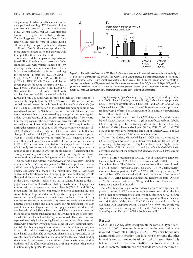
Figure 2. The facilitator effect of 10 nM CXCL12 on HVA Ca currents recorded in dopaminergic neurons of the substantia nigra inrat brain slices is prevented by 500 nM CGP 55845. A, HVA calcium current recorded in a dopaminergic neuron in response to avoltage step from �60 to �10 mV in the absence (control) or the presence of 10 nM CXCL12. Calcium currents were isolated by thecontinuous perfusion of 1 �M TTX, 4-aminopyridine, 10 mM tetraethylammonium, 2 mM CsCl, 50 �M APV, 10 �M CNQX, and 1 �M
gabazine. B, The effect of 10 nM CXCL12 on HVA Ca currents was significantly blocked by the CXCR4 antagonist AMD3100 (AMD, 200nM) and by 500 nM CGP 55845, the GABAB receptor antagonist (applied in a different set of neurons).
11646 • J. Neurosci., July 10, 2013 • 33(28):11643–11654 Guyon et al. • GABABR Agonists and Antagonists Interact with CXCR4

gands specifically bind to CXCR4 and quantify the binding affin-ity for these interactions.
Chemotactic assaysThere is a great deal of interest in stem cell therapy for the treat-ment of neurological disease (Peterson, 2004). It has previouslybeen reported that baclofen inhibited CXCL12-induced migra-tion of human peripheral blood mononuclear cells and suggestedthat the mechanism for this was heterologous desensitization ofseveral chemokine receptors (Duthey et al., 2010). In anotherreport, CXCL12-induced migration of hematopoietic stem andprogenitor cells was also blocked by GABA and baclofen, througha suppression of the CXCL12-induced calcium (Seidel et al.,2007). GABA has been shown to inhibit the migration of cancercells, although the mechanisms underlying these effects remainelusive (Ortega, 2003; Entschladen et al., 2004). To explore fur-ther the action of GABA and baclofen on tumoral cell migrationinduced by CXCL12, we used the human breast adenocarcinomacell line MDA-MB-231, which has previously been characterizedto express the CXCR4 receptor and to migrate toward CXCL12 ina migration assay (Vinader et al., 2011). Our RT-PCR and West-ern blots results are illustrated here (Fig. 1A,B) showing that bothCXCR4 RNA and protein and GABAB receptors are endoge-nously expressed in these cells. An agarose gel chemotaxis assaywas used to quantify cell migration as outlined in the Materialsand Methods section (Fig. 1C). As proof of concept, photo-graphic snapshots of the migration of cells under the agarosespots as controls (minus CXCL12), plus CXCL12, and plusCXCL12 and pre-incubation with the GABA antagonist CGP55845 are shown in Figure 1D. It should be noted that at theconcentration of antagonists used, the cells remained viable for
the duration of the experiment (as attested by their morphology),except for CGP 54626 (5 �M), which was thus excluded from thestudy. Preliminary experiments to optimize the assay found thata concentration of CXCL12 (100 nM) gave a 10-fold increase inmigration over basal, thus further experiments were performedwith this concentration (Fig. 1E). Pre-incubation with CXCR4antagonist AMD3100 and gp120/CD4 significantly inhibited theCXCL12-induced migration by, respectively, 48 and 51% but notback to basal levels. A similar inhibition of cell migration wasmeasured when cells were pre-incubated with GABAB receptorantagonist (CGP 55845) and agonists (baclofen and GABA).These results suggest that there may be interaction betweenCXCR4 and GABAB agents.
Brain slices experimentsWe previously reported patch-clamp whole-cell experimentsshowing that CXCR4 stimulation by CXCL12 chemokine posi-tively modulates HVA calcium currents in dopaminergic neu-rons in acute brain slices from rat substantia nigra (Guyon et al.,2008). HVA calcium currents (Fig. 2A) were prevented by 200 nM
AMD3100 (Fig. 2B), as previously described (Guyon et al., 2008).We investigated whether the CXCL12 HVA calcium currents(Fig. 2A) were also prevented by 500 nM 55845, which was signif-icantly reducing the chemotactism induced by CXCL12 onMDA-MB-231 cells. In whole-cell patch-clamped dopamineneurons, 500 nM CGP 55845 significantly blocked the potentiat-ing CXCL12 effect on HVA calcium currents (Fig. 2B), suggestingthat the effect of CGP 55845 also interfered with CXCR4 in mam-malian neurons, and that the use of GABAB pharmacologicalagents could interfere with CXCR4 receptors in the brain. GABAB
agonists could not be tested here because they depress HVA Ca
Figure 3. Electrophysiological recordings of oocytes expressing CXCR4, GIRK1, and GIRK2. A, Inward currents recorded in response to various concentrations of CXCL12 as indicated by the bars.B, Concentration–response curve of CXCL12. Currents were measured at the peak and plotted against log[CXCL12]. Values are the mean of several experiments as indicated by the numbers inparenthesis. The curve was fitted to a sigmoid curve using MicroCal Origin software, with EC50 of 0.49 � 0.02 nM and a Hill number of 1.87. C, Current–voltage relationship obtained in control, in thepresence of 1 nM CXCL12 and in the presence of 1 nM CXCL12 � 100 �M Ba 2�. D, Current–voltage relationship of CXCL12-evoked current (current recorded in 1 nM CXCL12 minus control current).E, The inward current activated by 1 nM CXCL12 was blocked by 100 nM AMD3100, which induced a small inward current by itself. The response to 1 nM CXCL12 partially recovered after washout ofAMD3100. F, The inward current activated by 1 nM CXCL12 was blocked by 30 nM gp120 applied together with 30 nM CD4, which induced a small inward current by itself. The response to 1 nM CXCL12partially recovered after washout of gp120/CD4.
Guyon et al. • GABABR Agonists and Antagonists Interact with CXCR4 J. Neurosci., July 10, 2013 • 33(28):11643–11654 • 11647

currents by stimulating GABAB receptorsendogenously expressed on neurons(Guyon and Leresche, 1995).
Therefore, as shown in the two exam-ples above, agents supposed to act onGABAB receptor also affect the CXCR4system. We investigated the possibilitythat GABAB agents could also interactwith CXCR4 receptor. To test this hy-pothesis, we first used a heterologousmodel of expression of wild-type CXCR4in Xenopus oocytes that lack GABAB re-ceptors (Uezono et al., 1998) or expressthem at low levels in a small subpopula-tion of cells (Yang et al., 2001).
Heterologous expression of CXCR4 onXenopus oocytesWe performed electrophysiology experi-ments on Xenopus oocytes expressingwild-type CXCR4 chemokine receptor to-gether with the GIRK channels (GIRK1and GIRK2) as a reporter of CXCR4 activ-ity. Activation by the CXCR4 ligandCXCL12 in high K� Ringer’s solution in-duced an inward current at holdingpotentials of �30 mV (Fig. 3A). The am-plitude of the current response dependedon the concentration of CXCL12, saturat-ing at �1 nM (n � 11), with half-maximalstimulation at 0.49 � 0.02 nM and Hillcoefficient of 1.87 (Fig. 3B).
To verify that the currents induced bychemokine perfusion resulted from theactivation of GIRK channels, we charac-terized the properties of these currents.The voltage dependence of the currentshowed a strong inward rectification, asexpected for GIRK (Fig. 3 C,D), and thecurrent showed a strong inhibition byBa2�, also consistent with GIRK (Fig. 3C).The selective CXCR4 antagonist AMD3100antagonizes the CXCL12-elicited GIRK cur-rent (Fig. 3E). AMD3100 (200 nM) induceda 76.67 � 3.12% decrease (n � 4, from2.15 � 0.49 to 0.52 � 0.16 �A, p � 0.02) in the current elicited by 1nM CXCL12, confirming that the chemokine activates GIRKthrough the CXCR4 receptor. It should be noted that AMD3100sometimes induced an inward current by itself (see Fig. 5E), butalways prevented the effect of CXCL12. We also tested anotherCXCR4 antagonist, the recombinant protein HIV-1 IIIB gp120 (co-applied with recombinant CD4 as described previously; Tran et al.,2005). As illustrated in Figure 3 F, the gp120/CD4 complex antago-nized the effects of CXCL12 by 80.5 � 11.4% (n � 10, from 1.86 �0.28 to 0.59 � 0.28 �A, p � 0.02), accompanied by a small directinduction of inward current (15.8 � 5.2%, n � 6, of the currentinduced by 1 nM CXCL12), as previously described (Madani et al.,1998). No response to CXCL12, AMD3100, or gp120/CD4 was ob-served in water-injected oocytes (n � 5) or in oocytes expressingonly the GIRK channels (GIRK1 and GIRK2) (n � 5; data notshown). Thus, we conclude that the genuine CXCR4 functionallyactivates the GIRK channel in oocytes.
Compounds acting on GABAB receptor act directlyon CXCR4We then tested the hypothesis that compounds acting on GABAB
receptor could act directly on CXCR4. Both GABAB antagonistsCGP 55845 and CGP 54626, baclofen, and GABA were inactiveon oocytes injected only with GIRK1 and GIRK2 (n � 3 each;data not shown), suggesting the lack of functional expression ofendogenous GABAB receptors on the batches of oocytes that weused (Yang et al., 2001). Surprisingly, in oocytes coexpressingCXCR4 and the G-protein-coupled GIRK channels (GIRK1 andGIRK2), both GABAB receptor antagonists CGP 55845 and CGP54626 induced an inward current similar to the one induced byCXCL12 (n � 30 and 16, respectively for CGP 55845 and 54626;Figs. 4, 5A1,B1). The effect of both GABAB receptor antagonistswas dose dependent (Fig. 4). The EC50s (see Table 1) were, re-spectively, 1.87 and 61.02 nM with Hill numbers of 1. Similarly,the GABAB receptor agonists baclofen and GABA were both ableto reversibly activate inward currents in a dose-dependent man-
0,0
0,2
0,4
0,6
0,8
1,0
Mea
n N
orm
aliz
ed C
urre
nt A
mpl
itude
Concentration (nM)0.01 0.1 1 10 100 1000 10000 100000
Bac
lofe
nG
AB
AC
GP
5584
5C
GP
5462
6
1 mM 10 mM
1 min
0.1 µA
50 mM 5 mM
1 mM 10 mM 50 mM
0.1 µA
0.1 µA
1 min
1 nM Mn 001 Mn 01 50 nM
1 min
100 nM 500 nM50 nM 10 nM
0.2 µA
1 min
CXCL12CGP 55845CGP54626BaclofenGABA
A
B
Figure 4. Dose-dependent effect on CXCR4 receptor of several pharmacological agents acting on GABAB receptor. A, Inwardcurrents recorded in oocytes expressing CXCR4, GIRK1, and GIRK2 in response to the drugs indicated on the left, at variousconcentrations as indicated by the bars. B, Concentration–response curves of several pharmacological agents acting on GABAB
receptor as compared with CXCL12. Currents were measured at the peak, normalized to the maximum response, and plottedagainst log[CXCL12]. Values are the mean of several experiments, which number (n) is indicated in Table 1. The curves were fittedto sigmoid curves using MicroCal Origin software. EC50 and a Hill number are also indicated in Table 1 (mean � SEM).
11648 • J. Neurosci., July 10, 2013 • 33(28):11643–11654 Guyon et al. • GABABR Agonists and Antagonists Interact with CXCR4
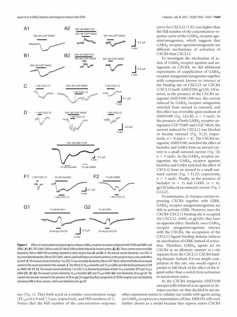
ner (Fig. 4). They both acted in a similar concentration range(EC50s of 6.9 and 7.5 �M, respectively, and Hill numbers of 1).Notice that the Hill number of the concentration–response
curve for CXCL12 (1.92) was higher thanthe Hill number of the concentration–re-sponse curve of the GABAB receptor ago-nists/antagonists, which suggests thatGABAB receptor agonists/antagonists usedifferent mechanisms of activation ofCXCR4 than CXCL12.
To investigate the mechanism of ac-tion of GABAB receptor agonists and an-tagonists on CXCR4, we did additionalexperiments of coapplication of GABAB
receptor antagonists/antagonists togetherwith compounds known to interact atthe binding site of CXCL12 on CXCR4(CXCL12 itself, AMD3100, gp120). Of in-terest, in the presence of the CXCR4 an-tagonist AMD3100 (200 nM), the currentinduced by GABAB receptor antagonistsswitched from inward to outward, andthis effect was reversible upon washout ofAMD3100 (Fig. 5A2,B2; n � 3 each). Inthe presence of both GABAB receptor an-tagonists CGP 55485 and CGP 54626, thecurrent induced by CXCL12 was blockedor became outward (Fig. 5C,D, respec-tively, n � 8 and n � 4). The CXCR4 an-tagonist, AMD3100, switched the effect ofbaclofen and GABA from an inward cur-rent to a small outward current (Fig. 5E;n � 3 each). As the GABAB receptor an-tagonists, the GABAB receptor agonistsbaclofen and GABA switched the effect ofCXCL12 from an inward to a small out-ward current (Fig. 5 F1,F2, respectively;n � 5 each). Finally, in the presence ofbaclofen (n � 3) and GABA (n � 6),gp120 induced an outward current (Fig. 5G1,G2).
To summarize, in Xenopus oocytes ex-pressing CXCR4 together with GIRK,GABAB receptor antagonists/agonists areable to activate GIRK. However, once theCXCR4-CXCL12 binding site is occupied(by CXCL12, AMD, or gp120), they havean opposite effect. Similarly, once GABAB
receptor antagonists/agonists interactwith the CXCR4, the occupation of theCXCL12 ligand-binding domain inducesan inactivation of GIRK instead of activa-tion. Therefore, GABAB agents act onCXCR4 in an allosteric manner at a siteseparate from the CXCL12-CXCR4 bind-ing domain. Indeed, if it was simple com-petition at this site, one would expect apartial or full block of the effect of the li-gand rather than a switch from activationto inactivation states.
As the CXCR4 antagonist AMD3100unexpectedly behaved as an agonist in Xe-nopus oocytes, we thus decided to use an-
other expression system to validate our results with agents actingon GABAB receptors in a mammalian cell line. HEK293 cells werefurther chosen as a model because they express native CXCR4
200 nM AMD 3100
30 nM gp120/CD4 30 nM gp120/CD4
F1
G1
1 nM CXCL121 nM CXCL12
1 nM CXCL12
500 nM CGP 55845
1 nM CXCL12 1 nM CXCL12
1 nM CXCL12
500 nM CGP 54626
200 nM AMD3100500 nM CGP 54626 500 nM CGP 54626
200 nM AMD3100
500 nM CGP 55845500 nM CGP 55845
B1
C
B2
D
500 nM CGP 54626
500 nM CGP 55845
F2
G2
10 µM baclofen 10 µM GABA
ABAG Mµ01nefolcab Mµ01
1 nM CXCL12 1 nM CXCL12
1 nM CXCL12
0.2 µA
0.1 µA
0.2 µA
0.2 µA
0.1 µA
0.1 µA
0.2 µA
2 min
1 min
1 min
1 min
1 min
1 min
1 min
A1 A2
Figure 5. Effects of various pharmacological agents acting on GABAB receptors on oocytes coinjected with CXCR4 and GIRK1 andGIRK2. A1, B1, CGP 55845 (500 nM) and CGP 54626 (500 nM) both induced an inward current. A2, B2, These currents were reversiblyblocked by 200 nM AMD3100 recordings obtained in other oocytes than A1 and B1. C, The inward current elicited by 1 nM CXCL12was reversibly blocked by 500 nM CGP 55845, which could itself induce an inward current as in the oocyte or have a very small effectby itself. D, The inward current elicited by 1 nM CXCL12 was reversibly blocked by 500 nM CGP 54626, which itself induced an inwardcurrent in the oocyte presented in this example. E, The effects of 10 �M baclofen and 10 �M GABA were blocked by perfusion of 200nM AMD3100. F1, F2, The inward current elicited by 1 nM CXCL12 as blocked by perfusion of both 10 �M baclofen (F1) and 10 �M
GABA (F2). G1, G2, The inward currents elicited by 10 �M baclofen (G1) and 10 �M GABA (G2) were blocked by 30 nM gp120. Thecurrent even became outward in the presence of 30 nM gp120 suggesting that a proportion of CXCR4 receptors was constitutivelyactivating GIRK in these oocytes, which was blocked by the gp120.
Guyon et al. • GABABR Agonists and Antagonists Interact with CXCR4 J. Neurosci., July 10, 2013 • 33(28):11643–11654 • 11649

although at very low levels (Busillo et al., 2010) and because aligand-binding assay is available in this cell line.
GABAB agents also interact with CXCR4 expressed inHEK293 cellsWhole-cell patch-clamp recordings were then performed inHEK293 cells transiently expressing CXCR4, GIRK1-F137S, andYFP and superfused with an extracellular solution containingraised [K�]. In cells transfected with CXCR4 and GIRK1-F137S,CXCL12 (1 nM) evoked currents that displayed a high degree ofinward rectification (Fig. 6 A, B). Barium (200 �M), a blockerof Kir channels, abolished the CXCL12-evoked current (Fig-ure 6A; n � 5).
The reversal potential for the CXCL12-evoked current was�23.4 � 4.6 mV (n � 5), which agrees closely with the calcu-lated equilibrium potential for K� of �25 mV under these re-cording conditions. At a holding potential of �60 mV, themaximum current evoked by 1 nM CXCL12 was �383.8 � 63.9pA (n � 42). This current was fully and reversibly blocked by 200nM AMD3100 (n � 5), which induced an outward current of�230 � 86 pA by itself (n � 5), suggesting that some CXCR4 wasconstitutively active. gp120/CD4 (3 nM each) also blocked 70.0 �10.4% (n � 7) of the current induced by 1 nm CXCL12. Interest-ingly, CXCL12-induced current was also significantly blocked by500 nM CGP 55 845 and CGP 54 626 (Fig. 6 C,E). These GABAB
antagonists had no effect when applied alone. Similarly, GABAand to a lesser extent baclofen also significantly blocked theCXCL12-induced current (Fig. 6D,E) while these GABAB recep-tor agonists had no effect when applied alone.
When transfected with GIRK� and YFP without CXCR4,some transfected HEK293 cells (6/17 tested) exhibited a smallinward current of �79.2 � 24.5 pA (n � 6) at �60 mV in re-sponse to CXCL12 (1 nM). No current was detected in response toAMD, gp120/CD4, CGP 55845, or CGP 54626 (n � 9 each). Onlya small amount of HEK293 cells transfected with GIRK only (2/7cells) presented a small inward current in response to baclofen(10 �M) or GABA (10 �M; between �25 and �75 pA) suggestingthat low amounts of GABAB receptors were present endoge-nously only in a subpopulation of HEK293 cells, as confirmed bythe RT-PCR or Western blot experiments that exhibited a smallband for both human GABABR2 and CXCR4 receptor (Fig.1A,B).
Baclofen and other GABAB agents directly interact withCXCR4 expressed in lipoparticlesTo substantiate the data above that GABAB agents are directlybinding the CXCR4 receptor, we used BSI, a molecular interac-tion photometer, to quantify ligand/receptor binding. We previ-ously reported binding specificity of the native pairs CXCR4/CXCL12 binding using red blood cell “ghosts” with humanSUP-T1 lymphoma T-cells expressing CXCR4 and BSI (Baksh etal., 2011). Here, we expressed the CXCR4 receptor using lipopar-
ticle technology: protein directly incorporated into virus-likeparticles with a lipid bilayer surface that provides concentratedprotein (50 –200 pM/mg) in the native conformation (Jones et al.,2008). Equilibrium dissociation constants were calculated usingsaturation analysis. To demonstrate that the refractive index (RI)change is not due simply to the introduction of the pairs, we useddopamine as a control in each experiment (at five concentrationsthat span the concentration range in all the other curves), andmeasured no RI change. In Figure 7A the saturation isothermmeasured binding of CXCL12 with a KD of 0.49 nM (� 0.16 nM),in agreement with a previously published report (Jones et al.,2008). Next, we measured binding of the two GABAB antagonistsCGP 55845 and CGP 54626 (Fig. 7B,C) calculating KD values of11 nM (�0.5 nM) and 35 nM (�6.2 nM), respectively. Finally, wemeasured binding of two GABAB agonists to CXCR4, baclofen,and GABA (Fig. 7D,E) calculating KD values of 10.3 � 2.8 �M
and 0.57 � 0.24 �M. To confirm that this binding is at a differentsite from that of CXCL12, we pre-incubated the lipoparticles withCXCL12 and performed a binding assay using baclofen. Figure 7Fshows that baclofen is still binding CXCR4 even when CXCL12 isbound and the affinity and signal magnitude for baclofen with thereceptor is enhanced by CXCL12 binding. To demonstrate thatthe observed binding was not due to CXCL12 interacting withbaclofen, the assay was performed in the absence of CXCR4, withno observed binding signal (Fig. 7F). These results are proof inprinciple that GABAB ligands directly bind CXCR4, and at a sitedistinct from the active site.
GABAB agents do not interact with CXCR4 at the CXCL12ligand binding siteTo check whether GABAB antagonists and agonists do not bindcompetitively with CXCL12, we used the Tag-lite receptor ligandtechnology, which is designed to show a binding between theligand and the receptor by a homogeneous and nonradioactiveassay on living cells (Fig. 8A). To check the ability of unlabeledGABAB ligands to bind to CXCR4 receptor, a competition assaywas first performed with the CXCR4 ligand d2-labeled and unla-beled GABAB ligands, testing a wide range of concentrationsof unlabeled compounds against a fixed concentration ofCXCL12-d2 ligand. None of the GABAB ligand was able to com-pete with the d2-labeled ligand while the d2-labeled CXCL12 wasdisplaced by the unlabeled CXCL12 with KI in accordance withexpected values (Fig. 8B). These data suggest that these ligandsdo not bind to the CXCL12-CXCR4 binding site. We alsotested whether the GABAB d2-labeled ligand (CGP 54626 de-rivative) could interact at the CXCL12 binding site of CXCR4receptors. The specific binding curve, obtained by subtractingthe nonspecific signal from the total binding signal, revealedthat the d2-labeled CGP 54626 derivative (used as a GABAB
fluorescent ligand) does not bind to the CXCR4 receptor at theCXCL12 binding pocket (Fig. 8C). Therefore, as hypothesized,GABAB agents do not interact at the binding site of CXCL12on CXCR4 but modulate the activation of the receptor byallosteric interaction.
DiscussionOur study demonstrates that GABAB agonists and antagonistsdirectly bind CXCR4 by allosteric action. This finding has impor-tant physiological implications for the immune and nervous sys-tems and is also highly relevant in pointing a novel moleculartarget to baclofen, a drug widely used in human health.
Table 1. EC50s and Hill numbers (mean � SEM) of dose-response curves obtainedby recording inward currents on oocytes expressing CXCR4, GIRK1, and GIRK2 inresponse to the different drugs indicated on the left as illustrated in Figure 4
Drug tested n EC50 Hill number
CXCL12 5 0.59 � 0.01 nM 1.92 � 0.19CGP 55845 4 1.87 � 0.12 nM 1.01 � 0.02CGP 54626 4 61.02 � 8.77 nM 1.11 � 0.09Baclofen 4 6.87 � 1.06 �M 0.98 � 0.06GABA 5 7.49 � 1.2 �M 0.97 � 0.06
11650 • J. Neurosci., July 10, 2013 • 33(28):11643–11654 Guyon et al. • GABABR Agonists and Antagonists Interact with CXCR4

Direct effect of GABAB receptor agents on CXCR4The human breast cancer cell line MDA-MB-231 expressing endog-enously CXCR4 allowed us to demonstrate the effects of bothGABAB receptor agonists and antagonists on native CXCR4 by usingthe chemotactic properties of the chemokine CXCL12. In neurons,the GABAB receptor antagonist blocked the effects of CXCL12 onHVA calcium currents. However, in neurons, it is hard to decipherthe relative effect of GABAB pharmacological agents on CXCR4 orGABAB receptor because neurons express both CXCR4 and GABAB
in similar amounts. To resolve whether these agents have a directeffect at CXCR4, we used X. laevis oocytes or HEK293 cells express-ing CXCR4, which have relatively no endogenous GABAB receptorexpression compared with the overexpressed CXCR4. These cellsallow for pharmacological studies although we acknowledge that thereceptor might behave differently in vivo. For example, we observedin TIRF experiments that CXCR4 does not form dimers when ex-
pressed in X. laevis oocytes (A. Guyon, un-published data) contrary to what isdescribed in mammalian cells. In oocytesexpressing CXCR4 receptors, GABAB ago-nists and antagonists both behaved as ago-nists as they were able to activate GIRKcurrent. This was not a direct effect on GIRKas the effect was absent in oocytes injectedwith GIRK only. These compounds actedon CXCR4 since they interfered withCXCL12 and AMD3100 for the activationof GIRK. In HEK293 cells, however, GABAB
agonists and antagonists both blocked theCXCL12-induced current without inducingany currents by themselves. Therefore, it islikely that in mammalian cells (neurons andimmune cells), GABAB receptor agents be-have as antagonists on CXCR4, and therebyblock the effects of CXCL12.
Similarly, AMD3100 behaved as an ago-nist in the Xenopus oocyte experiments.AMD3100 has previously been described tobehave as a partial agonist and has shownsome agonistic effects in other models(Zhang et al., 2002; Tran et al., 2005). Suchpartial agonist activity precludes the use ofthis agent as an antagonist of metastatic ac-tivity in mammalian carcinoma becausestimulation of CXCR4 at the surface of tu-mor cells could theoretically increase dis-semination. However, AMD3100 did notbehave as an agonist in HEK293 cells tran-siently transfected with CXCR4 and GIRK.Contradictorily, it induced an outward cur-rent, likely due to blocking the activity oftonically activated CXCR4. Similarly, in thechemotactic assay, AMD3100 blocked themigration of MDA-MB-231 cells toward theagarose drop containing CXCL12. Thus,AMD3100 behaves differently dependingon the cell type expressing CXCR4. Thiscould be due to distinct intracellular path-ways, to a different folding or conformationof the protein, or to heterologous dimeriza-tion depending on the tested model.
The BSI assays showed a direct interac-tion of GABAB receptor antagonists and
agonists with CXCR4, with KD values that are in the range of ourelectrophysiological results. The Tag-lite competition ligand-binding assay demonstrated that GABAB receptor antagonists/agonists do not interact at the ligand-binding domain forCXCL12 on CXCR4. This result is corroborated by the differencein Hill numbers observed in the concentration–response curvesbetween CXCL12 on one side (close to 2) and the GABAB agentson the other side (Hill number close to 1). This difference indi-cates a separate mode of action. It is hard to speculate on the siteand mechanisms of interaction of GABAB agents on CXCR4 andthese questions will be the topic of future studies. Using Tag-litetechnology, we found no fluorescent resonance energy transferinteraction between the CXCR4 terbium cryptate, fixed at theCXCL12 binding site, and the GABAB receptor-labeled ligands.Forster’s radius (R0, distance equivalent to 50% transfer effi-ciency) of 9 nm CXCR4 terbium cryptate-GABAB d2-labeled li-
Figure 6. Effects of various pharmacological agents acting on GABAB receptors on patch-clamp recordings from HEK293 cellstransiently expressing CXCR4 and GIRK channels. A, Current–voltage relationship in control conditions (leak current in the presenceof the extracellular solution with elevated KCl), in the presence of CXCL12, or in the presence of CXCL12 � 200 �M of the GIRKchannel blocker BaCl2, as indicated. B, CXCL12-induced current. Leak current has been subtracted. CXCL12 evoked an inwardlyrectifying current that had a reversal potential of �24.6 mV. C, Representative traces showing the effect of 1 nM CXCL12 before,during, and after the washout of 500 nM CGP 55845, as indicated by the bars. Cells were held at �25 mV and then stepped to �60mV for 100 ms every 2 s (see Materials and Methods). D, Representative traces showing the effect of 1 nM CXCL12 before, during,and after the washout of 10 mM GABA, as indicated by the bars. E, Histogram showing the percentage of decrease induced by thevarious compounds indicated on the GIRK current induced by 1 nM CXCL12 in HEK293 cells transiently expressing the CXCR4 andGIRK channel.
Guyon et al. • GABABR Agonists and Antagonists Interact with CXCR4 J. Neurosci., July 10, 2013 • 33(28):11643–11654 • 11651

gand pair, indicates that the allosteric site of GABAB ligands isthus far from the CXCL12 binding pocket on CXCR4. Anotherway to test this would be using the crystal structure of the receptorin the presence of the GABAB receptor antagonists/agonists toallow determining where these compounds interact on thechemokine receptor. Silencing CXCR4 expression throughsiRNA technology would also provide some clue to the relevanceof GABA–CXCR4 interactions in mammalian cells and whether aheterologous dimerization may be occurring. Other mechanismsof interactions between GABAB and CXCR4 are currently underinvestigation, and will address both direct interaction and activa-tion of second messenger cascade.
Physiological implicationThis study shows that GABA is able to block the effect of CXCL12on CXCR4. Thus, it is likely that when the GABAergic system isactivated, GABA released in the brain will antagonize the effect ofCXCL12 on its receptor CXCR4, and thus could influence thechemokine neurotransmission as well as the inflammatory re-sponse in the CNS. We now demonstrate that there is reciprocalcross talk between these two systems as it has previously beenshown that CXCR4 stimulation by CXCL12 can increase presyn-aptic GABA release (Guyon and Nahon, 2007).
Indeed, in dopaminergic neurons of the rat substantia nigra,we have previously shown that CXCR4 stimulation by CXCL12induces an increase of release of presynaptic neurotransmitter,particularly of GABA (Guyon et al., 2006). We have also shownthat CGP 55845 (500 nM) blocks the outward GIRK current in-
duced by CXCL12 (recorded in the presence of glutamate recep-tor blockers). At that time, we interpreted this result as an effectmediated through GABAB receptor stimulation by GABA spillingover following CXCL12 presynaptic stimulation and increase inGABAB release. However, in view of the present results, we canre-interpret our data. Indeed, the GIRK currents might have beenactivated by the stimulation of postsynaptic CXCR4 by CXCL12,which was then blocked by CGP 55484.
CXCR4 activation by CXCL12 has been shown to increasepresynaptic neurotransmitter release and particularly GABA re-lease in several neuronal populations (Guyon and Nahon, 2007;Bhattacharyya et al., 2008; Qu et al., 2008). If GABA can in turnblock the effects of CXCL12, this could represent a negative feed-back loop for presynaptic chemokine release. Indeed, when ap-plying CXCL12 for several minutes, a transient increase in thefrequency of spontaneous postsynaptic currents is frequently ob-served, followed by a reduced activity (Guyon et al., 2006, theirFigure 3). This reduction could be due to an antagonistic effect ofGABA, although desensitization of CXCR4 itself cannot be ex-cluded. Similarly, it has been shown that elevated concentrationsof CXCL12 exert more opposite effect than lower concentrationson the electrical activity of some neuronal populations that re-ceive GABA inputs (Guyon and Nahon, 2007). The antagonisticeffect of GABA released presynaptically in response to CXCL12could contribute to these biphasic effects. In the future, it will beof interest to search for putative effects of GABAB receptor li-gands on CXCR7, the other receptor for CXCL12 (Schonemeieret al., 2008), as well as on other chemokine receptors.
Figure 7. Representative plots of BSI signal versus ligand concentration for the determination of binding constants for CXCR4 to the following ligands: CXCL12 (A), CGP 55845 (B), CGP 54626 (C),baclofen (D), and GABA (E). Dopamine (tested at 5 different concentrations: 0.02, 2, 200, 2, and 200 nM) was used as a nonbinding control ligand for all five plots. The binding of baclofen to theCXCR4 –CXCL12 complex (0.2 nM CXCR4 � 20 nM CXCL12) is represented in F; also shown is the negative control of the interaction of baclofen with CXCL12. For all plots, error bars indicate SDs of themeasurements from three independent trials.
11652 • J. Neurosci., July 10, 2013 • 33(28):11643–11654 Guyon et al. • GABABR Agonists and Antagonists Interact with CXCR4

Putative applications in cancer treatment and inflammationBaclofen treatment was demonstrated to reduce the incidence ofsome carcinogen-induced gastrointestinal cancers in rats (Tat-suta et al., 1990) as well as human hepatocarcinoma cell growth(Wang et al., 2008). In contrast, baclofen promotes human pros-tate cancer cell migration (Azuma et al., 2003). As CXCR4 ishighly expressed in cancer cells, baclofen may have been actingthrough CXCR4 in these examples.
Similarly, it has been shown that GABA can affect cell prolifera-tion and have anti-inflammatory properties on fibroblasts, althoughthe mechanism of action of GABA was not elucidated (Han et al.,2007). We suggest that GABA may have acted through the CXCR4receptor, as CXCR4 is expressed on fibroblasts (Qu et al., 2008).Baclofen is currently used for the treatment of spasticity in patientswith spinal cord injury, cerebral palsy, traumatic brain injury, mul-tiple sclerosis, and other disorders (Plassat et al., 2004; Guglani andLodha, 2007; Kolaski and Logan, 2008; Rekand and Grønning,2011). Recently, it has been used in the treatment of alcohol depen-dence and withdrawal (Addolorato et al., 2006). The allosteric effectsof such agents at CXCR4 likely contribute to these beneficial effectsas CXCR4 often colocalizes with GABAB receptors.
As a conclusion, this study opens new perspectives on theputative use of baclofen and other GABAB agents acting atCXCR4 for their therapeutic potential to treat quite a broad rangeof diseases, such as ischemic stroke, brain tumors, HIV enceph-alopathy, and multiple sclerosis, as well as affecting stem cellmigration (Duthey et al., 2010) and other cancers.
ReferencesAddolorato G, Leggio L, Agabio R, Colombo G, Gasbarrini G (2006)
Baclofen: a new drug for the treatment of alcohol dependence. Int J ClinPract 60:1003–1008. CrossRef Medline
Azuma H, Inamoto T, Sakamoto T, Kiyama S, Ubai T, Shinohara Y, Maemura
K, Tsuji M, Segawa N, Masuda H, Takahara K, Katsuoka Y, Watanabe M(2003) Gamma-aminobutyric acid as a promoting factor of cancer me-tastasis; induction of matrix metalloproteinase production is potentiallyits underlying mechanism. Cancer Res 63:8090 – 8096. Medline
Baksh MM, Kussrow AK, Mileni M, Finn MG, Bornhop DJ (2011) Label-free quantification of membrane-ligand interactions using backscatteringinterferometry. Nat Biotechnol 29:357–360. CrossRef Medline
Banisadr G, Fontanges P, Haour F, Kitabgi P, Rostene W, Melik Parsadaniantz S(2002) Neuroanatomical distribution of CXCR4 in adult rat brain and its local-ization in cholinergic and dopaminergic neurons. Eur J Neurosci 16:1661–1671.CrossRef Medline
Bhattacharyya BJ, Banisadr G, Jung H, Ren D, Cronshaw DG, Zou Y, Miller RJ(2008) The chemokine stromal cell-derived factor-1 regulates GABAergicinputs to neural progenitors in the postnatal dentate gyrus. J Neurosci 28:6720–6730. CrossRef Medline
Bonavia R, Bajetto A, Barbero S, Pirani P, Florio T, Schettini G (2003) Chemokinesand their receptors in the CNS: expression of CXCL12/SDF-1 and CXCR4 andtheir role in astrocyte proliferation. Toxicol Lett 139:181–189. CrossRef Medline
Bowery NG (1993) GABAB receptor pharmacology. Annu Rev PharmacolToxicol 33:109 –147. CrossRef Medline
Busillo JM, Benovic JL (2007) Regulation of CXCR4 signaling. Biochim Bio-phys Acta 1768:952–963. CrossRef Medline
Busillo JM, Armando S, Sengupta R, Meucci O, Bouvier M, Benovic JL(2010) Site-specific phosphorylation of CXCR4 is dynamically regulatedby multiple kinases and results in differential modulation of CXCR4 sig-naling. J Biol Chem 285:7805–7817. CrossRef Medline
Carbajal KS, Schaumburg C, Strieter R, Kane J, Lane TE (2010) Migration ofengrafted neural stem cells is mediated by CXCL12 signaling through CXCR4in a viral model of multiple sclerosis. Proc Natl Acad Sci U S A 107:11068–11073. CrossRef Medline
Choi WT, An J (2011) Biology and clinical relevance of chemokines andchemokine receptors CXCR4 and CCR5 in human diseases. Exp Biol Med236:637– 647. CrossRef Medline
Comerford I, McColl SR (2011) Mini-review series: focus on chemokines.Immunol Cell Biol 89:183–184. CrossRef Medline
Doranz BJ, Berson JF, Rucker J, Doms RW (1997) Chemokine receptors as
Figure 8. Baclofen and other ligands of GABAB receptor do not bind at the CXCL12/SDF1 binding site on CXCR4. A, Principle of the Tag-lite receptor-ligand technology. The receptor is expressedat the cell surface with a SNAP tag and then labeled with a fluorescent donor dye (terbium cryptate) through an appropriate substrate. On the other side, the ligand is labeled with a red acceptor dye(d2). If the fluorescent donor dye labeled on the receptor is excited by a nitrogen laser or flash lamp (�340 nm) when the ligand binds to the receptor, there is a transfer of energy between the donordye to the acceptor dye, resulting in the latter dye emitting light, in a time-resolved manner, at 665 nm. The receptor-ligand binding can hence be monitored at 665 nm on a time-resolved mode.B, Competition assay run with d2-labeled CXCL12 at KD (12.5 nM). None of the unlabeled GABAB ligand was able to compete with the d2-labeled ligand while the d2-labeled CXCL12 was displacedby the unlabeled CXCL12 as expected. C, The specific binding curve was obtained by subtracting the nonspecific signal from the total binding signal. It revealed that the d2-labeled CGP 54626derivative (used as a GABAB fluorescent ligand) does not bind to the CXCR4 receptor at the CXCL12 binding pocket.
Guyon et al. • GABABR Agonists and Antagonists Interact with CXCR4 J. Neurosci., July 10, 2013 • 33(28):11643–11654 • 11653

fusion cofactors for human immunodeficiency virus type 1 (HIV-1). Im-munol Res 16:15–28. CrossRef Medline
Duthey B, Hubner A, Diehl S, Boehncke S, Pfeffer J, Boehncke WH (2010)Anti-inflammatory effects of the GABA(B) receptor agonist baclofen inallergic contact dermatitis. Exp Dermatol 19:661– 666. CrossRef Medline
Entschladen F, Drell TL 4th, Lang K, Joseph J, Zaenker KS (2004) Tumour-cell migration, invasion, and metastasis: navigation by neurotransmitters.Lancet Oncol 5:254 –258. CrossRef Medline
Froestl W (2010) Chemistry and pharmacology of GABAB receptor ligands.Adv Pharmacol 58:19 – 62. CrossRef Medline
Gabuzda D, Wang J (2000) Chemokine receptors and mechanisms of celldeath in HIV neuropathogenesis. J Neurovirol 6 [Suppl 1]:S24 –S32.Medline
Gao Z, Wang X, Wu K, Zhao Y, Hu G (2010) Pancreatic stellate cells in-crease the invasion of human pancreatic cancer cells through the stromalcell-derived factor-1/CXCR4 axis. Pancreatology 10:186 –193. CrossRefMedline
Guglani L, Lodha R (2007) Enteral baclofen in the management of tetanus-relatedspasms: case report and review of literature. J Trop Pediatr 53:139–141. Medline
Guyon A, Leresche N (1995) Modulation by different GABAB receptortypes of voltage-activated calcium currents in rat thalamocortical neu-rones. J Physiol 485:29 – 42. Medline
Guyon A, Nahon JL (2007) Multiple actions of the chemokine stromal cell-derived factor-1alpha on neuronal activity. J Mol Endocrinol 38:365–376.CrossRef Medline
Guyon A, Skrzydelsi D, Rovere C, Rostene W, Parsadaniantz SM, Nahon JL(2006) Stromal cell-derived factor-1alpha modulation of the excitabilityof rat substantia nigra dopaminergic neurones: presynaptic mechanisms.J Neurochem 96:1540 –1550. CrossRef Medline
Guyon A, Skrzydelski D, Rovere C, Apartis E, Rostene W, Kitabgi P, MelikParsadaniantz S, Nahon JL (2008) Stromal-cell-derived factor 1alpha/CXCL12 modulates high-threshold calcium currents in rat substantianigra. Eur J Neurosci 28:862– 870. CrossRef Medline
Han D, Kim HY, Lee HJ, Shim I, Hahm DH (2007) Wound healing activityof gamma-aminobutyric Acid (GABA) in rats. J Microbiol Biotechnol17:1661–1669. Medline
Johnson EA, Oldfield S, Braksator E, Gonzalez-Cuello A, Couch D, Hall KJ,Mundell SJ, Bailey CP, Kelly E, Henderson G (2006) Agonist-selectivemechanisms of mu-opioid receptor desensitization in human embryonickidney 293 cells. Mol Pharmacol 70:676 – 685. CrossRef Medline
Jones JW, Greene TA, Grygon CA, Doranz BJ, Brown MP (2008) Cell-freeassay of G-protein-coupled receptors using fluorescence polarization.J Biomol Screen 13:424 – 429. CrossRef Medline
Juarez J, Bendall L, Bradstock K (2004) Chemokines and their receptors astherapeutic targets: the role of the SDF-1/CXCR4 axis. Curr Pharm Des10:1245–1259. CrossRef Medline
Kaupmann K, Malitschek B, Schuler V, Heid J, Froestl W, Beck P, MosbacherJ, Bischoff S, Kulik A, Shigemoto R, Karschin A, Bettler B (1998)GABA(B)-receptor subtypes assemble into functional heteromeric com-plexes. Nature 396:683– 687. CrossRef Medline
Kolaski K, Logan LR (2008) Intrathecal baclofen in cerebral palsy: a decadeof treatment outcomes. J Pediatr Rehabil Med 1:3–32. Medline
Laviv T, Vertkin I, Berdichevsky Y, Fogel H, Riven I, Bettler B, Slesinger PA,Slutsky I (2011) Compartmentalization of the GABAB receptor signal-ing complex is required for presynaptic inhibition at hippocampal syn-apses. J Neurosci 31:12523–12532. CrossRef Medline
Lazarini F, Tham TN, Casanova P, Arenzana-Seisdedos F, Dubois-Dalcq M(2003) Role of the alpha-chemokine stromal cell-derived factor (SDF-1)in the developing and mature CNS. Glia 42:139 –148. CrossRef Medline
Li M, Ransohoff RM (2008) Multiple roles of chemokine CXCL12 in thecentral nervous system: a migration from immunology to neurobiology.Prog Neurobiol 84:116 –131. CrossRef Medline
Liu YL, Yu JM, Song XR, Wang XW, Xing LG, Gao BB (2006) Regulation ofthe chemokine receptor CXCR4 and metastasis by hypoxia-inducible fac-tor in non small cell lung cancer cell lines. Cancer Biol Ther 5:1320 –1326.CrossRef Medline
Madani N, Kozak SL, Kavanaugh MP, Kabat D (1998) gp120 envelope gly-coproteins of human immunodeficiency viruses competitively antago-nize signaling by coreceptors CXCR4 and CCR5. Proc Natl Acad Sci U S A95:8005– 8010. CrossRef Medline
Ortega A (2003) A new role for GABA: inhibition of tumor cell migration.Trends Pharmacol Sci 24:151–154. CrossRef Medline
Osawa Y, Xu D, Sternberg D, Sonnett JR, D’Armiento J, Panettieri RA, EmalaCW (2006) Functional expression of the GABAB receptor in human air-way smooth muscle. Am J Physiol Lung Cell Mol Physiol 291:L923–L931.CrossRef Medline
Peterson DA (2004) Stem cell therapy for neurological disease and injury.Panminerva Med 46:75– 80. Medline
Plassat R, Perrouin Verbe B, Menei P, Menegalli D, Mathe JF, Richard I(2004) Treatment of spasticity with intrathecal Baclofen administration:long-term follow-up, review of 40 patients. Spinal Cord 42:686 – 693.CrossRef Medline
Qu Y, Mao M, Li X, Zhang L, Huang X, Yang C, Zhao F, Xiong Y, Mu D(2008) Enhanced migration and CXCR4 over-expression in fibroblastswith telomerase reconstitution. Mol Cell Biochem 313:45–52. CrossRefMedline
Rane MJ, Gozal D, Butt W, Gozal E, Pierce WM Jr, Guo SZ, Wu R, GoldbartAD, Thongboonkerd V, McLeish KR, Klein JB (2005) Gamma-aminobutyric acid type B receptors stimulate neutrophil chemotaxis duringischemia-reperfusion. J Immunol 174:7242–7249. Medline
Rekand T, Grønning M (2011) Treatment of spasticity related to multiplesclerosis with intrathecal baclofen: a long-term follow-up. J Rehabil Med43:511–514. CrossRef Medline
Rempel SA, Dudas S, Ge S, Gutierrez JA (2000) Identification and localiza-tion of the cytokine SDF1 and its receptor, CXC chemokine receptor 4, toregions of necrosis and angiogenesis in human glioblastoma. Clin CancerRes 6:102–111. Medline
Rostene W, Guyon A, Kular L, Godefroy D, Barbieri F, Bajetto A, Banisadr G,Callewaere C, Conductier G, Rovere C, Melik-Parsadaniantz S, Florio T(2011) Chemokines and chemokine receptors: new actors in neuroendo-crine regulations. Front Neuroendocrinol 32:10 –24. CrossRef Medline
Salcedo R, Oppenheim JJ (2003) Role of chemokines in angiogenesis:CXCL12/SDF-1 and CXCR4 interaction, a key regulator of endothelialcell responses. Microcirculation 10:359 –370. CrossRef Medline
SchonemeierB,KolodziejA,SchulzS,JacobsS,HoelltV,StummR (2008) Regionalandcellular localizationoftheCXCl12/SDF-1chemokinereceptorCXCR7inthedeveloping and adult rat brain. J Comp Neurol 510:207–220. CrossRef Medline
Seidel J, Niggemann B, Punzel M, Fischer J, Zanker KS, Dittmar T (2007)The neurotransmitter GABA is a potent inhibitor of the stromal cell-derived factor-1alpha induced migration of adult CD133� hematopoi-etic stem and progenitor cells. Stem Cells Dev 16:827– 836. CrossRefMedline
Shepherd AJ, Loo L, Gupte RP, Mickle AD, Mohapatra DP (2012) Distinctmodifications in Kv2.1 channel via chemokine receptor CXCR4 regulateneuronal survival-death dynamics. J Neurosci 32:17725–17739. CrossRefMedline
Tatsuta M, Iishi H, Baba M, Nakaizumi A, Ichii M, Taniguchi H (1990)Inhibition by gamma-amino-n-butyric acid and baclofen of gastriccarcinogenesis induced by N-methyl-N-nitro-N-nitrosoguanidine inWistar rats. Cancer Res 50:4931– 4934. Medline
Tian J, Lu Y, Zhang H, Chau CH, Dang HN, Kaufman DL (2004) Gamma-aminobutyric acid inhibits T cell autoimmunity and the development ofinflammatory responses in a mouse type 1 diabetes model. J Immunol 173:5298–5304. Medline
Tran PB, Ren D, Miller RJ (2005) The HIV-1 coat protein gp120 regulatesCXCR4-mediated signaling in neural progenitor cells. J Neuroimmunol160:68 –76. CrossRef Medline
Uezono Y, Akihara M, Kaibara M, Kawano C, Shibuya I, Ueda Y, YanagiharaN, Toyohira Y, Yamashita H, Taniyama K, Izumi F (1998) Activation ofinwardly rectifying K� channels by GABA-B receptors expressed in Xe-nopus oocytes. Neuroreport 9:583–587. CrossRef Medline
Vinader V, Al-Saraireh Y, Wiggins HL, Rappoport JZ, Shnyder SD, PattersonLH, Afarinkia K (2011) An agarose spot chemotaxis assay for chemo-kine receptor antagonists. J Pharmacol Toxicol Methods 64:213–216.CrossRef Medline
Wang J, Loberg R, Taichman RS (2006) The pivotal role of CXCL12 (SDF-1)/CXCR4 axis in bone metastasis. Cancer Metastasis Rev 25:573–587.Medline
Wang T, Huang W, Chen F (2008) Baclofen, a GABAB receptor agonist,inhibits human hepatocellular carcinoma cell growth in vitro and in vivo.Life Sci 82:536 –541. CrossRef Medline
Wang Y, Huang J, Li Y, Yang GY (2012) Roles of chemokine CXCL12 and itsreceptors in ischemic stroke. Curr Drug Targets 13:166 –172. CrossRefMedline
11654 • J. Neurosci., July 10, 2013 • 33(28):11643–11654 Guyon et al. • GABABR Agonists and Antagonists Interact with CXCR4

Yang Q, Li ZW, Wei JB (2001) [Current responses mediated by endogenousGABA(B) and GABA(C) receptors in Xenopus oocytes]. Sheng Li XueBao 53:311–315. Medline
Zhang WB, Navenot JM, Haribabu B, Tamamura H, Hiramatu K, Omagari A,Pei G, Manfredi JP, Fujii N, Broach JR, Peiper SC (2002) A point muta-tion that confers constitutive activity to CXCR4 reveals that T140 is an
inverse agonist and that AMD3100 and ALX40 – 4C are weak partial ago-nists. J Biol Chem 277:24515–24521. CrossRef Medline
Zhao XP, Huang YY, Huang Y, Lei P, Peng JL, Wu S, Wang M, Li WH, ZhuHF, Shen GX (2010) Transforming growth factor-beta1 upregulates theexpression of CXC chemokine receptor 4 (CXCR4) in human breast can-cer MCF-7 cells. Acta Pharmacol Sin 31:347–354. CrossRef Medline
Guyon et al. • GABABR Agonists and Antagonists Interact with CXCR4 J. Neurosci., July 10, 2013 • 33(28):11643–11654 • 11654a