thephaget4proteinuvswdriveshollidayjunction … · thephaget4proteinuvswdriveshollidayjunction ......
TRANSCRIPT
The Phage T4 Protein UvsW Drives Holliday JunctionBranch Migration*
Received for publication, July 19, 2007, and in revised form, September 4, 2007 Published, JBC Papers in Press, September 5, 2007, DOI 10.1074/jbc.M705913200
Michael R. Webb, Jody L. Plank, David T. Long, Tao-shih Hsieh, and Kenneth N. Kreuzer1
From the Department of Biochemistry, Duke University Medical Center, Durham, North Carolina 27710
The phage T4 UvsW protein has been shown to play a crucialrole in the switch from origin-dependent to recombination-de-pendent replication in T4 infections through the unwinding oforigin R-loop initiation intermediates. UvsW also functionswith UvsX and UvsY to repair damaged DNA through homolo-gous recombination, and, based on genetic evidence, has beenproposed to act as a Holliday junction branch migrationenzyme. Here we report the purification and characterization ofUvsW. Using oligonucleotide-based substrates, we confirm thatUvsW unwinds branched DNA substrates, including X and Ystructures, but shows little activity in unwinding linear duplexsubstrates with blunt or single-strand ends. Using a novel Hol-liday junction-containing substrate, we also demonstrate thatUvsWpromotes the branchmigrationofHolliday junctions effi-ciently through more than 1000 bp of DNA. The ATP hydroly-sis-deficient mutant protein, UvsW-K141R, is unable to pro-moteHolliday junction branchmigration.However, bothUvsWand UvsW-K141R are capable of stabilizing Holliday junctionsagainst spontaneous branchmigrationwhenATP is not present.Using two-dimensional agarose gel electrophoresis we alsoshow that UvsW acts on T4-generated replication intermedi-ates, includingHolliday junction-containingX-shaped interme-diates and replication fork-shaped intermediates. Takentogether, these results strongly support a role for UvsW in thebranch migration of Holliday junctions that form during T4recombination, replication, and repair.
Homologous recombination plays essential, but seeminglyparadoxical, roles in promoting genetic diversity through mei-otic recombination and in maintaining genomic stability. Theimportance of recombination in genomic stability is ensured byits roles in the repair of DNA damage such as double strandbreaks and in the restart of stalled replication forks (for review,see Ref. (1)). For all of these processes, the Holliday junction(HJ)2 is a common feature. HJs can be formed through anenzyme-mediated process that involves the invasion of a single-
stranded portion of a DNA into a homologous sequence inanother DNA.HJsmay also be formed through regression of aninactive replication fork. The branch point of a HJ can migratethrough strand exchange, as long as both participating duplexsegments are homologous. Branch migration (BM) has beenshown to be catalyzed by a number of enzymes including RecG(2) and RuvAB (3) from Escherichia coli, a subset of eukaryoticRecQ enzymes (4, 5), and Rad54 from human cells (6).Bacteriophage T4 has been used as amodel biological system
since the early days of molecular biology and has been invalu-able in advancing our understanding ofmany fundamental bio-logical processes. The T4 genome encodes �300 different pro-teins, including all essential enzymes at the replication fork,making it a relatively simple organism to study (7). Further-more, T4 proteins show about as much sequence homology toeukaryotes as they do to prokaryotes (8). For example, T4 DNApolymerase (gp43) shows strong homology to the B familyDNApolymerases of the archaeal, eucaryal, and viral kingdoms (9);T4 thymidylate synthase and the replisome sliding clamp pro-tein (gp45) show extensive conserved regions with the corre-sponding proteins from Bacteria and Eucary; and a subunit ofthe DNA polymerase clamp loader, gp44, is homologous toeukaryotic replication factor C (7).Replication proceeds through two distinct pathways in T4
infections (10). Origin-dependent replication initiates atspecific replication origins in the early stages of infection byassembly of replication complexes onto persistent RNA-DNA hybrids (R-loops). Recombination-dependent replica-tion (RDR) predominates later in infection, and involves theassembly of replication fork complexes onto D-loop recom-bination intermediates. The same enzymes involved in RDRare also involved in double-stranded DNA break repair, andthus the processes of recombination, replication and DNArepair are all tightly interconnected (11, 12).The T4 UvsW gene was first identified through mutant phe-
notypes which included increased sensitivity to hydroxyurea,decreased recombination, increased sensitivity to UV, anddecreased UV mutability (13, 14). Some UvsW mutants haddissimilar effects on replication anddouble-strandbreak repair,indicating that UvsW might have distinct multiple functionsthat could be uncoupled (14, 15). Subsequently, it was shownthat the UvsW protein could unwind R-loops in an ATP-de-pendent manner in vitro. Furthermore, expression of UvsW atlate times of infection represses origin-dependent replicationpresumably by unwinding the origin R-loop intermediate (16).In addition, UvsW protein was found to possess a branched-DNA specific helicase activity that was ATP-dependent andabolished by the K141R mutation in the Walker A motif of a
* This work was supported by National Institutes of Health Grants R01GM066934, R01 GM29006, and 5T32CA009111-30. The costs of publicationof this article were defrayed in part by the payment of page charges. Thisarticle must therefore be hereby marked “advertisement” in accordancewith 18 U.S.C. Section 1734 solely to indicate this fact.
1 To whom correspondence should be addressed: Box 3711, Duke UniversityMedical Center, Durham, NC 27710. Tel.: 919-684-6466; Fax: 919-684-6525;E-mail: [email protected].
2 The abbreviations used are: HJ, Holliday junction; BM, branch migration;RDR, recombination-dependent replication; GST, glutathione S-transfer-ase; DTT, dithiothreitol; MOPS, 4-morpholinepropanesulfonic acid; ATP�S,adenosine 5�-O-(thiotriphosphate).
THE JOURNAL OF BIOLOGICAL CHEMISTRY VOL. 282, NO. 47, pp. 34401–34411, November 23, 2007© 2007 by The American Society for Biochemistry and Molecular Biology, Inc. Printed in the U.S.A.
NOVEMBER 23, 2007 • VOLUME 282 • NUMBER 47 JOURNAL OF BIOLOGICAL CHEMISTRY 34401
by guest on August 21, 2018
http://ww
w.jbc.org/
Dow
nloaded from
RecA-like domain of UvsW (15). Subsequent x-ray crystallog-raphy of theN-terminal region ofUvsW-K141R confirmed thatthe protein fits the structural profile of a superfamily II DNAhelicase and suggested unique structural domains for DNAbinding (17). UvsWhas also been shown to function as a 3�3 5�helicase and to promote single-stranded DNA annealing (18).TheUvsWprotein can complement E. coli recG rnhA double
mutants, most likely through unwinding of persistent R-loopsin the bacterial chromosome (15). Accordingly, theE. coliRecGhelicase shows very similar functional properties to UvsW,including R-loop unwinding, branched-DNA specific unwind-ing, and a weak 3�3 5� helicase activity, though there is onlylimited structural homology between the two (17, 19). RecGhasbeen shown to promote BM of HJs in vitro, and to facilitate theregression of stalled replication forks (20, 21).Double-strand break repair is only partially reduced in T4
mutants lacking gp49 (Endo VII; HJ cleaving endonuclease),but is completely lost in the uvsW/49 double mutant (15). Thissuggests that HJs can be processed by two different pathways ina T4 infection: Endo VII-catalyzed cleavage and a UvsW-pro-moted HJ resolving activity, most likely involving BM. To date,however, the enzyme(s) responsible for in vivo BM of HJs in T4has not been identified, although the strand exchange enzymeUvsX and the helicase gp41 (in conjunction with its loader pro-tein gp59) have been shown to promote 3-strand BM in vitro(22). The roles of UvsW protein in recombination, repair andmutagenesis, together with the similarities to RecG, suggestthat UvsW could be a HJ BM enzyme. Here we confirm thisprediction by showing that UvsW promotes the migration of aHJ through �1000 bp of DNA, using a novel HJ-containingsubstrate that should be generally useful. We also show thatUvsW can resolve HJs and replication fork intermediates com-posed of T4-modified DNA generated during T4 infection.
EXPERIMENTAL PROCEDURES
Enzymes—Ddahelicasewas a generous gift ofDr. StephenW.White (St. Jude Children’s Research Hospital, Memphis, TN).Restriction enzymes and T4 DNA ligase were purchased fromNew England Biolabs (Beverly, MA). RNase (DNase-free) wasobtained from Roche Applied Sciences (Indianapolis, IN) andPreScission protease was obtained from GE Healthcare (Buck-inghamshire, UK). Reverse gyrase was purified from Archaeo-globus fulgidus according to a published procedure (23).Expression Plasmid Construction—The plasmid pKCK47
carries the UvsW gene (GenBankTM accession AF158101.6;Tulane T4-like genome data base) as a GST fusion in apGEX-3X expression vector (Amersham Biosciences) (15).Repeated DNA sequencing of this construct as well as a PCRproduct of T4 genomicDNAconsistently showed anAG3GAsequence inversion at T4 map position 114045 (update 3/28/2003). Thus, the data base sequence was incorrect, and thecodon 457 should read GAC (aspartate) rather than AGC (ser-ine). The plasmid pKCK48 carries a point mutation that causesamino acid substitution K141R in UvsW. To produce the in-frame expression constructs described below, both plasmidswere modified by insertion of a cytosine two bases upstream ofthe UvsW start codon using a QuikChangeTM site-directedmutagenesis kit (Stratagene). The plasmids were then digested
with BamHI, and the resulting UvsW- and UvsW-K141R-con-taining fragments were ligated to BamHI-digested plasmidpGEX-6p-1 (Amersham Biosciences) producing the UvsW-containing plasmid pMRW47–6p, and the K141R-containingplasmid, pMRW48–6p. These constructs generated glutathi-one S-transferase (GST)-UvsW fusions under control of theIPTG-inducible Ptac promoter. The linker region of pGEX-6p-1 encodes a unique amino acid sequence that is cleaved byPrescission protease, allowing the selective cleavage of GSTfrom the fusion products. The residual peptide GPLGSTremains attached to theN-terminal end of the purified proteinsfollowing cleavage.A version of pGEX-6p-1 that had its ColE1-based origin of
replication replaced with an R6K � replication origin was con-structed as follows. Plasmid pGEX-6p-1 was first digested withAlwNI and PflMI, producing two fragments, the smaller ofwhich contained only the ColE1-based replication origin andflanking, non-coding, sequences. To obtain an R6K � replica-tion origin sequence with PflMI- and AlwNI-ligatable ends, theR6K �-containing plasmid pGPS4 (New England Biolabs) wasused as a template for PCR amplification using primers flankingthe origin sequence (position 369 through 678) but designed toproduce PflMI- and AlwNI-digestible ends. The purified PCRproduct was then digested with PflMI and AlwNI, repurified,and ligated to the purified, large AlwNI/PflMI pGEX-6p-1digestion fragment (minus the ColE1 replication origin). Theresulting plasmid (pMRW3) was transformed into a � pro-tein-expressing strain of E. coli, BW23322 (�(argF-lac)169,�uidA4::pir-116, rpoS396(Am), endA9(del-ins)::FRT, rph-1,hsdR514, creC510, robA1), selected with ampicillin and con-firmed by DNA sequence analysis.To produce a UvsW-expressing version of pMRW3, both
pMRW3andpMRW47–6pwere digestedwith BsaI andEcoNI.The gel-purified fragment containing the R6K � replication ori-gin from pMRW3 was then ligated to the UvsW-containingfragment of pMRW47–6p using T4 DNA ligase. The resultingplasmid (pMRW7) was obtained after transformation intoBW23322 and confirmed by sequence analysis.UvsW Protein Purification—E. coli strain BW23322 contain-
ing the pMRW7 plasmid was grown at 37 °C in 1 liter of Luriabroth (LB) containing carbenicillin (100 �g/ml) with vigorousshaking to anA600 of �1.0, at which point IPTGwas added to 1mM. After 1 h, cells were harvested by centrifugation at 4,000�g (4 °C) and resuspended in 8 volumes of PBSX (10 mMNa2HPO4, 2 mM KH2HPO4, 3 mM KCl, 500 mM NaCl, pH 7.3)containing one Complete™ (Mini) Protease Inhibitor Mixturetablet (Roche Applied Science). Resuspended cells were frozenin dry ice/ethanol and either stored at �80 °C or used immedi-ately as follows. After thawing, the cells were treated with Tri-tonX-100 (0.2%) and lysozyme (1mg/ml), refrozen and thawed,and then sonicated. Cell debris was removed by centrifugationat 45,000 � g for 20 min, followed by filtration through a0.22-�mfilter. ATP (2mM) andMg2� (4mM)were added to thehomogenate which was incubated at 37 °C for 5 min. Glutathi-one-Sepharose 4B resin was then added and gently mixed for1.5 h at 4 °C. This suspension was placed in a column, washedthree times with PBSX, and then treated with RNase (5 �g) bymixing one bed volume of an RNase-TE buffer (10 mM Tris-
UvsW-promoted Holliday Junction Migration
34402 JOURNAL OF BIOLOGICAL CHEMISTRY VOLUME 282 • NUMBER 47 • NOVEMBER 23, 2007
by guest on August 21, 2018
http://ww
w.jbc.org/
Dow
nloaded from
HCl, pH 7.6; 0.25 mM EDTA) with the resin and incubating for5 min at 37 °C. The column was then washed three times withHPB (290 mM Na2HPO4, 110 mM NaH2PO4, pH 7.2) and oncewith PPCB (50 mM Tris-HCl (pH 7.0), 150 mM NaCl, 1 mMEDTA, 1 mM DTT), and then PreScission protease (20 units inone bed volume of PPCB) was added, and the protease-con-taining column was incubated for 12 h at 4 °C. Cleaved pro-tein was eluted with PBSX, and fractions were checked forpurity using UV absorbance and SDS-PAGE with Coomassiestaining. Selected fractions were dialyzed against 20 mM Tris(pH 7.6), 100 mM NaCl, 1 mM DTT, 0.5 mM EDTA, and 50%glycerol. Protein concentration was determined (Bio-Radprotein assay), and samples were stored at �20 °C. The over-all yield was �600 �g.
The UvsW-K141R protein was prepared as follows. A 0.5-liter LB culture of BL21/pLysS containing pMRW48–6p wasgrown to an A600 of �1.1 and induced with IPTG at 0.5 mM.The protein purification was essentially the same as thatused for UvsW, except that K141R was eluted from the col-umn using HPB. The overall yield was 1.4 mg.Oligonucleotide Substrates—DNA unwinding substrates
(Table 1) were made by annealing the following oligonucleo-tides in the appropriate combinations, generally as described(24, 25). A0 (5�-ACGCTGCCGAATTCTGGCTTGCTAAAG-GATAGGTCGAATTTCTCATTTT-3�), B0 (5�-CAAAGTA-AGAGCTTCTCGAGCTGCGCTAGCAAGCCAGAATTCG-GCAGCGT-3�), C0 (5�-TCTTTGCCCAAATGCAGGTTCA-CCCGCGCAGCTCGAGAAGCTCTTACTTTG-3�), D0 (5�-AAAATGAGAAAATTCGACCTATCCTTGGGTGAACC-TGCATTTGGGCAAAGA-3�), E0 (5�-AAAATGAGAAAA-TTCGACCTATCCTTGCGCAGCTCGAGAAGCTCTTA-CTTTG-3�), a2 (5�-AAGGATAGGTCGAATTTTCTCAT-TTT-3�), b2 (5�-TAGCAAGCCAGAATTCGGCAGCGT-3�),c2 (5�-GCGCAGCTCGAGAAGCTCTTACTTTG-3�), d1 (5�-AAATGAGAAAATTCGACCTATCCTT-3�).All oligonucleotides were purified by denaturing PAGE with
gel extraction prior to annealing and, after annealing, by non-denaturing PAGE with electroelution and dialysis into 10 mMTris-HCl (pH 7.6) containing 5 mM MgCl2. Appropriate oligo-nucleotides were 5�-end labeled with [�-32P]ATP and polynu-cleotide kinase; only one strand of each substrate was labeled(indicated by *). The molar concentrations of the final DNAsubstrates were estimated by relating the specific activity of thelabeled oligonucleotide to the activity of the purified substrates.Gel referencemarkerswere generated by denaturing the appro-priate substrate at 100 °C for 5 min.Oligonucleotide-based Unwinding Assay—Reactions were
carried out in 20 �l of helicase reaction buffer (30mMTris-HCl(pH 7.5), 40 mM sodium acetate, 1 mM DTT, 5% glycerol, and0.1 mg/ml bovine serum albumin) containing MgCl2 and ATPas indicated. Typically, substrates were added to yield concen-trations of 0.5 nM (10 fmol per reaction), followed immediatelyby the addition of enzyme or control solution. Reactions wereincubated at 37 °C for 15 min and then terminated by additionof a 2-�l stop buffer (100 mM Tris-HCl (pH 7.5), 50 mM EDTA,2% SDS, 5 mg/ml proteinase K, 50% glycerol, 0.1% bromphenolblue, and 0.1% xylene cyanol) containing 50 nM of the unlabeledversion of the substrate used for that particular reaction. The
samples were incubated for 30 min at 30 °C and then separatedon 7.5% polyacrylamide gels using 0.5� TBE (44.5 mM Tris-HCl, 44.5mMborate, 1mMdisodiumEDTA) at 4 °C (typically at12.5 V/cm). Gels were fixed in 10% acetic acid/methanol, driedbriefly, exposed to a PhosphorImager screen overnight andvisualized using PhosphorImager and ImageQuant software(Molecular Dynamics).DNA-trioxsalen Cross-linking—DNA substrates (25–50 ng/�l)
were treated with trioxsalen (final concentration 0.2 mM) andwere exposed to a long-wave UV source for 20 min at roomtemperature.Double Holliday Junction Synthesis—The Double Holliday
Junction Substrate 2 (DHJS-2) was synthesized utilizing meth-ods developed by Plank and Hsieh (26). Briefly, A/B and B/Alarge heterodimers were prepared by annealing and linkingpurified ssDNA, usingArchaeoglobus fulgidus reverse gyrase asdescribed (27). The reactions were then stopped by the addi-tion of EDTA to 10 mM and SDS to 1%, and incubated at80 °C for an additional 5 min. KCl was added to 500 mM, andthe solutionwas cooled on ice for 15min to precipitate the KDSand protein. The precipitate was removed by centrifugation at20,000 � g for 20 min, and the cleared supernatant containingtheDNAwas loaded onto aQiagenDEAE column. The columnwas washed with a solution containing 10 mM MOPS (pH 7.0),1 M NaCl, 4 M urea, and 30% ethanol to remove any remainingssDNA circles (28), and the large heterodimers were theneluted per kit instructions (Qiagen, Inc.). The two large het-erodimers were then annealed and linked to each other usingthe same reaction conditions as above. This reaction was thenstopped with the addition of EDTA to 10 mM and SDS to 1%,and the DNA was extracted with phenol/chloroform and pre-cipitated with ethanol. The substrate was then dissolved in 10mM Tris (pH 7.9), 0.1 mM EDTA and spectrophotometricallyquantified.Holliday Junction Branch Migration Assay—DHJS-2 was
digested simultaneously with BamHI andAlwNI for 1 h at 30 °Cin NEBuffer 2 (10 mM Tris-HCl (pH 7.9), 10 mMMgCl2, 50 mMNaCl, 1 mM DTT). BM assays were carried out at 37 °C usingthe digested DHJS-2 in 20 �l of helicase reaction buffer (30 mMTris-HCl, pH 7.5, 40 mM sodium acetate, 1 mM DTT, 5% glyc-erol, and 0.1 mg/ml bovine serum albumin) containing MgCl2and ATP (or analogue) at the concentration indicated. Typi-cally, DNA substrate concentrations were 7.5–15 ng/�l andsubstrate addition was followed immediately by the addition ofenzymeor control solution. Reactionswere terminated by addi-tion of 2 �l of (10�) stop buffer (20% Ficoll, 1% SDS, 100 mMMgCl2, 10 �g/ml ethidium bromide, 0.1% bromphenol blue,and 0.1% xylene cyanol) and 2 �l of proteinase K (5 mg/ml).Following incubation at 30 °C for 30 min, the products wereseparated on a 1.2% agarose gel containing 0.5 �g/ml ethidiumbromide. Gels were run overnight at 4 °C at 4 V/cm, destained,and the DNA was visualized using an Alphaimager (AlphaInnotech Corp.).T4 Infection and DNA Purification—Bacterial cells (E. coli
CAG12135 derivative: acrA::Tn10-kan, recA::Tn10-cam, andrecD) were grown with vigorous shaking at 37 °C to an A560 of0.5 (�4 � 108 per ml), and infected with T4 strain K10-49am[amB262 (gene 38), amS29 (gene 51), nd28 (denA), rIIPT8 (rII-
UvsW-promoted Holliday Junction Migration
NOVEMBER 23, 2007 • VOLUME 282 • NUMBER 47 JOURNAL OF BIOLOGICAL CHEMISTRY 34403
by guest on August 21, 2018
http://ww
w.jbc.org/
Dow
nloaded from
denB deletion), amE727 (gene 49)] at amultiplicity of 6 PFUpercell. After 4 min at 37 °C without shaking, cells were incubatedfor an additional 28 min with shaking. Infected cells from 1 mlof culture were collected by centrifugation at 13,000 rpm for 2min, and immediately resuspended in 300 �l of SDS lysis buffer(50 mM Tris-HCl (pH 7.8), 10 mM EDTA, 100 mM NaCl, 0.2%SDS). Proteinase Kwas added to 0.5mg/ml, and the suspensionwas incubated at 65 °C for 2 h. Total nucleic acid was extractedsequentially with phenol/chloroform-isoamyl alcohol (24:1),and dialyzed overnight at 4 °C against TE buffer (10 mM Tris-HCl, pH 7.8; 1 mM EDTA).Two-dimensional Gel Analysis of T4 Replication Inter-
mediates—Purified DNA from T4 infections was treated withthe restriction enzyme PacI in 1� restriction buffer (50 mMNaCl, 10 mM Tris-HCl (pH 7.8), 3.5 mMMgCl2, 1 mMDTT, 0.1mg/ml bovine serum albumin) at 37 °C overnight. The restric-tion digestion reaction was then supplemented to give the fol-lowing concentrations: 1 mM ATP, 20 mM Tris-HCl, pH 7.8,and 68 nM purified UvsW (unless otherwise indicated). Reac-tionswere incubated at 37 °C for the indicated time, followed bysequential DNA extraction with phenol/chloroform-isoamylalcohol (24:1).Two-dimensional agarose gel electrophoresis was carried
out according to the procedure of Friedman and Brewer (29).Briefly, the first-dimension gel was a 0.4% agarose gel run in0.5� TBE buffer for 30 h at 0.75 V/cm at room temperature.The desired gel lanewas sliced from the first-dimension gel andcast across the top of the second-dimension 1% agarose gel,which was run in 0.5� TBE buffer containing 0.3 �g/mlethidium bromide for 16 h at 4.5 V/cm at 4 °C. For Southernhybridization, agarose gels were transferred to a Nytran Super-Charge nylon transfer membrane (Schleicher & Schuell Bio-science, Inc) by the downward sponge method (30). The probefor the Southern blots consisted of a PCR fragment containingT4 origin ori(34) (T4 map positions 149.172–152.033 kb)labeled with [�-32P]dATP by using the Random Primed DNALabeling kit (Roche Applied Sciences).
RESULTS
Cloning and Expression of UvsW—UvsW and UvsW-K141Rhad been cloned and expressed as GST fusion proteins previ-ously by this laboratory (15). The purified GST-UvsW wild-type fusion protein possesses a branched-DNA specific andATP-dependent helicase activity, and the lysine to argininesubstitution (in the Walker A motif) abolished both ATPaseand helicase activities.We sought untagged versions of the proteins for further bio-
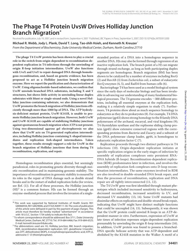
chemical characterization. To this end we cloned UvsW andthe UvsW-K141R mutant into a GST fusion expression vectorcontaining a PreScission protease (Amersham Biosciences)recognition site in the GST linker region. PreScission proteasealso has a GST fusion tag, allowing it to be used on-column tocleave glutathione-bound fusion proteins containing the recog-nition site. The cleaved protein product is then eluted withoutthe need for glutathione, leaving the protease bound to the col-umn. This strategy worked well for generating substantialamounts of the highly purified UvsW-K141R mutant protein(Fig. 1, lane 1). Attempts to obtain the wild-type UvsW protein
using this system, however, were hampered by plasmid insta-bility and low copy number. BecauseUvsW is known to unwindR-loops at replication initiation sites, we reasoned that replac-ing the R-loop-dependent ColE1-based plasmid origin with anR6K �-based replication origin might improve UvsW produc-tion. The R6K � origin uses the � initiator protein with noR-loop intermediate (31). The plasmidwas found to be stable instrain BW23322, which encodes a mutated version of � (pir-116) that results in an increased plasmid copy number (32).Initially, we found that UvsW was highly contaminated with
RNA and a co-purifying protein which was identified by massspectral analysis to be the chaperonin GroEL. Both contami-nants were effectively eliminated by including an RNase treat-ment and an incubation with ATP/Mg2� during the purifica-tion process (33). Using these techniques and additionalmodifications of the standard extraction procedure, we wereeventually able to obtain �600 �g of highly purified, solubleUvsW protein from 1 liter of culture (Fig. 1, lane 2).Unwinding of Oligonucleotide Substrates—The GST-UvsW
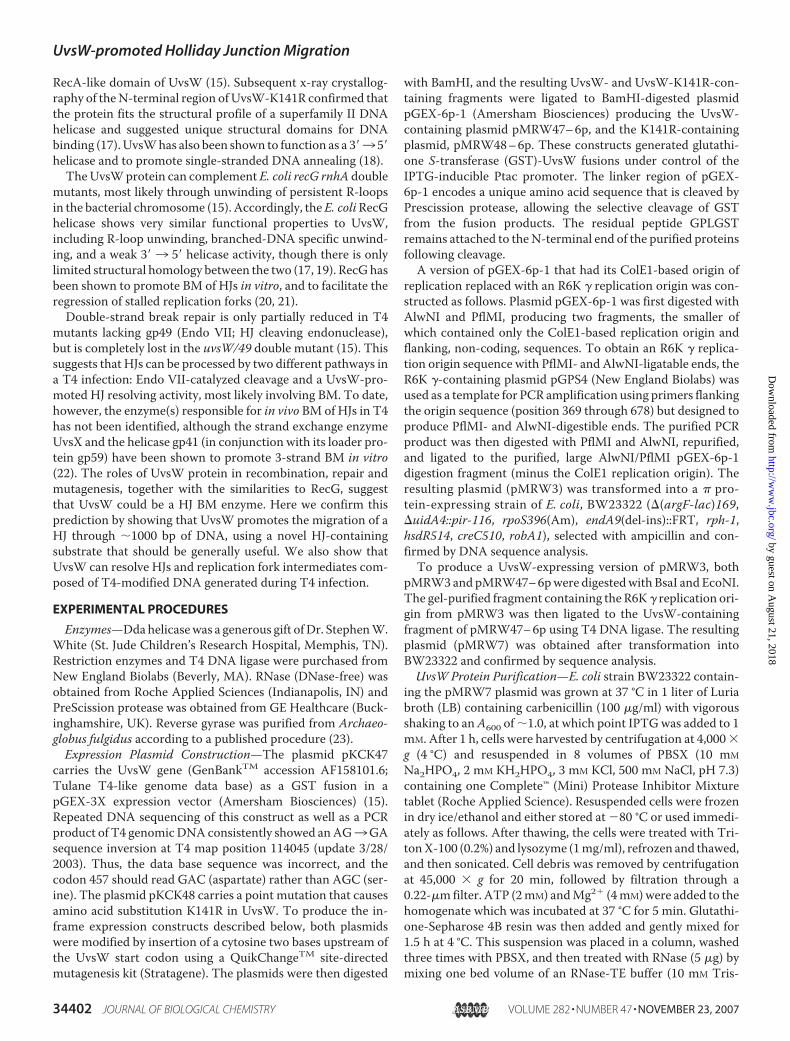
fusion protein was previously shown to unwind a blunt-ended,fully duplex, branched-DNAY substrate in the presence ofATP(15). We began by analyzing the unwinding activity in moredetail using the oligonucleotide-based substrates shown in Fig.2. As in previous studies (15), UvsW was able to unwind aduplex DNA Y substrate, but not a blunt-ended linear DNAsubstrate (Fig. 2). We did not detect significant unwinding ofdouble-stranded substrates with either 5� or 3� single-strandedoverhangs (the latter contrary to an earlier report (18)), or dou-ble-stranded DNAwith a flayed single-stranded end (Fig. 2). Incontrast, UvsW efficiently unwound the static X-junction intoflayed duplexes (Fig. 2B). At an enzyme/substrate ratio of 2:1,
1 2 3
2 5 01 5 0
1 0 0
7 5
5 0
3 7
2 5
2 0
KDa
FIGURE 1. Purified UvsW proteins. Purified proteins were subjected to SDS-PAGE and then stained with Coomassie Blue. PreScission protease-cleavedUvsW-K141R is in lane 1, PreScission protease-cleaved wild-type UvsW is inlane 2, and the GST-UvsW fusion protein eluted with glutathione withoutPreScission protease cleavage is in lane 3. All subsequent experiments wereperformed using the PreScission protease-cleaved products shown in lanes 1and 2.
UvsW-promoted Holliday Junction Migration
34404 JOURNAL OF BIOLOGICAL CHEMISTRY VOLUME 282 • NUMBER 47 • NOVEMBER 23, 2007
by guest on August 21, 2018
http://ww
w.jbc.org/
Dow
nloaded from

UvsW unwound essentially 100% of the static X-junctions inless than 1 min (data not shown). As expected, the 5� 3 3�helicase Dda was able to unwind the 5� single-strand overhangsubstrate and the flayed substrate, but was essentially inactiveon the other substrates.UvsW Catalyzes Branch Migration of Holliday Junctions—
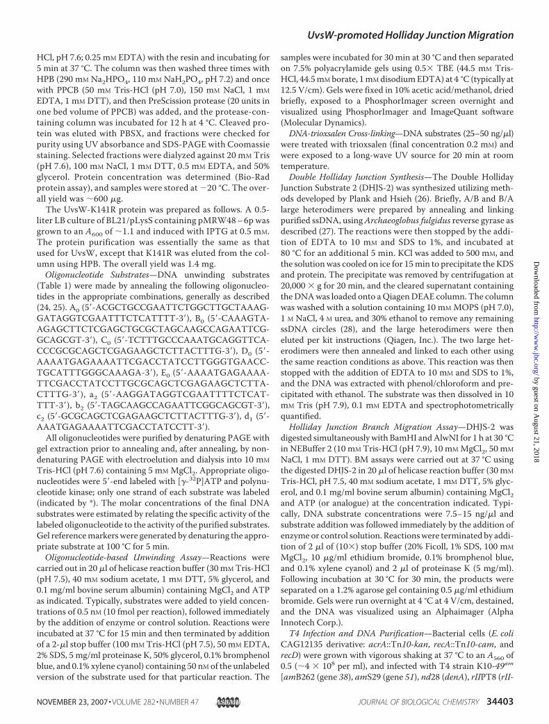
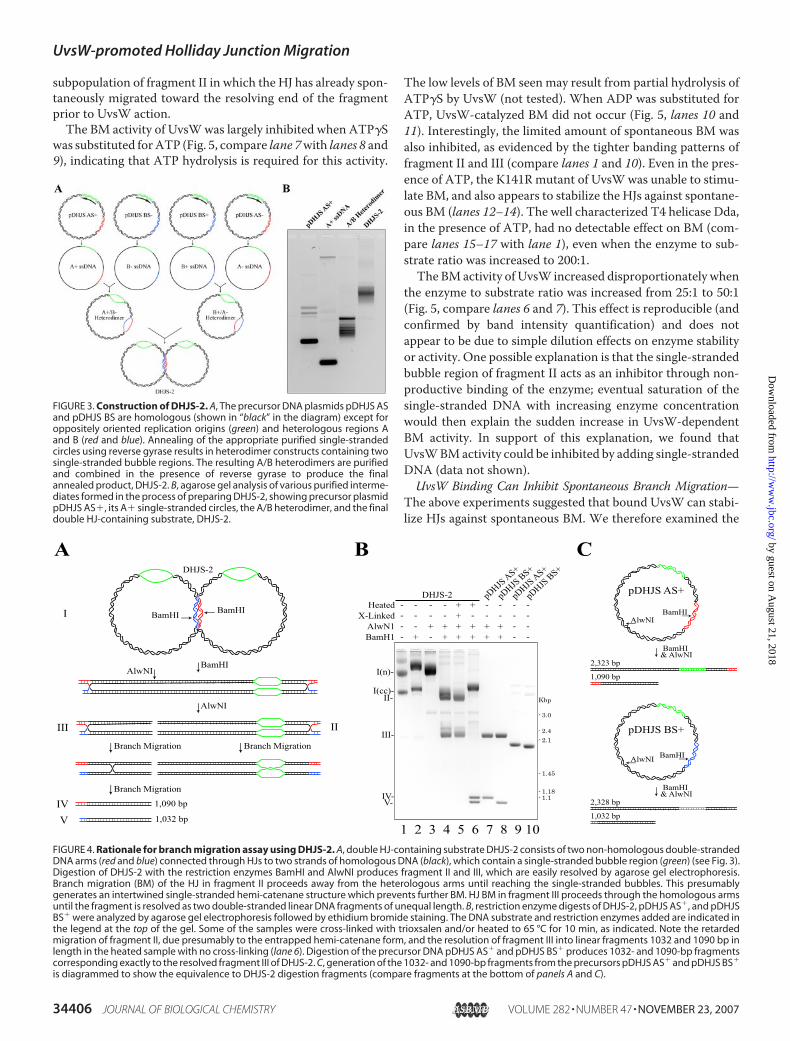
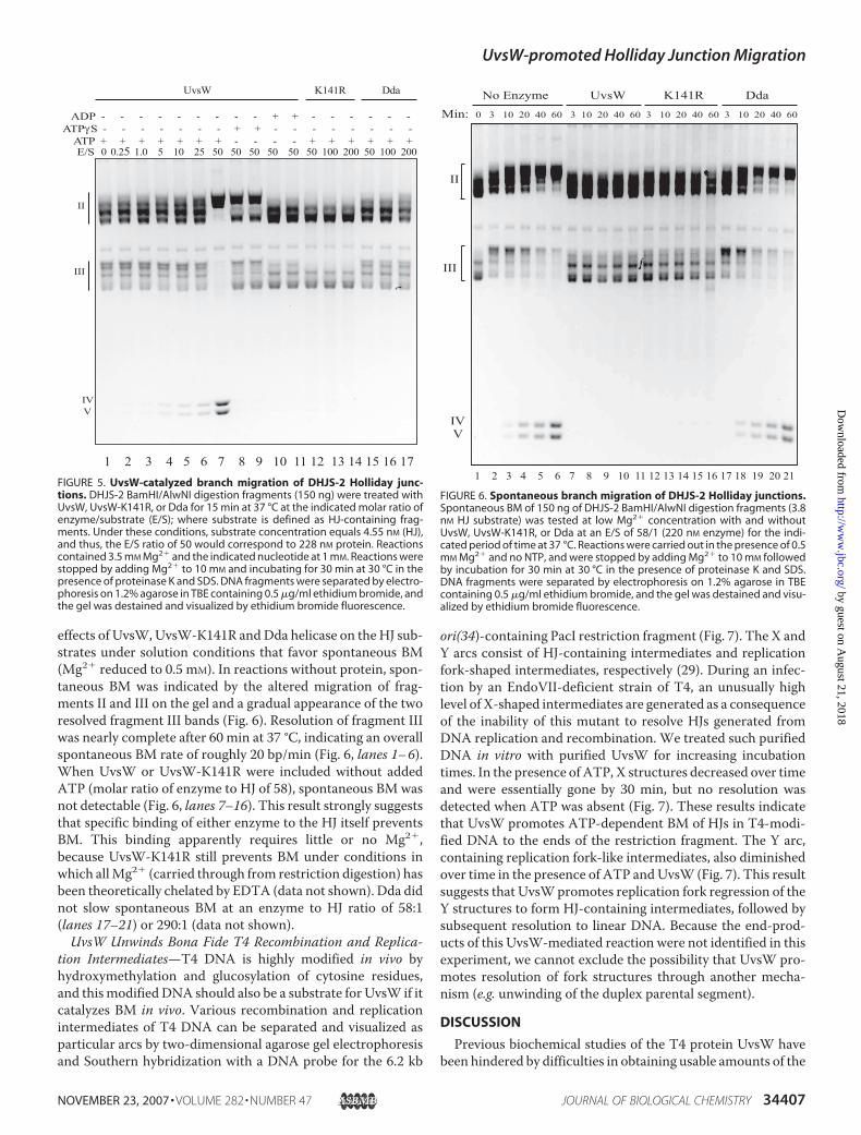
The above results suggest that UvsW may catalyze DNA BM.We directly tested BM activity using a novel substrate andrecently developed method. An in vitro synthesized substrate,DHJS-2, consists of two double-stranded DNA circles com-bined to form two HJs (Fig. 3) (26). The two DNA circles arelargely homologous except for the �300-bp duplex regionsbetween the two HJs (red and blue) and two �450-base single-stranded bubble segments (green; identical, non-complemen-tary sequence). BM of the HJs in intact DHJS-2 is prevented bytopological constraints. However, digestion of DHJS-2 withrestriction enzymes AlwNI and BamHI generates two frag-ments, both of which contain a HJ that can migrate in onedirection only (through the two homologous arms) (Fig. 4A).The smaller fragment (III in Fig. 4) can branchmigrate through�1000 bp of DNA before resolving into two linear DNAduplexes of unequal length, 1090 and 1032 bp. The larger frag-ment from DHJS-2 digestion (II in Fig. 4) can branch migratethrough �350 bp before encountering a non-complementarysingle-stranded bubble region where, we believe, BM termi-nates as an irreversible hemi-catenane.DHJS-2 can be made in substantial quantities and can there-
fore be used at concentrations sufficient for visualizing withethidium bromide staining after agarose gel separation. Fig. 4Billustrates some of the properties of this substrate relevant toour BM assay. Untreated DHJS-2 migrates as two main bands,representing covalently closed and nicked forms (lane 1). Upondigestionwith BamHI andAlwNI, the two singleHJ-containingfragments II and III are produced (lane 4). When heated brieflyto 65 °C, the HJs migrate spontaneously so that the larger frag-ment (II) becomes trapped as a hemi-catenane migrating moreslowly, and fragment III resolves into the two expected 1032-and 1090-bp duplex fragments (lane 6). Digestion of the pre-cursor plasmids pDHJS AS� and pDHJS BS� provides duplexmarkers that exactly co-migrate with the resolved III fragments(lanes 7 and 8), excluding the possibility that the resolved bandsresult from melting of the heterologous ends. As expected,inter-strand crosslinking of fragments II and III with trioxsalen(immediately following digestion) prevents BMuponheating ofthe fragments (compare lanes 5 and 6).Spontaneous BM of the HJs in DHJS-2 can occur as soon as
the topological constraints have been removed by restrictionenzyme digestion. The rate of spontaneous migration is verysensitive to temperature, ionic strength, and Mg2� concentra-tion (34). Under our conditions spontaneous BMwas robust inthe absense of Mg2�, much slower at Mg2� levels as low as 2–3mM, and undetectable at Mg2� concentrations of 10 mM orhigher. Spontaneous BM during long periods of gel electro-phoresis was prevented by running at 4 °C and includingethidium bromide (0.5 �g/ml in both gel and buffer).WhenDHJS-2 fragments II and III were incubated for 15min
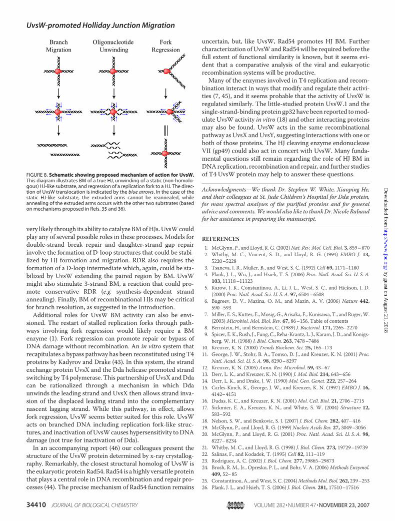
at 37 °C in the presence of 3.5mMMg2� and 1mMATP, there isno discernible resolution of fragment III into the two small
products (Fig. 5, lane 1). However, limited spontaneous BMapparently occurred, resulting in some of fragments II and IIImigrating slightly slower in the gel (compare Fig. 5, lane 1 withFig. 4, lane 4 and 5; also see below). Increasing concentrationsof UvsWallowed complete resolution of fragment III to the twoexpected small fragments and fragment II to the slowly migrat-ing hemi-catenane form (Fig. 5, lanes 2–7). The resolutionproducts of fragment III can be detected as early as 1 min afterstart of incubation with UvsW, suggesting that UvsW can pro-mote BM at rates of roughly 20 bp/sec. One caveat, however, isthat the earliest resolution products could be derived from a
UvsW (nM)Dda (nM)
Heated
--+
10--
100--
-200-
--+
10--
100--
-200-
---
10--
100--
-200-
--+
---
---
* *
**
*
*
**
1 2 3 4 5 6 7 8 9 10 11 12 13 14 15
A
a b c
UvsW (nM)Dda (nM)
Heated
--+
10--
100--
-200-
--+
10--
100--
-200-
---
10--
100--
-200-
--+
---
---
**
*
*
*
*
*
1 2 3 4 5 6 7 8 9 10 11 12 13 14 15
B
d e f
FIGURE 2. UvsW- and Dda-catalyzed unwinding of oligonucleotide sub-strates. A, non-branched DNA substrates were tested for unwinding by UvsWand Dda. The oligonucleotide composition of the substrates were A0b2*(a),A0d1*(b), and a2d1* (c) (see “Experimental Procedures”). The three substrates(0.5 nM), 32P-labeled at the positions indicated by the asterisk, were treatedwith the indicated amounts of UvsW or Dda for 15 min at 37 °C in reactionbuffer containing 3.5 mM Mg2� and 1 mM ATP. Stop buffer containing SDS andproteinase K was added, the samples were incubated for 30 min at 30 °C, andthe products were separated on a 7.5% polyacrylamide gel and visualizedusing phosphorimaging. B, branched DNA substrates were tested forunwinding by UvsW and Dda using exactly the same conditions. The oligo-nucleotide composition of the substrates (by subpanel), were A0B0*(d),A0B0*E0 (e), and A0B0*C0D0 (f). The unwinding of A0B0*C0D0 (subpanel f) pro-duces doublet bands presumably representing the two possible labeledproducts, A0B0* and B0*C0.
UvsW-promoted Holliday Junction Migration
NOVEMBER 23, 2007 • VOLUME 282 • NUMBER 47 JOURNAL OF BIOLOGICAL CHEMISTRY 34405
by guest on August 21, 2018
http://ww
w.jbc.org/
Dow
nloaded from
subpopulation of fragment II in which the HJ has already spon-taneously migrated toward the resolving end of the fragmentprior to UvsW action.The BM activity of UvsWwas largely inhibited when ATP�S
was substituted forATP (Fig. 5, compare lane 7with lanes 8 and9), indicating that ATP hydrolysis is required for this activity.
The low levels of BM seen may result from partial hydrolysis ofATP�S by UvsW (not tested). When ADP was substituted forATP, UvsW-catalyzed BM did not occur (Fig. 5, lanes 10 and11). Interestingly, the limited amount of spontaneous BM wasalso inhibited, as evidenced by the tighter banding patterns offragment II and III (compare lanes 1 and 10). Even in the pres-ence of ATP, the K141R mutant of UvsWwas unable to stimu-late BM, and also appears to stabilize the HJs against spontane-ous BM (lanes 12–14). The well characterized T4 helicase Dda,in the presence of ATP, had no detectable effect on BM (com-pare lanes 15–17 with lane 1), even when the enzyme to sub-strate ratio was increased to 200:1.The BMactivity of UvsW increased disproportionately when
the enzyme to substrate ratio was increased from 25:1 to 50:1(Fig. 5, compare lanes 6 and 7). This effect is reproducible (andconfirmed by band intensity quantification) and does notappear to be due to simple dilution effects on enzyme stabilityor activity. One possible explanation is that the single-strandedbubble region of fragment II acts as an inhibitor through non-productive binding of the enzyme; eventual saturation of thesingle-stranded DNA with increasing enzyme concentrationwould then explain the sudden increase in UvsW-dependentBM activity. In support of this explanation, we found thatUvsWBMactivity could be inhibited by adding single-strandedDNA (data not shown).UvsW Binding Can Inhibit Spontaneous Branch Migration—
The above experiments suggested that bound UvsW can stabi-lize HJs against spontaneous BM. We therefore examined the
FIGURE 3. Construction of DHJS-2. A, The precursor DNA plasmids pDHJS ASand pDHJS BS are homologous (shown in “black” in the diagram) except foroppositely oriented replication origins (green) and heterologous regions Aand B (red and blue). Annealing of the appropriate purified single-strandedcircles using reverse gyrase results in heterodimer constructs containing twosingle-stranded bubble regions. The resulting A/B heterodimers are purifiedand combined in the presence of reverse gyrase to produce the finalannealed product, DHJS-2. B, agarose gel analysis of various purified interme-diates formed in the process of preparing DHJS-2, showing precursor plasmidpDHJS AS�, its A� single-stranded circles, the A/B heterodimer, and the finaldouble HJ-containing substrate, DHJS-2.
- 3.0
- 2.4- 2.1
- 1.45
- 1.18- 1.1
Kbp
DHJS-2
BamHIBamHI
AlwNI
AlwNI
BamHI
I
III II
IVV
I(n)-
I(cc)-II-
III-
IV-V-
BamH1 +- + + + + +- - -AlwN1 + + + + + +- - - -
X-Linked +- - - - - - - - -Heated + +- - - - - - - -
pDHJS AS+
pDHJS AS+
pDHJS BS+
pDHJS BS+
DHJS-2
A B
Branch Migration
Branch Migration
Branch Migration
pDHJS AS+
BamHIAlwNI
BamHI & AlwNI
2,323 bp
1,090 bp
pDHJS BS+
BamHIAlwNI
BamHI & AlwNI
2,328 bp
1,032 bp1,090 bp
1,032 bp
C
1 2 3 4 5 6 7 8 9 10FIGURE 4. Rationale for branch migration assay using DHJS-2. A, double HJ-containing substrate DHJS-2 consists of two non-homologous double-strandedDNA arms (red and blue) connected through HJs to two strands of homologous DNA (black), which contain a single-stranded bubble region (green) (see Fig. 3).Digestion of DHJS-2 with the restriction enzymes BamHI and AlwNI produces fragment II and III, which are easily resolved by agarose gel electrophoresis.Branch migration (BM) of the HJ in fragment II proceeds away from the heterologous arms until reaching the single-stranded bubbles. This presumablygenerates an intertwined single-stranded hemi-catenane structure which prevents further BM. HJ BM in fragment III proceeds through the homologous armsuntil the fragment is resolved as two double-stranded linear DNA fragments of unequal length. B, restriction enzyme digests of DHJS-2, pDHJS AS�, and pDHJSBS� were analyzed by agarose gel electrophoresis followed by ethidium bromide staining. The DNA substrate and restriction enzymes added are indicated inthe legend at the top of the gel. Some of the samples were cross-linked with trioxsalen and/or heated to 65 °C for 10 min, as indicated. Note the retardedmigration of fragment II, due presumably to the entrapped hemi-catenane form, and the resolution of fragment III into linear fragments 1032 and 1090 bp inlength in the heated sample with no cross-linking (lane 6). Digestion of the precursor DNA pDHJS AS� and pDHJS BS� produces 1032- and 1090-bp fragmentscorresponding exactly to the resolved fragment III of DHJS-2. C, generation of the 1032- and 1090-bp fragments from the precursors pDHJS AS� and pDHJS BS�
is diagrammed to show the equivalence to DHJS-2 digestion fragments (compare fragments at the bottom of panels A and C).
UvsW-promoted Holliday Junction Migration
34406 JOURNAL OF BIOLOGICAL CHEMISTRY VOLUME 282 • NUMBER 47 • NOVEMBER 23, 2007
by guest on August 21, 2018
http://ww
w.jbc.org/
Dow
nloaded from
effects of UvsW, UvsW-K141R andDda helicase on theHJ sub-strates under solution conditions that favor spontaneous BM(Mg2� reduced to 0.5 mM). In reactions without protein, spon-taneous BM was indicated by the altered migration of frag-ments II and III on the gel and a gradual appearance of the tworesolved fragment III bands (Fig. 6). Resolution of fragment IIIwas nearly complete after 60 min at 37 °C, indicating an overallspontaneous BM rate of roughly 20 bp/min (Fig. 6, lanes 1–6).When UvsW or UvsW-K141R were included without addedATP (molar ratio of enzyme to HJ of 58), spontaneous BM wasnot detectable (Fig. 6, lanes 7–16). This result strongly suggeststhat specific binding of either enzyme to the HJ itself preventsBM. This binding apparently requires little or no Mg2�,because UvsW-K141R still prevents BM under conditions inwhich allMg2� (carried through from restriction digestion) hasbeen theoretically chelated by EDTA (data not shown). Dda didnot slow spontaneous BM at an enzyme to HJ ratio of 58:1(lanes 17–21) or 290:1 (data not shown).UvsW Unwinds Bona Fide T4 Recombination and Replica-
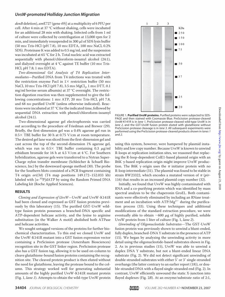
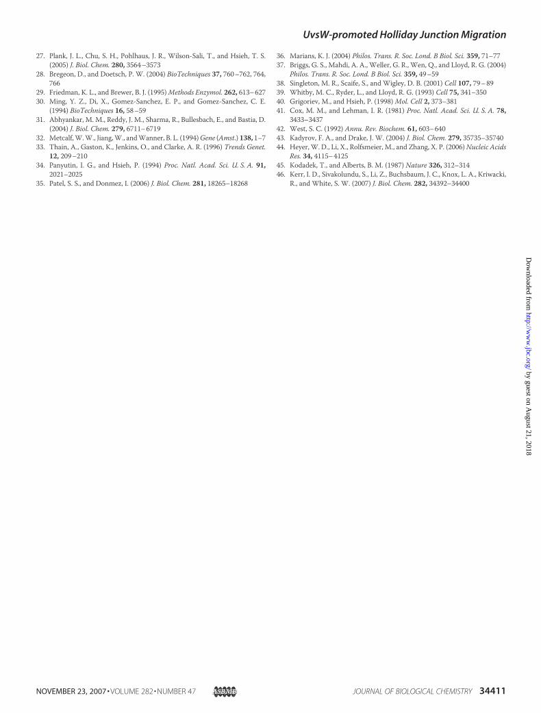
tion Intermediates—T4 DNA is highly modified in vivo byhydroxymethylation and glucosylation of cytosine residues,and thismodified DNA should also be a substrate for UvsW if itcatalyzes BM in vivo. Various recombination and replicationintermediates of T4 DNA can be separated and visualized asparticular arcs by two-dimensional agarose gel electrophoresisand Southern hybridization with a DNA probe for the 6.2 kb
ori(34)-containing PacI restriction fragment (Fig. 7). The X andY arcs consist of HJ-containing intermediates and replicationfork-shaped intermediates, respectively (29). During an infec-tion by an EndoVII-deficient strain of T4, an unusually highlevel of X-shaped intermediates are generated as a consequenceof the inability of this mutant to resolve HJs generated fromDNA replication and recombination. We treated such purifiedDNA in vitro with purified UvsW for increasing incubationtimes. In the presence of ATP, X structures decreased over timeand were essentially gone by 30 min, but no resolution wasdetected when ATP was absent (Fig. 7). These results indicatethat UvsW promotes ATP-dependent BM of HJs in T4-modi-fied DNA to the ends of the restriction fragment. The Y arc,containing replication fork-like intermediates, also diminishedover time in the presence of ATP andUvsW (Fig. 7). This resultsuggests that UvsWpromotes replication fork regression of theY structures to form HJ-containing intermediates, followed bysubsequent resolution to linear DNA. Because the end-prod-ucts of this UvsW-mediated reaction were not identified in thisexperiment, we cannot exclude the possibility that UvsW pro-motes resolution of fork structures through another mecha-nism (e.g. unwinding of the duplex parental segment).
DISCUSSION
Previous biochemical studies of the T4 protein UvsW havebeen hindered by difficulties in obtaining usable amounts of the
ADP - - - - - - - - - + + - - - - - -ATPγS - - - - - - - + + - - - - - - - -
ATP + + + + + + + - - - - + + + + + + E/S 0 0.25 1.0 5 10 25 50 50 50 50 50 50 100 200 50 100 200
UvsW K141R Dda
II
III
IVV
1 2 3 4 5 6 7 8 9 10 11 12 13 14 15 16 17FIGURE 5. UvsW-catalyzed branch migration of DHJS-2 Holliday junc-tions. DHJS-2 BamHI/AlwNI digestion fragments (150 ng) were treated withUvsW, UvsW-K141R, or Dda for 15 min at 37 °C at the indicated molar ratio ofenzyme/substrate (E/S); where substrate is defined as HJ-containing frag-ments. Under these conditions, substrate concentration equals 4.55 nM (HJ),and thus, the E/S ratio of 50 would correspond to 228 nM protein. Reactionscontained 3.5 mM Mg2� and the indicated nucleotide at 1 mM. Reactions werestopped by adding Mg2� to 10 mM and incubating for 30 min at 30 °C in thepresence of proteinase K and SDS. DNA fragments were separated by electro-phoresis on 1.2% agarose in TBE containing 0.5 �g/ml ethidium bromide, andthe gel was destained and visualized by ethidium bromide fluorescence.
II
IVV
No Enzyme UvsW K141R DdaMin: 0 3 10 20 40 60 3 10 20 40 60 3 10 20 40 60 3 10 20 40 60
III
1 2 3 4 5 6 7 8 9 10 11 12 13 14 15 16 17 18 19 20 21
FIGURE 6. Spontaneous branch migration of DHJS-2 Holliday junctions.Spontaneous BM of 150 ng of DHJS-2 BamHI/AlwNI digestion fragments (3.8nM HJ substrate) was tested at low Mg2� concentration with and withoutUvsW, UvsW-K141R, or Dda at an E/S of 58/1 (220 nM enzyme) for the indi-cated period of time at 37 °C. Reactions were carried out in the presence of 0.5mM Mg2� and no NTP, and were stopped by adding Mg2� to 10 mM followedby incubation for 30 min at 30 °C in the presence of proteinase K and SDS.DNA fragments were separated by electrophoresis on 1.2% agarose in TBEcontaining 0.5 �g/ml ethidium bromide, and the gel was destained and visu-alized by ethidium bromide fluorescence.
UvsW-promoted Holliday Junction Migration
NOVEMBER 23, 2007 • VOLUME 282 • NUMBER 47 JOURNAL OF BIOLOGICAL CHEMISTRY 34407
by guest on August 21, 2018
http://ww
w.jbc.org/
Dow
nloaded from

purified protein. Our laboratory previously purified a GST-UvsW fusion protein (15) and more recently, a hexahistidine-tagged version of UvsWwas successfully purified (18). Here wereport a purification scheme based on GST affinity tagging andon-column affinity tag cleavage. This method allowed us topurify 600 �g of un-tagged, wild-type UvsW for biochemicalstudies. An important feature of the method involves substitu-tion of theColE1 origin of replication of the plasmid vectorwithan R6K � replication origin, so that plasmid replication occurswithout an R-loop initiation intermediate.
We confirmed the branched-DNA specific helicase activity ofUvsW using oligonucleotide basedsubstrates. Under our conditions,UvsW showed no helicase activityon three non-branched DNA sub-strates, including duplex DNA oli-gomerswith 3� or 5� single-strandedoverhangs and a duplex with bluntends. UvsW has been reported byNelson and Benkovic (18) tounwind duplex DNA oligomerswith a 3� single-stranded overhang.An additional discrepancy is thatNelson and Benkovic found a strongstimulation of UvsW-promotedbranched DNA unwinding by gp32,but we found that gp32 was notstimulatory for either UvsW-medi-ated oligonucleotide unwinding orBM activity (data not shown). Theunwinding activities of UvsWreported by Nelson and Benkovic inthe absence of gp32 were dramati-cally weaker than in our experi-ments, and the oligonucleotide sub-strates were different between thetwo studies. Additional experi-ments are necessary to resolvethese discrepancies and determinewhether UvsW is strictly dependenton a branch point for activity.While UvsW showed no unwind-
ing activity using a duplex substratewith flayed (non-complementary)single-stranded ends (Fig. 2B, lanes3 and 4), it was capable of unwind-ing the Y shaped substrate con-structed by annealing this flayedsubstrate with an oligonucleotidecomplementary to the flayed ends(Fig. 2B, lanes 8 and 9). The activitywas both ATP-dependent and sen-sitive to Mg2� concentration (datanot shown). Unwinding activitywith this substrate was incompleteunder our standard reaction condi-tions and was unexpectedly inhib-
ited by increasing the concentration of enzyme. This effect wasreproducible and may be relevant to a proposed mechanism ofunwinding/BM (see below). In comparison, unwinding of theHJ-like substrate was fast and very efficient. This substrate isbest described as a static X-junction because, unlike a true HJ,the branch point cannotmigrate due to non-homologous arms.Unwinding of this substrate produced two different detectableflayed products when the substrate was labeled on only onestrand (Fig. 2B, lanes 13 and 14). This result indicates that theenzyme has no preference for how it orients on the branch
0 min 5 min 10 min 30 min
30 min, -ATP 30 min, -UvsW Schematic
Y-arc
X-arc
1x Spot
Line of Linears
A
B
0 min 5 min 10 min 30 min 30 min 30 min -ATP -UvsW
Resolution of Replication Intermediates Over Time
Y-ShapesX-Shapes
Frac
tion
of T
otal
RIs
0.05
0.01
0.02
0.03
0.04
0
0.06
FIGURE 7. Resolution of T4 X- and Y-shaped replication intermediates by purified UvsW. Purifed DNA froma K10-49am infection was digested with PacI, then treated with purified UvsW for the time and conditionsindicated. A, replication intermediates were analyzed by two-dimensional gel electrophoresis (as describedunder “Experimental Procedures”). The first-dimension gel was run from left to right, and the second-dimen-sion gel was run from top to bottom. Replication intermediates from the 6.2-kb PacI fragment containing T4origin ori(34) were visualized by Southern hybridization with a PCR-generated probe. B, amounts of eachreplication intermediate (X and Y) were determined using ImageQuant software (Molecular Dynamics) bydrawing polygonal boundaries around the X-and Y-arcs. These amounts are expressed graphically as a fractionof the total replication intermediates in the corresponding gel. Total replication intermediates are defined asthe sum of all quantifiable DNA on the blot, including X- and Y-arcs, linear monomer (1X spot) and line oflinears.
UvsW-promoted Holliday Junction Migration
34408 JOURNAL OF BIOLOGICAL CHEMISTRY VOLUME 282 • NUMBER 47 • NOVEMBER 23, 2007
by guest on August 21, 2018
http://ww
w.jbc.org/
Dow
nloaded from

point. In addition, no single-stranded products were formedfrom the static-X junction, consistentwith the lack of activity ofUvsW on flayed or blunt-ended duplex substrates. We con-clude that UvsW strongly prefers branched DNA with at leasttwo duplex arms, with the HJ-like substrate being most highlypreferred.Based on genetic studies, UvsWwas implicated in the gener-
ation or processing of recombination intermediates such asHJs(15). The branched-DNA specific unwinding activity andstrong preference for the static-X structure described herein isconsistent withUvsWacting as aHJ BMenzyme.We examinedthis possibility using a novel BM assay developed as amodifica-tion of a previously described method (26). The basis for thisassay is a double HJ containing substrate, DHJS-2, synthesizedin vitro. The twoHJs are constrained frommigrating by regionsof heterology in one direction and by topological constraints inthe other. Upon digestion with the appropriate restrictionenzymes, two HJ-containing fragments are released and BMwithin these fragments can be readily detected by formation ofresolution products. Using this assay, we demonstrated thatUvsW efficiently promotes BM of HJs through at least 1000 bpunder our conditions. This activity is very sensitive to [Mg2�],with concentrations greater than 3.5mM (when [ATP]� 1mM)becoming progressively more inhibitory, and complete inhibi-tion evident at about 10mMMg2� (data not shown). This enzy-matic activity is dependent on ATP, cannot be supported byADP, and is only weakly supported by ATP�S (Fig. 5).TheUvsW-K141Rmutant contains amutation in theWalker
A motif and is totally inactive for BM (Fig. 5). This mutationinactivates one of two RecA-like domains found in UvsW, sig-nificantly inhibiting ATP hydrolysis (15). The loss of BM activ-ity in this mutant is consistent with models in which two func-tional RecA domains are required for translocation of amonomeric helicase (35). TheUvsW-K141Rmutant appears tostabilize HJs against spontaneous BM in the presence of ATParguing that K141R can bind HJs but cannot translocate(branch migrate). Similarly, in the absence of ATP, either wild-type UvsW or UvsW-K141R also strongly inhibit spontaneousBM. These results argue that specific binding of UvsW to HJsdoes not require ATP binding or hydrolysis, nor does it require[Mg2�] higher than 0.5 mM.
We have also shown that purified UvsW can resolve bothT4-modified DNA X and Y structures generated during a T4infection (Fig. 7). The X structures are presumably HJs gener-ated through either RDR or recombinational repair, while the Ystructures are simple replication fork intermediates. Under ourin vitro conditions with no additional proteins, X structurespresumably resolve into linear forms when the HJ migrates offthe end of the restriction fragment. The diminishing intensityof the Y arc with UvsW incubation time parallels that of the Xarc, implying that these structures are also being resolved byUvsW (Fig. 7B). One explanation is that the replication fork struc-tures are regressed by UvsW into HJ X structures, which are thenresolved into linearmonomeric forms. Regression of stalled repli-cation forks has been inferred to be one pathway through whichstalled replication forks are processed in E. coli (1, 36).
The E. coli RecG protein has also been shown to catalyze BMand regression of stalled replication fork-like structures in vitro
(20, 37). While UvsW and RecG share little structural homol-ogy, the remarkably similar functional properties of the twosuggest that they catalyze BM through a similar mechanism.The crystal structure of RecG suggested a model of RecG-pro-moted HJ BM (37, 38). In this model, RecG binds the HJ in anorientation that directs translocation along one arm. TwohomologousDNAside arms are directed throughDNAbindingchannels in the enzyme that are only large enough for onestrand of each arm, and as the enzyme translocates along thedirectional arm, it unwinds and subsequently anneals the sec-ond strand of each side arm to each other. An important com-ponent of the model is a protein wedge domain, which directsHJ binding and steers the second strands of the side arms tofacilitate their annealing. As previously described by others (37,38), this mechanism can explain HJ BM, regression of replica-tion forks into HJs, and unwinding of branched oligonucleo-tides (see Fig. 8). While no structure similar to the RecG wedgedomain has been reported for UvsW, the crystal structure of aUvsW-branched DNA complex has yet to be determined and afunctionally related domain may ultimately be found. Thismodel is based on only one functional UvsW (or RecG) unitbinding each HJ, oriented with respect to one directional arm.The activity would presumably be more efficient if two func-tional enzymes bound per HJ, as long as they were on opposingarms. However, if two enzymes bind such that one is on a direc-tional arm and the other on a side arm, translocation will beeffectively blocked. This model might explain the resultsobserved with the Y oligonucleotide substrate used in thisstudy, where unwinding was inhibited at higher enzyme con-centrations. The Y structure that we used might allow bindingof more than one enzyme per junction, but since there are notruly opposing arms in this structure, such binding would becounterproductive for unwinding.In our BM assay we observed essentially complete resolution
of a HJ-containing structure (fragment III) by apparent unidi-rectional migration through �1000 bp of DNA. If we assumethat UvsW binds the HJs of our substrate randomly withrespect to orientation on the HJ, there will be a 50% probabilityof moving the HJ in either direction. When bound in the pro-ductive orientation, complete resolution would occur if theenzyme is highly processive. If UvsW activity is more distribu-tive, multiple cycles of productive and counterproductive BMwould occur before BM happens to reach the DNA ends andrelease the product fragments. Clearly, the rate of UvsW-cata-lyzed BMwould be affected by conditions that affect processiv-ity. For these reasons, and the fact that the starting position ofBM in our substrate may not be fixed, measuring the absoluterate of UvsW-promoted BM using our assay is problematic.Nevertheless, assuming the HJs in our substrate are relativelyimmobile prior to the assay and that the enzyme is highly pro-cessive, we calculated an approximate rate of UvsW-promotedBM of 20 bp/s (data not shown). For comparison, the rate ofRecG- and RuvAB-promoted BM has been estimated to be8–40 bp/s (39) and 10 bp/s (40), respectively, while the rate ofRecA-driven strand exchange was estimated to be 3–10 bp/s(41, 42).UvsW acts as a regulator of R-loop initiated replication and
also plays roles in T4 replication, recombination and repair,
UvsW-promoted Holliday Junction Migration
NOVEMBER 23, 2007 • VOLUME 282 • NUMBER 47 JOURNAL OF BIOLOGICAL CHEMISTRY 34409
by guest on August 21, 2018
http://ww
w.jbc.org/
Dow
nloaded from
very likely through its ability to catalyze BMofHJs. UvsWcouldplay any of several possible roles in these processes. Models fordouble-strand break repair and daughter-strand gap repairinvolve the formation of D-loop structures that could be stabi-lized by HJ formation and migration. RDR also requires theformation of a D-loop intermediate which, again, could be sta-bilized by UvsW extending the paired region by BM. UvsWmight also stimulate 3-strand BM, a reaction that could pro-mote conservative RDR (e.g. synthesis-dependent strandannealing). Finally, BM of recombinational HJs may be criticalfor branch resolution, as suggested in the Introduction.Additional roles for UvsW BM activity can also be envi-
sioned. The restart of stalled replication forks through path-ways involving fork regression would likely require a BMenzyme (1). Fork regression can promote repair or bypass ofDNA damage without recombination. An in vitro system thatrecapitulates a bypass pathway has been reconstituted using T4proteins by Kadyrov and Drake (43). In this system, the strandexchange protein UvsX and the Dda helicase promoted strandswitching by T4 polymerase. This partnership of UvsX andDdacan be rationalized through a mechanism in which Ddaunwinds the leading strand and UvsX then allows strand inva-sion of the displaced leading strand into the complementarynascent lagging strand. While this pathway, in effect, allowsfork regression, UvsW seems better suited for this role. UvsWacts on branched DNA including replication fork-like struc-tures, and inactivation ofUvsWcauses hypersensitivity toDNAdamage (not true for inactivation of Dda).In an accompanying report (46) our colleagues present the
structure of the UvsW protein determined by x-ray crystallog-raphy. Remarkably, the closest structural homolog of UvsW isthe eukaryotic proteinRad54. Rad54 is a highly versatile proteinthat plays a central role in DNA recombination and repair pro-cesses (44). The precise mechanism of Rad54 function remains
uncertain, but, like UvsW, Rad54 promotes HJ BM. Furthercharacterization ofUvsWandRad54will be required before thefull extent of functional similarity is known, but it seems evi-dent that a comparative analysis of the viral and eukaryoticrecombination systems will be productive.Many of the enzymes involved in T4 replication and recom-
bination interact in ways that modify and regulate their activi-ties (7, 45), and it seems probable that the activity of UvsW isregulated similarly. The little-studied protein UvsW.1 and thesingle-strand-binding protein gp32 have been reported tomod-ulate UvsW activity in vitro (18) and other interacting proteinsmay also be found. UvsW acts in the same recombinationalpathway as UvsX andUvsY, suggesting interactions with one orboth of those proteins. The HJ cleaving enzyme endonucleaseVII (gp49) could also act in concert with UvsW. Many funda-mental questions still remain regarding the role of HJ BM inDNAreplication, recombination and repair, and further studiesof T4 UvsW protein may help to answer these questions.
Acknowledgments—We thank Dr. Stephen W. White, Xiaoping He,and their colleagues at St. Jude Children’s Hospital for Dda protein,for mass spectral analyses of the purified proteins and for generaladvice and comments.Wewould also like to thankDr. Nicole Rabaudfor her assistance in preparing the manuscript.
REFERENCES1. McGlynn, P., and Lloyd, R. G. (2002)Nat. Rev. Mol. Cell. Biol. 3, 859–8702. Whitby, M. C., Vincent, S. D., and Lloyd, R. G. (1994) EMBO J. 13,
5220–52283. Tsaneva, I. R., Muller, B., and West, S. C. (1992) Cell 69, 1171–11804. Plank, J. L., Wu, J., and Hsieh, T. S. (2006) Proc. Natl. Acad. Sci. U. S. A.
103, 11118–111235. Karow, J. K., Constantinou, A., Li, J. L., West, S. C., and Hickson, I. D.
(2000) Proc. Natl. Acad. Sci. U. S. A. 97, 6504–65086. Bugreev, D. V., Mazina, O. M., and Mazin, A. V. (2006) Nature 442,
590–5937. Miller, E. S., Kutter, E., Mosig, G., Arisaka, F., Kunisawa, T., and Ruger,W.
(2003)Microbiol. Mol. Biol. Rev. 67, 86–156, Table of contents8. Bernstein, H., and Bernstein, C. (1989) J. Bacteriol. 171, 2265–22709. Spicer, E. K., Rush, J., Fung, C., Reha-Krantz, L. J., Karam, J. D., andKonigs-
berg, W. H. (1988) J. Biol. Chem. 263, 7478–748610. Kreuzer, K. N. (2000) Trends Biochem. Sci. 25, 165–17311. George, J. W., Stohr, B. A., Tomso, D. J., and Kreuzer, K. N. (2001) Proc.
Natl. Acad. Sci. U. S. A. 98, 8290–829712. Kreuzer, K. N. (2005) Annu. Rev. Microbiol. 59, 43–6713. Derr, L. K., and Kreuzer, K. N. (1990) J. Mol. Biol. 214, 643–65614. Derr, L. K., and Drake, J. W. (1990)Mol. Gen. Genet. 222, 257–26415. Carles-Kinch, K., George, J. W., and Kreuzer, K. N. (1997) EMBO J. 16,
4142–415116. Dudas, K. C., and Kreuzer, K. N. (2001)Mol. Cell. Biol. 21, 2706–271517. Sickmier, E. A., Kreuzer, K. N., and White, S. W. (2004) Structure 12,
583–59218. Nelson, S. W., and Benkovic, S. J. (2007) J. Biol. Chem. 282, 407–41619. McGlynn, P., and Lloyd, R. G. (1999) Nucleic Acids Res. 27, 3049–305620. McGlynn, P., and Lloyd, R. G. (2001) Proc. Natl. Acad. Sci. U. S. A. 98,
8227–823421. Whitby, M. C., and Lloyd, R. G. (1998) J. Biol. Chem. 273, 19729–1973922. Salinas, F., and Kodadek, T. (1995) Cell 82, 111–11923. Rodriguez, A. C. (2002) J. Biol. Chem. 277, 29865–2987324. Brosh, R. M., Jr., Opresko, P. L., and Bohr, V. A. (2006)Methods Enzymol.
409, 52–8525. Constantinou, A., andWest, S. C. (2004)MethodsMol. Biol. 262, 239–25326. Plank, J. L., and Hsieh, T. S. (2006) J. Biol. Chem. 281, 17510–17516
BranchMigration
Oligonucleotide Unwinding
ForkRegression
FIGURE 8. Schematic showing proposed mechanism of action for UvsW.This diagram illustrates BM of a true HJ, unwinding of a static (non-homolo-gous) HJ-like substrate, and regression of a replication fork to a HJ. The direc-tion of UvsW translocation is indicated by the blue arrows. In the case of thestatic HJ-like substrate, the extruded arms cannot be reannealed, whileannealing of the extruded arms occurs with the other two substrates (basedon mechanisms proposed in Refs. 35 and 36).
UvsW-promoted Holliday Junction Migration
34410 JOURNAL OF BIOLOGICAL CHEMISTRY VOLUME 282 • NUMBER 47 • NOVEMBER 23, 2007
by guest on August 21, 2018
http://ww
w.jbc.org/
Dow
nloaded from
27. Plank, J. L., Chu, S. H., Pohlhaus, J. R., Wilson-Sali, T., and Hsieh, T. S.(2005) J. Biol. Chem. 280, 3564–3573
28. Bregeon, D., and Doetsch, P. W. (2004) BioTechniques 37, 760–762, 764,766
29. Friedman, K. L., and Brewer, B. J. (1995)Methods Enzymol. 262, 613–62730. Ming, Y. Z., Di, X., Gomez-Sanchez, E. P., and Gomez-Sanchez, C. E.
(1994) BioTechniques 16, 58–5931. Abhyankar, M. M., Reddy, J. M., Sharma, R., Bullesbach, E., and Bastia, D.
(2004) J. Biol. Chem. 279, 6711–671932. Metcalf,W.W., Jiang,W., andWanner, B. L. (1994)Gene (Amst.) 138, 1–733. Thain, A., Gaston, K., Jenkins, O., and Clarke, A. R. (1996) Trends Genet.
12, 209–21034. Panyutin, I. G., and Hsieh, P. (1994) Proc. Natl. Acad. Sci. U. S. A. 91,
2021–202535. Patel, S. S., and Donmez, I. (2006) J. Biol. Chem. 281, 18265–18268
36. Marians, K. J. (2004) Philos. Trans. R. Soc. Lond. B Biol. Sci. 359, 71–7737. Briggs, G. S., Mahdi, A. A., Weller, G. R., Wen, Q., and Lloyd, R. G. (2004)
Philos. Trans. R. Soc. Lond. B Biol. Sci. 359, 49–5938. Singleton, M. R., Scaife, S., and Wigley, D. B. (2001) Cell 107, 79–8939. Whitby, M. C., Ryder, L., and Lloyd, R. G. (1993) Cell 75, 341–35040. Grigoriev, M., and Hsieh, P. (1998)Mol. Cell 2, 373–38141. Cox, M. M., and Lehman, I. R. (1981) Proc. Natl. Acad. Sci. U. S. A. 78,
3433–343742. West, S. C. (1992) Annu. Rev. Biochem. 61, 603–64043. Kadyrov, F. A., and Drake, J. W. (2004) J. Biol. Chem. 279, 35735–3574044. Heyer,W. D., Li, X., Rolfsmeier, M., and Zhang, X. P. (2006)Nucleic Acids
Res. 34, 4115–412545. Kodadek, T., and Alberts, B. M. (1987) Nature 326, 312–31446. Kerr, I. D., Sivakolundu, S., Li, Z., Buchsbaum, J. C., Knox, L. A., Kriwacki,
R., and White, S. W. (2007) J. Biol. Chem. 282, 34392–34400
UvsW-promoted Holliday Junction Migration
NOVEMBER 23, 2007 • VOLUME 282 • NUMBER 47 JOURNAL OF BIOLOGICAL CHEMISTRY 34411
by guest on August 21, 2018
http://ww
w.jbc.org/
Dow
nloaded from
KreuzerMichael R. Webb, Jody L. Plank, David T. Long, Tao-shih Hsieh and Kenneth N.
The Phage T4 Protein UvsW Drives Holliday Junction Branch Migration
doi: 10.1074/jbc.M705913200 originally published online September 5, 20072007, 282:34401-34411.J. Biol. Chem.
10.1074/jbc.M705913200Access the most updated version of this article at doi:
Alerts:
When a correction for this article is posted•
When this article is cited•
to choose from all of JBC's e-mail alertsClick here
http://www.jbc.org/content/282/47/34401.full.html#ref-list-1
This article cites 46 references, 22 of which can be accessed free at
by guest on August 21, 2018
http://ww
w.jbc.org/
Dow
nloaded from