the structural basis of multifunctionality in … · the structural basis of multifunctionality in...
TRANSCRIPT
1
THE STRUCTURAL BASIS OF MULTIFUNCTIONALITY IN A B12 PROCESSING ENZYME† Markos Koutmos, Carmen Gherasim, Janet L Smith and Ruma Banerjee*
Department of Biological Chemistry and the Life Sciences Institute, University of Michigan Medical Center, Ann Arbor, MI 48109-0600
Running head: Structure of Human CblC *Corresponding Author: Tel: (734)-615-5238, email: [email protected] †This work was supported by grants from the National Institutes of Health (DK45776 to R.B., GM048533
to J.L.S.)
An early step in the intracellular processing of B12 involves CblC, which exhibits dual reactivity, catalyzing the reductive decyanation of cyanocobalamin (vitamin B12) and the dealkylation of alkylcobalamins (e.g. methylcobalamin; MeCbl). Insights into how the CblC scaffold supports this chemical dichotomy have been unavailable despite it being the most common locus of patient mutations associated with inherited cobalamin disorders that manifest in both severe homocystinuria and methylmalonic aciduria. Herein, we report structures of human CblC, with and without bound MeCbl, which provide novel biochemical insights into its mechanism of action. Our results reveal that CblC is the most divergent member of the NADPH-dependent flavin reductase family and can use FMN or FAD as a prosthetic group to catalyze reductive decyanation. Furthermore, CblC is the first example of an enzyme with glutathione transferase activity that has a sequence and structure unrelated to the GST superfamily. CblC thus represents an example of evolutionary adaptation of a common structural platform to perform diverse chemistries. The CblC structure allows us to rationalize the biochemical basis of a number of pathological mutations associated with severe clinical phenotypes. A network of trafficking proteins tailor and escort B12, a reactive and rare cofactor, from its point of entry into the cell to its two client enzymes in mammals, methionine synthase and methylmalonyl-CoA mutase (1). An early step in this processing pathway involves accepting the B12 cargo as it exits the lysosome and converting the varied incoming derivatives into a common intermediate that can be partitioned for synthesis
into the two biologically active forms, methylcobalamin (MeCbl)1 and 5´-deoxyadenosylcobalamin (AdoCbl). This step is catalyzed by the CblC protein (2,3), the product of the cblC locus, a hotbed of mutations in the group of inherited cobalamin disorders (4). CblC is unique among B12 proteins in being able to manipulate the cobalt-carbon bond in B12 in both a homolytic and heterolytic fashion. It catalyzes the reductive decyanation of cyanocobalamin (CNCbl or vitamin B12) in the presence of a flavoprotein oxidoreductase (3). Remarkably, when presented with an alkylcobalamin, CblC also catalyzes the nucleophilic displacement of the alkyl group by glutathione (2) (Fig 1a). The lack of obvious sequence similarity between CblC and any protein of known structure has limited mechanistic insights into the structural basis of its remarkable chemical versatility. In this study, we report the crystal structures of human CblC in the apo- (2.0 Å) and B12-bound (1.95 Å) forms, which reveal the presence of a large cavity for housing B12 and an N-terminal flavodoxin nitroreductase domain. The latter suggested that CblC might be a flavin-dependent B12 processing enzyme, which was confirmed by the demonstration that CblC catalyzes decyanation of CNCbl in the presence of FADH2. The structures provide a framework for understanding the biochemical penalties associated with patient mutations leading to methylmalonic aciduria and homocystinuria.
EXPERIMENTAL PROCEDURES Expression and Purification of CblC Constructs-Truncated CblC (1-244) ∆C38 (construct 1) was cloned into the pET28b(+) vector (Novagen) between the NcoI and XhoI restriction sites using full-length CblC as a template and the following primers:
http://www.jbc.org/cgi/doi/10.1074/jbc.M111.261370The latest version is at JBC Papers in Press. Published on June 22, 2011 as Manuscript M111.261370
Copyright 2011 by The American Society for Biochemistry and Molecular Biology, Inc.
by guest on July 5, 2018http://w
ww
.jbc.org/D
ownloaded from
2
5´CGTCTCCCATGGAGCCGAAAGTCGCAGAGCTGAAGCAG-3´ (forward) and 5´-ATGTGACTCGAGACTAGGCTTCTCTGAGGGCTGAG-3´ (reverse). To aid with phasing, the quadruple methionine mutant L48M/V83M/V107M/I164M CblC (1-244) ∆C38 (construct 2) was generated using the QuickChange site-directed mutagenesis kit (Stratagene) using the following sense mutagenic primers: L48M:(5´-CTGCCAGGACCTACCATGGCCTT CCT-3´), V83M: (5´-CCAGTGGACCAGTGTATGGCC TACCA-3´), V107M:(5´-ATTGCTGACTACGAGATGCACC CCAA-3´), and V164M: (5´-GCCATCCGAGGGGTAATGCTG CTGC-3´).
CblC (1-238) ∆C44 (construct 3) was generated by LIC cloning into pMCSG7 (5). t-CblC (1-244) (construct 1) was expressed and purified as previously described for full-length CblC [2] with the following modifications. Cells were resuspended in 50 mM potassium phosphate pH 7.8, 300 mM KCl, 20 mM imidazole buffer containing 0.2 mg/ml lysozyme and one tablet of EDTA-free complete protease inhibitor (Roche). Following sonication, the cell free extract was subjected to Ni-NTA chromatography, followed by size exclusion chromatography on a Superdex 200 column. Purification of CblC (1-237) ∆C45, construct 3, containing a cleavable N-terminal 6xHis tag, included an additional step of tag removal by TEV protease prior to size exclusion chromatography. For crystallization, purified recombinant proteins were exchanged in 50 mM Tris pH 8.0, 1 mM TCEP and concentrated to ~10-15 mg/ml. For enzymatic studies, CblC proteins were exchanged into 100 mM Hepes pH 8, 150 mM KCl, 10% glycerol or 50 mM Tris pH 8. Selenomethionine-labeled (SeMet) CblC (construct 2) was expressed in an identical manner to constructs 1 and 3 with the exception that the cells were grown in minimal media supplemented with 50 mg/L of SeMet. CblC Expression in Murine Tissues and Protein Stability Studies-Western blot analysis using CblC antibody generated in-house was performed to determine the predominant form of the CblC protein in murine tissues. The tissues were homogenized in liquid nitrogen, resuspended in an
equal volume of lysis buffer [0.1 M sodium phosphate (pH 7.4), containing 0.1% Triton X-100, 10 µl/ml protease inhibitor mixture (Sigma) including 25 µg/ml tosyllysine chloromethyl ketone, 25 µg/ml phenylmethylsulfonyl fluoride, 27 µg/ml aprotinin, and 10 µg/ml leupeptin] and stored at −80°C until further use. The lysate was centrifuged at 4 °C for 30 min at ≈14,000 × g, and the protein concentration in the supernatant was determined by the Bradford assay (Bio-Rad). Alternatively, the pulverized tissue homogenate powder was boiled directly in loading buffer. Protein (200 µg/lane) samples were separated by electrophoresis on a 10% polyacrylamide gel followed by transfer to a PVDF membrane and the expression of CblC was probed by using the respective primary and secondary antibodies and detected by using the chemiluminescent horseradish peroxidase system (Pierce).
The stability of full-length and t-CblC was monitored in a turbidity assay where the change in absorbance at 600 nm was recorded for each protein (0.5 mg/ml) incubated at temperatures ranging from 25ºC to 75 ºC. Tm was determined using equation 1. f= a/(1+exp(-(x-Tm)/b)) (1) CblC Dealkylation and Decyanation Activities-Dealkylation and decyanation were monitored as previously published [2,3]. In addition, decyanation of CNCbl by t-CblC was examined in the absence of MSR/NADPH using either FMNH2 (30-40 µM) or FMN/FAD (30-40 µM)/NADPH (200 µM) as source of reducing equivalents. Reduced flavins were generated prior to use by photochemical reduction in the presence of 10-20 mM sodium oxalate (6). Binding of FMN and FAD to t-CblC by protein fluorescence-Flavin binding to apo-t-CblC was determined from quenching of protein fluorescence monitored at 340 nm following excitation at 280 nm. Fluorimetric titrations were carried out at RT (20 ºC) in 1 ml quartz cuvettes on a Shimadzu RF-5301 PC spectrofluorometer and the emission spectra at 300-500 nm were monitored with excitation and emission slit widths of 3 and 5 mm, respectively. Aliquots of 0-50 µM flavin were titrated in 0.1 µM protein in 100 mM Hepes pH 8, 150 mM KCl, and 10% glycerol. The change in protein fluorescence was recorded after allowing 5 min for equilibration. The binding constant was determined using equation 2.
by guest on July 5, 2018http://w
ww
.jbc.org/D
ownloaded from
3
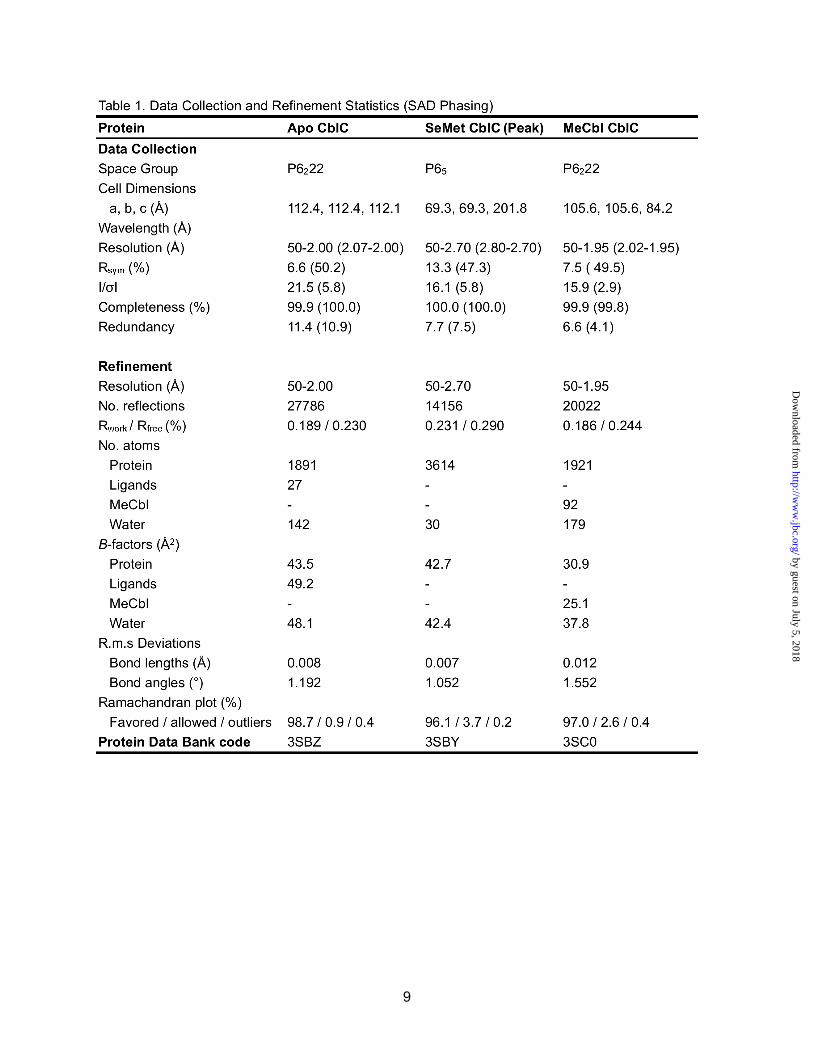
∆F= ∆FmaxL /(KD+L) (2) Crystallization and cryoprotection-Three constructs were used for crystallization. Construct 1, t-CblC (1-244), was a C-terminal His-tagged CblC-∆C38 that crystallized in the apo-form even in the presence of various Cbl forms. Construct 2, t-CblC (1-244), is a variant of construct 1 with four methionine substitutions at positions L48, V83, V107, and V164. The 3 Met residues in wild-type CblC did not provide sufficient anomalous signal for phasing with data from crystals of the SeMet variant. Thus, we engineered the extra methionines in Construct 2, which afforded diffraction quality crystals with a sufficient anomalous signal for phasing (see details below). Construct 3, t-CblC-∆C44 (1-238), is a “tag-less” construct derived from an N-terminal His-tagged construct after TEV-protease cleavage. After the removal of the His-tag, the CblC protein was concentrated to 25-30 mg/ml and MeCbl was added to a final concentration of 0.75-1.0 mM. The mixture was then passed over a size exclusion (S200) column to remove any free cofactor. The final concentration of MeCbl•CblC for crystallography was 10-15 mg/ml. All the steps described above were performed in the dark to avoid photolysis of the cofactor. Construct 3 was engineered to address the problem of crystallization of a MeCbl•CblC complex using Construct 1 with its C-terminal His tag. All protein samples were concentrated to ~10 mg/ml in 50 mM Tris pH 8.0 and 1 mM TCEP before crystallization at 4 °C by the vapor diffusion method from 1:1 mixtures of protein solution with reservoir solution in sitting drop plates (Corning and Intel). The reservoir solution for Construct 1 was 0.9 M sodium malonate, 5% Jeffamine 600 pH 7.0, 0.1 M Hepes pH 6.7, and for Construct 2 was 0.96 M sodium malonate, 10% Jeffamine 600 pH 7.0, 0.1 M Hepes pH 6.7. The resulting crystals of apo-t-CblC and the SeMet-apo-t-CblC were cryoprotected for a few minutes by soaking in 0.8 M sodium malonate, 5% Jeffamine 600 pH 7.0, 0.1 M Hepes pH 6.7 and 20% v/v glycerol prior to flash cooling in liquid N2. Apo t-CblC ∆C38 (Construct 1) crystals were of space group P6222 (a=b= 112.5, c= 112.1) with 1 molecule in the asymmetric unit (Matthews’ coefficient VM=3.2 Å3/Da for 1 molecules per ASU, 61.3% solvent content), while crystals of Apo SeMet-t-CblC ∆C38 (Construct 2) were of
space group P65 (a=b= 69.3, c= 201.8) with 2 molecules in the asymmetric unit (Matthews’ coefficient VM=2.4 Å3/Da for 2 molecules per ASU, 49.0% solvent content).
Co-crystallization of t-CblC-∆C44 (Construct 3) with MeCbl was achieved using a reservoir solution of 2.0 M ammonium sulfate, and 0.1 M Tris.HCl pH 8.5. The resulting colored crystals of MeCbl•t-CblC were cryoprotected by a 1 h soak in 1.6 M ammonium sulfate, 0.1 M Tris.HCl pH 8.5, and 25% v/v glycerol prior to flash cooling in liquid N2. MeCbl•t-CblC-∆C44 (Construct 3) crystals were of space group P6222 (a=b= 105.6, c= 84.2, α=β= 90.0 γ= 120.0) with 1 molecule in the asymmetric unit (Matthews’ coefficient VM=3.5 Å3/Da for 1 molecules per ASU, 65.3% solvent content). Data collection and structure determination- Diffraction data were collected at 100 K for all t-CblC crystals on beamline GM/CA-CAT 23-ID-D at the Advanced Photon Source (APS), Argonne National Laboratory (Argonne, IL). Data were recorded on a Mar300 detector and processed with HKL200 (7). Phenix (8) AutoSol was used to identify the selenium sites and calculate density-modified 2.9-Å experimental maps based on a single-wavelength SAD dataset from one crystal of SeMet “enriched” protein. Specifically, 13 of the 14 Se sites were located, and used for SAD phasing, using phenix.hyss. Subsequently, Phaser (9) was used to calculated experimental phases, followed by density modification by RESOLVE (10), including twofold averaging (FOM 0.35 before and 0.65 after density modification). The experimental density map showed clear features of the protein backbone and well defined side-chains. Modeling of the electron density was done manually using COOT (11). Restrained refinement with isotropic individual B-factors was performed using REFMAC5 (12) of the CCP4 suite (13). The final SeMet-CblC model is missing residues 135-145, and 230-244 in chain A, and 1-4 and 233-244 in chain B. The final SeMet-CblC model was used to solve the other structures by molecular replacement with Phaser using the 2.0 Å apo-t-CblC data set and the 1.95 Å MeCbl•t-CblC data set. Both molecular replacement solutions were refined with PHENIX, including rigid body refinement of the individual domains, followed by simulated annealing in torsional space, individual atomic refinement, and restrained individual B-
by guest on July 5, 2018http://w
ww
.jbc.org/D
ownloaded from
4
factor adjustment with maximum-likelihood targets. Initial density allowed for the MeCbl cofactor to be modeled. The geometric quality of the models was assessed with MolProbity (14). PyMOL (15) was used to create molecular images.
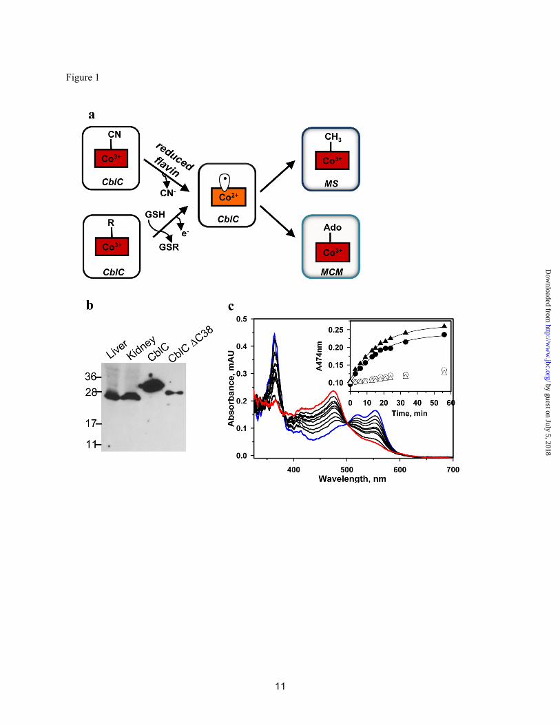
RESULTS AND DISCUSSION Truncated CblC is the Predominant Cellular Form of the Protein-Human CblC is 282 amino acids in length; however, the last ~40 residues are predicted to be highly unstructured and are absent in non-mammalian CblCs (Fig. S1). The cblC gene is predicted to be alternatively spliced by ECgene (16) to generate products that are either 245 or 282 amino acids in length and, despite the relatively large number of patient mutations (>700 alleles) mapped to CblC (4,17), no disease-causing mutations have been confirmed beyond residue 2452. These data beg the question as to whether the truncated form of CblC predominates intracellularly. Western blot analysis of murine tissue reveals the presence of a single band with a molecular mass of ~26 kDa, corresponding to the truncated human protein, t-CblC (28.9 kDa for the His-tagged recombinant form), and in excellent agreement with the calculated size for murine t-CblC (27 kDa) (Fig. 1b). This result indicates that t-CblC is the predominant if not only, form of CblC. The decyanation activity of t-CblC (1-244) is similar to full-length CblC while the dealkylation activity is ~2-fold higher (2,3). The full-length protein (Tm =41.5+0.5 ˚C) is slightly less stable than t-CblC (Tm= 46.6+1.5 ˚C).
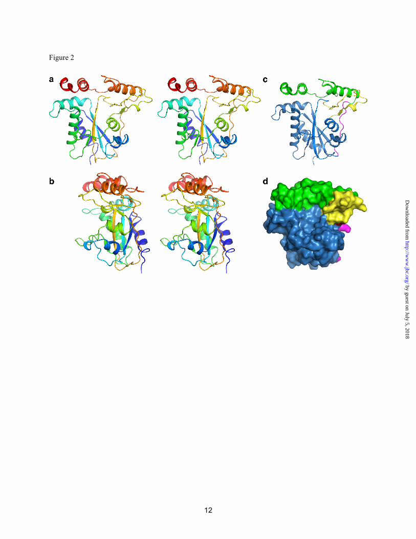
Crystallization of CblC-The apo- and Cbl-bound forms of human t-CblC were crystallized, and phases were obtained through Se anomalous diffraction for apo-CblC and by molecular replacement for the Cbl-bound form (Table 1). CblC is comprised of two modules (N-terminal 1-172; C-terminal 183-244) connected by a long linker (residues 173-182) (Fig. 2). The N-terminal core module exhibits a fold characteristic of the NADP oxidase/flavin reductase family, and contains a four-stranded antiparallel β-sheet flanked by α-helices and a short antiparallel two-stranded β-sheet (Fig. S2). The C-terminal module (cap) is composed of four helices that cap the core N-terminal module (Fig. 2a). The fourth helix is ordered in apo-CblC but disordered in the
MeCbl•t-CblC complex. A large cavity is formed at the interface of the cap and core of CblC (Fig. 2). CblC was predicted to house a C-terminal TonB-like domain (residues 185-282) (4) found in bacterial 2-protein transporter systems. However, the C-terminal cap domain does not exhibit a TonB-like fold.
CblC has a flavin reductase fold-Although CblC has the fold of a flavin reductase, it has very low sequence identity (<12%) and poor structure alignment with other members of the flavin reductase family. Two of the closest structural homologues of CblC are also examples of non-canonical flavin reductases, BluB (18) (rmsd = 4.2 Å based on 117 residues, 9% identity) and iodotyrosine deiodinase (IYD) (19) (rmsd = 3.0 Å based on 102 residues, 7% identity) (Fig. S3a). Both enzymes are members of a sub-family with novel catalytic activities atypical of flavin reductases; BluB cannibalizes flavin to synthesize the cobalamin tail and IYD uses mono- and di-iodotyrosine to salvage iodine in the thyroid. Although CblC possesses the overall flavin reductase fold, it is the most divergent member of the flavin reductase family, and is the first member of a new subfamily. CblC catalyzes the dealkylation of alkylcobalamins, a reaction that is flavin independent. Decyanation of CNCbl requires reducing equivalents, and the diflavin oxidoreductases, methionine synthase reductase (MSR) and novel reductase 1 (NR1), can couple to CblC (3). The structural similarity of CblC to members of the flavin reductase family suggests that it has intrinsic flavin oxidoreductase activity, consistent with a report of reduced NADH-dependent cobalamin reductase activity in cblC cell lines (20) and the reported decyanation activity in the presence of NADPH and FAD (21).
We characterized the reductive decyanation of CNCbl in the presence of FMN/FAD and NADPH (Fig. 1c). The observed decyanation rate (k = 0.07 ± 0.01 min-1), is comparable to those observed in the presence of MSR (0.10 ± 0.004 min-1) and NR1 (0.068 ± 0.007 min-1) (3). Intrinsic protein fluorescence quenching was used to monitor binding of FMN (KD= 4.4 ± 1.1 µM) and FAD (1.4 ± 0.3 µM) to apo-t-CblC (Fig. 4).
Unlike other flavin reductases, which bind flavin at a dimer interface, CblC is a monomer in
by guest on July 5, 2018http://w
ww
.jbc.org/D
ownloaded from
5
solution and in crystals. The inability of CblC to dimerize can be explained structurally. Other flavin reductases form inter-subunit β-sheets in which each subunit contributes a β-strand to the central sheet of the partner subunit. Helix HD and the following loop (LDE) block formation of this contact in CblC (Figs. S2, S3). Attempts to soak either FMN or FAD into apo- and MeCbl-bound t-CblC crystals were unsuccessful. However, based on structural alignments with other flavin reductases, we can model a probable flavin-binding site (Fig. S3). In the apo-t-CblC structure, the predicted flavin pocket is blocked by loop L3F (Fig. S3a). The putative flavin site becomes accessible upon Cbl binding, which induces structural changes as described below. B12-binds to CblC in a “base-off” conformation-The structure of t-CblC in the presence of MeCbl reveals that Cbl binds in the large cavity between core and cap in a “base-off” form, i.e. where the endogenous lower axial base, dimethylbenzimidazole, is not a ligand to the cobalt as predicted by absorption spectroscopy (3) (Fig. 3). On the other hand, CblC has been reported to bind CNCbl in both the “base-on” (3) and “base-off” (21) state. This discrepancy is explained by the observation that the conversion from the “base-on” to CblC-bound “base-off” state is slow (Fig. 4b). The pKa for protonation of dimethylbenzimidazole in CNCbl is 0.1 and governs the “base-on”/”base-off” equilibrium in solution (22). By binding the cofactor in the “base-off” form, the protein enhances the reactivity of Co for reductive decyanation. By removing the electron-releasing nitrogen ligand, the “base-off” state facilitates cobalt reduction (23), providing a chemical rationale for binding the substrate in this conformation.
The cobalamin tail resides in a crevice on the surface of CblC (Fig. 3a,b), which is formed between helix HF and the two-stranded anti-parallel β-sheet (Fig. S3). Contacts in the crevice are largely hydrophobic with only two hydrogen bonds between the cofactor tail and residues Tyr129 and Gln131 (Fig. 3c, Table S1). The corrin ring binds in the large cavity with its non-alkylated, “lower ligand” side against the two-stranded anti-parallel β-sheet. This is in striking contrast to the mammalian B12-dependent
enzymes, which bind cobalamin in a “base-off/His on” conformation, wherein a histidine residue in a conserved D-X-H-X-X-G(X41)SXL(X26-28)GG motif serves as a lower axial ligand to the cobalt. CblC lacks the His motif and does not provide a lower ligand. The cofactor is secured at the cavity wall by ten polar interactions between the side chains of the corrin ring and backbone and side chain atoms from both the core and cap of CblC (Fig. 3, Table S1). The “upper ligand” face of the corrin ring is oriented towards the unfilled portion of the active site cavity. The cobalamin “cage” is further defined on the top by part of the cap module (helix I, and loop LIJ) and on the floor by loop L3F. Electron density is clear for the upper axial methyl group of MeCbl. The large cavity is only partially filled by MeCbl. The cavity size explains the latitude of CblC to accommodate a structural diversity of Cbl derivatives ranging from a methyl to a 5´-deoxyadenosyl group (24). The cavity volume is large enough to also accommodate glutathione adjacent to the Co alkyl ligand (Fig. S4), consistent with the glutathione-dependent dealkylation activity of CblC. Conformational Changes Induced by B12 Binding-MeCbl binding results in three conformational changes in CblC (Fig. 3d). The most significant is movement of loop L3F towards Cbl, creating the floor of the binding pocket. In the apo-t-CblC structure, the L3F loop blocks the putative flavin binding site, and the conformational change suggests that Cbl binding induces formation of or stabilizes the flavin pocket. This is consistent with the higher affinity of CblC for FMN in the presence (1.5 ± 0.2 µM) versus absence (0.5 ± 0.1 µM) of CNCbl. The affinity of CblC for flavin might be further modulated by interaction between CblC and the next protein in the B12 trafficking pathway, e.g. CblD.
A second difference is the repositioning of the LIJ loop in the cap to form part of the Cbl-binding pocket. Finally, helix HL in the C-terminal cap is disordered in the MeCbl•t-CblC structure. Although our attempts to build into electron density for this region were unsuccessful, the cap region is clearly in a different and more flexible conformation in the MeCbl•t-CblC structure. Glutathione was modeled into the MeCbl•t-CblC structure by placing the sulfur near the methyl ligand of MeCbl (Fig. S4). We speculate that
by guest on July 5, 2018http://w
ww
.jbc.org/D
ownloaded from
6
glutathione binding may order the flexible C-terminus of the cap. Although CblC exhibits glutathione transferase (GST) activity, it lacks both the conserved G-site for glutathione binding and the thioredoxin domain with distinct βαβαββα topology, which are hallmarks of the GST superfamily (25,26). Structural Insights into Pathogenic Mutations in CblC-Our data allow us to rationalize the potential biochemical penalties associated with a number of patient mutations (Fig. S1). Most of the pathological mutations map to residues (Fig. S1 and Fig. 5) that can be grouped into two major categories: positions important for cobalamin binding and for structural integrity. The significance of another category of residues that are mutated in a small subset of patients is not obvious. Most mutations within this group are benign with the notable exception of R161G/Q. Arg161 is proximal to but does not contact the upper axial ligand of cobalamin. Arg161 may be important for glutathione binding and/or the dealkylation activity. Interestingly, none of the patient mutations map to the putative FMN binding site. We speculate that mutations in the FMN binding site might be “silent” since CblC can use alternative reducing partners like MSR and NR1. Summary-Our study provides structural insights into how catalytic versatility is embedded within
the CblC scaffold enabling catalysis of radically distinct chemical transformations. The CblC structure reveals that it is the most divergent member of the NADPH-dependent flavin reductase family and suggests that the flavin site is formed on demand, i.e. when Cbl is bound. The cavernous Cbl-binding site explains the relaxed specificity for the Cbl upper axial ligand (24), an advantage for a processing enzyme that accepts varied B12 derivatives entering the cytoplasm. The CblC structure illustrates that its GST activity is housed in a scaffold that is unrelated to the GST superfamily. Hence, CblC represents a novel architectural solution for both the decyanation and dealkylation activities. Accession Numbers-Atomic structure factors and coordinates are deposited at the Protein Data Bank under accession codes 3SBY (SeMet-t-CblC), 3SBZ (apo-t-CblC), 3SC0 (MeCbl•t-CblC). Acknowledgements-We thank Rowena G. Matthews for critical discussions and for her input on the manuscript. We acknowledge GM/CA CAT at the Advanced Light Source for beam time. We thank W.C. Brown and J. Delproposto (LSI, University of Michigan, Ann Arbor, MI) for cloning CblC in one of the expression plasmids (Construct 3).
by guest on July 5, 2018http://w
ww
.jbc.org/D
ownloaded from
7
REFERENCES
1. Banerjee, R., Gherasim, C., and Padovani, D. (2009) Current Op Chem Biol 13, 477-484 2. Kim, J., Hannibal, L., Gherasim, C., Jacobsen, D. W., and Banerjee, R. (2009) J Biol Chem 284,
33418-33424 3. Kim, J., Gherasim, C., and Banerjee, R. (2008) Proc. Natl. Acad. Sci. U.S.A. 105, 14551-14554 4. Lerner-Ellis, J. P., Tirone, J. C., Pawelek, P. D., Dore, C., Atkinson, J. L., Watkins, D., Morel, C. F.,
Fujiwara, T. M., Moras, E., Hosack, A. R., Dunbar, G. V., Antonicka, H., Forgetta, V., Dobson, C. M., Leclerc, D., Gravel, R. A., Shoubridge, E. A., Coulton, J. W., Lepage, P., Rommens, J. M., Morgan, K., and Rosenblatt, D. S. (2006) Nat. Genet. 38, 93-100
5. Stols, L., Gu, M., Dieckman, L., Raffen, R., Collart, F. R., and Donnelly, M. I. (2002) Protein Expr Purif 25, 8-15
6. Massey, V., and Hemmerich, P. (1977) J Biol Chem 252, 5612-5614 7. Otwinowski, Z., and Minor, W. (1997) Methods Enzymol 276, 307-326 8. Adams, P. D., Afonine, P. V., Bunkoczi, G., Chen, V. B., Davis, I. W., Echols, N., Headd, J. J., Hung,
L. W., Kapral, G. J., Grosse-Kunstleve, R. W., McCoy, A. J., Moriarty, N. W., Oeffner, R., Read, R. J., Richardson, D. C., Richardson, J. S., Terwilliger, T. C., and Zwart, P. H. (2010) Acta Crystallogr D Biol Crystallogr 66, 213-221
9. McCoy, A. J., Grosse-Kunstleve, R. W., Adams, P. D., Winn, M. D., Storoni, L. C., and Read, R. J. (2007) J Appl Crystallogr 40, 658-674
10. Terwilliger, T. C. (2000) Acta Crystallogr D Biol Crystallogr 56, 965-972 11. Emsley, P., and Cowtan, K. (2004) Acta Crystallogr D Biol Crystallogr 60, 2126-2132 12. Murshudov, G. N., Vagin, A. A., and Dodson, E. J. (1997) Acta Crystallogr D Biol Crystallogr 53,
240-255 13. (1994) Acta Crystallogr D Biol Crystallogr 50, 760-763 14. Davis, I. W., Leaver-Fay, A., Chen, V. B., Block, J. N., Kapral, G. J., Wang, X., Murray, L. W.,
Arendall, W. B., 3rd, Snoeyink, J., Richardson, J. S., and Richardson, D. C. (2007) Nucleic Acids Res 35, W375-383
15. DeLano, W. L. (2002) The PyMOL Molecular Graphics System DeLano Scientific, Palo Alto, CA, USA
16. Kim, P., Kim, N., Lee, Y., Kim, B., Shin, Y., and Lee, S. (2005) Nucleic Acids Res 33, D75-79 17. Lerner-Ellis, J. P., Anastasio, N., Liu, J., Coelho, D., Suormala, T., Stucki, M., Loewy, A. D., Gurd,
S., Grundberg, E., Morel, C. F., Watkins, D., Baumgartner, M. R., Pastinen, T., Rosenblatt, D. S., and Fowler, B. (2009) Hum Mutat 30, 1072-1081
18. Taga, M. E., Larsen, N. A., Howard-Jones, A. R., Walsh, C. T., and Walker, G. C. (2007) Nature 446, 449-453
19. Thomas, S. R., McTamney, P. M., Adler, J. M., Laronde-Leblanc, N., and Rokita, S. E. (2009) J Biol Chem 284, 19659-19667
20. Pezacka, E. H. (1993) Bioch. Biophys. Acta 1157, 167-177 21. Froese, D. S., Zhang, J., Healy, S., and Gravel, R. A. (2009) Mol Genet Metab 98, 338-343 22. Brown, K. L., and Hakimi, J. M. (1984) J. Am. Chem. Soc. 106, 7894-7899 23. Lexa, D., and Saveant, J.-M. (1983) Acc. Chem. Res. 16, 235-243 24. Hannibal, L., Kim, J., Brasch, N. E., Wang, S., Rosenblatt, D., Banerjee, R., and Jacobsen, D. W.
(2009) Mol Genet Metabol 97, 260-266 25. Allocati, N., Federici, L., Masulli, M., and Di Ilio, C. (2009) FEBS J 276, 58-75 26. Oakley, A. J. (2005) Curr Opin Struct Biol 15, 716-723
by guest on July 5, 2018http://w
ww
.jbc.org/D
ownloaded from
8
FOOTNOTES
1Abbreviations used. MeCbl: methylcobalamin, AdoCbl: 5´-deoxyadenosylcobalamin, CNCbl:
cyanocobalamin or vitamin B12, t-CblC: truncated CblC.
2Of the three mutations described in reference 6 beyond residue 245, two (R267Q and S271G) are benign
and one (a 9 amino acid deletion between S264-P272) is downstream of a frameshift terminating mutation
on the same allele.
by guest on July 5, 2018http://w
ww
.jbc.org/D
ownloaded from
10
FIGURE LEGENDS
Fig. 1. The predominance of t-CblC in tissues and the reactions it catalyzes. (a) Decyanation and dealkylation reactions catalyzed by CblC. (b) The predominant form of CblC in murine liver and kidney extracts (~26 kDa) migrates at a molecular weight consistent with its truncation (28.9 kDa with His tag versus 32.8 kDa with His-tag for full-length CblD). (c) t-CblC (50 µM) catalyzes conversion of 35 µM CNCbl (blue) to cob(II)alamin (red) in the presence of 40 µM FMNH2. The inset shows the kinetics of decyanation in the presence of t-CblC/FMNH2 (●), t-CblC/MSR (▲) or in the absence of the enzyme with FMNH2 (○) or MSR (∆ ). MS and MCM denote methionine synthase and methylmalonyl-CoA mutase, respectively.
Fig. 2. Structure of apo-CblC. (a) and (b) stereoview of apo-t-CblC in two orientations, related by a 90o rotation. Cartoon (c) and space filling (d) representations of the arrangement of the apo-t-CblC modules. The core module is shown in blue, the cap is displayed in green, the linker is in pink, and the 2 β-sheet anti-parallel insertion of the core domain is shown in yellow.
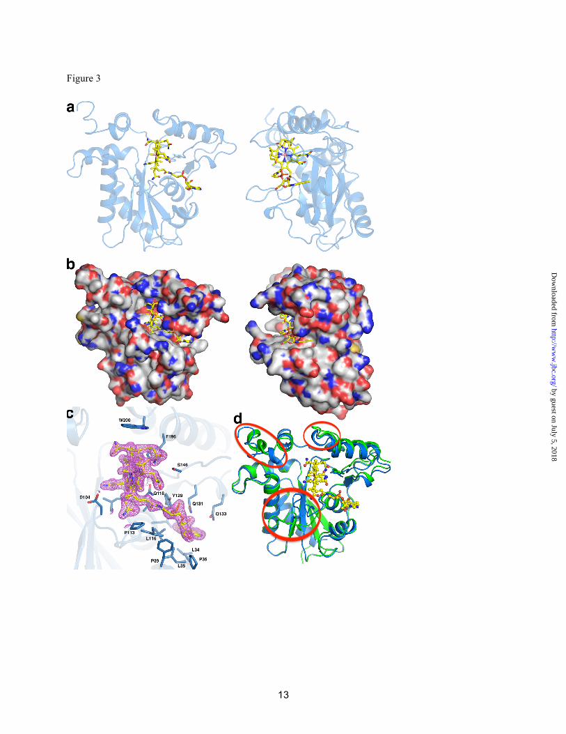
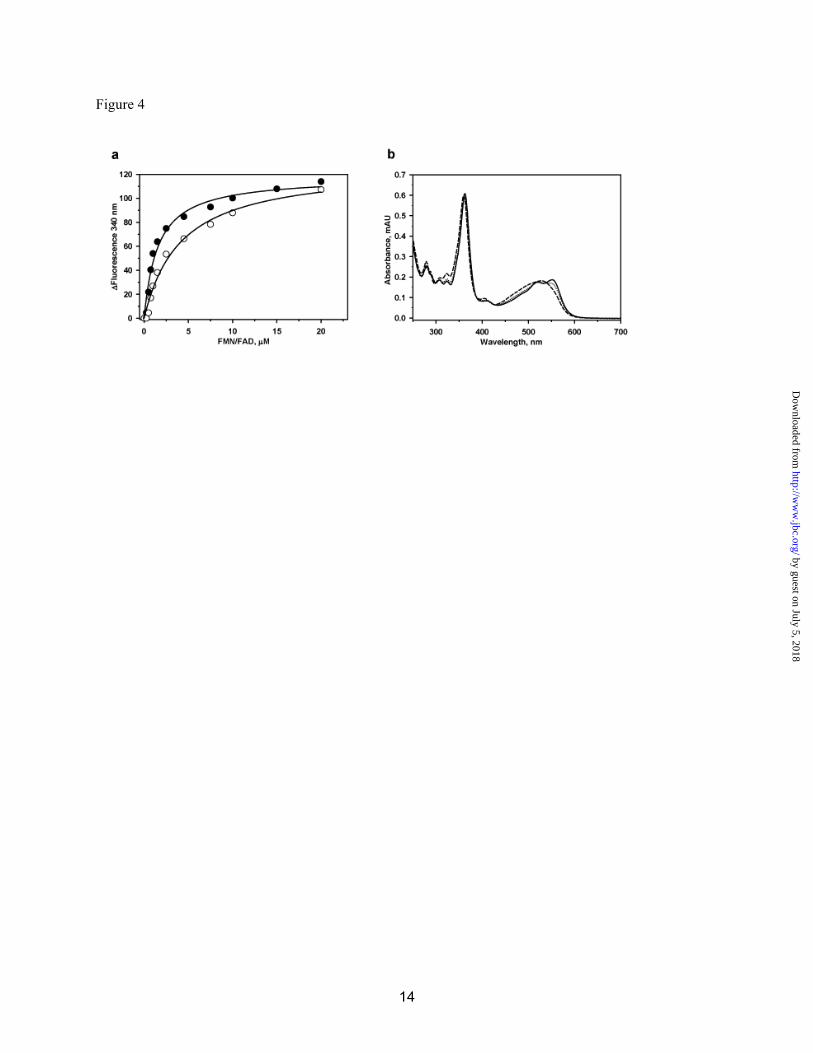
Fig. 3. Structure of holo-CblC. MeCbl•t-CblC shown in two orientations and displayed in (a) cartoon and (b) space-filling representations. (c) 2Fo-Fc electron density contoured at 1.5s (magenta) and stick model (yellow) of MeCbl is shown with its immediate protein environment. Residues in close proximity to MeCbl are shown as sticks (blue). (d) Superposition of the apo- (green) and MeCbl- (blue) t-CblC structures in which the three major differences are encircled in red. Fig. 4. Binding of flavin and CNCbl to CblC. (a) FMN (○) and FAD (●) to apo-t-CblC binding to CblC. A solution of t-CblC was titrated with increasing concentrations of FMN or FAD, respectively as described under Methods. Upon excitation at 280 nm, changes in the emission spectra were recorded between 300 and 450 nm. The binding constant for each flavin was determined by plotting the changes at 340 nm as a function of ligand concentration as described under Methods. (b) Time-dependent spectral change from “base-on” to “base-off” CNCbl in the presence of t-CblC. t-CblC (50 µM) was incubated with 30 µM CNCbl and changes in the absorption spectra were recorded over a period of time. The initial spectra of CNCbl-bound to t-CblC ( - ) changed over time to an intermediate spectrum at 3 min (…) and finally to the spectrum of “base-off’ CNCbl-bound t-CblC after 20 min (---). No further changes were observed after this time point. The authenticity of the “base-off” CNCbl-bound CblC was confirmed by comparing the spectra to that of CNCbl in 7%HCl (not shown).
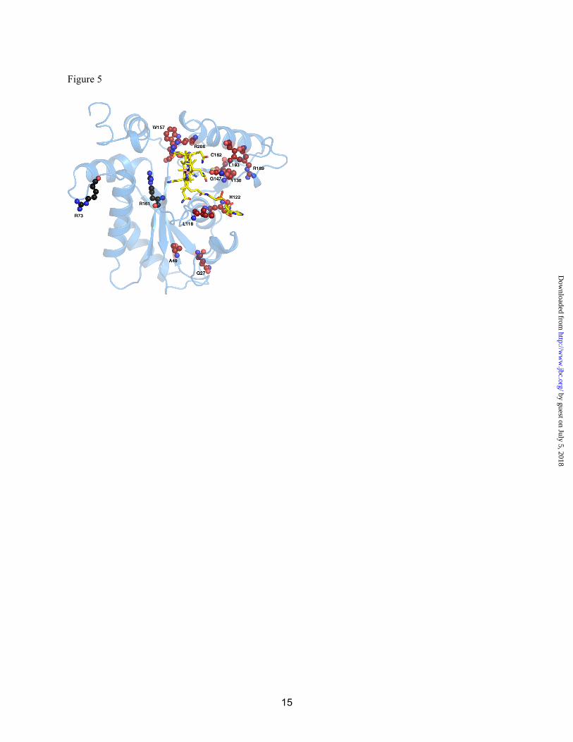
Fig. 5. Patient mutations mapped onto the MeCbl•t-CblC structure. Residues are shown in ball and stick with the ones in red representing residues involved in Cbl binding or structural integrity. The functions of residues in black are not obvious and we speculate, that R73 due to its surface exposure, might be involved in protein-protein interactions while R161 might be involved in GSH binding due to its vicinity to the GSH putative binding site.
by guest on July 5, 2018http://w
ww
.jbc.org/D
ownloaded from
Markos Koutmos, Carmen Gherasim, Janet L. Smith and Ruma BanerjeeThe structural basis of multifunctionality in a B12 processing enzyme
published online June 22, 2011J. Biol. Chem.
10.1074/jbc.M111.261370Access the most updated version of this article at doi:
Alerts:
When a correction for this article is posted•
When this article is cited•
to choose from all of JBC's e-mail alertsClick here
Supplemental material:
http://www.jbc.org/content/suppl/2011/06/22/M111.261370.DC1
by guest on July 5, 2018http://w
ww
.jbc.org/D
ownloaded from