mycofabrication, mechanistic aspect and multifunctionality of metal nanoparticles
TRANSCRIPT
Mycofabrication, mechanistic aspect and Multifunctionality of Metal
Nanoparticles - Where are we? And where should we go?
Mahendra Rai*, Alka Yadav and Aniket Gade
Department of Biotechnology SGB Amravati University, Amravati.; E-mail: [email protected]
* Address for correspondence: Dr. Mahendra K Rai, Professor and Head, Department of Biotechnology, S.G.B.
Amravati University, Amravati – 444 602 (Maharashtra), India; Telephone No :+91-721-2662206 Ext 267; Fax No:
+91-721-2662135, 2660949; E-mail: [email protected]; [email protected]
Fungi consists of varied group of heterotrophs, which due to their unique properties show applications in different fields.
Nanotechnology covers diverse fields of science and technology, and the fabrication of nanoparticles using the biological
route is the need of the day. The introduction of biological agents for the synthesis of nanoparticles has encouraged the
researchers to search for the efficiency of different systems to synthesize metallic nanoparticles. Among the different
systems harnessed for their potential of synthesizing nanoparticles, the fungal system has emerged as an efficient system
synthesizing metal nanoparticles both intra- and extracellularly. The fungus mediated nanoparticles present
monodispersity, dimensions and stability. Also, the system is eco-friendly and economically viable for the synthesis of
nanoparticles.
The present review focuses on different concepts and mechanism involved in the synthesis of metal nanoparticles, present
status, multiple applications, and different areas of research.
Keywords: Fungi, Nanotechnology, nanoparticles, intracellular, extracellular.
1.1 Introduction
Nanotechnology is a multidisciplinary science comprising various aspects of research and technology [1]. Nanoparticles
are metal particles in the size range of 1-100nm and form building blocks of nanotechnology [2]. Metal nanoparticles
like gold, silver and platinum have gained considerable attention in recent times due to their fundamental and
technological interest. These nanoparticles have unique catalytic, electronic and optical properties distinct from the
metallic particles [3]. In recent times, many methods have been designed to synthesize nanoparticles such as physical
method, chemical method and biological methods [3], [4], [5]. The physical and chemical methods involve the use of
strong chemical reducing agents such as sodium borohydride and weak reducing agents like sodium citrate, alcohols,
use of gamma rays and UV rays, etc. [6]. Studies have reported that the biological methods depict an inexpensive and
eco-friendly route for synthesis of nanoparticles. Till date synthesis of nanoparticles have been demonstrated by the use
of biological agents like bacteria, fungi, yeast and plants [3]. A number of bacteria like Bacillus subtilis [7],
Pseudomonas stutzeri [8], Thermonospora sp. [9], Shewanella algae [10], Lactobacillus strains [11], etc. have been
studied for the synthesis of metallic nanoparticles. Yeast have also been explored for the biosynthesis of nanoparticles
including Candida glabrata [12], Schizosaccaharomyces pombe [13], MKY3 [14] etc. While, a number of plants like
Medicago sativa [15], Pelargonium graveolans [16], Azadirachta indica [17], Triticum [18], Cinnamomum camphora
[19], Capsicum annum [20] have been used for the fabrication of metal nanoparticles.
The synthesis of nanoparticles by fungi, and their subsequent application, particularly in medicine are studied under
Myconanotechnology. Myconanotechnology is the interface between ‘Mycology’ and ‘Nanotechnology’ and has
considerable potential, partly due to the wide range and diversity of the fungi [21]. When focusing on the synthesis of
nanoparticles using fungi, it was observed that nanoparticles of good monodispersity and well dimensions could be
synthesized. As fungi are found to secrete high amount of protein they might result in the significant mass productivity
of nanoparticles. The fungal proteins are capable of hydrolyzing metal ions. In addition to this, fungi are easy to isolate
and culture. Moreover, the downstream processing and the handling of fungal biomass are less complex than the
synthetic methods [22].
Mycofabrication can be defined as the synthesis of metal nanoparticles using fungi. The fungal system in recent
times has emerged as “Bionanofactories” synthesizing nanoparticles of silver, gold, platinum and CdS etc.
Fungi can accumulate metal ions by physico-chemical and biological mechanisms including extracellular binding by
metabolites and polymers, binding to specific polypeptides, and metabolism-dependent accumulation [23]. The possible
use of fungi has gained much importance, as they are easy to culture in bulk. Also, the extracellular secretion of
enzymes has an added advantage in the downstream processing and handling of biomass [24] when compared to the
bacterial fermentation process which involves use of sophisticated instruments to obtain clear filtrate from the colloidal
broth [25]. Moreover, fungi are excellent secretors of protein compared to bacteria and actinomycetes, resulting into
higher yield of nanoparticles [25]. Thus, using these dissimilatory properties of fungi, it could be extensively used for
the rapid and eco-friendly biosynthesis of metal nanoparticles. The present review focuses on the synthesis of metal
nanoparticles using fungi, various applications, and the present status of research involving its future impressions.
_______________________________________________________________________________________
1.2 Where we are in fungus mediated nanoparticle synthesis?
The biological route for the synthesis of nanoparticles implying the use of microorganisms is advantageous over the
traditional methods, as biological synthesis is cost-efficient, environment-friendly and simple method. The main
advantage of the biological route is its ability to manipulate nanoparticle properties by gaining control over the size and
shape of nanoparticles [26], [27]. The feasibility of the fungal system for the synthesis of metal nanoparticles has been
successfully demonstrated (Table1). The fungal system shows the capability of both intracellular and extracellular
synthesis of nanoparticles [22], [26], [28]. In the recent past, research work using the fungal system has been carried out
using both aspects of intracellular and extracellular methods for synthesis of nanoparticles of gold, CdS, silver, silica,
titania, zirconia, etc. ([29], [30], [31], [32], [33]). Moreover, a number of fungal species like Verticillium, Phoma sp.,
sp. Fusarium oxysporum, Aspergillus fumigatus, Trichoderma asperellum, have been explored ( [29], [31], [33], [34],
[27]) for the synthesis of metal nanoparticles.
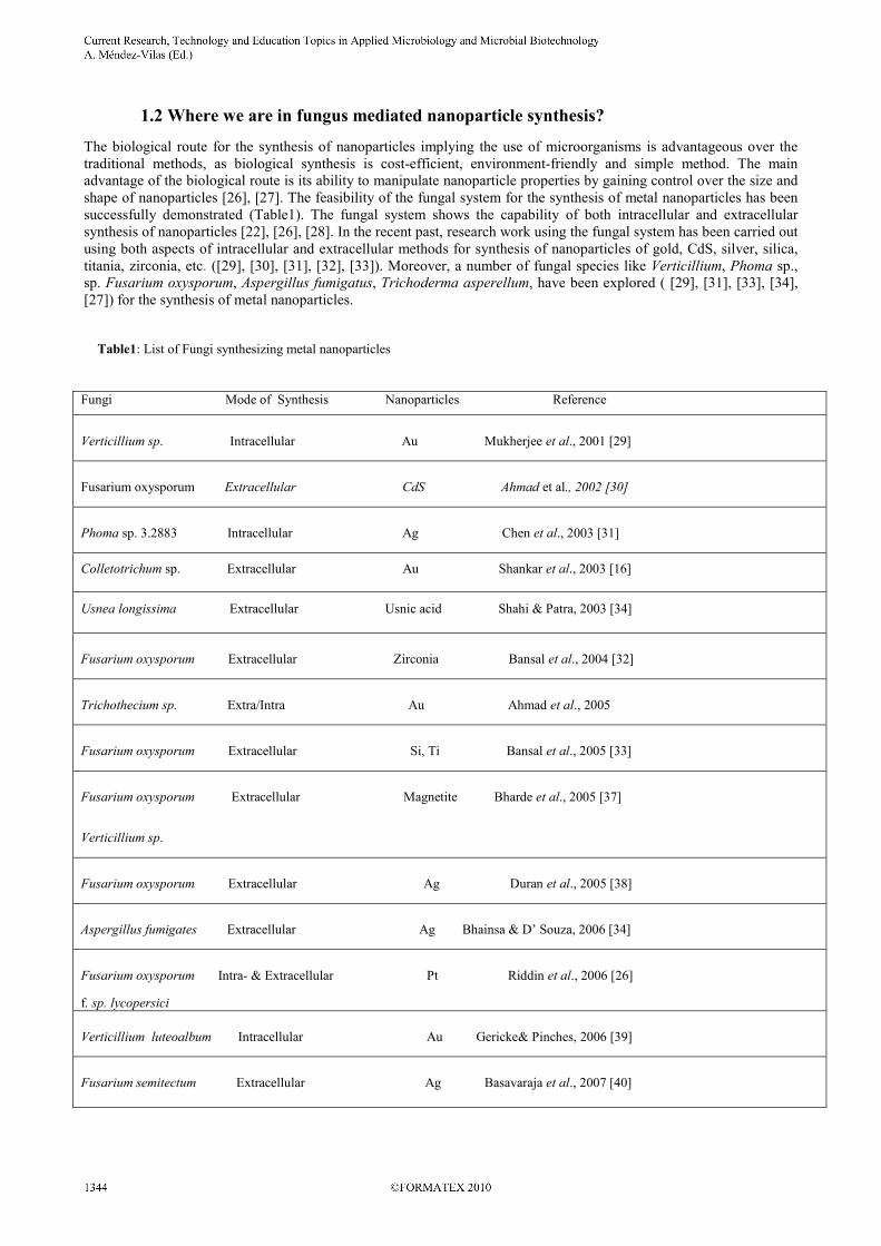
Table1: List of Fungi synthesizing metal nanoparticles
Fungi Mode of Synthesis Nanoparticles Reference
Verticillium sp. Intracellular Au Mukherjee et al., 2001 [29]
Fusarium oxysporum Extracellular CdS Ahmad et al., 2002 [30]
Phoma sp. 3.2883 Intracellular Ag Chen et al., 2003 [31]
Colletotrichum sp. Extracellular Au Shankar et al., 2003 [16]
Usnea longissima Extracellular Usnic acid Shahi & Patra, 2003 [34]
Fusarium oxysporum Extracellular Zirconia Bansal et al., 2004 [32]
Trichothecium sp. Extra/Intra Au Ahmad et al., 2005
Fusarium oxysporum Extracellular Si, Ti Bansal et al., 2005 [33]
Fusarium oxysporum Extracellular Magnetite Bharde et al., 2005 [37]
Verticillium sp.
Fusarium oxysporum Extracellular Ag Duran et al., 2005 [38]
Aspergillus fumigates Extracellular Ag Bhainsa & D’ Souza, 2006 [34]
Fusarium oxysporum Intra- & Extracellular Pt Riddin et al., 2006 [26]
f. sp. lycopersici
Verticillium luteoalbum Intracellular Au Gericke& Pinches, 2006 [39]
Fusarium semitectum Extracellular Ag Basavaraja et al., 2007 [40]
_______________________________________________________________________________________
One of the earliest reports of the synthesis of nanoparticles by fungi was demonstrated by the fungus, Verticillium sp.
[29]. Gold nanoparticles were synthesized intracellularly by growing the fungal cells in a defined medium and then
transferred to aqueous auric chloride solution, the pale yellow color of the fungal cells changed to vivid purple over 24
hours. The UV visible spectra of the fungal cells confirmed the synthesis of gold nanoparticles by depicting an
absorption peak at 540nm, characteristic for gold nanoparticles. The TEM (Transmission Electron Microscopy) and
Higher magnification TEM studies demonstrated the presence of gold nanoparticles with an average size of 20-28nm on
the cell wall as well as in the cytoplasm. The gold nanoparticles were mostly spherical while some were triangular or
hexagonal.
The synthesis of CdS quantum dots by the fungus Fusarium oxysporum was depicted by extracellular enzymatic
reduction of sulphate ions [30]. The CdS quantum dots were supposed to be formed by the reaction of Cd2+
ions with
sulphate ions and the enzymatic reduction of sulphate ions to sulphide ions thus concluding that the fungus plays the
role of a bio-reducing agent (enzyme sulphate reductase). The semiconductor nanoparticles were monodisperse in size
from 5 to 20nm. The XRD analysis of particles showed the nanocrystalline nature of nanoparticles with Bragg
reflections characteristic for hexagonal CdS particles.
Freeze-dried fungal biomass could also be exploited for the synthesis of silver nanoparticles [31]. Freeze-dried
mycelium of Phoma sp. 3.2883 was treated with silver nitrate solution for 50 hours. Adsorption assays indicated that the
mycelium had absorbed some 13mg of silver and TEM micrographs showed the presence of a large number of silver
nanoparticles of around 70nm within the fungal mycelium.
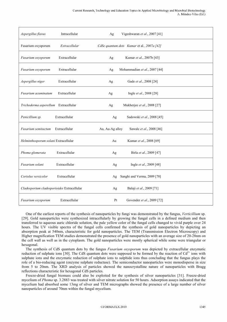
Aspergillus flavus Intracellular Ag Vigeshwaran et al., 2007 [41]
Fusarium oxysporum Extracellular CdSe quantum dots Kumar et al., 2007a [42]
Fusarium oxysporum Extracellular Ag Kumar et al., 2007b [43]
Fusarium oxysporum Extracellular Ag Mohammadian et al., 2007 [44]
Aspergillus niger Extracellular Ag Gade et al., 2008 [24]
Fusarium acuminatum Extracellular Ag Ingle et al., 2008 [28]
Trichoderma asperellum Extracellular Ag Mukherjee et al., 2008 [27]
Penicillium sp. Extracellular Ag Sadowski et al., 2008 [45]
Fusarium semitactum Extracellular Au, Au-Ag alloy Sawale et al., 2008 [46]
Helminthosporum solani Extracellular Au Kumar et al., 2008 [69]
Phoma glomerata Extracellular Ag Birla et al., 2009 [47]
Fusarium solani Extracellular Ag Ingle et al., 2009 [48]
Coriolus versicolor Extracellular Ag Sanghi and Verma, 2009 [70]
Cladosporium cladosporioides Extracellular Ag Balaji et al., 2009 [71]
Fusarium oxysporum Extracellular Pt Govender et al., 2009 [72]
_______________________________________________________________________________________

An endophytic fungus Colletotrichum sp. isolated from Pelargonium graveolens also depicted synthesis of spherical
gold nanoparticles [16]. The XRD study showed formation of stable gold nanoparticle aggregates. While, the TEM
studies revealed synthesis of predominantly spherical nanoparticles and some aggregated into larger irregular structures
with no well-defined morphology. The smaller spherical nanoparticles ranged in size from 8 to 40nm. In the study it
was found that the fungal proteins were responsible for the stabilization of gold nanoparticles.
Shahi and Patra [35] demonstrated synthesis of bioactive nanoparticles of usnic acid from the lichen-forming fungus
Usnea longissima in a defined medium. The bioactive nanoparticles were uniform in shape and formulated for the
preparation of nanoemulsion by dissolving the usnic acid particles in oleic acid and acetone. The nanoemulsion showed
activity against dermatophytes. While performing the in vitro investigation minimum inhibitory concentration was
found to be 0.1µl/ml for the test fungi, viz., Epidermophyton floccosum, Microsporum audouinii, M.canis, M.gypseum,
M.nanum, Trichophyton mentagrophytes, T. rubrum, T. tonsurans, and T. violaceum. The in vivo investigation (for
control of fungal infections) was performed on human skin for testing its irritant activity and long term toxicity on
human skin. The nanoemulsion did not show any irritation or adverse effect at 5% concentration up to 3 weeks. After
topical application of nanoemulsion on human skin improvements were started from first week. Fifty percent showed
moderate improvement and 30% mild improvement. In second week, 30% and 25% depicted significant and moderate
improvement respectively. While, in the third week 60% patients showed completely diminished fungal infections and
30% significant improvement. Thus, the nanoemulsion prepared by bioactive nanoparticles showed the potential for
curing dermatophytic infections in humans.
Zirconia is an oxide of great potential so is titania and silica. Fusarium oxysporum has shown the potential for the
synthesis of zirconia, titania and silica nanoparticles. Bansal et al. [32] demonstrated the synthesis of quasi-spherical
zirconia nanoparticles. The fungal protein similar to silicatein was found to be responsible for the synthesis of zirconia
nanoparticles. Extracellular synthesis of zirconia nanoparticles was done by exposing the fungal biomass to aqueous
solution of zirconium hexafluoride anions at room temperature. After 24 hrs of reaction subsequent bright field and dark
field TEM images of the reaction mixture were taken which showed formation of spherical shape nanoparticles with
quasi-spherical morphology (3 to 11nm in size). The X-ray Diffraction (XRD) analysis of the thin film of the solution
cast nanoparticles showed Bragg reflections characteristic for crystalline zirconia. The Fourier Transform Infrared
Spectra (FTIR) of these biogenic nanoparticles depicted Zr-O-Zr vibrational band which was followed by the
disappearance of the protein amide I and II bands thus improving the crystallinity of biogenic particles. To find out the
proteins secreted by the fungus which were responsible for the hydrolysis of the aqueous anionic metal complex, SDS-
PAGE (Sodium dodecyl sulfate polyacrylamide gel electrophoresis) was carried out which clearly showed the initiation
of two extracellular proteins in the presence of zirconium hexafluoride anions. These extracellular proteins were eluted
from the gel and on testing showed positive results for hydrolysis of zirconium hexafluoride anions to zirconia
nanoparticles. In another study Bansal et al. [33] reported the synthesis of titania and silica nanoparticles. Fusarium
oxysporum was used for the extracellular synthesis of silica and titania nanoparticles. The biosynthesized nanoparticles
were quasi-spherical in shape also, the FTIR analysis showed the presence of Si-O-Si and Ti-O-Ti stretching vibration
and amide bonds which were considered to be responsible for the synthesis on silica and titania nanoparticles.
Ahmad et al. [36] reported that the fungus Trichothecium sp. could synthesize gold nanoparticles by both extra and
intracellular method. The fungal biomass when kept in stationary condition resulted in rapid extracellular synthesis
(Mycelial mat of Trichothecium sp. was obtained and separated from culture broth and washed thrice in distilled water
and resuspended in 100ml aq. auric chloride solution) of gold nanoparticles whereas; when the biomass was kept in
shaking condition on a rotary shaker it resulted in intracellular synthesis of nanoparticles. The TEM photographs of the
extracellulary synthesized gold nanoparticles showed a number of individual gold nanoparticles along with few
aggregates. The morphological study of these particles depicted presence of polygons (specially triangles and hexagons)
as well as some polydisperse spheres and rods. The selected Area Electron Diffraction (SAED) pattern showed the
Scherrer ring pattern which is characteristic of face centered cubic (fcc) structure of gold. While, the TEM micrographs
of the intracellulary synthesized gold nanoparticles showed formation of small particles with spherical morphology. The
SAED pattern of these gold particles showed diffuse rings with lattice spacing which were in agreement with that of
gold. The intracellular formation of gold nanoparticles was also studied using the XRD study. The XRD data revealed
presence of intense peaks (111, 200, 220, 311) corresponding to Bragg reflections of gold. The mean size of these
particles was calculated using the Debye-Scherrer equation which determined that the average size of these gold
nanoparticles was 13nm. Thus from the present study, the authors concluded that when the reaction conditions are
changed the enzymes and proteins, which are released in the stationary phase do not release in shaking condition and
hence result in the intracellular and extracellular synthesis of gold nanoparticles.
Isolates of Fusarium oxysporum and Verticillium sp. were exploited for the synthesis of magnetite nanoparticles [37].
The fungal filtrates was challenged with 2:1 molar mixture of K3[Fe(Cn)6] and K4[Fe(Cn)6]. The TEM images of
nanoparticles synthesized using F. oxysporum showed irregular particles with quasi-spherical morphology in size range
of 20-50nm. The SAED analysis of individual particles showed crystalline nature of nanoparticles while, the XRD
analysis showed well-defined Bragg reflections characteristic of ferric oxide. The FTIR analysis of these biosynthesized
nanoparticles showed absorbance peak at 522, 568, 627 cm-1
which were attributed to Fe-O-Fe stretching mode
vibrations and absorption peaks at 1638 and 1540 cm-1
were attributed to the presence of proteins. The TEM images of
_______________________________________________________________________________________
the particles synthesized using Verticillium sp. depicted number of octahedrally shaped iron oxide particles in size range
of 100 to 400nm. The SAED analysis confirmed that the particles were magnetite. The XRD pattern of iron oxide
nanoparticles also showed number of Bragg reflections characteristic of ferric oxide. The FTIR analysis showed
absorption bands around 522 and 627 cm-1
which were characteristic of Fe-O-Fe vibration modes.
Duran et al. [38] demonstrated the synthesis of silver nanoparticles using Fusarium oxysporum. The authors
concluded that the enzyme nitrate reductase might be responsible for the reduction of silver ions and the subsequent
synthesis of silver nanoparticles. Extracellular biosynthesis of silver nanoparticles using Aspergillus fumigatus was
demonstrated by Bhainsa & D’Souza [34]. The synthesis of silver nanoparticles was monitored by UV-visible
spectrophotometry, XRD and TEM analysis. TEM micrograph of biosynthesized silver nanoparticles taken after 72 hrs
of incubation showed variable shape with majority of them spherical in shape along with some triangular in size range
of 5-25nm. The XRD spectrum of the nanoparticles showed intense peaks in consent with the Bragg reflections of
crystalline silver.
Gericke and Pinches [39] have demonstrated intracellular synthesis of gold nanoparticles. The fungus Verticillium
luteoalbum was exploited for the synthesis of gold nanoparticles. TEM micrographs of the biosynthesized nanoparticle
showed particle morphologies including spherical, triangular, hexagonal and other shapes. Large variation in the size of
particles was observed which varied from a few to approximately 100nm. To obtain better control over size and shape,
fungal biomass was grown for 24, 48, 72 hrs and exposed to auric chloride solution for 24 hrs. It was observed from the
results that the age of the cells at the time of exposure to AuCl4- solution did not have any significant effect on the shape
of nanoparticles however, a decrease in number of particles was observed with fungal biomass. The pH of the reaction
solution was also found to play an important role in the particle synthesis. TEM images of the particle synthesized at pH
3, 5, 7 and 9 showed particles with shape morphologies including triangles, hexagons, spheres and rods. The EDS
spectrum of the nanoparticles indicated that the Au nanoparticles were mainly composed of Au with trace amounts of C,
O, Na, Si and Al. Thus, the synthesis of gold nanoparticles was found to be related to the incubation temperature.
Increased temperature resulted in faster particle growth rate.
Riddin et al. [26] demonstrated Response Surface Methodology (RSM) which consists of a central design to
determine the optimal conditions like temperature, pH and concentration of hydrogen hexachloroplatinate (H2PtCl6) for
the synthesis of platinum nanoparticles. For the intercellular synthesis (RSM1), fungal biomass (Fusarium oxysporum
sp. lycopersici) was suspended in varying concentrations (1.31, 4.11, 8.22, 12.33, 15.13) of H2PtCl6 and placed in water
bath at required temperature (24.8, 35, 50, 65, 75.2◦C) and the pH was adjusted (3.6, 5.0, 7.0, 9.0, 10.4) with Na2CO3.
The nanoparticle formation was observed for a period of 72 hours for any color change from yellow to dark brown.
Similarly, for extracellular synthesis (RSM2) the fungal cell free extract was treated with H2PtCl6. The results of above
experiments were evaluated using the RSM equation. According to the first order model of RSM1 it was observed that
the optimum yield of nanoparticles was found to be 3.23mg/l at pH 3.6, temperature 75.2◦Cand H2PtCl6 concentration of
15.13mM. While, in case of RSM2 model predicted that the optimum yield of nanoparticles was found to be 4.85mg/l at
pH 10.41, temperature75.2◦C and H2PtCl6 concentration of 15.13mM.
The TEM analysis of the extracellular solution (0, 2, 16, 24, 48 & 72 hrs) showed different geometrically shaped
nanoparticles with varying morphologies after incubation up to 72 hrs. The TEM analysis of particles synthesized at
65◦C depicted lower amounts of particles while, particles synthesized at 35
◦C gave a yield of 0.35mg/l nanoparticles.
The wide variation observed in the TEM morphologies of nanoparticles signifies that control over experimental
parameters like temperature and pH would help in determination of particle concentration.
Basavaraja et al. [40] reported extracellular synthesis of highly stable and crystalline silver nanoparticles using the
fungus F. semitactum. The fungal cell filtrate was treated with 1mM silver nitrate, the color of the cell filtrate changed
after 24 hrs of reaction from colourless to brown. The UV-vis spectral analysis of the samples of the reaction mixture
indicated surface plasmon band at 420nm which increased in intensity with time interval of 40 to 120 hrs. For the
verification of the UV-vis spectral analysis the XRD analysis of the samples were studied which depicted diffraction
signals (111, 200, 220 & 311) corresponding to the face centered cubic structure of silver. The mean diameter of the
particles was calculated using Scherrers equation. The average particle size of the silver nanoparticles was found to be
35nm. For further investigation of shape and size of nanoparticles TEM analysis of silver nanoparticles was carried out.
The TEM micrographs of silver nanoparticles showed that the nanoparticles were isolated with majority of them
spherical in shape with size of 10-60nm. The FTIR analysis of silver nanoparticles illustrated two bands at 1640 and
1540 cm-1
and were identified as amide I and amide II which arise due to carbonyl stretch and –N-H stretch vibrations
of the amide linkages of proteins. These amide groups and peptide linkages of proteins have found to possess the
capability to bind metal hence, in this case the proteins perhaps formed a coat over the silver nanoparticles and helped
prevent aggregation of nanoparticles.
The fungus Aspergillus flavus when treated with aqueous silver ions accumulated silver nanoparticles on its cell wall
[41]. The TEM micrographs showed the synthesis of monodisperse silver nanoparticles. The nanoparticles were protein
stabilized which were assumed to be fungal proteins.
Synthesis of highly luminescent CdSe quantum dots at room temperature using F. oxysporum was demonstrated by
Kumar et al. [42]. For the experimental purpose, the fungus F. oxysporum was challenged with aq. CdCl2, SeCl4
solution. The synthesis of CdSe nanoparticles was depicted after 96 hrs of reaction with a change in coloration to
_______________________________________________________________________________________

reddish brown. The UV-vis spectra of the reaction mixture showed strong surface plasmon peak at 370nm. Further, the
FTIR analysis showed intense peaks at 100, 111, 220, 311 & 222 corresponding to Bragg reflections of CdSe particles.
The TEM analysis of the CdSe particles showed polydisperse particles in size range of 9-15nm with average size
11±2nm with spherical morphology. The X-ray photoelectron spectroscopy of the particles evidently showed presence
of Cd, Se, C, O, N and Na as prominent elements. Nitrate reductase mediated method for the synthesis of silver
nanoparticles has been employed by Kumar et al. [43]. The nitrate reductase enzyme was purified from the fungal cell-
filtrate of Fusarium oxysporum by column chromatography, and was then used in an anaerobic reaction with silver
nitrate, 4-hydroxyquinoline and NADPH. The synthesis yielded individual silver particles of 10-25nm and aggregates.
The particles were well dispersed, crystalline and consisted predominantly of Ag, C, O, N and Na.
Photobiological synthesis of silver nanoparticles was investigated using F. oxysporum [44]. The UV-vis spectra
depicted plasmon peak at 440nm corresponding presence of large metallic silver nanoparticles. The TEM and SEM
micrographs of silver nanoparticles illustrated synthesis of spherical nanoparticles with size 10-80nm. The EDS analysis
of the samples also confirmed that the nanoparticles were of metallic silver. Plant pathogenic fungus Aspergillus niger
isolated from soil can serve as an efficient fabricator of silver nanoparticles [24]. The fungus Aspergillus niger was
isolated from soil and used for the extracellular synthesis of silver nanoparticles. The biosynthesized silver
nanoparticles were characterized with the help of UV-visible spectroscopy and TEM analysis. The UV-visible spectra
showed an absorption peak around 420nm, while the TEM analysis confirmed the synthesis of spherical nanoparticles
with a size of 20nm. Elemental Spectroscopy Imaging (ESI) was done to find out the protein content in the silver
nanoparticles. The results of ESI depicted presence of fungal protein around silver nanoparticles thus, increasing the
stability of silver nanoparticles. Also, the antibacterial activity of biosynthesized silver nanoparticles (10µg/ml) was
evaluated against Escherichia coli and Staphylococcus aureus. To assess antibacterial activity, silver nanoparticles were
treated against S. aureus and E.coli and characterized by TEM analysis which depicted the presence of elemental silver
in the bacterial membrane while, some nanoparticles successfully penetrated the bacterial cells and completely
disrupted the bacterial membrane therefore, proving the potency of silver nanoparticles as an efficient antibacterial
agent.
Another plant pathogen Fusarium acuminatum isolated from infected ginger was also successfully exploited for
extracellular synthesis of silver nanoparticles [28]. The plant pathogen when challenged with aqueous silver nitrate
solution (1mM) depicted synthesis of silver nanoparticles which was further characterized by UV-visible study and
TEM analysis. The optical spectrum showed plasmon resonance at 420nm, while the TEM analysis demonstrated
synthesis of spherical nanoparticles in the range of 5-40 nm with average diameter of 13 nm. The synthesis of silver
nanoparticles was observed to be due to the enzyme nitrate reductase present in the fungal cell filtrate which was also
confirmed by using a specific substrate disc for nitrate reductase (Nitrate Reagent Discs DD 041, Hi-media, Mumbai,
India). The color of the disc turned reddish from white when treated with the fungal cell filtrate indicating the presence
of nitrate reductase in the fungal filtrate. The mycofabricated silver nanoparticles were also evaluated for antibacterial
activity against human pathogenic bacteria like Escherichia coli, Salmonella typhi, Staphylococcus epidermidis and
Staphylococcus aureus using well diffusion method. Silver nanoparticles proved to be toxic to each of the above species
and the effect was found to be 1.4–1.9X stronger than that of pure silver ions. In the present study, S. aureus showed the
maximum zone of inhibition as compared to other bacteria.
Trichoderma asperellum a non-pathogenic fungi also possess capability to synthesize silver nanoparticles was
depicted by Mukherjee et al. [27]. The biosynthesis of silver nanoparticles on treatment of fungal cell filtrate with 1mM
silver nitrate was easily identified by the color change from yellow to dark brown within 24 hours. The UV-vis spectra
exhibited intense peak at 410nm corresponding to the surface plasmon frequency of nanocrystalline silver. The XRD
patterns of the reaction mixture showed diffraction pattern at 111 & 220 planes which were in agreement with the face
centered cubic structure of metallic silver. Typical high resolution TEM micrographs of silver nanoparticles showed
synthesis of highly stable nanoparticles with size range of 13-18nm.
A hypothetical mechanism for the synthesis of silver nanoparticles was corroborated according to the FTIR study of
silver nanoparticles. The FTIR spectra of silver nanoparticles depicted intense peak at 1240cm-1
corresponding to
stretching vibrations of Ag-N bonds and two broad bands at 1350 and 1565 cm-1
attributed to symmetric and
asymmetric C=O stretching vibration of CO2. Selective enhancement of these Raman bands indicated that C=O bonds
and Ag-N bonds lie perpendicular to the nanosilver surface and gets associated with the formation of a cap over
nanoparticles. Also, the symmetric and asymmetric stretching bonds of CO2 significantly broaden due to distortion of
the respective bond angles and bond lengths which further support in the encapsulation of silver nanoparticles. The
band at 240 cm-1
confirmed the formation of a chemical bond between silver nanoparticles and the nitrogen of amino
groups.
Sadowski et al. [45] exploited the fungus, Penicillium sp. For the extracellular synthesis of silver nanoparticles the
fungal cell filtrate was treated in the dark with Ag+ ions for the biosynthesis process. The reaction mixture showed color
change from colorless to brown which intensified with the increase in incubation period. The UV-vis spectral analysis
showed absorption peak around 440nm corresponding to larger silver nanoparticles. Further analysis of the particles
was done by laser diffraction study, SEM and measurement of zeta potential. The laser diffraction study showed that the
reaction mixture contained polydisperse nanoparticles ranging from hundreds of nanometers to micrometers. The SEM
_______________________________________________________________________________________
micrographs of the nanoparticles were in accordance with the laser diffraction study and also depicted that the
nanoparticles were partially aggregated. The effect of pH on the zeta potential was investigated in natural condition
with pH close to 8. The zeta potential of the nanoparticles was found to be equal to -26.3±0.2mv thus concluding that
silver nanoparticles possess negative zeta potential.
Gold (AuNP) and Gold-Silver (Au-AgNP alloy) nanoparticles were synthesized by extracellularly treating the fungus
F. semitactum with Au and Ag+ ions [46]. The AuNP and Au-AgNP nanoparticles were characterized by XRD, TEM
and FTIR studies. The XRD study of the AuNP nanoparticles showed Bragg reflections 911, 200, 220, 3110
corresponding to the face centered cubic structure of silver. The XRD data of Au-AgNP also showed Bragg reflections
corresponding to both Au and Ag patterns. The mean particle size of AuNP and Au-AgNP nanoparticles was calculated
using Scherrers equation which revealed the average particle size of particles, 25 and 18nm respectively. The bright
field TEM images of AuNP showed polydisperse spherical shaped particles in the size range of 18-80nm. While, the
bright field TEM images of Au-AgNP showed variable sizes of polydisperse spherical nanoparticles in the size range of
10-35nm with an average size of 20nm. The FTIR analysis of AuNP and Au-AgNP particles showed peaks at 1643,
1543, 1405 and 1075cm-1
corresponding to the presence of amide I and amide II bonds responsible for the bioreduction
of metal ions. N-H vibrations were found to be responsible for the formation of a cap over nanoparticles and prevent
agglomeration.
Combined effects of biosynthesized silver nanoparticles from Phoma glomerata [47], was evaluated in combination
with commercially available antibiotics using disc diffusion method. Further, the data was statistically analyzed by
evaluation of increase in fold area. The silver nanoparticles were synthesized by treating the fungal filtrate with 1mM
silver nitrate solution and analysis of samples using UV-visible, FTIR, and SEM analysis. The UV-visible spectra
depicted absorption spectra around 440nm. SEM study showed spherical nanoparticles in size range of 60-80nm with
some aggregates. The FTIR analysis confirmed capping of silver nanoparticles by biomolecules. The combined effect of
silver nanoparticles was appraised against three human pathogenic bacteria (S. aureus, E. coli and P. aeruginosa). The
antibacterial activities of ampicillin, gentamycin, streptomycin and vancomycin were comprehensively increased in
combination with silver nanoparticles against E. coli, P. aeruginosa and S. aureus. In contrast, the synergistic activity
observed was better in E. coli and P. aeruginosa than S. aureus.
A phytoapthogen F. solani (USM-3799) was harnessed for the extracellular biosynthesis of silver nanoparticles by
Ingle et al. [48]. The biosynthesized nanoparticles were characterized using FTIR and TEM study. The FTIR spectra of
the silver nanoparticles depicted presence of functional groups like C-N, C-O-C, amide linkages and –COO-. These
functional were found to play an important role in the capping of nanoparticles and their further stability in aqueous
solution. The TEM images of the nanoparticles further revealed the synthesis of Polydisperse spherical nanoparticles in
the size range of 5-35nm with average size of 16.23nm.
Fungus mediated synthesis of silver nanoparticles was reported using the fungus Penicillium(J3 strain) [49]. Ten
different strains of Penicillum (J1, J2, J3, F4, F16, MEA F16, MEA F5, MEA P22, MEA W4 and MEA W18) were
screened for the synthesis of silver nanoparticles. The fungal cell filtrate of each of the ten strains of fungus was treated
with Ag+ ions and observed for change in coloration. The J3 strain of Penicillum showed change in coloration from
colorless to brown while the other test strains did not. Hence, the J3 strain of the fungus was further analyzed. The UV-
vis spectra of the samples showed absorbance peak at 425nm implying the bioreduction of Ag+ ions. The SEM
micrographs of the fungi also depicted synthesis of silver nanoparticles. The synthesis of silver nanoparticles was
further confirmed by TEM analysis which depicted presence of spherical nanoparticles in the size range of 10-100nm
with average size of 60nm.
1.3 Mechanistic Aspects
Fungal cell wall and cell wall sugars are likely to play an important role in the reduction of metal ions [29]. The fungal
cell wall is a dynamic structure, which changes and modifies at different stages in the life cycle of a fungus. It is
composed of a microfibrillar component located to the inner side of the wall and usually embedded in an amorphous
matrix material. The prime components of the fungal cell wall include β-linked glucans and chitin, while the matrix
consists mainly of polysaccharides that are mostly water-soluble [50].
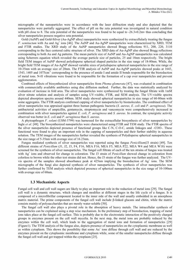
The fungal cell wall also plays a pivotal role in the absorption of heavy metals. The intracellular synthesis of
nanoparticles can be explained using a step wise mechanism. In the preliminary step of bioreduction, trapping of metal
ions takes place at the fungal cell surface. This is probably due to the electrostatic interaction of the positively charged
groups in enzymes present on the cell wall mycelia. In the next step, the metal ions are probably reduced by the
enzymes within the cell wall, which leads to the aggregation of metal ions and formation of nanoparticles [29]
(Figure1). The TEM analysis of the fungus also, depicts presence of nanoparticles on the cytoplasmic membrane as well
as within cytoplasm. This shows the possibility that some Au+ ions diffuse through cell wall and are reduced by the
enzymes present on the cytoplasmic membrane and cytoplasm while, some of the smaller nanoparticles diffuse through
the fungal cell wall and get trapped within the cytoplasm [25].
_______________________________________________________________________________________
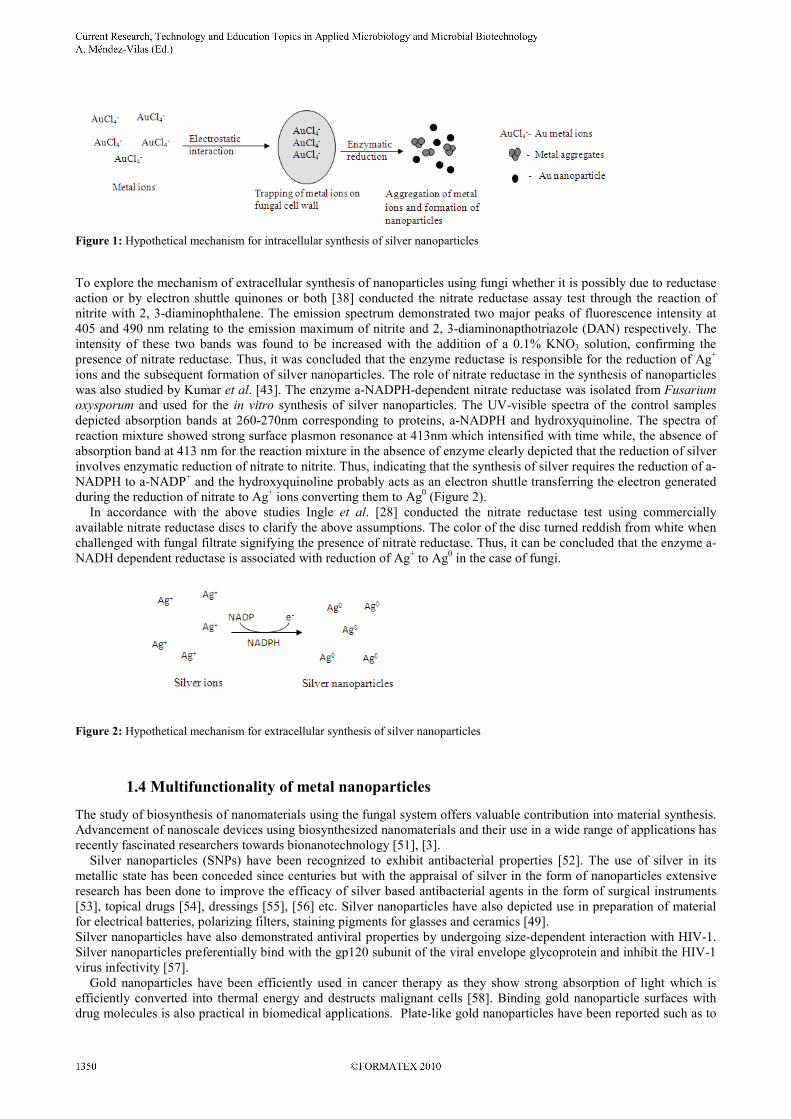
Figure 1: Hypothetical mechanism for intracellular synthesis of silver nanoparticles
To explore the mechanism of extracellular synthesis of nanoparticles using fungi whether it is possibly due to reductase
action or by electron shuttle quinones or both [38] conducted the nitrate reductase assay test through the reaction of
nitrite with 2, 3-diaminophthalene. The emission spectrum demonstrated two major peaks of fluorescence intensity at
405 and 490 nm relating to the emission maximum of nitrite and 2, 3-diaminonapthotriazole (DAN) respectively. The
intensity of these two bands was found to be increased with the addition of a 0.1% KNO3 solution, confirming the
presence of nitrate reductase. Thus, it was concluded that the enzyme reductase is responsible for the reduction of Ag+
ions and the subsequent formation of silver nanoparticles. The role of nitrate reductase in the synthesis of nanoparticles
was also studied by Kumar et al. [43]. The enzyme a-NADPH-dependent nitrate reductase was isolated from Fusarium
oxysporum and used for the in vitro synthesis of silver nanoparticles. The UV-visible spectra of the control samples
depicted absorption bands at 260-270nm corresponding to proteins, a-NADPH and hydroxyquinoline. The spectra of
reaction mixture showed strong surface plasmon resonance at 413nm which intensified with time while, the absence of
absorption band at 413 nm for the reaction mixture in the absence of enzyme clearly depicted that the reduction of silver
involves enzymatic reduction of nitrate to nitrite. Thus, indicating that the synthesis of silver requires the reduction of a-
NADPH to a-NADP+ and the hydroxyquinoline probably acts as an electron shuttle transferring the electron generated
during the reduction of nitrate to Ag+ ions converting them to Ag
0 (Figure 2).
In accordance with the above studies Ingle et al. [28] conducted the nitrate reductase test using commercially
available nitrate reductase discs to clarify the above assumptions. The color of the disc turned reddish from white when
challenged with fungal filtrate signifying the presence of nitrate reductase. Thus, it can be concluded that the enzyme a-
NADH dependent reductase is associated with reduction of Ag+ to Ag
0 in the case of fungi.
Figure 2: Hypothetical mechanism for extracellular synthesis of silver nanoparticles
1.4 Multifunctionality of metal nanoparticles
The study of biosynthesis of nanomaterials using the fungal system offers valuable contribution into material synthesis.
Advancement of nanoscale devices using biosynthesized nanomaterials and their use in a wide range of applications has
recently fascinated researchers towards bionanotechnology [51], [3].
Silver nanoparticles (SNPs) have been recognized to exhibit antibacterial properties [52]. The use of silver in its
metallic state has been conceded since centuries but with the appraisal of silver in the form of nanoparticles extensive
research has been done to improve the efficacy of silver based antibacterial agents in the form of surgical instruments
[53], topical drugs [54], dressings [55], [56] etc. Silver nanoparticles have also depicted use in preparation of material
for electrical batteries, polarizing filters, staining pigments for glasses and ceramics [49].
Silver nanoparticles have also demonstrated antiviral properties by undergoing size-dependent interaction with HIV-1.
Silver nanoparticles preferentially bind with the gp120 subunit of the viral envelope glycoprotein and inhibit the HIV-1
virus infectivity [57].
Gold nanoparticles have been efficiently used in cancer therapy as they show strong absorption of light which is
efficiently converted into thermal energy and destructs malignant cells [58]. Binding gold nanoparticle surfaces with
drug molecules is also practical in biomedical applications. Plate-like gold nanoparticles have been reported such as to
_______________________________________________________________________________________
slice through the cells after surface functionalization with drugs or labelling the particles with various desired carrier
biomolecules. Additionally, the infrared absorption of the anisotropic particles could also be utilized for hyperthermia in
various therapeutic treatments like for cancer and in architectural applications like infrared-absorbing optical coatings
[59]. But recently gold nanoparticles have come up as alternative materials to assist in the purification of water as
selective photothermal agents using visible light to control microorganisms in water [60].
Optical properties of non-spherical gold nanoparticles are easy to tune along with particle size and shape hence, show
a wide range of applications like optical labelling for biosensing events and biomedical labelling. Thus, it is speculated
that these non-spherical gold nanoparticles could play a major role in future cancer diagnosis and therapy [61]. Gold
nanoparticles are also found to play important role in areas like pollution control (e.g. water & air filter), chemical
processing, fuel cell technology (e.g. solar & fuel cells) [62].
Zirconia nanoparticles due to their intrinsic physico-chemical properties are used as resistant coating tool, in high
temperature engine components and as catalyst [32]. Titanium nanoparticles have been used in sunscreen, cosmetics
[33]. Samarium nanoparticles have paved a new application of nanotechnology in nuclear medicine for detection and
treatment of cancerous tumours [63]
Alginate nanoparticles have been used to develop a natural polymer based nanoparticle drug delivery system for four
key Anti-Tuberculosis drugs (Rifampicin, Isoniazid, Pyrazinamide and Ethambutol). The efficiency of drug
encapsulated alginate nanoparticles was tested against TB-infected mice, which depicted complete bacterial clearance
from lungs, liver and spleen [64]. Platinum nanoparticles are noteworthy in the industrial production of fuel cells [26].
Magnetite nanomaterials synthesized using the biosynthetic approach can be used as precursors instead of cyanide
complexes to avoid cyanide toxicity to the environment [65]. Magnetic nanoparticles are also useful in the removal of
environmental contaminants from water [66]. Semiconductor CdSe quantum dots due to their optical and electronic
properties can be used as luminescent probes [42].
Antimony oxide (Sb2O3) nanoparticles are applicable as conductive materials, effective catalyst, functional filler and
optical material [67].
Recent application of nanoparticles includes areas like biological sensing and labelling and imaging of live cells and
tissues [68]. Future applications of nanoparticles involve development, transport and detection of digital information for
security of home land, high speed data communication and computing components [61].
1.5 Future Perspectives
Nanotechnology is one of the most significant areas of research and its impact is felt in our present day life. With the
advancement of the technology nanoparticles are expected to solve large scale problems using nanoscale solutions.
The biosynthetic route for the synthesis of metal nanoparticles using fungi is a simple process involving the reaction of
fungal culture with aqueous solutions of metal ions. But there are a number of questions, which need to be addressed.
The synthesis process points out that there are a number of reducing agents involved in the reduction of metal ions and
corresponding formation of nanoparticles. These reducing agents also affect the size and shape of nanoparticles hence,
there is a need to investigate the exact mechanism involved in the biosynthesis of nanoparticles. Studies on the synthesis
of nanoparticles of specific size and shape depend on different factors like temperature and light intensity. Biosynthetic
approach for nanoparticle synthesis also needs to focus on the shape selectivity and size monodispersity of
nanoparticles. Studying the novel shape and size dependent physical and chemical properties of nanoparticles and their
subsequent interaction could help in development of a new range of photonic and electronic devices that can control and
manipulate light at nanoscale. Establishment of low-cost recovery techniques to make the synthesis process
commercially feasible also needs to be undertaken.
1.6 Conclusion
The fungi are now known to be efficient tool for synthesis of nanoparticles by both intra- and extracellular methods.
The fungal system has shown its compatibility over other groups of organisms as the handling of fungal biomass and its
downstream processing is much simpler. A number of metallic nanoparticles including silver, gold, titanium, silica,
zirconium and platinum have been successfully synthesized using the fungal system. The fungal-derived nanoparticles
have depicted a wide range of applications in different fields of science including medicines, pharmaceutical industry,
agriculture, electronics, etc. But there are certain areas which need to be worked out before exploring the complete
potential. An exact mechanism of synthesis of nanoparticles is yet to be discovered. Understanding the exact
mechanism involved in the synthesis of nanoparticles and the effect of different factors on the reduction of metal ions
will help in developing low-cost techniques for the synthesis and recovery of nanoparticles.
Thus, sketching different practicalities and reducing agent involved in the synthesis of nanoparticles, would help in
understanding the fungal system as one of the most efficient systems for harnessing nanoparticles.
_______________________________________________________________________________________
References
[1] Uskokovic, V. Nanomaterials and Nanotechnologies: Approaching the crest of this big wave. Current Nanoscience. 2008; 4: 119-
129.
[2] Rai, M, Yadav, A, Gade, A. Silver nanoparticles: as a new generation of antimicrobials. Biotechnology Advance. 2009a; 27: 76-
83.
[3] Rai, M, Yadav, A, Gade, A. Current trends in Phytosynthesis of metal nanoparticles. Critical Reviews in Biotechnology. 2008;
28(4): 277-284.
[4] Jana, NR, Pal, T, Sau, T K, Wang, Z L. Seed- mediated growth method to prepare cubic copper nanoparticles. Current Science.
2000; 79(9): 1367-1370.
[5] Kohler, JM, Hubner, U, Romanus, H, Wagner, J. Formation of star-like and core-shell Au-Ag nanoparticles during two and three
step preparation in batch and microfluidic systems. Journal of Nanomaterial. 2007; 1155: 98134-98141.
[6] Nair, L S and Laurencin, C T. Silver nanoparticles: Synthesis and Therapeutic applications. Journal of Biomedical
Nanotechnology. 2007; 3: 301-316.
[7] Southam, G and Beveridge, T J. The in vitro formation of placer gold by bacteria. Geoch Cosmoch Acta. 1994; 58: 4527-4530.
[8] Klaus, T, Granqvist, C G, Joerger, R, Olsson, E. Silver-based crystalline nanoparticles microbially fabricated. Proceedings of the
National Academy of Science. 1999; 96(24): 13611-13614.
[9] Ahmad, A, Senapati, S, Khan, M I, Kumar, R, Sastry, M. Extracellular biosynthesis of monodisperse gold nanoparticles by a
novel extremophilic actinomycete Thermonospora sp. Langmuir. 2003; 19: 3550-3553.
[10] Konishi, Y, Nomura, T, Tsukiyama, T, Saioth, N. Microbial preparation of gold nanoparticles by anaerobic bacterium.
Transactions Material Research Society Japan. 2004; 29: 2341-2343.
[11] Prasad, K, Jha, A K, Kulkarni, A R. Lactobacillus assisted synthesis of Titanium nanoparticles. Nanoscale Research Letters.
2007; 2: 248-250.
[12] Dameron, C T, Reese, R N, Mehra, R K, Korton, A R, Caroll, P J, Steigerwald, M L, Brus, L E, Winge, D R. Biosynthesis of
cadmium sulfide quantum semiconductor crystallites. Nature. 1989; 338: 596-597.
[13] Kowshik, M, Deshmukh, N, Kulkarni, S K, Paknikar, K M, Vogel, W, Urban, J. Microbial synthesis of Semiconductor CdS
nanoparticles their characterization and their use in fabrication of an ideal diode. Biotechnology and Bioengineering. 2002;
78(5): 583-588.
[14] Kowshik, M A,Ashataputre, S, Kharrazi, S, Kulkarni, S K, Paknikari, K M, Vogel, W, Urban, J. Extracellular synthesis of
silver nanoparticles by a silver-tolerant yeast strain MKY3. Nanotechnology. 2003; 14: 95-100.
[15] Gardea –Torresedey, J L, Armendariz, V, Herreira, I, Parsons, J G, Peralta- Videa, J R, Teimann, K J, Torresday, K J. Binding
of silver (I) ions by alfalfa biomass (Medicago sativa): batch, pH, time, temperature and ionic strength studies. Journal of
Hazardous Substance Research. 2003; 4: 1-15.
[16] Shankar, S S, Ahmad, A, Pasricha, R, Sastry, M. Bioreduction of chloroaurate ions by Geranium leaves and its endophytic
fungus yields gold nanoparticles of different shapes. Journal of Material Chemistry. 2003; 13: 1822-1826.
[17] Shankar, SS, Ahmad, A, Rai, A, Sastry, M. Rapid synthesis of Au Ag and bimetallic Au core-Ag shell nanoparticles by using
neem (Azadirachta indica) leaf broth. Journal of Colloids and Interface Science. 2004; 275(5): 496-502.
[18] Armendariz, V, Gardea- Torresdey, J L, Herrera, I, Moller, A D, Jose-Yacaman, M, Peralta-Videa, J R, Troiani, H. HRTEM
characterization of gold nanoparticles produced by wheat biomass. Revista Mexicana de Fisica. 2004; 50: 7–11.
[19] Huang, J, Chen, C, He, N, Hong, J, Lu, Y, Qingbiao, L, Shao, W, Sun, D, Wang, X H, Wang, Y, Yiang, X. Biosynthesis of
silver and gold nanoparticles by novel sundried Cinnamomum camphora leaf. Nanotechnology. 2007; 18: 105-106.
[20] Li, S, Qui, L, Shen, Y, Xie, A, Yu, X, Zhang, L, Zhang, Q. Green synthesis of silver nanoparticles using Capsicum annum L
extract. Green Chemistry. 2007; 9: 852-858.
[21] Rai, M, Yadav, A, Bridge, P, Gade, A. Myconanotechnology: A new and emerging science. In: Rai, M K and Bridge, P D. eds.
Applied Mycology. New York, CAB International. 2009b; 14: 258-267.
[22] Mandal, D, Bolander, M E, Mukhopadhyay, D, Sarkar, G, Mukherjee, P. The use of microorganisms for the formation of metal
nanoparticles and their application. Applied Microbiology and Biotechnology. 2006; 69: 485-492.
[23 ] Holan, Z R and Volesky, B. Accumulation of cadmium, lead and nickel by fungal and wood biosorbents. Applied Biochemistry
and Biotechnology. 1995; 53(2): 133-146.
[24] Gade, A K, Bonde, P, Ingle, A P, Marcato, P D, Duran, N, Rai, M K. Exploitation of Aspergillus niger for synthesis of silver
nanoparticles. Journal of Biobased Material and Bioenergy. 2008; 2: 243-247.
[25] Sastry, M, Ahmad, A, Khan, M I, Kumar, R. Biosynthesis of metal nanoparticles using fungi and actinomycetes. Current
Science. 2003; 85 (2): 162-170.
[26] Riddin, T. L., Gericke, M., Whiteley, C. G. Analysis of the inter- and extracellular formation of platinum nanoparticles by
Fusarium oxysporum f sp lycopersici using response surface methodology. Nanotechnology. 2006; 17: 3482- 3489.
[27] Mukherjee, P, Roy, M, Mandal, B P, Dey, G K, Mukherjee, P K, Ghatak, J, Tyagi, A K, Kale, S P. Green synthesis of highly
stabilized nanocrystalline silver particles by a non-pathogenic and agriculturally important fungus asperellum. Nanotechnology.
2008; 19: 103-110.
[28] Ingle, A, Gade, A, Pierrat, S, Sonnichsen, C, Rai, M K. Mycosynthesis of silver nanoparticles using the fungus Fusarium
acuminatum and its activity against some human pathogenic bacteria. Current Nanoscience. 2008; 4: 141-144.
[29] Mukherjee, P, Ahmad, A, Mandal, D, Senapati, S, Sainkar, S R, Khan, M I, Ramani, R, Parischa, R, Ajayakumar, P V, Alam,
M, Sastry, M, Kumar, R. Bioreduction of AuCl4- ions by the fungus Verticillium sp and surface trapping of the gold
nanoparticles formed. Angewante Chemie International Edition. 2001; 40(19): 3585-3588.
[30] Ahmad, A, Mukherjee, P, Mandal, D, Senapati, S, Khan, M I, Kumar, R, Sastry, M. Enzyme mediated extracellular biosynthesis
of CdS nanoparticles by the fungus Fusarium oxysporum. Journal of American Chemical Society. 2002; 124: 12108-12109.
[31] Chen, J C, Lin, Z H, Ma, X X. Evidence of the production of silver nanoparticles via pretreatment of Phoma sp 32883 with
silver nitrate. Letters in Applied Microbiology. 2003; 37: 105-108.
_______________________________________________________________________________________
[32] Bansal, V, Rautray, D, Ahamd, A, Sastry, M. Biosynthesis of zirconia nanoparticles using the fungus Fusarium oxysporum.
Journal of Material Chemistry. 2004; 14: 3303- 3305.
[33] Bansal, V, Rautray, D, Bharde, A, Ahire, K, Sanyal, A, Ahmad, A, Sastry, M. Fungus-mediated biosynthesis of silica and titania
particles. Journal of Material Chemistry. 2005; 15: 2583-2589.
[34] Bhainsa, K C and D’Souza, S F. Extracellular biosynthesis of silver nanoparticles using the fungus Aspergillus fumigates.
Colloids and Surfaces B: Biointerface. 2006; 47: 160-164.
[35] Shahi, S K and Patra, M. Microbially synthesized bioactive nanoparticles and their formulation active against human pathogenic
fungi. Reviews Advanced Material Science. 2003; 5: 501-509.
[36] Ahmad, A, Senapati, S, Khan, M I, Kumar, R, Sastry, M. Extra-/Intracellular Biosynthesis of Gold Nanoparticles by an
Alkalotolerant Fungus Trichothecium sp. Journal of Biomedical Nanotechnology. 2005; 1: 47-53.
[37] Bharde, A, Rautray, D, Bansal, V, Ahmad, A, Sarkar, I, Yusuf, S M, Sanyal, M, Sastry, M. Extracellular Biosynthesis of
Magnetite using fungi. Small. 2006; 2(1): 135-141.
[38] Duran, N, Marcato, P D, Alves, O L, DeSouza, G, Esposito, E. Mechanistic aspects of biosynthesis of silver nanoparticles by
several Fusarium oxysporum strains. Journal of Nanobiotechnology. 2005; 3: 1-8.
[39] Gericke, M and Pinches, A. Microbial production of gold nanoparticles. Gold Bulletin. 2006; 39(1): 22-28.
[40] Basavaraja, S, Balaji, S D, Lagashetty, A, Rajasab, A H, Venkataraman, A. Extracellular biosynthesis of silver nanoparticles
using the fungus Fusarium semitectum. Material Research Bulletin. 2007; 43(5): 1164-1170.
[41] Vigneshwaran, N, Ashtaputre, M, Nachane, R P, Paralikar, K M, Balasubramanya, H. Biological synthesis of silver
nanoparticles using the fungus Aspergillus flavus. Material Letters. 2007; 61(6): 1413-1418.
[42] Kumar, A S, Ansary, A A, Ahmad, A, Khan, M I. Extracellular Biosynthesis of CdSe Quantum Dots by the Fungus Fusarium
oxysporum. Journal of Biomedical Nanotechnology. 2007a; 3: 190-194.
[43] Kumar, S A, Abyaneh, M K, Gosavi, S W, Kulkarni, S K, Pasricha, R, Ahmad, A, Khan, M I. Nitrate reductase-mediated
synthesis of silver nanoparticles from AgNO3. Biotechnology Letters. 2007b; 29: 439-445.
[44] Mohammadian, A, Shaojaosadati, S A, Rezee, M H. Fusarium oxysporum mediates photogeneration of silver nanoparticles.
Scientia Iranica. 2007; 14(4): 323-326.
[45] Sadowski, Z, Maliszewska, G B, Polowczyk, I, Kozlecki, T. Synthesis of silver nanoparticles using microorganisms. Material
Science-Poland. 2008; 26(2): 419-425.
[46] Sawale, B D, Salimath, B, Deshpande, R, Bedre, M D, Prabhakar, B K, Venkataraman, A. Biosynthesis and stabilization of Au
and Au–Ag alloy nanoparticles by fungus, Fusarium semitectum. Science Technology Advanced Materials.2008;
doi:10.1088/1468-6996/9/3/035012.
[47] Birla, S S, Tiwari, V V, Gade, A K, Ingle, A P, Yadav, A P, Rai, M K. Fabrication of silver nanoparticles by Phoma glomerata
and its combined effect against Escherichia coli Pseudomonas aeruginosa and Staphylococcus aureus. Letters in Applied
Microbiology. 2009; 27: 76-83.
[48] Ingle, A, Rai, M K, Gade, A, Bawaskar, M. Fusarium solani: a novel biological agent for the extracellular synthesis of silver
nanoparticles. Journal of Nanoparticle Research. 2009; doi 101007/s11051-008-9573-y.
[49] Maliszewska, I, Szewczyk, K, Waszak, K. Biologiacl synthesis of silver nanoparticles. Journal of Physics-Conference Series.
2009; 146: 120-125.
[50] Pebberdy, J. F. Fungal cell walls- A review. In: Kuhn, P J, Trinci, A P J, Jung, M J, Goosey, M W, Coping L G. eds.
Biochemistry of cell walls and membranes in fungi. Germany, Springer- Verlag. 1990; pp 5-30.
[51] Mohanpuria, P, Rana, N K, Yadav, S K. Biosynthesis of nanoparticles: technological concepts and future applications. Journal
of Nanoparticle Research. 2008; 7: 9275-9280.
[52] Morones, J R, Elechiguerra, J L, Camacho, J B, Ramirez, J T. The bactericidal effect of silver nanoparticles. Nanotechnology.
2005; 16: 2346-2353.
[53] Furno F, Morley KS, Wong B, Sharp BL, Arnold PL, Howdle SM. Silver nanoparticles and polymeric medical devices: a new
approach to prevention of infection? Journal of Antimicrobial Chemotherapy. 2004; 54:1019–24.
[54] Tian J, Wong K K Y, Ho C. M., Lok C N, Yu W Y, Che C M, Chiu J F, Tam P K H Topical Delivery of Silver Nanoparticles
Promotes Wound Healing. ChemMedChem. 2006; 00: 171-180.
[55] Duran, N, Alves, O L, De Souza, G I H, Esposito, E, Marcato, P D. Antibacterial effect of silver nanoparticles by fungal process
on textile fabrics and their effluent treatment. Journal of Biomedical Nanotechnology. 2007; 3: 203-208.
[56] Tripathi, A, Chandrasekaran, N, Raichur, A M, Mukherjee, A. Antibacterial applications of silver nanoparticles by aqueous
extract of Azadirachta indica (Neem) leaves. Journal of Biomedical Nanotechnology. 2009; 5(1): 93-98.
[57] Elechiguerra, J L, Burt, J L, Morones, J R, Camacho-Bragado, A, Gao, X, Lara, H H, Yacaman, M J. Interaction of silver
nanoparticles with HIV-1. Journal of Nanobiotechnology. 2005; 3 (6): doi: 101186/1477-3155-3-6.
[58] El-Sayed, HI, Huang, X, El-Sayed, M A. Selective laser photo-thermal therapy of epithelial carcinoma using anti- EGFR
antibody conjugated gold nanoparticles. Cancer Letters. 2006; 239: 129-135.
[59] Jagannathan, R, Poddar, P, Prabhune, A. Cephalexin-Mediated Synthesis of Quasi-Spherical and Anisotropic Gold
Nanoparticles and Their in Situ Capping by the Antibiotic. Journal of Physical Chemistry. 2007; 111: 6933-6938.
[60] El-Adly, A A, El-Senousy, W M, Samhan, F A, Mohamed, M B. Photothermal Efficieny of Gold Nanorods in Controlling
Microorganisms in Water. Journal of Applied Science and Research. 2008; 4(12): 1811-1816.
[61] Delapierre-Treguer, M, Majimel, J, Mornet, S, Duguet, E, Ravaine E. Synthesis of non-spherical gold nanoparticles. Gold
Bulletin. 2008; 41(2): 195-207.
[62] Saifuddin, N, Wong, C W, Yasumira Nur, A A. Rapid biosynthesis of silver nanoparticles using culture supernatant of bacteria
with Microwave irradiation. E- Journal of Chemistry. 2009; 6(1): 61-70.
[63] Ascencio, J A, Rincon, A C, Canizal, G. Synthesis and theoretical analysis of samarium nanoparticles: Perspectives in nuclear
medicine. Journal of Physical Chemistry B. 2005; 109(18): 8806-8812.
_______________________________________________________________________________________
[64] Ahmad, R, Pandey, R, Sharma, S, Khuller, G K. Alginate Nanoparticles as Antituberculosis Drug Carriers: Formulation
Development Pharmacokinetics and therapeutic potential. Indian Journal of Chest Diseases and Allied Science. 2006; 48: 171-
176.
[65] Bharde, A, Rautray, D, Bansal, V, Ahmad, A, Sarkar, I, Yusuf, S M, Sanyal, M, Sastry, M. Extracellular Biosynthesis of
Magnetite using fungi. Small. 2006; 2(1): 135-141.
[66] Gong, P, Li, H, He, X, Wang, K, Hu, J, Tan, W, Zhang, S, Yang, X. Preparation and antibacterial activity of Fe3O4@Ag
nanoparticles. Nanotechnology. 2007; 18: 604-611.
[67] Jha, A K, Prasad, K, Prasad, K. A green low cost biosynthesis of Sb2O3 nanoparticles. Biochemistry and Engineering Journal.
2009; 43(3): 303-306.
[68] De, M, Ghosh, P S, Rotello, V M. Applications of nanoparticles in Biology. Advanced Materials. 2009j; 20(22): 4225-4241.
[69] Kumar, S A, Peter, Y A, Nadaeu, J L. Facile biosynthesis, separation, conjugation of gold nanoparticles to doxorubicin.
Nanotechnology. 2008; doi:10.1088/0957-4484/19/49/495101.
[70] Sanghi, R and Verma, P. Biomimetic synthesis and characterisation of protein capped silver nanoparticles. Bioresorce
Technology. 2009; 100 (1): 501-504.
[71] Balaji, D S, Basavaraja, S, Deshpande, R, Bedre, D, Prabhakar, B K, Venkataraman, A. Extracellular biosynthesis of
functionalized silver nanoparticles by strains of Cladosporium cladosporioides fungus. Colloids and Surfaces B: Biointerfaces.
2009; 68(1): 88-92.
[72] Govender, Y, Riddin, T, Gericke, M, Whiteley, C G. Bioreduction of platinum salts into nanoparticles: a mechanistic
perspective. Biotechnology Letters. 2009; doi 10.1007/s10529-008-9825-z.
_______________________________________________________________________________________