the ictal interictal continuum -...
TRANSCRIPT
The ictal interictal continuum
Disclosures:
Research Support: Columbia University Clinical Translational Science Award
(CTSA), Irving Institute for Clinical and Translational Research
Jan Claassen, MD, PhD Division of Critical Care Neurology
Columbia University College of Physicians & Surgeons New York, NY 10032
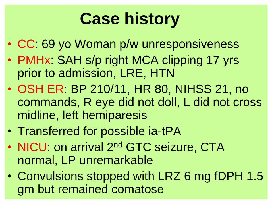
• CC: 69 yo Woman p/w unresponsiveness
• PMHx: SAH s/p right MCA clipping 17 yrs prior to admission, LRE, HTN
• OSH ER: BP 210/11, HR 80, NIHSS 21, no commands, R eye did not doll, L did not cross midline, left hemiparesis
• Transferred for possible ia-tPA
• NICU: on arrival 2nd GTC seizure, CTA normal, LP unremarkable
• Convulsions stopped with LRZ 6 mg fDPH 1.5 gm but remained comatose
Case history

Emergent cEEG
Loaded with Valproic Acid 30 mg/kg
EEG post clinically successfully treated GCSE
• 20 - 48% electrographic seizures
• 14% NCSE
• Most without any clinical signs DeLorenzo Epil 1998, Treiman NEJM 1998
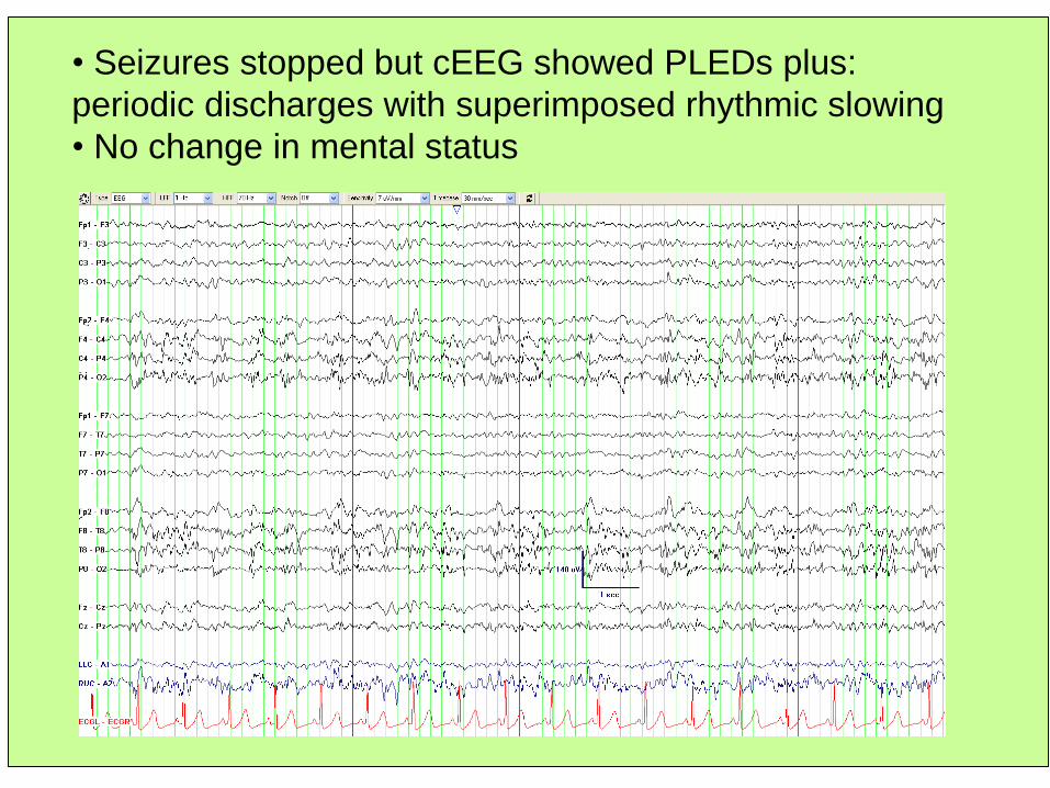
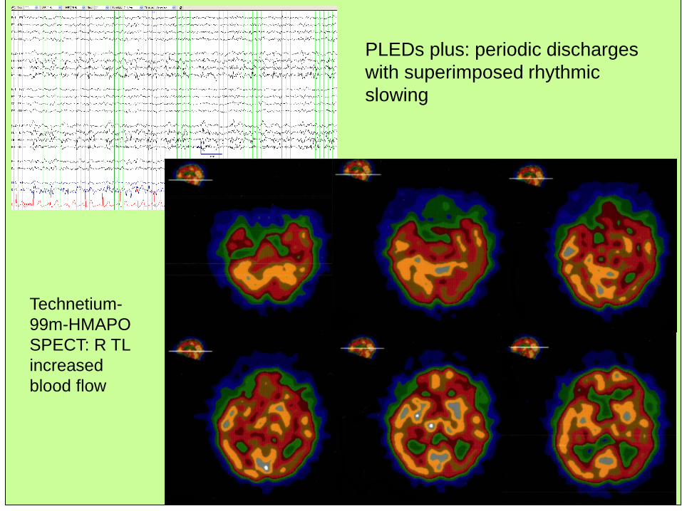
• Seizures stopped but cEEG showed PLEDs plus:
periodic discharges with superimposed rhythmic slowing
• No change in mental status
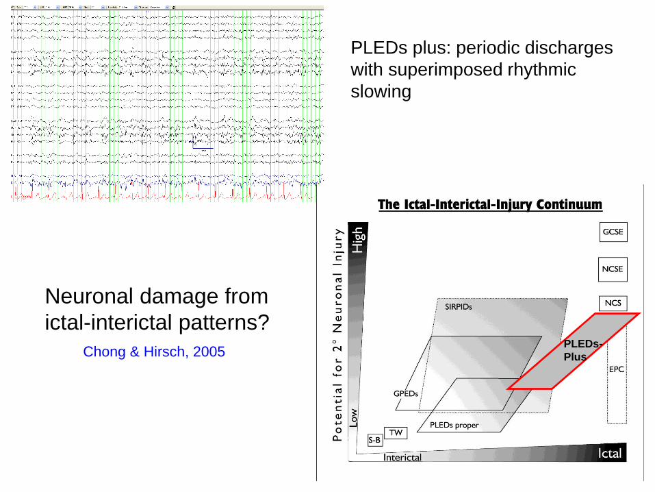
PLEDs plus: periodic discharges
with superimposed rhythmic
slowing
PLEDs-
Plus
Neuronal damage from
ictal-interictal patterns?
Chong & Hirsch, 2005
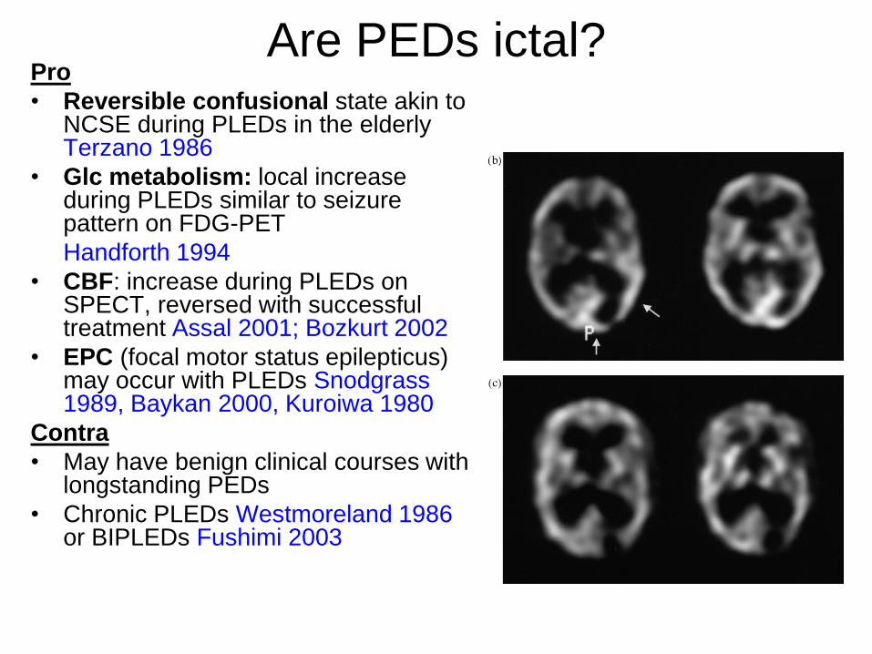
Are PEDs ictal? Pro
• Reversible confusional state akin to NCSE during PLEDs in the elderly Terzano 1986
• Glc metabolism: local increase during PLEDs similar to seizure pattern on FDG-PET
Handforth 1994
• CBF: increase during PLEDs on SPECT, reversed with successful treatment Assal 2001; Bozkurt 2002
• EPC (focal motor status epilepticus) may occur with PLEDs Snodgrass 1989, Baykan 2000, Kuroiwa 1980
Contra
• May have benign clinical courses with longstanding PEDs
• Chronic PLEDs Westmoreland 1986 or BIPLEDs Fushimi 2003
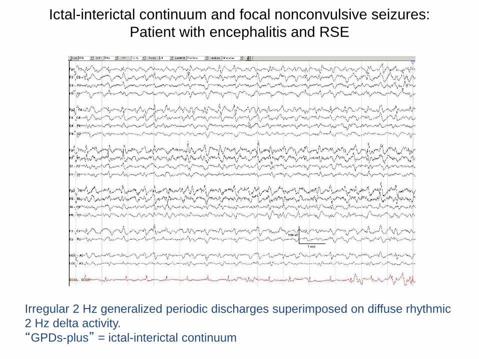
Ictal-interictal continuum and focal nonconvulsive seizures:
Patient with encephalitis and RSE
Irregular 2 Hz generalized periodic discharges superimposed on diffuse rhythmic
2 Hz delta activity.
“GPDs-plus” = ictal-interictal continuum
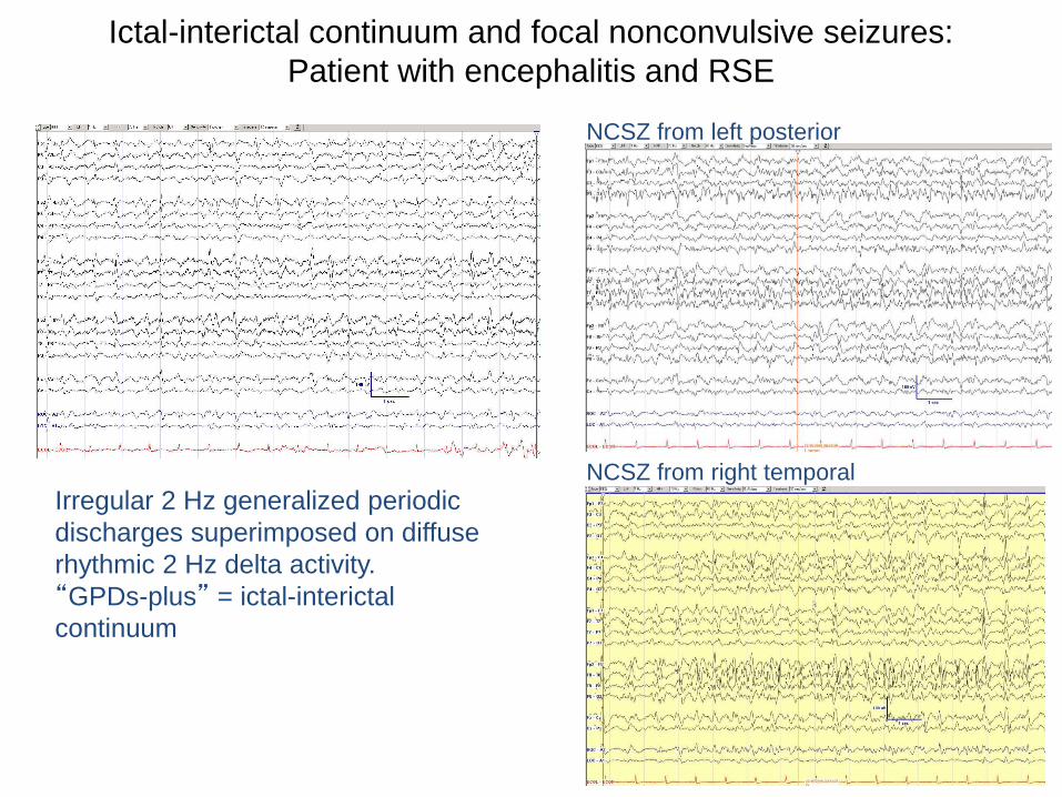
Ictal-interictal continuum and focal nonconvulsive seizures:
Patient with encephalitis and RSE
Irregular 2 Hz generalized periodic
discharges superimposed on diffuse
rhythmic 2 Hz delta activity.
“GPDs-plus” = ictal-interictal
continuum
NCSZ from left posterior
NCSZ from right temporal
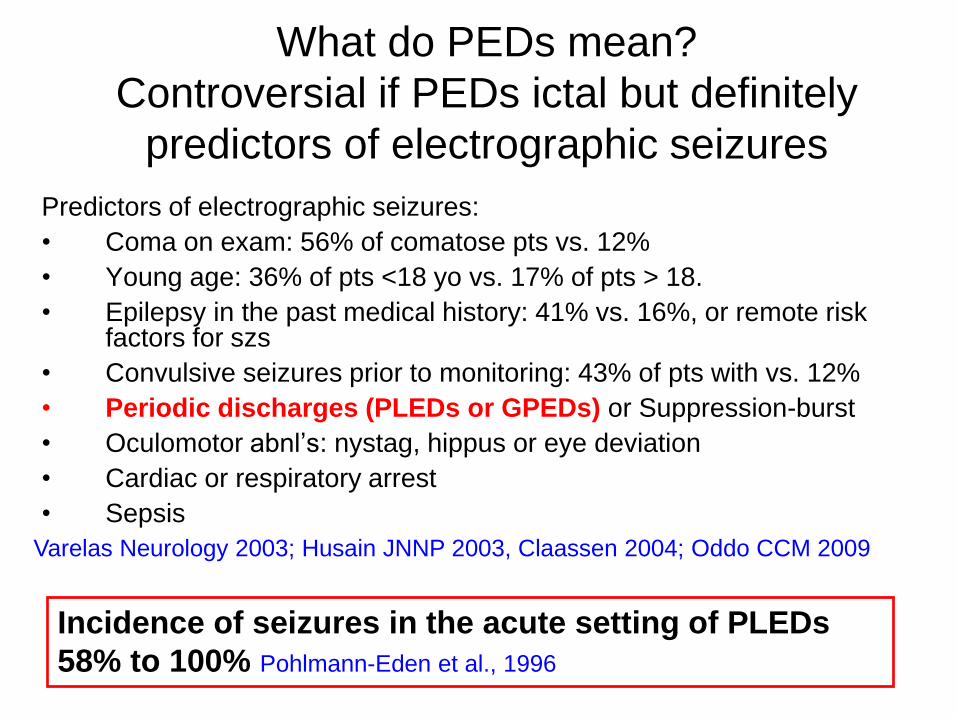
What do PEDs mean?
Controversial if PEDs ictal but definitely
predictors of electrographic seizures
Predictors of electrographic seizures:
• Coma on exam: 56% of comatose pts vs. 12%
• Young age: 36% of pts <18 yo vs. 17% of pts > 18.
• Epilepsy in the past medical history: 41% vs. 16%, or remote risk factors for szs
• Convulsive seizures prior to monitoring: 43% of pts with vs. 12%
• Periodic discharges (PLEDs or GPEDs) or Suppression-burst
• Oculomotor abnl’s: nystag, hippus or eye deviation
• Cardiac or respiratory arrest
• Sepsis
Varelas Neurology 2003; Husain JNNP 2003, Claassen 2004; Oddo CCM 2009
Incidence of seizures in the acute setting of PLEDs
58% to 100% Pohlmann-Eden et al., 1996
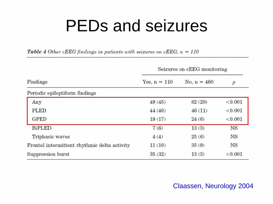
PEDs and seizures
Claassen, Neurology 2004
PLEDs plus: periodic discharges
with superimposed rhythmic
slowing
Technetium-
99m-HMAPO
SPECT: R TL
increased
blood flow
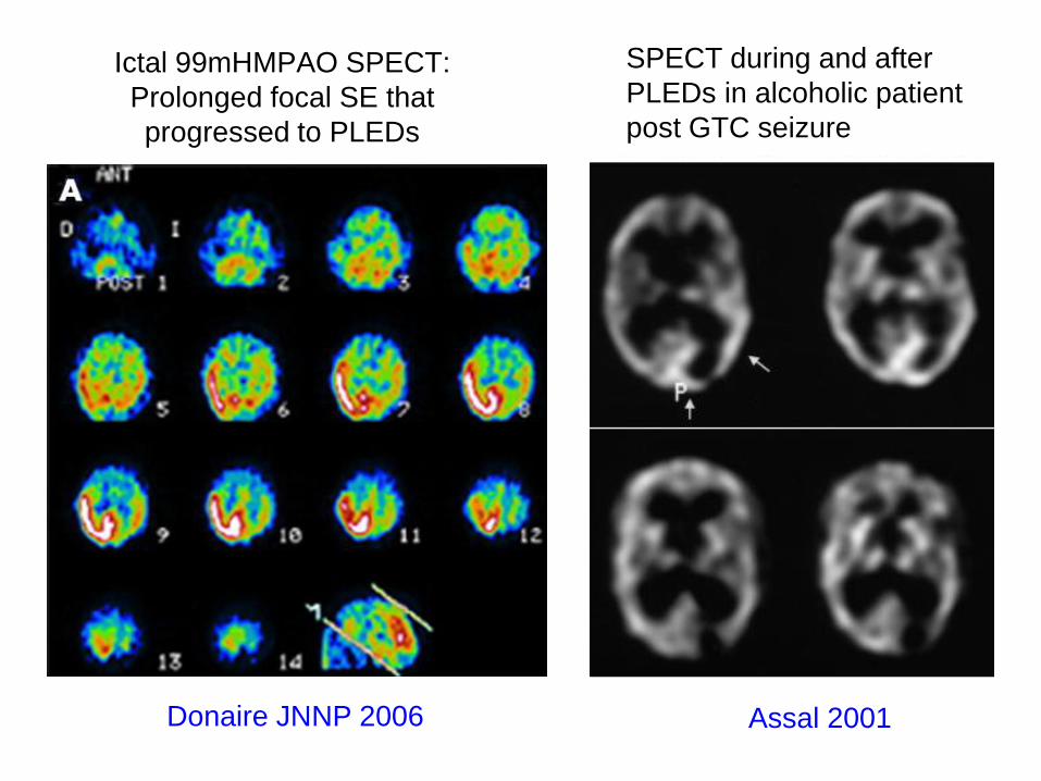
Ictal 99mHMPAO SPECT:
Prolonged focal SE that
progressed to PLEDs
Donaire JNNP 2006
SPECT during and after
PLEDs in alcoholic patient
post GTC seizure
Assal 2001
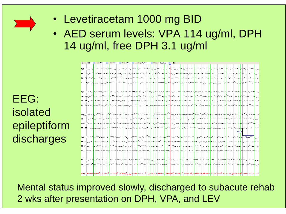
• Levetiracetam 1000 mg BID
• AED serum levels: VPA 114 ug/ml, DPH 14 ug/ml, free DPH 3.1 ug/ml
EEG:
isolated
epileptiform
discharges
Mental status improved slowly, discharged to subacute rehab
2 wks after presentation on DPH, VPA, and LEV
Done!
Successful rehabilitation: she was tapered
off all AEDs for a secondary cognitive
deterioration and returned to her
neurological baseline
Happy ending, but …
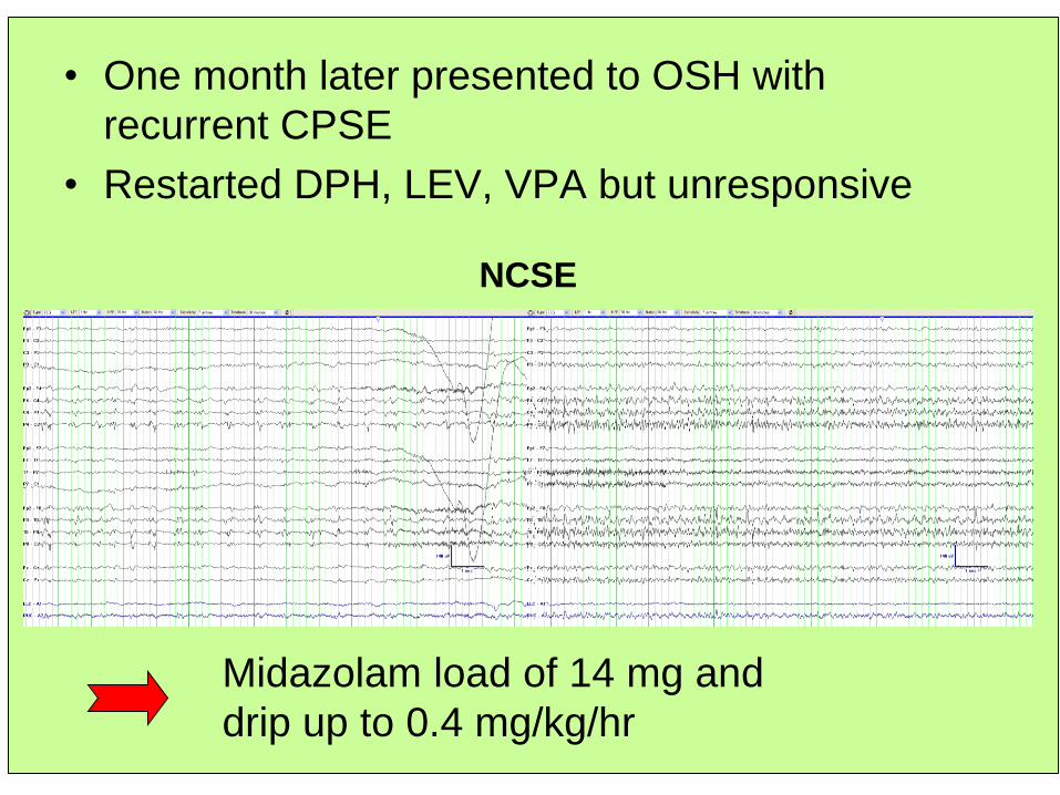
• One month later presented to OSH with
recurrent CPSE
• Restarted DPH, LEV, VPA but unresponsive
Midazolam load of 14 mg and
drip up to 0.4 mg/kg/hr
NCSE
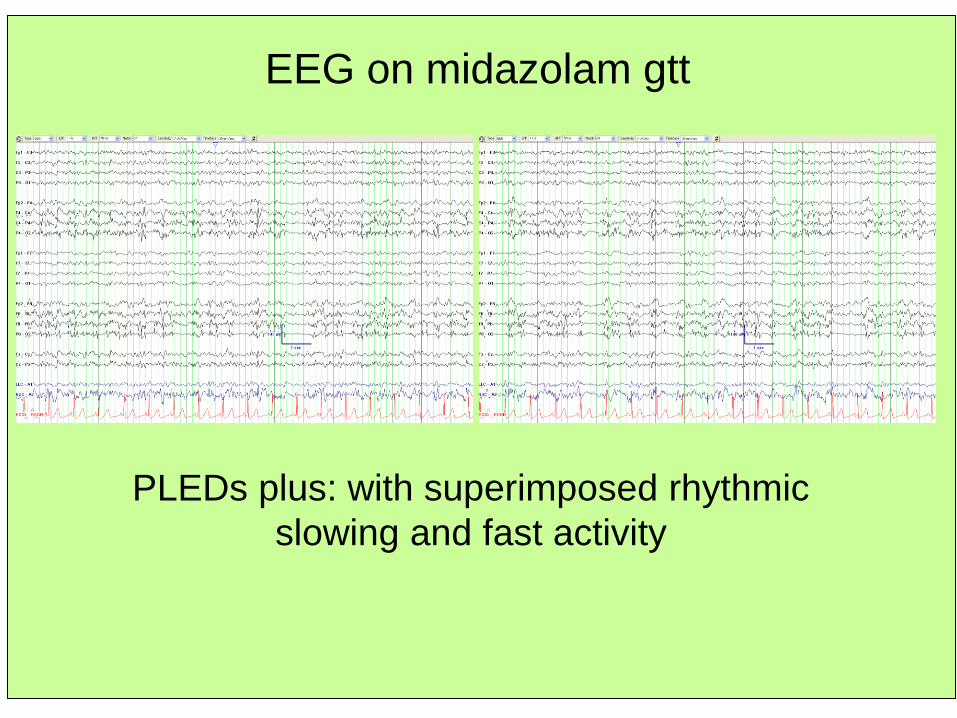
PLEDs plus: with superimposed rhythmic
slowing and fast activity
EEG on midazolam gtt
MTT
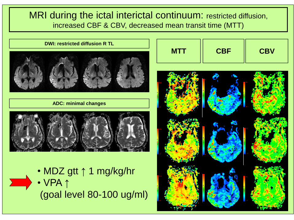
MRI during the ictal interictal continuum: restricted diffusion,
increased CBF & CBV, decreased mean transit time (MTT)
CBF
CBV
DWI: restricted diffusion R TL
ADC: minimal changes
• MDZ gtt ↑ 1 mg/kg/hr
• VPA ↑
(goal level 80-100 ug/ml)
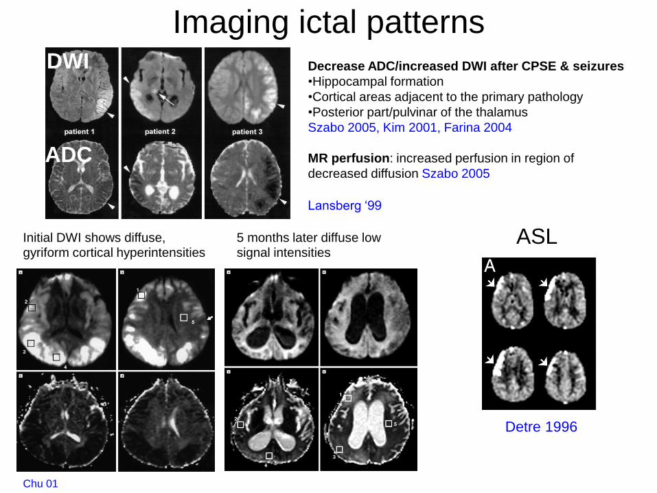
Imaging ictal patterns DWI
ADC
Lansberg ‘99
Decrease ADC/increased DWI after CPSE & seizures
•Hippocampal formation
•Cortical areas adjacent to the primary pathology
•Posterior part/pulvinar of the thalamus
Szabo 2005, Kim 2001, Farina 2004
MR perfusion: increased perfusion in region of
decreased diffusion Szabo 2005
Detre 1996
Initial DWI shows diffuse, gyriform cortical hyperintensities
Chu 01
5 months later diffuse low signal intensities
ASL
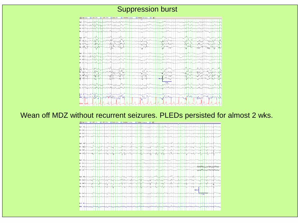
Suppression burst
Wean off MDZ without recurrent seizures. PLEDs persisted for almost 2 wks.
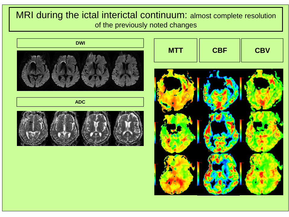
MRI during the ictal interictal continuum: almost complete resolution
of the previously noted changes
DWI
ADC
MTT
CBF
CBV
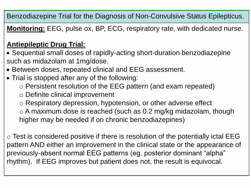
Benzodiazepine Trial for the Diagnosis of Non-Convulsive Status Epilepticus.
Monitoring: EEG, pulse ox, BP, ECG, respiratory rate, with dedicated nurse.
Antiepileptic Drug Trial:
Sequential small doses of rapidly-acting short-duration benzodiazepine
such as midazolam at 1mg/dose.
Between doses, repeated clinical and EEG assessment.
Trial is stopped after any of the following:
o Persistent resolution of the EEG pattern (and exam repeated)
o Definite clinical improvement
o Respiratory depression, hypotension, or other adverse effect
o A maximum dose is reached (such as 0.2 mg/kg midazolam, though
higher may be needed if on chronic benzodiazepines)
o Test is considered positive if there is resolution of the potentially ictal EEG
pattern AND either an improvement in the clinical state or the appearance of
previously-absent normal EEG patterns (eg. posterior dominant “alpha”
rhythm). If EEG improves but patient does not, the result is equivocal.
• Due to thrombocytopenia VPA stopped
• Recurrence of NCSE -> Phenobarbital started
• Neuro: encephalopathic
• While weaning phenobarbital started on LTG
• Left > right facial twitching developed with PLEDs
proper
• Benzodiazepine trial: 1 mg midazolam repeated
doses of (total of 4 mg) -> facial twitching resolved
and PLEDs gone but otherwise no neuro
improvement = Equivocal
•Serial NSE levels throughout hospital course did
not correlate with EEG activity
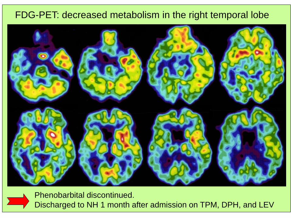
FDG-PET: decreased metabolism in the right temporal lobe
Phenobarbital discontinued.
Discharged to NH 1 month after admission on TPM, DPH, and LEV
Outcome: 2 months after discharge
• continuing to improve in an active
rehabilitation program
• Interactive
• able to recognize family members
• moves all four extremities
• mobilized to a chair
• undergoing ventilator weaning
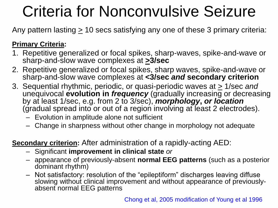
Criteria for Nonconvulsive Seizure Any pattern lasting > 10 secs satisfying any one of these 3 primary criteria:
Primary Criteria:
1. Repetitive generalized or focal spikes, sharp-waves, spike-and-wave or sharp-and-slow wave complexes at >3/sec
2. Repetitive generalized or focal spikes, sharp waves, spike-and-wave or sharp-and-slow wave complexes at <3/sec and secondary criterion
3. Sequential rhythmic, periodic, or quasi-periodic waves at > 1/sec and unequivocal evolution in frequency (gradually increasing or decreasing by at least 1/sec, e.g. from 2 to 3/sec), morphology, or location (gradual spread into or out of a region involving at least 2 electrodes). – Evolution in amplitude alone not sufficient
– Change in sharpness without other change in morphology not adequate
Secondary criterion: After administration of a rapidly-acting AED: – Significant improvement in clinical state or
– appearance of previously-absent normal EEG patterns (such as a posterior dominant rhythm)
– Not satisfactory: resolution of the “epileptiform” discharges leaving diffuse slowing without clinical improvement and without appearance of previously-absent normal EEG patterns
Chong et al, 2005 modification of Young et al 1996
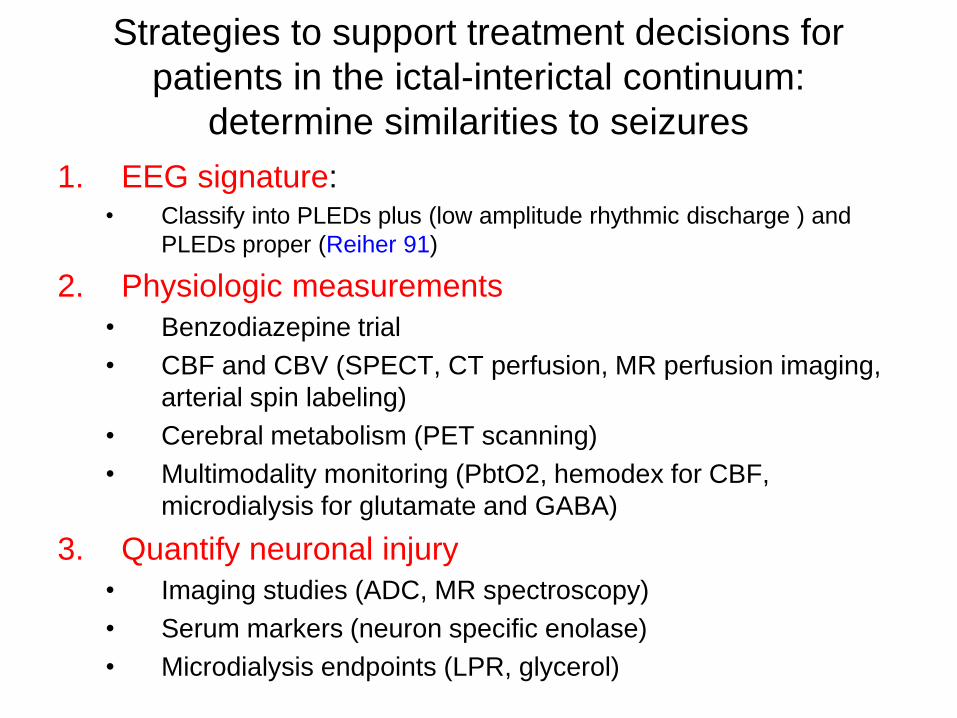
Strategies to support treatment decisions for
patients in the ictal-interictal continuum:
determine similarities to seizures
1. EEG signature: • Classify into PLEDs plus (low amplitude rhythmic discharge ) and
PLEDs proper (Reiher 91)
2. Physiologic measurements
• Benzodiazepine trial
• CBF and CBV (SPECT, CT perfusion, MR perfusion imaging,
arterial spin labeling)
• Cerebral metabolism (PET scanning)
• Multimodality monitoring (PbtO2, hemodex for CBF,
microdialysis for glutamate and GABA)
3. Quantify neuronal injury
• Imaging studies (ADC, MR spectroscopy)
• Serum markers (neuron specific enolase)
• Microdialysis endpoints (LPR, glycerol)
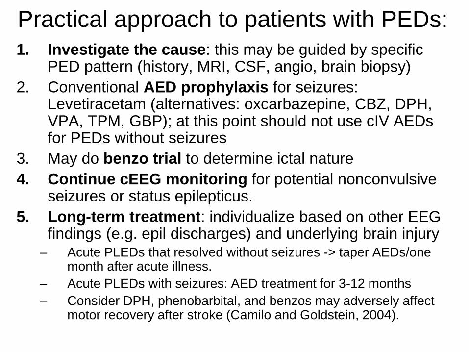
Practical approach to patients with PEDs:
1. Investigate the cause: this may be guided by specific PED pattern (history, MRI, CSF, angio, brain biopsy)
2. Conventional AED prophylaxis for seizures: Levetiracetam (alternatives: oxcarbazepine, CBZ, DPH, VPA, TPM, GBP); at this point should not use cIV AEDs for PEDs without seizures
3. May do benzo trial to determine ictal nature
4. Continue cEEG monitoring for potential nonconvulsive seizures or status epilepticus.
5. Long-term treatment: individualize based on other EEG findings (e.g. epil discharges) and underlying brain injury
– Acute PLEDs that resolved without seizures -> taper AEDs/one month after acute illness.
– Acute PLEDs with seizures: AED treatment for 3-12 months
– Consider DPH, phenobarbital, and benzos may adversely affect motor recovery after stroke (Camilo and Goldstein, 2004).
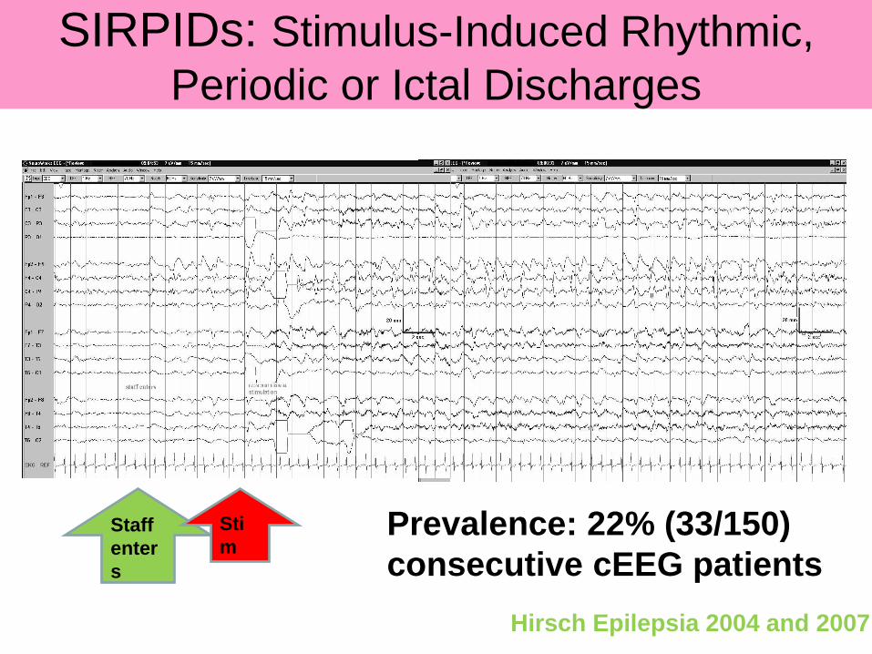
SIRPIDs: Stimulus-Induced Rhythmic,
Periodic or Ictal Discharges
Hirsch Epilepsia 2004 and 2007
Staff
enter
s
Sti
m Prevalence: 22% (33/150)
consecutive cEEG patients
Division Critical Care Neurology
• J Michael Schmidt, PhD
• Stephan A. Mayer, MD
• NICU Fellows
• NICU nurses
• Neurology residents
Department of
Neurosurgery
• E Sander Connolly, MD
• Neurosurgery residents
Acknowledgements Depart. of Biomedical Informatics
• Adler Perotte, MD
• Samantha Kleinberg, PhD
• David Albers, PhD
• George Hripcsak, MD, MS
Comprehensive Epilepsy Center
• Bin Tu, MD
• EEG Technicians
• Epilepsy attendings
• Epilepsy fellows