ictal patterns in generalized epilepsy - id&a :: medical ... scalp patterns...ictal patterns in...
TRANSCRIPT
Journal o{Clinical N<'urophysiology10(3):268-280. Raven Press. Ltd.. New York@ 1993 American Electroencephalographic Society
LUhele5.5
Ictal Patterns in Generalized Epilepsy
Ivo Drury and Thomas R. Henry
EEG and Epilepsy Laboratories and the Department of Neurology. University of Michigan Medical School.Ann Arbor, Michigan. USA
Summary: Ictal EEG may be of great benefit in facilitating accurate classifica-tion of the underlying seizure disorder in some patients and thus guiding furtherinvestigation and management. Ictal recordings in patients with generalizedepilepsies are protean in their manifestations and yet may have considerableoverlap. Classification is only possible through careful synthesis of all availableclinical and electrophysiological data. Although the underlying pathophysio-logical mechanisms of the generalized epilepsies remain uncertain, evidencefrom EEG recordings tends to support Gloor's concept of corticoreticular epi-lepsy. KeyWords: Ictal EEG-Corticoreticular epilepsy- Idiopathic generalizedepilepsy-Symptomatic generalized epilepsy.
This article discusses ictal patterns recorded fromscalp EEG in patients with various forms of general-ized epilepsy. We emphasize the correlation of ictalsemiology with electrographic changes, the value ofictal over interictal recordings in accurate classifica-tion of the epilepsies, some of the limitations of scalpEEG recording of ictal activities, and the major pit-falls in recording and interpreting ictal discharges.
CLASSIFICATION OF THEGENERALIZED EPILEPSIES
The Commission on Classification and Terminol-ogy of the International League Against Epilepsy hasproposed revised classifications of both epileptic sei-zures (1981) and the epilepsies and epileptic syn-dromes (1989). It defines the generalized epilepsiesand syndromes as epileptic disorders with general-ized seizures, Le.,"seizures in which the first clinical
Address correspondence and reprint requests to Dr. I. Drury atEEG Laboratory, University HospitallB300/0036, 1500E. Medi-cal Center Drive, Ann Arbor, M148109, U.SA
--
changes indicate initial involvement of both hemi-spheres. . .. The ictal electroencephalographic pat-terns initially are bilateral." There are two majortypes of generalized epilepsies: idiopathic general-ized epilepsy with an EEG expression of a general-ized, bilateral, synchronous, symmetrical dischargewith no known or suspected etiology other than apossible hereditary predisposition and those gen-eralized epilepsies secondary to some underlyingprocess either identified (symptomatic) or unidenti-fied (cryptogenic). The classification utilizes inter-ictal and in some cases ictal EEG data in describingthese epileptic syndromes; yet, the vast majority ofpatients with seizures have their epilepsy classifiedand treatment instituted without resort to recordedictal data. Despite some inherent limitations, e.g.,the occurrence of complex partial seizures in somepatients with symptomatic generalized epilepsy, orknowing whether patients with nocturnal general-ized tonic-clonic seizures might have a focal onset ornot, the classification represents the best availablescheme for categorizing patients with seizures toguide investigation and management.
268
-- -- - ---

ICTAL PATTERNS IN GENERAliZED EPILEPSY 269
PATHOPHYSIOLOGY
An understanding of the mechanisms involved inthe production of epileptiform discharges on scalpEEG would be helpful in interpreting interictal andictal recordings, but, despite much clinical andexperimental review, the responsible mechanismsremain uncertain. The bilaterally synchronous andsymmetric spike-and-wave discharges on the scalpsuggest a deep-seated generator. An early conceptwas that of centrencephalic epilepsy, arising fromMorison and Dempsey's (1942) work on the specificand nonspecific thalamic activating systems. Jasper~and Droogleever-Fortuyn (1946) produced bisyn-chronous spike-and-wave discharges in the cat byelectrical stimulation of the intralaminar nucleus ofthe thalamus at 3 Hz. Similarly, alumina implanta-tions into the intralaminar nucleus of the thalamusand reticular formation of the midbrain of the catproduced typical 3-Hz spike-wave activity with sei-zures (Guerrero-Gigueroa et aI., 1963). In a study ofdepth recording in six children with absence sei-zures, Williams (1953) proposed that the 3-Hz spike-wave activity had its origin in the thalamus and thenincreased in voltage until it reached the cortex, whichthen set up a reverberating cortical-thalamic circuit.In some patients with symptomatic generalized epi-lepsy, Velasco et aI. (1989) described spike-wavecomplexes in the centromedian nucleus of the thala-mus preceding bilateral surface cortical dischargesduring symptoms of nonconvulsive generalized sei-zures.
Other clinical and experimental work, however,suggested that the cortex played a primary role.TOkel and Jasper (1952)described generalized spike-wave discharges in patients with lesions in anteriorparasagittal areas and mesial lesions of the brain.Marcus and Watson (1966) using bilaterally symmet-rical cobalt foci in the monkey cortex produced syn-chronous spike-wave discharges if the corpus callo-sum was intact. Niedermeyer et aI. (1969), in depthrecordings in patients with generalized spike-and-wave, found focal discharges in the frontal lobe intwo patients. Bancaud et aI. (1974) showed that elec-trical stimulation of human mesial frontal cortexproduced clinical and EEG activity indistinguish-able from spontaneous spike-wave bursts and ab-sence. Working with the photosensitive West Africanbaboon Papio papio, Naquet et aI. (1972) showedthat the discharges of the spontaneous and photical-ly induced seizures have a frontorolandic origin.
From his experimental work, Gloor has provideda unitary hypothesis, termed the corticoreticular epi-
lepsies. Large intramuscular injections of penicillinin the cat produced a transient epileptogenic statewith generalized synchronous discharges duringwhich the cat stares, blinks his eyes, and has myo-clonic twitches of the face and brachial musculature.These findings could be reproduced by the applica-tion of a weak solution of penicillin to wide areas ofthe cortex bilaterally but not by its application to thethalamus or its intralaminar nuclei. Discharges andseizures can be triggered by low-frequency, thalamicstimulation, which, in the normal animal, producesspindles and recruiting responses. Glooret aI. (1977)proposed a state of mild generalized cortical hyper-excitability with epileptiform discharges and sei-zures triggered by incoming thalamocortical volleys.The report of Bickford et aI. (1955) seems supportiveof Gloor's hypothesis. An ll-year-old girl with astory of absence seizures since age 2 developed otherseizures more suggestive of a partial onset after headtrauma. She showed typical 3-Hz spike-and-wavedischarges and focal sharp waves in the right frontalregion, some of which triggered generalized spike-and-wave activity. After depth electrode recordingsfrom both frontal and subcortical regions and withone electrode implanted in the thalamus, it was con-cluded that there was no evidence to support thespike being initiated in the thalamic region. Withbrief electrical stimuli of the depth contacts, longepisodes of spike-and-wave activity occurred accom-panied by the typical symptoms of a petit mal sei-zure. There was no evidence to suggest that thesedischarges were being initiated exclusively from anysingle region, cortical, subcortical, or thalamic.These authors' conclusions were that the findings inthis patient supported an abnormally facilitateddiffuse thalamocortical system as the basic "electro-pathology" in absence seizures.
Many spike-wave discharges seen during sleepoccur in conjunction with K complexes, which aremaximal over the Fz electrode rather than their moreusual Cz maximum. It appears that arousal stimuligenerate both the K complexes and the spike-wavedischarges. The same mechanism is probably at playduring wakefulness, but the K complexes are notavailable as visual guides. Gloor and Testa (1974) feltthat this cortical hyperexcitability was particularlyprominent at times when the ascending reticularactivating system was inactive. Some clinical obser-vations provide supportive data. Studying an 8-year-old girl with absence seizures, Cleeland and Booker(1967) found that the seizures were best developedduring drowsiness and by partial sensory restriction.
1. C/in. Neurophysiol.. Vol. 10. No. 3. 1993
- - - - - - - --- -

270 I DRURY AND T R. HENRY
In a study of 16children with symptomatic general-ized epilepsy, Papini et ai. (1984) found that, of 406recorded seizures, 53.9% occurred during inactivewakefulness, 31.5%during drowsiness, and only 8.1%during active wakefulness.
Thus, the corticoreticular epilepsies may dependon two anatomical areas-the modulating influenceof the thalamus and mesencephalic reticular forma-tion on a diffusely hyperexcitable cortex. This ap-pears a likely mechanism, particularly in absenceseizures. As Gloor emphasizes, it represents just oneend of the spectrum with the diffuse hyperexcitabil-ity perhaps due to an inherited or biochemical trait.Diffuse cortical hyperexcitability could result fromdecreased inhibitory neurotransmission, as suggest-ed by the observation in primary generalized epilep-sy of a modest (approximately 15%)generalized de-crease in cortical density of central benzodiazepinereceptors, which are associated with the predominantcerebral mediator of inhibitory neurotransmission,the GABAergic chloride ionophore (Savic et aI., 1990).Other cases of corticoreticular epilepsies, arisingfrom more localized disturbances such as a tumor,trauma, or following infection, may have a lessimportant genetically determined influence. Theunderlying basis for the EEG findings in the symp-tomatic generalized epilepsies may be even morecomplex. In infantile spasms, a number of lines ofevidence suggest that the primary abnormality maylie in the pons. Sleep studies have shown a markeddecrease in rapid eye movement (REM) sleep inchildren with infantile spasms compared to normalinfants and a decreased total sleep time, and reversalof the REM sleep abnormality only in infants whoseEEG and clinical picture improved with adrenocor-ticotrophic hormone or prednisone (Hrachovy et aI.,1981). Neuropathological changes in the pons havebeen described in infants who had infantile spasms(Morimatsu et aI., 1972),although this has not beensubstantiated by other workers (Jellinger, 1987).Lower concentrations of the serotonin metabolite5-hydroxyindoleacetic acid in the cerebrospinal flu-id (CSF) of infants with infantile spasms comparedto age-matched controls (Silverstein and Johnston,1984) suggest a state of supersensitivity of serotoninreceptors resulting in diminished presynaptic sero-tonin turnover and release from serotonin-contain-ing neurons in the raphe region of the pons. How-ever, the work of Chug ani and colleagues (1992),usingpositron emission tomography in infants with infan-tile spasms, suggests that the primary lesion in infan-tile spasms is a focal or diffuse cortical abnormality,
1. Clin. Neurophysiol.. Vol. 10. No.3. /993
which at a critical stage of maturation causes ab-normal functional interactions with brainstem raphenuclei. Older infants and children with symptomaticgeneralized epilepsies, with or without precedinginfantile spasms, are most likely to have conditionswith diffuse or multi focal cortical insults. The pres-ence of independent focal spikes in the frontal ortemporal regions particularly and the manner inwhich some patients with symptomatic generalizedepilepsy will display slow spike-and-wave activity onone EEG but multiple independent spike foci on an-other (Markand. 1977) supports the primary insultbeing cortical in these cases.
SEIZURES OF THE IDIOPATHICGENERALIZED EPILEPSIES
The seizures that appear in these epileptic syn-dromes, which are also called primary generalized orintrinsic generalized epilepsies, typically presentafter the age of4 years and consist of three major types:absence, convulsive seizures either tonic-clonic orclonic-tonic-clonic type, and myoclonic.
Absence Seizures
The classic interictal EEG of absence seizures isthe presence of a normal background activity super-imposed on which are bilaterally symmetric andsynchronous 3-Hz spike-and-wave discharges usu-ally with a superior frontal maximum. In some chil-dren with absence seizures, paroxysms of interictaloccipital rhythmic delta activity provide an excep-tion to the general rule that background activity isnormal in the primary generalized epilepsies. Theseparoxysms should not be mistaken for partial sei-zures. Additionally, medication effects and otherfactors not inherent in the epilepsy itself may causeabnormalities of background activity. The general-ized spike-and-wave discharges begin abruptly at afrequency of3.5-4 Hz, gradually slowing to 2.5-3 Hz.As the burst progresses, the spike discharges maybecome lower in amplitude. The discharges arereadily provoked by both hyperventilation andhypoglycemia. The distinction between interictaland ictal activities may be merely a measure of theduration of the discharge, since the morphology andtopography of the generalized spike-wave is identi-cal in either case. Those discharges that last longerthan 3 s have a readily recognized clinical accom-paniment with an arrest of movement, a vacantappearance to the eyes, and then return to previous
--

ICTAL PATTERNS IN GENERALIZED EPILEPSY
Fpl-F3
F3-C3
C3-P3
PJ.OI
Fp2-F4
F4-C4
C4-P4
P4-02
FIG. 1. Recorded absence seizure in a 19-year-old woman withjuvenile absence epilepsy. Spike-wave frequency slows from 3.5Hz to 2.5 Hz during this simple staring spell.
activities after the discharge has ceased (Fig. 1). Onoccasion, absence seizures may be associated withminor motor activity with either increases or de-creases in postural tone, mild clonic activity, auto-matisms, autonomic phenomena, or a combinationof some of these features. In testing auditory reac-tion times, Browne et al. (1974) found them to benormal in the 1s before a paroxysm, but only 45% ofthem were normal at the start of a paroxysm and only4% of reaction times were normal in the first secondof the discharge. The stimulus mode may be a factorin some differences described in the literature in re-sponsiveness to different sensory inputs. Orrenquoted by Browne (1983) found several patients withgreater responsiveness to auditory than visual cluesduring spike-wave paroxysms. Some authorities(Mirsky and VanBuren, 1965;Geller and Geller, 1970)have shown impaired visual attentiveness 0.5 s be-fore the start of a discharge. Others (Orren, 1978)have shown decreased visual evoked potential amp-litude before discharges. Absence seizures may alsooccur with other types of discharges, such as gener-alized irregular spike-and-wave bursts, with slowspike-and-wave activity (Gomez and Westmoreland,
271
1987), or with generalized paroxysmal fast activity(Lee, 1983).
Generalized Tonic-Clonic Seizures
In some forms of primary generalized epilepsywith generalized tonic-clonic seizures, the baselineEEG recording will be normal, whereas other pa-tients will show bursts of generalized 3-Hz spike-and-wave, generalized irregular polyspike-wave orbursts offrontal intermittent rhythmic delta activity.Immediately preceding the tonic phase of the sei-zure, there may be preictal bursts of generalizedpolyspike-wave activity associated with bilateralmassive myoclonus (Gastaut and Broughton, 1972).The seizure will commence with a brief period ofextreme voltage attenuation with or without super-imposed low-voltage, high-frequency generalizedparoxysmal fast activity, which gradually becomesmore discernible after being initially obscured by theprominent tonic phase of the seizure (Fig. 2, left).Often, the first detectable EEG change is the appear-ance of paroxysmal fast activity at about 10 Hz withrapidly increasing amplitude (Gastaut and Brough-ton, 1972). After approximately 10s, the paroxysmalfast activity becomes intermixed with rhythmic slowactivities, which gradually become more prominentas the paroxysmal fast activity wanes. This blend ofrhythmic slowing and the fragments of generalizedparoxysmal fast activity creates a polyspike-and-wave appearance. As the slower EEG rhythm reachesapproximately 4 Hz, the tonic muscle contractionsgive way to interruptions in muscle tone manifestingthe clonic phase of the seizure. The last clonicmovement is followed by profound voltage suppres-sion whose duration depends on the length of the
C3.Cz
Cz-C4 '
~"\I'¥It~I}i1i\"~---'V'-""'v-'-/v' :::.-N/VI""!'t'?h" ~~
~
IWI~.Wt~~~I1"J~~
,~""'N~'~.~
~
Mrt~)r~~~N~~JI~ ~_..i~""'~
,~,.,~PZ.P4P4-T6
FIG.2. Fourdiscontinuous EEG samples recorded during a 65-s generalized tonic-clonic seizure in a 39-year-old man with post-traumaticsymptomatic generalized epilepsy. The first sample shows frequent generalized spike-wave activity interictally. followed by generalized hy-pertonus. The second sample shows complete myogenic obscuration ofEEG activity during generalized hypertonus. while myogenic andkinesogenic artifacts obscure the third EEG sample during generalized clonus. Lower-amplitude generalized jerks occur synchronouslywith spike-wave complexes during the fourth sample. The fourth sample also demonstrates severe postictal suppression ofEEG activity (inartifact-free channels).
J Clin. Neurophysiol.. Vol. 10. No. 3. /993

272 1 DRURY AND T. R. HENRY
preceding seizure or seizures and any coexistingcauses of encephalopathy (Fig. 2, right). The returnof cerebral activity is initially in the delta frequencyrange and gradually increases in amplitude and fre-quency until restitution of the normal backgroundactivities ofthe patient. The duration of the postictalphase varies significantly and tends to be longer inyoung children or in patients who have had multipleseizures.
Clonic-Tonie-Clonic Seizures
In these seizures, the tonic and the second clonicphase are similar to the findings already describedfor the generalized tonic-clonic seizures. In eightpatients with clonic-tonic-clonic seizures describedby Delgado-Escueta and Enrile-Bascal (1984), theictal EEG manifestations consisted of diffuse 10-16-Hz spikes during the myoclonic jerks, which evolvedinto a tonic extensor posture. During the later clonicphase, when extension periodically relaxed, diffusehigh-voltage rapid spikes were interrupted by quies-cence on the EEG.
Myoclonic Seizures
In the idiopathic generalized epilepsies, myoclonicjerks are typically associated with high-amplitude,very rapid to-15-Hz spikes with or without accom-panying slow waves. They may occur alone or pre-cede generalized tonic-clonic seizures. In juvenilemyoclonic epilepsy, the EEG during myoclonic jerkswas reported by Delgado-Escueta and Enrile-Bascal(1984) as showing high-amplitude to-16-Hz spikes,sometimes preceded by diffuse irregular 2-5-Hzspike-wave complexes and followed by irregular I-3-Hz slow waves. Most children with absence seizureswho have prominent myoclonus ultimately prove tohave a condition other than an idiopathic general-ized epilepsy. The two major syndromes that occurin this context, the myoclonic-astatic seizures ofDoose (1970) and myoclonic absence (Tassinari andBureau, 1985),will be discussed under the symptom-atic generalized epilepsies.
Syndromes of the IdiopathicGeneralized Epilepsies
The epileptic syndromes that constitute the idio-pathic epilepsies are characterized by the occurrenceof different types of generalized seizures, sometimesof more than one type, with an interictal EEG thattypically shows normal background activities and
1. Clin. Neurophysiol.. Vol. /0. No.3. /993
- - - ---
FpI.F3F3-C3
CJ.PJ
P3-01 . jFp2-F4 -
F4-C4
C4-P.
P4-01
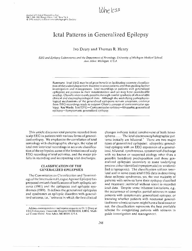
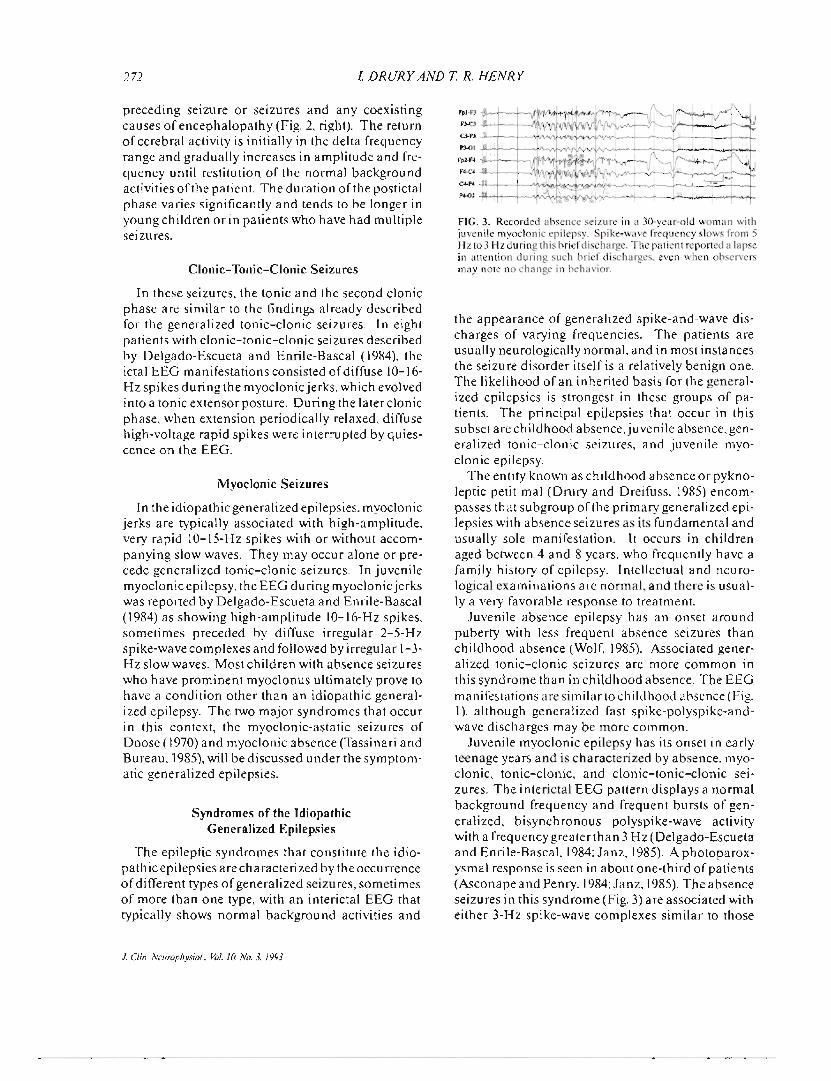
FIG. 3. Recorded ahsence seizure in a 30-year-old woman withjuvenile myoclonic epilepsy. Spike-wave frequency slows from 5Hz to 3 Hz during this hrief discharge. The patient reported a lapsein attention during such hrief discharges. even when ohserversmay note no change in hehavior.
the appearance of generalized spike-and-wave dis-charges of varying frequencies. The patients areusually neurologically normal, and in most instancesthe seizure disorder itself is a relatively benign one.The likelihood of an inherited basis for the general-ized epilepsies is strongest in these groups of pa-tients. The principal epilepsies that occur in thissubset are childhood absence, juvenile absence, gen-eralized tonic-clonic seizures, and juvenile myo-clonic epilepsy.
The entity known as childhood absence or pykno-leptic petit mal (Drury and Dreifuss, 1985) encom-passes that subgroup of the primary generalized epi-lepsies with absence seizures as its fundamental andusually sole manifestation. It occurs in childrenaged between 4 and 8 years, who frequently have afamily history of epilepsy. Intellectual and neuro-logical examinations are normal, and there is usual-ly a very favorable response to treatment.
Juvenile absence epilepsy has an onset aroundpuberty with less frequent absence seizures thanchildhood absence (Wolf, 1985). Associated gener-alized tonic-clonic seizures are more common inthis syndrome than in childhood absence. The EEGmanifestations are similar to childhood absence (Fig.1), although generalized fast spike-polyspike-and-wave discharges may be more common.
Juvenile myoclonic epilepsy has its onset in earlyteenage years and is characterized by absence, myo-clonic, tonic-clonic, and clonic-tonic-clonic sei-zures. The interictal EEG pattern displays a normalbackground frequency and frequent bursts of gen-eralized, bisynchronous polyspike-wave activitywith a frequency greater than 3Hz (Delgado-Escuetaand Enrile-Bascal, 1984;Janz, 1985). Aphotoparox-ysmal response is seen in about one-third of patients(Asconape and Penry, 1984;Janz, 1985).The absenceseizures in this syndrome (Fig. 3) are associated witheither 3-Hz spike-wave complexes similar to those
272 I DRURY AND T. R. HENRY
preceding seizure or seizures and any coexistingcauses of encephalopathy (Fig. 2, right). The returnof cerebral activity is initially in the delta frequencyrange and gradually increases in amplitude and fre-quency until restitution of the normal backgroundactivities of the patient. The duration of the postictalphase varies significantly and tends to be longer inyoung children or in patients who have had multipleseizures.
Clonic-Tonie-Clonic Seizures
In these seizures, the tonic and the second clonicphase are similar to the findings already describedfor the generalized tonic-clonic seizures. In eightpatients with clonic-tonic-clonic seizures describedby Delgado-Escueta and Enrile-Bascal (1984), theictal EEG manifestations consisted of diffuse 10-16-Hz spikes during the myoclonic jerks, which evolvedinto a tonic extensor posture. During the later clonicphase, when extension periodically relaxed, diffusehigh-voltage rapid spikes were interrupted by quies-cence on the EEG.
Myoclonic Seizures
In the idiopathic generalized epilepsies, myoclonicjerks are typically associated with high-amplitude,very rapid 1O-15-Hz spikes with or without accom-panying slow waves. They may occur alone or pre-cede generalized tonic-clonic seizures. In juvenilemyoclonic epilepsy, the EEG during myoclonic jerkswas reported by Delgado-Escueta and Enrile-Bascal(1984) as showing high-amplitude 1O-16-Hz spikes,sometimes preceded by diffuse irregular 2-5-Hzspike-wave complexes and followed by irregular I-3-Hz slow waves. Most children with absence seizureswho have prominent myoclonus ultimately prove tohave a condition other than an idiopathic general-ized epilepsy. The two major syndromes that occurin this context, the myoclonic-astatic seizures ofDoose (1970) and myoclonic absence (Tassinari andBureau, 1985),will be discussed under the symptom-atic generalized epilepsies.
Syndromes of the IdiopathicGeneralized Epilepsies
The epileptic syndromes that constitute the idio-pathic epilepsies are characterized by the occurrenceof different types of generalized seizures, sometimesof more than one type, with an interictal EEG thattypically shows normal background activities and
J Clin. Neurophysiol.. Vol. /0. No.3. /993
- - --
FpI.F3D-CJ
0-P3
P3-01"
f. -
f4-C4
CA.P.
J'4.02
FIG. 3. Recorded absence seizure in a 30-year-old woman withjuvenile myoclonic epilepsy. Spike-wave frequency slows from 5Hz to 3 Hz during this brief discharge. The patient reported a lapsein attention during such brief discharges. even when observersmay note no change in behavior.
the appearance of generalized spike-and-wave dis-charges of varying frequencies. The patients areusually neurologically normal, and in most instancesthe seizure disorder itself is a relatively benign one.The likelihood of an inherited basis for the general-ized epilepsies is strongest in these groups of pa-tients. The principal epilepsies that occur in thissubset are childhood absence, juvenile absence, gen-eralized tonic-clonic seizures, and juvenile myo-clonic epilepsy.
The entity known as childhood absence or pykno-leptic petit mal (Drury and Dreifuss, 1985) encom-passes that subgroup of the primary generalized epi-lepsies with absence seizures as its fundamental andusually sole manifestation. It occurs in childrenaged between 4 and 8 years, who frequently have afamily history of epilepsy. Intellectual and neuro-logical examinations are normal, and there is usual-ly a very favorable response to treatment.
Juvenile absence epilepsy has an onset aroundpuberty with less frequent absence seizures thanchildhood absence (Wolf, 1985). Associated gener-alized tonic-clonic seizures are more common inthis syndrome than in childhood absence. The EEGmanifestations are similar to childhood absence (Fig.1), although generalized fast spike-polyspike-and-wave discharges may be more common.
Juvenile myoclonic epilepsy has its onset in earlyteenage years and is characterized by absence, myo-clonic, tonic-clonic, and clonic-tonic-clonic sei-zures. The interictal EEG pattern displays a normalbackground frequency and frequent bursts of gen-eralized, bisynchronous polyspike-wave activitywith a frequency greater than 3Hz (Delgado-Escuetaand Enrile-Bascal, 1984;Janz, 1985). A photoparox-ysmal response is seen in about one-third of patients(Asconape and Penry, 1984;Janz, 1985).The absenceseizures in this syndrome (Fig. 3) are associated witheither 3-Hz spike-wave complexes similar to those
--- - - --

ICTAL PATTERNS IN GENERALIZED EPILEPSY 273
seen in pyknolepsy or rapid polyspike-wave com-plexes slowing to 3Hz during unconsciousness (Del-gado-Escueta and Enril-Bascal, 1984;Janz, 1985). Ina clinical and video/EEG study of these absences(Panayiotopoulos et aI., 1989), the clinical manifes-tations were subtle even during prolonged dischargesand often apparent only ifthe patient was involved insome type of activity during the ictus. The EEGmanifestations were variable but usually consistedof polyspike-wave complexes of rather irregular fre-quency rather than the smooth 3-Hz spike-wavecomplexes of pure absence. Myoclonic jerks in thissyndrome are most likely to occur in the transitionfrom sleep to wakefulness and during photic stimu-lation.
Other types of idiopathic generalized epilep-sies occur, and many patients who appear to have anidiopathic generalized epilepsy may not fit neatly intoany specific category. For instance, the Commissionon Classification and Terminology of the Interna-tional League Against Epilepsy (1989), which pro-posed the revised classification of epilepsies and epi-leptic syndromes, recognizes the entity of epilepsieswith grand mal seizures on awakening. These pa-tients have generalized 3-Hz or more rapid spike-wave discharges on EEG but no evidence of absenceseizures or myoclonic jerks.
SEIZURES OF THE SYMPTOMATICGENERALIZED EPILEPSIES
These epileptic syndromes may present at any agebut commonly occur in infancy or early childhood.Their clinical and EEG features may vary signifi-cantly, to a much greater extent than the idiopathicgeneralized epilepsies and are chiefly dependent onthe age of the child. Seizures that occur in thesesyndromes may be considered under the followingcategories: convulsive, nonconvulsive, and infantilespasms and myoclonic seizures.
Convulsive Seizures
The EEG appearance of tonic-clonic seizures hasalready been described.
The classic EEG accompaniment of tonic seizuresis generalized paroxysmal fast activity (GPFA)(Blume et aI., 1973; Markand, 1977; Beaumanoir,1985). This pattern typically occurs during sleepwhen many bursts are not associated with any clini-cal change, whereas in wakefulness most bursts areictal. The discharges consist of bursts of 1O-25-Hzspikes averaging 3-5 s, preceded or followed by gen-
Fpl.Al
Fp2-A2
F3-Al
F4-A2
C3-AIC4.A2
FIG.4. Three-second-long train ofGPFAassociated with arousaland brief tonic activity in a 19-year-old man with symptomaticgeneralized epilepsy from tuberous sclerosis. Note how the dis-charge ends in a slow spike-and-wave.
eralized sharp-and-slow-wave complexes (Fig. 4)(Brenner and Atkinson, 1982). There is little changein the frequency of the spikes during the bursts, buttheir amplitude may decrease as the discharge pro-gresses (Fig. 5). The discharges typically have anamplitude maximum at the superior frontal elec-trodes, are bilaterally synchronous but may have ashifting voltage emphasis between the two hemi-spheres. and rarely may occur unilaterally. Activa-tion procedures such as hyperventilation and photicstimulation do not provoke G PFA. Blume et al. (1973)also described tonic seizures occurring with electro-decremental responses.
Nonconvulsive Seizures
Atypical absence seizures are characterized by amore gradual onset and cessation than pure absenceseizures and are often accompanied by a slight de-crease in body tone (Beaumanoir, 1985). Their EEGaccompaniment consists of slow spike-and-waveactivity unchanged from the baseline tracing, ormore hypersynchronous slow spike-and-wave activ-ity (Fig. 6) (Markand, 1977).
Atonic and myoclonic seizures that may be clinic-ally indistinguishable may be associated with a morerapid high-voltage spike-and-wave activity some-times accompanied by brief electrodecremental re-sponses (Fig. 7) (Gastaut and Broughton, 1972;Blume, 1987) or without any identified change onscalp EEG (Gastaut and Broughton, 1972;Markand,1977; Blume, 1987). In an analysis of 239 drop at-tacks in 45 patients with symptomatic generalizedepilepsy recorded with CCTV-EEG techniques, Egliet al. (1985) observed that the majority of these dropattacks were not in fact atonic seizures but pure tonicseizures, which they termed axial spasms. A sudden
J Clin. Neurophysiol.. Vol. 10. No.3. 1993
--

274 I DRURY AND T. R. HENRY
Fpl-FJ
FJ.CJ
CJ-PJ IPJ-OI
Fp2-F4
F4-C4
C4-P4
P4-01
flexion of the hips, upper trunk. and head led to a fallproduced by the rapidity and severity of the hipflexion. These episodes could occur alone or in con-nection with other seizure manifestations, such asabsences. In the pure axial spasms, there were nodisturbances of consciousness and no significantEEG changes. When axial spasms were combinedwith other seizure manifestations, they were pre-ceded by an absence seizure and generalized spike-wave activity with the onset of the spasm coincidingwith the last generalized spike. Egli et al. (1985) be-lieve that axial spasms represent a more mature formof infantile spasms. Akinetic seizures characterizedby a complete lack of mobility despite preservationof muscle tone and associated with an impairment ofconsciousness have similar or identical EEG dis-charges to those seen in atonic seizures (Gastaut andBroughton, 1972).
Infantile Spasms and Myoclonic Seizures
Gibbs and Gibbs (1952) coined the term hypsar-rhythmia to describe the high-amplitude irregularasynchronous delta activity with multiple indepen-dent and shifting spike foci, which is the classic EEGfinding in this condition. Gibbs and Gibbs (1952)described in their atlas diffuse spike discharges as-
Fpl.F)FJ-CJ
C3-PJ
PJ-OI
F..cz
c.-..
Fp2-F4F4-C4
C.~P4
P4-01
FIG. 6. Recorded atypical absence seizure in a 33-year-old pa-tient with symptomatic generalized epilepsy. Background EEGshowed diffuse slowing and generalized slow spike-and-wave ac-tivity. The atypical absence seizure occurred during the right-hand portion of the sample and was associated with a loss ofawareness.
J Clin. Neurophysiol.. Vol. 10. No. 3. 1993
FIG. S. Recorded tonic seizure with generalized paroxysmalfast activity (GPFA). maximal right hemisphere. in a 19-year-old patient with mental retardation. a severe right hemiparesis.and symptomatic generalized epilepsy. The GPFA in thissample consists of rhythmic alpha-like activity.
sociated with the jerking and quivering movementsof the infantile spasms. It was quickly appreciatedthat the most common EEG accompaniment to thespasms was an abrupt generalized reduction in volt-age, termed electrodecremental by Bickford andKlass (1960). The complex motor phenomena thatmay occur during infantile spasms were first docu-mented by Pampiglione (1964). He demonstratedthat these spasms were associated with activity inboth flexor and extensor muscles, and that, evenwhen the muscle activity appeared to be extremelybrief. it was usually accompanied by a group ofmuscle action potentials, thus differing from theactivities seen in myoclonic seizures. Kellaway andcolleagues (1979) reported an analysis of over 5,000spasms in 24 infants with a synchronized video andpolygraphic recording system. In their study, mixedspasms with both flexor and extensor activity werethe most common type, accounting for 42% of thetotal, followed by spasms in flexion (34%)and exten-sion (22%). These authors were the first to describethe akinesia and attenuated responsiveness that couldeith er foIIow a spas m or occu r independen tly as a sei-zure without a preceding spasm. These events theytermed an "arrest."
Spasms in West's syndrome tend to cluster and areespecially likely to occur soon after arousal fromsleep. The spasms seem least likely to occur when thechild has been stimulated through measurement
FpI.FJ
FJ-CJ
C3-PJ
PJ-OI
Fp2-F4
F4-C4
C4-P4
P4-02
FIG. 7. Recorded generalized atonic seizure in a 39-year-oldwoman with post-traumatic generalized epilepsy. The patient'shead dropped during the generalized electrodecremental EEGpattern. which followed a single generalized spike-wave complex.More prolonged electrodecrements were associated with addi-tional axial atonia.
- ----

ICTAL PATTERNS IN GENERALIZED EPILEPSY
FpI.FJ
FJ.CJ
CJ.PJ
PJ.<)I
Fz-Cz
c.....
Fp2-F4
F4-C<
C4-P4
J'4.02
FIG. 8. Recorded infantile spasm in a 6-month-old girl. Thebackground EEG showed hypsarrhythmia. maximal over theright hemisphere. The spasm began with a generalized flexionmovement associated with high-amplitude sharp and slow-waveactivity followed by an abrupt decrement in voltage with promi-nent myogenic activity over the frontal regions.
and application of EEG electrodes. For these rea-sons and because seizure activity may be very subtlein these children, it is the policy in our laboratory torecord EEGs in children with known or suspectedinfantile spasms only as 2-h-long video/EEG record-ings. This allows both for an adequate transitionthrough states of wakefulness, drowsiness, sleep, andarousal and also facilitates the close correlation ofbehavioral activities with EEG changes.
Kellaway et al. (1979) identified 11 different ic-tal EEG patterns accompanying infantile spasms.About 80% of these events, however, consisted of ageneralized, frontally dominant slow-wave or sharp-and-slow-wave complex with or without an aftergo-ing period of abrupt voltage attenuation (Fig. 8). Al-most 38%of the over 5,000 events were accompaniedby a generalized slow-wave transient followed byvoltage attenuation. There was no correlation be-tween the type of spasm and the ictal pattern seen.
Fpl.AI~~'-""'v~
Fp2-AI
F3-AI
F4-AI
CJ.AI
C4-AI
PJ.AI
P4-AI
OI.AI
01.AI
FIG.9. EEG recording during sleep in a S-month-old child withcryptogenic infantile spasms. Waking EEG showed typicalhypsarrhythmic pattern. Sleeping sample showed greater inter-hemispheric synchrony and an alternating pattern with periods ofvoltage attenuation. These epochs of voltage attenuation shouldnot be confused with the electrodecremental pattern seen duringmany infantile spasms.
275
FIG. 10. Recorded true myoclonic jerk in a IS-month-old girlwith cryptogenic infantile spasms and hypsarrhythmia. Hypsar-rhythmia pattern was more prominent over the right hemisphere.The myoclonic jerk was accompanied by a rapid generalizedpolyspike-and-wave discharge without any electrodecrementalresponse and was clinically distinct from the child's infantilespasms.
However, "arrest" attacks and asymmetrical seizureswere most likely to be accompanied by a diffuse volt-age attenuation with superimposed fast activity, al-though the number of these events seen was verysmall. Electrodecremental episodes may occur inthe awake or sleeping state without evidence of aclinical seizure or any recorded change in the elec-tromyographic or other polygraphic channels (Kell-away et aI., 1979),and, conversely, in the presence ofmarked spasms, no obvious EEG changes may beseen on occasion (Pampiglione, 1964). In our experi-ence and that of others (Westmoreland and Gomez,1987), it is relatively common to see an alternatepattern during sleep in children with hypsarrhythmiathat approximates a burst-suppression pattern (Fig.9). Bursts of high-amplitude, rather asynchronousslowing are followed by 1-2 s of abrupt voltage atten-uation with this pattern repeating itself regularlyth rough out the sleeping tracing. Such activity shouldnot be viewed as indicative of underlying seizureactivity. Transient normalization ofthe EEG activityfor age may occur after a flurry of spasms (Kellawayet aI., 1979).
Patients with infantile spasms may also have myo-clonic jerks. As illustrated in Fig. 10,the myoclonicjerks are associated with a different EEG patternthan infantile spasms, with high-amplitude rapidspike or polyspike discharges without an ensuingelectrodecremental pattern. Partial seizures may
1. Clin. Neurophysiol.. Vol. /0. No.3. 1993
- - -

276 I DRURY AND T. R. HENRY
FpI.F7F7.T3
TJ.TS
TS.OI
C4-P4
P4-02
Fp2-FS
FIG. 11. Recorded partial seizure in the same child as in Fig. 10.This partial seizure was associated with the child rolling both hereyes back and appearing unresponsive. Its EEG accompanimentconsisted of high-amplitude spike-and-wave activity widely dis-tributed over the right hemisphere.
also occur in children with infantile spasms (Fig.11).
In patients with progressive myoclonus epilepsy,the myoclonus may be very prominent during thelater stages of the illness. Although associated withvery frequent generalized spike-wave activity, thereneed not be a precise temporal correlation betweenthe myoclonus and epileptiform activity, at least inLafora disease (Yen et aI., 1991),and probably in oth-er forms of progressive myoclonic epilepsy also.
Myoclonus may also be a prominent manifesta-tion in two other epileptic syndromes, the myoclonic-astatic seizures of Doose (1970) and myoclonic ab-sence (Tassinari and Bureau, 1985). In the Doosesyndrome, the interictal EEG is dominated by thetaactivity with a parietal maximum and bisynchron-ous frontally dominant fast polyspike-and-waveactivity. Myoclonic jerks may be prominent and areassociated with short bursts of high-frequency poly-spike-and-wave. The astatic or myoclonic-astaticseizures are associated with 2-3-Hz spike-and-waveactivity. GPFA occurs in sleep. Myoclonic absenceis characterized by absence seizures with prominentmyoclonus and sometimes with tonic activity butwith the typical 3-Hz spike-wave activity that is seenin pyknolepsy. The attacks are precipitated by hy-perventilation. No other seizure types or EEG dis-charges such as GPFA occur initially. Ultimately,myoclonic absence seizures disappear and other sei-zure types typical of the symptomatic generalizedepilepsies emerge.
J Clin. Neurophysiol.. Vo/. 10. No.3. /993
Syndromes of SymptomaticGeneralized Epilepsy
The clinical manifestations in patients with symp-tomatic generalized epilepsies are commonly age-dependent. In West's syndrome, first described byDr. W. J. West, a general practitioner in England, inhis own son in 1841, there is a strong correlationbetween the occurrence of infantile spasms and theappearance of hypsarrhythmia on EEG, but infan-tile spasms may appear without a hypsarrhythmicEEG. Nevertheless, the concordance of infantilespasms and hypsarrhythmia together is higher thanany other epileptic electroclinical syndrome irre-spective of age or etiology.
Symptomatic generalized epilepsies commonlymanifest themselves in older children and adults asthe Lennox-Gastaut syndrome characterized bymental retardation, multiple generalized seizuretypes often refractory to anticonvulsant therapy, andslowspike-and-wave discharges on EEG. The inter-ictal EEG manifestations of this syndrome, however,are imprecise, vary from patient to patient, and mayeven vary within the same patient depending on thestate of arousal or from one study to another. Themajor seizure types seen in this syndrome are atypi-cal absence (Fig. 6), tonic (Fig. 5), atonic (Fig. 7), andmyoclonic (Fig. 12), but generalized tonic-clonic,akinetic, clonic (Fig. 13), and complex partial sei-zures (Fig. 14) also occur. Many patients show fre-quent prolonged trains of slow spike-and-wave activ-ity. In most instances, these are not accompanied byany recognizable clinical change, although responsetesting may be limited due to mental dullness. Theremay even be significant variation of the latency ofresponses during periods with no slow spike-and-wave activity (Erba and Cavazzuti, 1977).
The epileptic syndromes of Doose (1970)and myo-
Fpl-F3
F3.C3
CJ.P3
PJ.OI
Fp2-F4
F4-C4
P4-02
FIG. 12. Recorded generalized myoclonic seizure in a 22-year-old woman with cryptogenic symptomatic generalized epilepsy.Several generalized jerks occurred during the period of general-ized spikes on EEG. but jerks were irregular and did not appearsynchronous with spikes.

ICTAL PATTERNS IN GENERALIZED EPILEPSY
Fpl-FJ
D-CJ
C3-P3
no,
Fp2-F4
F4-C4
UP<
~2
FIG. 13. Recorded generalized clonic seizures in a 23-year-oldman with generalized epilepsy secondary to early head trauma.Atypical absence seizures predominated in childhood. but exclu-sively generalized clonic seizures have occurred since adoles-cence. During briefer seizures. there is low-amplitude faciocervi-cal clonus synchronously with higher-amplitude. symmetricsomatic jerks. so that this unobscured EEG recording demon-strates generalized spike-wave complexes that are synchronouswith the jerks.
clonic absence (Tassinari and Bureau, 1985)that areconsidered by some authorities to be distinct entities,represent in our opinion forms of symptomatic gen-eralized epilepsy that begin in the latter half of thefirst decade of life in children who were previouslyneurologically normal but then evolve to look simi-lar to those children with Lennox-Gastaut syn-drome.
Children with multiple independent spike foci ontheir interictal EEG usually have the same clinicalfeatures and many of the same ictal events as chil-dren with slow spike-and-wave discharges. In someinstances, as reported by Markand (1977), children
Fpl.F7."..~..,~~. . . ., fvF7.T3~~---------TJ.T5':'~
5SI'JtI'#.;-~~ ~
="-"0 $ . ~:~~~-'~FPI'FJ,~ II,.." J'" ~f(FJ-C3~ . ,~t...-.....
C3-P3~PJ.OI
F'-C'Jv/'.Jo,fV'A~\\~~Cz.pz' ~
FP2-F4V""'~~'.w-J ~~'\_~'III.i"~""""""~~
F4-C4"vv' '" C.C4-P4~P4-02~FpZ-n
T4-T6
TO>02
I..
FIG. 14. Recorded partial seizure in a patient with symptomaticgeneralized epilepsy. a l4-year-old boy who had herpes simplexencephalitis at age 9. Interictal EEG showed generalized spike-and-wave and multiple independent spike discharges. This sei-zure commenced with a generalized myoclonic jerk followed by arhythmic ictal discharge in both temporal regions without anyclinical accompaniment.
277
with symptomatic generalized epilepsy may showslowspike-and-wave discharges on one EEG record-ing or in a particular state and multiple independentspike foci on another. Burnstine et al. (1991) reportedscalp ictal EEG recordings in four children withmultiple independent spike foci. Almost all seizureswere manifested by a tonic fencing posture. The ictalonset consisted of a generalized electrodecrementalpattern in 48% followed by rhythmic activity such asrepetitive sharp waves or spikes or repetitive thetaactivity either focally or over both hemispheres, or in50%began with rhythmic activity either focal, hemi-spheric, or bihemispheric and in 2% of cases with afocal electrodecremental pattern. The head and armmovements associated with the seizures were stereo-typed for each patient and did not vary with the site ofictal onset or the site of subsequent rhythmic activity.Three of these four patients subsequently underwentepilepsy surgery with improved seizure control in allthree.
NON CONVULSIVE GENERALIZEDSTATUS EPILEPTICUS
Nonconvulsive generalized status epilepticus(NCGSE) is a state of altered mental status charac-terized chiefly by a slowing of behavior sometimesprogressing to stupor, often with prominent psychia-tric manifestations and with an EEG accompani-ment of nearly continuous generalized epileptiformactivity. The occurrence of status epilepticus in chil-dren with pyknoleptic petit mal is exceptionally rare.When it occurs in young persons with idiopathicgeneralized epilepsy, most will have generalizedtonic-clonic seizures in addition to absence seizures(Niedermeyer and Khalifeh, 1965). It may also pre-sent without a prior history of seizures, particularlyin middle to later life and in women. It is in this agegroup that psychiatric manifestations tend to bemost common, sometimes resulting in admission topsychiatric services before the organic basis of thecondi tion is recognized (Lee, 1985).
The ictal EEG manifestations are remarkablyconsistent across a number of different case series(Lee, 1985; Guberman et aI., 1986; Dunne et aI., 1987).Discharges consist of continuous, or else intermit-tent but frequent, generalized spike-wave or poly-spike-wave discharges varying in frequency from I to4 Hz. The discharges usually abate promptly withthe administration of intravenous benzodiazepinesalong with a rapid return of the patient's mentalstatus to baseline (Fig. 15).
J. C/in. Neurophysiol.. Vol. 10. No.3. /993
--

-- ..-.-
ICTAL PAITERNS IN GENERAliZED EPILEPSY
tems are often adequate to demonstrate generalizedictal patterns. Widespread myogenic ("muscle"),kinesogenic ("movement"), and other artifacts are asignificant problem in recording generalized sei-zures (Fig. 2). Recording with greater numbers ofelectrodes can provide more sites that are not ob-scured by artifacts during some portion of a general-ized seizure.
Difficulties in distinguishing interictal from ictalgeneralized EEG discharges have been noted in thepreceding sections. Behavioral testing performedduring and after discharges can often permit thisdistinction. Simultaneous audio and video monitor-ing during scalp EEG recording is required for ade-quate documentation of unresponsiveness and otheraspects of ictal and postictal behavior. Behavioraltesting during monitored events is usually most com-petently performed by EEG technologists or othermedical professionals (Riley et aI., 1980). Parents,spouses, and other companions often can be trainedrapidly to perform adequate ictal and postictal be-havioral testing. Behavioral testing during seizuresshould at a minimum include assessment of respon-siveness to voice and touch, presentation of a mem-ory item, and subsequent evaluation of memory forthe item after any postictal behavioral alteration hasresolved. It is also useful for the observer to recordobservations of ictal behaviors, including unelicitedas well as tested behaviors.
Adequate documentation of behavior is not pro-vided by ACR, because notes kept by patients andtheir companions may not be consistently reliable indocumenting the quality of behavioral testing andoften are not sufficiently objective in reported obser-vations. ACR may be particularly useful for patientswhose spells do not readily occur in the hospitalenvironment and who have attentive parents orothers to follow them about with a notebook in whichto record behavioral observations. Even in the bestof circumstances, uncertainty as to the precise tem-poral relationship of behavioral and ACR-recordedEEG changes is likely. Outpatient short-term andinpatient long-term EEG audio and video monitor-ing remain the techniques of choice for determiningictal behavioral-electrographic correlations.
Induction of seizures during monitoring is usuallyindicated in order to obtain a favorable number ofseizures per day. Tapering of antiepileptic drugs(AEDs) is widely employed in order to induce epilep-tic seizures and perhaps also psychogenic pseudo-epileptic seizures in some individuals. Considerableknowledge of AED pharmacokinetics and pharma-
--- -
279
codynamics is desirable in order to obtain diagnos-tically useful recordings safely. In particular, overlyrapid withdrawal of barbiturates and benzodiaze-pines is capable of provoking epileptic seizures inpersons who have never previously experienced epi-leptic seizures. Such withdrawal seizures are gen-eralized from onset, so could lead to mistaken diag-nosis of primary generalized epilepsies. Other meansof physiological induction of epileptic seizures in-clude sleep deprivation, hyperventilation, photicstimulation, and sleep induction. Despite all suchmeasures, some patients will not have seizures dur-ing any reasonable period of monitoring.
REFERENCES
Asconape J. Penry JK. Some clinical and EEG aspects of benignjuvenile myoclonic epilepsy. Epilepsia 1984;25:108-14.
Bancaud J. Talairach P. Morel M. et al. "Generalized" epilepticseizures elicited by electrical stimulation of the frontal lobein man. Electroencephalogr Clin Neurophysiol 1974;37:275-82.
Beaumanoir A The Lennox-Gastaut syndrome. In: Roger J.Dravet C. Bureau M. et al.. eds. Epileptic syndromes in infancy.childhood and adolescence. London: John Libbey. 1985:89-99.
Bickford RG. Keith HM. MacCarty CS. Some observations onthe mechanisms of petit mal. Trans Am Neurol Assoc 1955;80:13-5.
Bickford RG. Klass D. Scalp and depth electrographic studies ofeIectro-decremental seizures. Electroencephalogr Clin Neuro-physioI1960;12:263.
Blume wr. David RB. Gomez MR. Generalized sharp-and-slow-wave complexes. Associated clinical features and long-termfollow-up. Brain 1973;96:289-306.
Blume wr. Lennox-Gastaut syndrome. In: LOders HO. LesserRP. eds. Epilepsy. Electroclinicalsyndromes. London: Spring-er-Verlag. 1987:73-92.
Brenner RP. Atkinson R. Generalized paroxysmal fast activity:electroencephalographic and clinical features. Ann Neurol1982;11:386-90.
Browne TR. Absence (petit mal) seizures. In: Browne TR. Feld-man RG. Epilep~y. Boston: Little. Brown. 1983:61-74.
Browne TR. Penry JK. Porter RJ. Dreifuss FE. Responsivenessbefore. during and after spike-and-wave paroxysms. Neurolo-gy 1974;24:659-65.
Burnstine H. Vining EPG. Uematsu S. Lesser RP. Multifocalindependent epileptiform discharges in children: ictal corre-lates and surgical therapy. Neurology 1991;41:1223-8.
Chugani HT. Shewmon DA Sankar R. Chen VC. Phelps ME.Infantile spasms: II. Lenticular nuclei and brainstem acti-vation on positron emission tomography. Ann Neuroll992;31:212-9.
Cleeland CS. Booker HE. Petit mal evoked by arousal duringsensory restriction. Arch NeuroI1967J7:324-30.
Commission on Classification and Terminology of the Interna-tional League Against Epilepsy. Proposal for revised clinicaland electroencephalographic classification of epilepticseizures. Epilepsia 1981;22:489-501.
Commission on Classification and Terminology of the Interna-tional League Against Epilepsy. Proposal for revised classi-ficationofepilepsiesand epilepticsyndromes.Epilepsia1989;30:389-99.
J. Clin. Neurophysiol.. Vol. 10. No.3. /993

280 l DRURY AND T. R. HENRY
Delgado-Escueta AV. Enrile-Bascal F. Juvenile myoclonic epi-lepsy of Janz. Neurology 1984;34:285-94.
Doose H. Gerken H. Leonhart R. VOlzke F. VOlzC. Centrence-phalic myoclonic-astatic petit mal. Neuropodiatrie 1970;2:59-78.
Drury I. Dreifuss FE. Pyknoleptic petit mal. Acta Neurol Scand1985:72:353-62.
Dunne JW. Summers QA. Stewart-Winne EG. Nonconvulsivestatus epilepticus: a prospective study in an adult generalhospital. Quart J Med 1987:238:117-26. .
Egli M. Mothersilll. O'Kane M. O'Kane F. The axial spasm-the predominant type of drop seizure in patients with sec-ondary generalized epilepsy. Epilepsia 1985:26:401-15.
Erba G. Cavazzuti V. Ictal and interictal response latency inLennox-Gastaut syndrome. Electroencephalogr Clin Neuro-physioI1977;42:717.
Gastaut H. Broughton R. Epileptic seizures. Clinical and electro-graphic features. diagnosis and treatmelll. Springfield. IL:Charles C Thomas. 1972.
Geller M. Geller A. Brief amnestic effects of spike-wave dis-charges. Neurology 1970:20:1089-95.
Gibbs FA. Gibbs EL. Atlas of electroencephalography.vol 2. Read-ing. MA: Addison-Wesley. 1952.
Gloor P. Quesney LF. Zumstein H. Pathophysiology of general-ized epilepsy in the cat: the role of cortical and subcorticalstructures. II. Topical application of penicillin to the cerebralcortex and subcortical structures. Electroencephalogr ClinNeurophysioI1977:43:79-94.
Gloor P. Testa G. Generalized penicillin epilepsy in the cat: ef-fects of intracarotid and intravertebral pentylenetetrazoland amobarbital injections. Electroencephalogr Clin Neuro-physioI1974:36:499-515.
Gomez MR. Westmoreland BF. Absence seizures. In: LOdersHO. Lesser RP. eds. Epilepsy. Electroclinical.\vndromes. Lon-don: Springer-Verlag. 1987:105-29.
Guberman A. Cantu-Reyna G. Stuss D. Broughton R. Noncon-vulsive generalized status epilepticus: clinical features.neurophysiological testing and long-term follow-up. Neural-0KI' 1986:36: 1284-91.
Guerrero-Gigueroa R. Barros A.deBalbian V. Heath RG. Experi-mental "petit mal" in kittens. Arch Neural 1963:9:297-306.
Hrachovy RA. Frost JD. Kellaway P. Sleep characteristics in in-fantile spasms. NeuroloKI' 1981:31:688-94.
Janz D. Epilepsy with impulsive petit mal (juvenile myoclonicepilepsy). Acta Neurol ScaI1lI1985;72:449-59.
Jasper HH. Droogleever-Fortuyn J. Experimental studies on thefunctional anatomy of petit mal epilepsy. Res Puhl A.\:\ocResNen' Melli Dis 1946:26:272-98.
Jellinger K. Neuropathological aspects of infantile spasms. BrainDel' 1987;9:349-57.
Kellaway P. Hrachovy RA. Frost JD. Zion T. Precise character-ization and quantification of infantile spasms. Ann Neurol1979;6:214-8.
Lee SI. Electroencephalography in infantile and childhood epi-lepsy. In: Dreifuss FE. ed. Pediatric epileptology: classifica-tion and managemelll o.fseizures in the child. Boston: Wright-PSG. 1983:33-63.
Lee SI. Nonconvulsive status epilepticus. Ictal confusion in laterlife. Arch Neuro/1985:42:778-81.
Marcus EM. Watson CWo Bilateral synchronous spike wave elcc-trographic patterns in the cat: interaction of bilateral corticalfoci in the intact. the bilateral cortical callosal and adien-
cephalic preparation. Arch Neuro/1966;14:60I-10.Markand ON. Slow spike-wave activity in EEG and associated
features: often called "Lennox" or "Lennox-Gastaut'. syn-drome. Neurology 1977:27:746-57.
J. Clin. Neurophysiol.. Vol. /0. No.3. /993
Mirsky AF. VanBuren JM. On the nature of the "absence" incentrencephalic epilepsy: a study of behavioral. electroen-cephalographic and autonomic factors. ElectroencephalogrClin NeurophysioI1965;18:334-48.
Morimatsu Y. Murofushi R. Handa T. Shinoara T. Shiraki H.Pathological studies of several physically and mentallyhandicapped-with special reference to 4 cases of infantilespasms. Shinkei Kenkyu No Shimpo 1972;16:465-70.
Morison RS. Dempsey EW. A study of thalamocortical relations.Am J PhysioI1942:135:281-92.
Naquet R. Menini C. Catier J. Photically induced epilepsy inPapio papio. The initiation of discharges and the role of thefrontal cortex and of the corpus callosum. In: Petsche H.Brazier MAB. eds. Synchronization o.fEEG activity in epilepsies.Wien: Springer-Verlag. 1972:347-66.
Niedermeyer E. Khalifeh R. Petit mal status ("spike-wave stupor"):an electro-clinical appraisal. Epilepsia 1965;6:250-62.
Niedermeyer E. Laws ER. Walker AE. Depth EEG findings inepileptics with generalized spike wave complexes. ArchNeuro/1969:21:51-8.
Orren MM. Evoked potential studies in petit mal epilepsy. In:Cobb WA. van Duyn H. cds. COlllemporary clinicalneuro-physioloKI'. Amsterdam: Elsevier. 1978:251-7. (EEG Suppl34.)
Pampiglione G. West's syndrome (infantile spasms)-a poly-myographic study. Arch Dis Child 1964:39:571-5.
Panayiotopoulos CPo Obeid T. Waheed G. Absences in juvenilemyoclonic epilepsy: a clinical and video-electroencephalo-graphic study. Ann Neuro/1989:25:391-7.
Papini M. Pasquinelli A. Armellini M. Orlandi D. Alertness andincidence of seizures in patients with Lennox-Gastaut syn-drome. Epilepsia 1984;25:161-7.
Riley TL. Berndt T. The role of the EEG technologist in delineat-ing pseudoseizures. Am J EEG Techno/1980:20:89-96.
Risinger MW. Engel J Jr. Van Ness Pc. Henry TR. Crandall PH.Ictal localization of temporal lobe seizures with scalp-sphenoidal recordings. NeuroloKv 1989:39:1288-93.
Savic I. Widen L. Thorell JO. Blomqvist G. Ericson K. Roland P.Cortical benzodiazepine receptor binding in patients withgeneralized and partial epilepsy. Epilepsia 1990:31:724-30.
Silverstein F. Johnston MV. Cerebrospinal nuid monoaminemetabolites in patients with infantile spasms. Neurology1984:34: 102-5.
Tassinari CA. Bureau M. Epilepsy with myoclonic absences.In: Roger J. Dravet C. Bureau M. et al.. eds. Epileptic syn-dromes in infanCl'.childhood and adolescence. London: JohnLibbey. 1985:12'1-9.
Tomson T. Svanborg E. Wedlund J-E. Nonconvulsive status epi-lepticus: high incidence of complex partial status. Epilepsia1986:27:276-85.
TOkel K. Jasper H. The electroencephalogram in parasagittalle-sions. Electroencephalogr Clin NeurophysioI1952;4:481-94.
Velasco M. Velasco F. Velasco AL. Lujan M. del Meracado N.Epileptiform EEG activities of the centromedian thalamicnuclei in patients with intractable partial motor. complexpartial and generalized seizures. Epilepsia 1989:30:295-306.
Westmoreland BF. Gomez MR. Infantile spasms (West's syn-drome). In: Loders HO. Lesser RP. eds. Epilepsy. Electro-clinical syndromes. London: Springer-Verlag. 1987:49-71.
Williams D. A study of thalamic and cortical rhythms in petit mal.Brain 1953:76:50-69.
Wo1fP. Juvenile absence epilepsy. In: Roger J. Dravet C. BureauM. et al.. eds. Epileptic syndromes in iI!fancy. childhood andadolescence. London: John Libbey. 1985:242-6.
Yen C. Beydoun A. Drury I. Longitudinal EEG studies in a kin-dred with Lafora disease. Epilepsia 1991:32:895-9.