the effects of mechanical vibration on human chondrocytes
TRANSCRIPT

Marquette Universitye-Publications@Marquette
Master's Theses (2009 -) Dissertations, Theses, and Professional Projects
The Effects of Mechanical Vibration on HumanChondrocytes In VitroBradley J. GauthierMarquette University
Recommended CitationGauthier, Bradley J., "The Effects of Mechanical Vibration on Human Chondrocytes In Vitro" (2016). Master's Theses (2009 -). Paper369.http://epublications.marquette.edu/theses_open/369

THE EFFECTS OF MECHANICAL VIBRATION ON HUMAN CHONDROCYTES
IN VITRO
by
Brad Gauthier, D.D.S.
A Thesis submitted to the Faculty of the Graduate School,
Marquette University,
in Partial Fulfillment of the Requirements for
the Degree of Master of Science
Milwaukee, Wisconsin
August 2016

ABSTRACT
THE EFFECTS OF MECHANICAL VIBRATION OF HUMAN CHONDROCYTES
IN VITRO
Brad Gauthier, D.D.S.
Marquette University, 2016
Introduction: Inflammation is the biological basis of temporomandibular joint disorders
(TMD), when severe it can lead to osteoarthritis. One of the physical therapies used to
manage this condition is mechanical vibration, as has been used in medicine for many
years as a non-pharmacological therapy. Recently an FDA approved dental device called
AcceleDent has been introduced to increase the rate of tooth movement and decrease
pain. As the device-generated vibration transmits to the TMJ, it is important for us to
investigate whether mechanical vibration influences TMJ on its biologic basis –
chondrocytes under normal and inflammatory conditions.
Materials and Methods: Human chondrocyte cell line C-28/I2 cells were maintained in
DMEM supplemented with 10% FBS. The cells were plated at a density of 5x10⁵/well to
24-well plates for differentiation or 6-well plates for gene expression studies. For
differentiation study, the cells were assigned to 4 subgroups i.e. osteogenic, normal,
osteogenic + IL-1β (1ng/mL), and normal + IL-1β (1ng/mL) and subjected to vibration of
0 Hz (control) or 0.3g/30Hz. The vibration was applied for 1 hour per day for 21
consecutive days with medium refreshed every 3 days, followed by Alizarin Red staining.
For gene expression study, the cells were subjected to vibration of 0 Hz (control) or
0.3g/30Hz with or without IL-1β (10ng/mL) for 1 hour, followed by quantitative
Polymerized Chain Reaction (qPCR) to evaluate gene expression levels of SOX9 and
MMP13 genes. One-way ANOVA was used to statistically test the difference between
experimental groups (P ≤ 0.05 considered significant).
Results: 0.3g/30Hz vibration exhibited a strong positive influence on the chondrocyte
differentiation, while IL-1β showed a minimal effect. Vibration increased SOX9 mRNA
expression by 1.28 fold when compared to controls. IL-1β decreased SOX9 mRNA
expression by 0.65 fold which was partially recovered by the vibration to 0.83 fold.
Vibration decreased MMP13 mRNA expression by 0.89 fold when compared to controls.
IL-1β increased MMP13 mRNA expression by 1.44 fold, which was slightly recovered
by the vibration to 1.30 fold.
Conclusions: Mechanical vibration (0.3g/30Hz) is able to increase differentiation of
human chondrocytes under normal and inflammatory conditions. Mechanical vibration
anabolically regulates gene expressions of SOX9 (upregulated) and MMP13
(downregulated), and can partially recover the catabolic changes of SOX9 and MMP13
induced by inflammation. Mechanical vibration (0.3g/30Hz) does not appear to harm
human chondrocytes in vitro and may help control or reduce inflammation.

i
ACKNOWLEDGMENTS
Brad Gauthier, D.D.S.
In no particular order I would like to thank Dr. Liu for being my thesis committee
leader and dedicating so much time and effort into this project. Without his mentorship
none of this would have been possible. I would like to thank Dr. Makky for her help with
the experimental set-up and oversight of the project. I’d also like to thank Dr. Bradley for
serving on my thesis committee.
I would also like to thank Dr. Fei Wang for her tireless help with conducting
experiments and collecting data.
I would also like to thank my co-resident, Dr. Megan DesRoches for her support
and motivation to complete our study.
Lastly I’d like to thank my family and friends, especially my wife Lauren who
has continuously stood by me in my endless pursuit of knowledge.

ii
TABLE OF CONTENTS
ACKNOWLEDGMENTS ................................................................................................... i
LIST OF TABLES ............................................................................................................. iii
LIST OF FIGURES ........................................................................................................... iv
CHAPTER 1: INTRODUCTION ........................................................................................1
CHAPTER 2: LITERATURE REVIEW .............................................................................5
CHAPTER 3: MATERIALS AND METHODS ...............................................................17
CHAPTER 4: RESULTS ...................................................................................................24
CHAPTER 5: DISCUSSION .............................................................................................37
CHAPTER 6: CONCLUSION ..........................................................................................40
BIBLIOGRAPHY ..............................................................................................................41

iii
LIST OF TABLES
Table 1. Representation of 24-well division were for study of cell differentiation……..20
Table 2. Experimental design for evaluation gene expression. .........................................21
Table 3. Sequences of primers used. ..................................................................................22
Table 4. Representation of 24-well plates division for study of cell differentiation. ........25
Table 5. SOX9 expression CT values from RT-qPCR analysis…………………………28
Table 6. RT-qPCR findings for SOX9…………………………………………………..28
Table 7. MMP13 expression CT values from RT-qPCR analysis…………….………….30
Table 8. RT-qPCR findings for MMP13………………………………………...…….....30
Table 9. Descriptive statistics for SOX9 gene expression……………………………….32
Table 10. One-way ANOVA for SOX9 analysis………………………………………...32
Table 11. Tukey HSD analysis of SOX9 gene expression………………………………33
Table 12. Descriptive statistics for MMP13 gene expression……………………………34
Table 13. One-way ANOVA for MMP13 analysis………………………………………34
Table 14. Tukey HSD analysis of SOX9 gene expression………………………………35

iv
LIST OF FIGURES
Figure 1. The major mechanotransduction pathways in chondrocytes .............................9
Figure 2. Mechanical vibration system….…………………..........................................18
Figure 3. Vibration plate with accelerometer attached. ...................................................19
Figure 4. Sham group, subjected to no mechanical vibration. ........................................25
Figure 5. Group subjected to 30 Hz mechanical vibration ..............................................25
Figure 6. End-point PCR testing of housekeeping gene, GAPDH. .................................26
Figure 7. End-point PCR testing of primers SOX9 and MMP13 ....................................27
Figure 8. Graph representing relative fold difference in SOX9 expression when
compared with controls. ...................................................................................29
Figure 9. Graph representing relative fold difference in MMP13 expression when
compared with controls. ...................................................................................31
1
CHAPTER 1
INTRODUCTION
Inflammation is a common protective response by the body. However, when
inflammation is chronic in nature it becomes pathologic. In dentistry some patients
present with chronic or acute inflammation of the temporomandibular joint (TMJ). The
temporomandibular joint is located bilaterally between the temporal bone and mandible.
It is unique in that it allows for both hinge and gliding movements, and is therefore
considered a ginglymoarthrodial joint. Some dental patients have complaints of pain,
clicking/popping, limited functioning, headaches, or a combination of these. Clinicians
must perform thorough examination along with proper diagnostic imaging to make the
correct diagnosis. Treatment is very complex because of the varied etiology of
temporomandibular disorders (TMD). One of the more common etiologies of TMD is
inflammation, which is seen in degenerative joint disease, or osteoarthritis (OA).
Symptoms of OA include pain, stiffness, and swelling (arthritis.org). Currently there is no
cure for OA. OA primarily affects joints of the knees and hips in the elderly population.
This disease is not only limited to these specific locations in the elderly but also other
areas of the body including the TMJ region in people of all ages. Treatment methods for
OA of the TMJs seek to alleviate pain and allow for improved functioning. Some
treatments include medications, physical therapy, occlusal appliances, stress reduction,
and surgery. Since inflammation appears to be one of the main pathogenic mechanisms of
articular cartilage breakdown, anti-inflammatory medications are often prescribed as a
way to help slow the progression of OA. Medications must be evaluated not only

2
regarding their ability to slow the progression of disease but also their effect on the
metabolism and reparative nature of the cell involved. Greco et. al found that Naproxen, a
non-steroidal anti-inflammatory drug (NSAID) decreased IL-1β induced catabolic
activity on chondrocytes. They also found that Naproxen reduced the expression of genes
involved in matrix production such as collagen 2A1 in resting conditions. Physical
therapy is also used often in combination with medications to treat OA. Dentists often
prescribe occlusal appliances, such as splints in an attempt to reduce proprioception and
help patients find a more stable joint position, centric relation. This approach also helps
reduce the wear and tear of the dentition from parafunction. Stress is also considered a
contributing factor to the progression of temporomandibular joint disorders and thus
stress reduction may help patients with symptoms. Surgery is typically the last approach
taken to treat TMD when all other methods have failed. Although these methods prove
useful in some patients, they may only provide temporary relief for others. Research in
the future should be directed at determining the molecular mechanism at which OA is
initiated and progresses to help provide better options for treating OA. At the base of the
molecular mechanism is the chondrocyte, a specialized cell type found in articular
cartilage tissues. Chondrocytes are unique cells that are required for articular cartilage
formation. These cells are derived from mesenchymal stem cells and are regulated by
multiple cytokines and transcription factors (Lin Z, et. al, 2006). They do not contain
blood vessels and nerves and are therefore not well equipped to repair or regenerate.
Identifying molecules for chondroprotection is currently a priority in modern medicine
(Greco K, 2011). Chondrocytes are found throughout the entire body including the
temporomandibular joints. These joints are of particular importance to dental
3
professionals and the population who suffer from TMD. The National Institutes of Health
estimated that in 1996, in the United States more than 10 million people suffered from
TMD (NIH, 1996). Due to the increase in the aging and general population that number is
likely getting even higher today. It is critical for practitioners to develop a proper
classification of the varied types of TMD as well as to target on the etiological factors
contributing to the disorder.
Mechanical vibration has been studied and used as treatment for muscle and bone
deficiencies especially in post-menopausal women suffering from osteoporosis.
Mechanical vibration has been shown to enhance bone mineral density in astronauts who
have been in space for an extended period of time (LeBlanc A, et. al, 2000). Being in
space vastly reduces the amount of peripheral loading that can be done and the
mechanical vibration serves as a way to load tissues. Vibration does not only affect
muscle and bone but a multitude of tissues in the body. Mechanical vibration has been
shown to enhance chondrocyte proliferation (Kaupp JA, Waldman SD, 2008).
Mechanoreceptors located within the chondrocytes seem to elicit a cellular response; the
exact mechanism of this response is not fully understood. A better understanding of this
mechanism may gain insight into the functioning of chondrocytes and provide clinicians
with better options to treat diseases of articular cartilage damage. Whole body vibration
(WBV) continues to be a well-researched area in medicine and may prove to be
promising for treating a variety of diseases in both medical and dental fields.
Mechanical vibration has recently been introduced to the field of orthodontics. A
commercially available device called AcceleDent (by OrthoAccel Inc.) claims to increase
the rate of orthodontic tooth movement and decrease pain by applying cyclical vibrations

4
to the dentition (www.acceledent.com). There is however conflicting evidence about the
effectiveness of applying vibration to both increase the rate of orthodontic tooth
movement and decrease pain experienced during orthodontic treatment. Besides focusing
on the effectiveness of this product it is also critical to review the safety of it. The device
is FDA-cleared but little evidence exists regarding its effects on TMJ. This device is
currently used by orthodontists and dentists who commonly see patients who could suffer
from TMD. When applied, the vibration from AccleDent device is not only localized to
the dentition but also transmitted to the surrounding tissues including the TMJ (Liu D,
2012). It would be doing an injustice to patients being treated with this device to not
further evaluate the effects of mechanical vibration on the temporomandibular joints.
Since mechanical vibration has been shown to have a positive influence on chondrocyte
proliferation (Kaupp, J.A., Waldman, S.D. 2008), we would like to evaluate the effects of
mechanical vibration applied at the same frequency used by AcceleDent, to determine
whether this vibration has a positive, negative, or no influence on these cells, in vitro.
Based on current knowledge, we hypothesize that mechanical vibration when
applied under certain circumstances can possibly be used as a therapy for treating patients
with TMD. The proposed mechanism under investigation is that vibration reduces
inflammation within the temporomandibular joint. Our aim is to investigate whether there
is a response at the cellular level to mechanical vibration of human chondrocytes in vitro.

5
CHAPTER 2
LITERATURE REVIEW
Inflammatory process
Inflammation is famously known for its four cardinal signs as described by Celsus
in 30 A.D.: rubor, calor, dolor, and tumor. These are also known as redness, heat, pain,
and swelling, respectively. The goal of the body is to achieve tissue integrity and
homeostasis (Martin 1997; Singer and Clark, 1999). The process to reach this
homeostasis includes inflammation, tissue formation, and tissue remodeling. Cells
interact with one another as well as cell matrices to perform this task (Eming, Krieg,
2007). People have been dealing with inflammation since the beginning of time. In an
attempt to treat this condition people would use extracts from willow leaves, which
contain salicilin. Salicylic acid was chemically synthesized in 1860 in Germany and 39
years later Bayer’s research director Dr. Heinrich Dresser introduced it in 1899 as
Aspirin. Today there are multiple drugs used to treat inflammation, most notably a group
known as NSAIDs, or non-steroidal anti-inflammatory drugs. Inflammation is caused by
a release of chemical mediators from tissues and migrating cells. Some of these mediators
include prostaglandins, leukotrienes, histamine, bradykinin, platelet-activating factor, and
interleukin-1 (Vane J, and Botting R, 1987). NSAIDs work primarily by inhibiting
cyclooxygenase (COX), which is an enzyme that catalyzes prostaglandins. There are two
main COX isoenzymes: COX-1 and COX-2. COX-1 is responsible for catalyzing
prostaglandins that protect the stomach and kidney. COX-2 is responsible for responding
to inflammatory stimuli (Vane J, and Botting R, 1998).
6
Osteoarthritis (OA) is the most common type of arthritis. It is also known as
degenerative joint disease and occurs when cartilage breaks down. This disease is
typically slowly progressing and seen most often in the elderly population. Patients with
OA have a breakdown of cartilage between the bones in the joint. Also, the affected
bones tend to slowly get bigger. Symptoms of OA include joint pain, stiffness, decreased
function, and swelling (rheumatology.org). Etiologies include those of both a
biochemical and biomechanical nature. When mechanical loading of a joint is well below
or above the normal physiologic range cartilage destruction tends to occur. Genetic,
dietary, estrogen levels, bone mineral density, muscle weakness, obesity, and joint laxity
have all been reported as risk factors relating to osteoarthritis (Ann Intern Med. 2000).
There are many factors that lead to degradation of cartilage including direct or indirect
modulation of anabolic and catabolic factors. Matrix metalloprotease (MMP) 13 is one of
the key catabolic factors in OA because of its ability to degrade collagen and other matrix
components (Liang Z, et. al, 2012). MMP13 is one of the most abundant proteinases and
has been found to regulate cell migration, alterations in the ECM, and apoptosis in
growth plate cartilage (Malemud CJ, 2006). Daily knee loading has been shown to
suppress cartilage destruction in mice with surgically induced osteoarthritis. MMP13
levels were found to be elevated in mice with OA. Knee loading reduced MMP13 activity
and thus reduced cartilage destruction (Hamamura K, et. al, 2013). Alcaraz M, et. al
recommended selective inhibitors of matrix metalloproteinases, aggrecanases, and other
proteinases as a therapeutic approach in treating OA. They also recommended promoting
anabolic factors by using growth factors and other regulatory molecules.

7
OA is typically thought of affecting the knee and hip joints. However, in the field
of dentistry OA has been studied in regard to the temporomandibular joints. Some
findings of OA associated with the TMJ include mandibular condyle flattening,
deformity, sclerosis, degeneration of the disc, and areas of erosion of the articular
cartilage of both the condyle and temporal bone (Gidarakou K, et. al, 2003). Dental
professionals are evaluating and treating these joints everyday. One common sign is disc
displacement, which often presents as a clicking sound on opening or closing of the
mandible. This displacement may progress to OA of the TMJ over time (Westesson P, et.
al, 1984). Kubota et. al reported that IL-1β levels in synovial fluid of the TMJ have a
positive correlation with OA change. Their findings suggest that IL-1β levels in the TMJ
may be an important marker for early detection of bone deterioration that are not
detectable by radiographs. Another study by Alstergren et. al showed that IL-1β in the
synovial fluid is associated with pain and hyperalgesia of the TMJ region. They
concluded that IL-1β seems to be a warning signal of tissue destruction. IL-1β is often
used in experiments to create an environment that mimics inflammation. Some treatment
options for OA of the TMJ include medications such as Ibuprofen and Glucosamine
sulfate. One study comparing these two medications showed that both have a positive
influence with regard to pain suffered from OA (Thie N, et. al, 2001). This indicates that
anti-inflammatory medications are one of the best options when the TMD is
inflammatory in nature. It is very difficult to make a definitive diagnosis of TMD because
there are many different presentations and etiologies of the pain and dysfunction. In a
meta-analysis by Kim MR, et. al, they stated that a reliable and valid diagnostic
classification system for TMD is needed to conduct better research in the future. In

8
general, TMD falls under two categories in which the disorder is believed to have
originated: joint and muscular. Joint problems arise from within the joint while muscular
problems often involve the muscles of mastication. It is extremely difficult to diagnose
the type of TMD a patient is suffering from, and may be even more difficult to treat. As
mentioned previously medications are one form of treatment. Other common types of
treatment involve occlusal appliances such as splints, stress reduction, physical therapy,
and surgery. For patients undergoing orthodontic or dental treatment it is necessary to
perform a thorough exam prior to initiating treatment, this must include the
temporomandibular joint regions. It should also be of interest to the clinician whether the
prescribed treatment plan and appliances used have an influence on the TMJs.
Chondrocytes
Chondrocytes are a special type of cell found in articular cartilage tissue. They are
crucial for cartilage formation and functionality. Chondrocytes are derived from
pluripotent mesenchymal stem cells and regulated by various cytokines and transcription
factors (Lin Z et. al, 2006). Human chondrocytes are typically round cells, they represent
about 5-10% of the total cartilage volume (Hunziker EB, et. al, 2002). Adult cartilage
does not contain blood vessels or nerves and is therefore not well equipped for wound
healing and regeneration. Constant mechanical loading is necessary for normal
functioning of the joints where cartilage covers the surfaces of bones (Liu J, et. al, 2001).
When the constant mechanical loading is diminished the cartilage will breakdown, as is
the case with osteoarthritis. Many studies have looked at mechanical vibration as a means

9
to produce constant loading to cartilage. Several of these studies have shown beneficial
results but are difficult to make generalizations because of differences in experimental
design (i.e. frequency, magnitude, subjects, etc.). The exact mechanism where an external
mechanical stimulus leads to changes in internal cell signaling is not fully understood.
Mechanical stresses have been shown to directly alter cellular processes including gene
expression, signal transduction, growth, and differentiation in vitro (Chen CS, Ingber DE,
1999). There are a number of potential mechanoreceptors that respond to vibration within
chondrocytes such as integrins, connexins, stretch-activated ion channels, and cilia (Lee,
HS and Salter, DM). These receptors help communicate the cells extracellular
environment to the internal environment.
Figure 1: The major mechanotransduction pathways in chondrocytes (Lee, HS and
Salter, DM).

10
A primary function of chondrocytes is to maintain the extracellular matrix (ECM)
through catabolic and anabolic processes. There are two main components to the ECM,
type II collagen and aggrecan. Type II collagen gives cartilage its tensile strength while
aggrecan provides osmotic resistance to resist compressive loads (Knudson CB and
Knudson W, 2001). Severe compressive loading can damage the ability of the
chondrocyte to repair the ECM. Chondrogenesis occurs during embryo development.
The process of chondrogenesis is regulated by multiple transcription factors, of which
SOX9 is considered to be the main regulator of chondrocyte differentiation. This
transcription factor is required to maintain the chondrocytic phenotype (Bi W, et. al,
1999). An experiment by Papadopoulou et. al involved animals who were subjected to
either a hard (normal loading) or soft (unloading) diet. They postulated that reduced
levels of SOX9 immunoexpression could be associated with increased loading
conditions. In yet another study, Xiong et. al found enhanced SOX9 expression in
condylar cartilage of rats that underwent mandibular advancement (unloaded condition).
SOX9 promotes cell survival and activates the genes for many cartilage specific
components and regulatory factors (Lefebvre V, 2016). In articular cartilage,
chondrocyte maturation is arrested which prevents further differentiation towards a
terminal hypertrophic state. In OA articular chondrocyte homeostasis is disrupted and
chondrocytes begin to undergo endochondral ossification. MMPs also act as an initiating
factor leading to endochondral ossification, vascular invasion, and altered bioavailability
of growth factors and chondrocyte apoptosis (Borzi et. al, 2010). MMP13 has been
shown to have direct angiogenic activities that are associated with OA severity and
clinical disease activity (Bonnet CS et. al 2005; Walsh DA et. al, 2007). A potential

11
therapy to treat OA could be to target MMP13 in hopes to maintain homeostasis and
prevent unwanted vascularization of mature articular cartilage.
Studies using human primary articular chondrocytes are limited because they are
difficult to obtain, the cartilage cannot be controlled, and the number of cells is not
adequate. After the primary cells are isolated they show very little proliferative capacity
(Sabatini, 2004). If they do show proliferative activity it usually indicates a loss of
differentiated phenotype. The loss of phenotype includes a change in cell shape and also
a change in the pattern of gene expression (Stokes DG, et. al, 2002). To avoid these
limitations most experimental designs utilize immortalized chondrocyte cell lines. Thus a
good model for studying human cartilage is to use immortalized human chondrocytes.
Viruses SV40-TAg and HPV-16 have been used to immortalize these cells. One of the
negative effects of using these viruses to create study models is the stable integration of
immortalizing genes disrupts the normal cell-cycle control but does not stabilize
expression of the type II collagen gene (COL2A1). This is the most sensitive marker of
the differentiated chondrocyte phenotype (Sabatini, 2004). Three immortalized cell lines
are commonly used: T/C-28a2, C-28/I2, and T/C-28a4. The latter two are derived from
the first. These cells are said to retain their morphology and maintain continuous
proliferation in monolayer culture (Finger F, et. al, 2003). C-28/I2 cells display the
highest levels of matrix anabolic and catabolic genes and are therefore preferred to be
used for investigating anabolic and catabolic activity and regulation. Of the three above
mentioned cell lines, C-28/I2 showed the highest SOX9 levels, indicating that this cell
line most closely resembles primary chondrocytes. However, Finger F. et. al also states
that none of these cell lines appear to be a direct substitute for primary chondrocytes.

12
Temporomandibular Joint
Temporomandibular joint (TMJ) is where mandible articulates with cranium,
specifically temporal bone. This joint is present on both the right and left sides and
functions as a single unit. TMJ is very complex as it allows for both hinge and gliding
movements, therefore considered as a ginglymoarthrodial joint. Located between the
condyle of mandible and glenoid fossa of temporal bone is an articular disc. This disc is
composed of dense fibrous connective tissue. Attached to the posterior aspect of the disc
is an area known as retrodiscal tissues. This is a region of loose connective tissue that is
highly vascularized and innervated. Capsular ligaments are attached both anterior-
superiorly and anterior-inferiorly to the disc. These anterior attachments are composed
primarily of collagen fibers. Another anterior attachment to the disc is made with the
superior head of the lateral pterygoid muscle. The capsular ligament is also attached
medially and laterally to the disc, which separates the TMJ into a superior joint space and
inferior joint space. These spaces are composed of highly specialized endothelial cells
forming a synovial lining. The synovial lining produces synovial fluid, which acts as a
lubricant as well as a facilitator for providing metabolic requirements (Okeson, 2008).
The surfaces of condyle and temporal fossa are composed of four layers or zones.
The most intimate layer with the bony articulations is termed the articular zone. This is
the functional surface. It is unique from other joints because it is composed of dense
fibrous connective tissue and not hyaline cartilage. Some benefits to this are the dense
fibrous connective tissue is less susceptible to the effects of aging and it has a better
ability to repair itself. The second zone is the proliferative zone. This area contains

13
undifferentiated mesenchymal tissue. This area responds to the functional loads placed on
the joints. The third zone, or the fibrocartilaginous zone, allows resistance to compressive
and lateral forces. The fourth and last zone is known as the calcified cartilage zone. In
this zone there are chondrocytes and chondroblasts. When these cells die their contents
are evacuated and replaced by bone cells (Okeson, 2008).
The articular cartilage is composed of chondrocytes and intercellular matrix. The
chondrocytes are responsible for producing collagen, proteoglycans, glycoproteins, and
enzymes that help form the matrix. The chondrocytes play a critical role for proper
functioning and maintenance of the TMJs (Okeson, 2008).
Clinical applications of mechanical vibration
Significant bone loss is often seen in astronauts who have been in space for
extended periods of time, due primarily to a lack of peripheral loading (LeBlanc A, et. al,
2000). Vibration therapy is used in medicine as a non-pharmacological analogue of
physical activity. Its purpose is to promote bone and muscle strength in individuals who
are frail (Thompson W, et. al, 2014). With a growing number of the aging population
come many challenges to the healthcare field. One such challenge is treating
osteoporosis. Osteoporosis is defined as a systemic skeletal disease characterized by low
bone mass and micro-architectural deterioration of bone tissue, with a consequent
increase in bone fragility and susceptibility to fracture (Consensus Development
Conference, 1993). Typically as people age their mobility and muscle function decreases.
An ideal treatment would be to increase the loading of bones by increasing the amount of

14
exercise an individual does. Exercise and other types of physical activity however
become extremely difficult for frail individuals. The theory of vibration therapy is to
target the musculoskeletal system in ways that mimic physical activity. This type of
therapy is very beneficial since it does not involve the use of any pharmacological agents.
Many elderly patients are taking multiple medications and would not be subjected to
additional drugs, which may have adverse effects. One study evaluated the effects of low-
intensity whole body vibration (WBV) on bone mineral density (BMD) in humans (Rubin
C, et. al, 2004). The study specifically looked at postmenopausal women subjected to
2x10 minutes of low intensity (0.2g, 30Hz) and a control group (placebo plate). The
control group was found to have lost 2% femoral neck BMD while the treatment group
gained 0.04% BMD. Another study looked at the ability of WBV to augment the anabolic
effects of dynamic exercise. They found that the vibration group had an enhanced effect
of training to increase lumbar BMD (Gomez-Cabello A, et. al, 2014). The experimental
group was also found to suffer less falls than the control group. Studies have shown that
children with immobility-associated disabilities may also benefit from low intensity
vibration therapy (Ward K, et. al, 2004). There appear to be many indications for
mechanical vibration in medicine. The mechanotransduction pathways need to be further
studied to gain a better understanding of which patients specifically would benefit the
most from mechanical vibration.
In the field of orthodontics vibration has recently been introduced as an adjunctive to
decrease treatment times and reduce the pain experienced from orthodontic treatment.
Conflicting evidence exists regarding the effectiveness of using mechanical vibration to
decrease treatment times. A study by Leethanakul C, et. al concluded that in combination

15
with orthodontic force, vibratory stimuli increased the secretion of IL-1β in gingival
crevicular fluid and also appeared to increase the bone resorption and accelerate
orthodontic tooth movement. The vibrational source for their study was an electric
toothbrush vibrating at 125Hz and patients were instructed to apply the vibrational source
to specific teeth for a minimum of 5 minutes, 3 times per day for 2 months. There are
other sources that can be used to apply vibrational force to the dentition. AcceleDent is an
FDA cleared, class II medical device that applies mechanical vibration to the dentition
with the goal of speeding up orthodontic tooth movement and decreasing the amount of
pain with treatment. In a study by Pavlin D, et. al, they found that vibrational forces
applied at 0.25N (25g) and a frequency of 30Hz to the dentition led to an increase in the
speed of orthodontic tooth movement. The study involved space closure rates in
maxillary first premolar extraction cases. They concluded a significant difference in tooth
movement between the vibration and non-vibration groups (1.16 mm/month vs. 0.79
mm/month). Kau CH, et. al reported seeing 2-3 mm/month of tooth movement with
mechanical vibration. Their study did not include control groups but they stated that
“conventional wisdom regarding normal rates of tooth movement are about 1 mm of
movement per month.” Bowman SJ, et. al concluded that AcceleDent reduced the time
needed for dental alignment and leveling. He also stated that there was a 30% increase in
the rate of tooth movement during leveling of the mandibular arch. However, in a
prospective randomized clinical trial Woodhouse NR, et. al found no evidence showing
that vibrational force can significantly increase the rate of tooth movement during
orthodontic alignment. They conducted their study using three groups: AcceleDent group,
a sham group with a non-vibrating replica of the AcceleDent device, and fixed appliances

16
only. All cases were first premolar extraction cases. They evaluated the rates of
orthodontic teeth for alignment in the mandibular arch with fixed appliances and found
no statistical difference between the three groups. Albeit there is conflicting evidence
regarding the effectiveness of mechanical vibration as a supplement to orthodontic
treatment, seemingly more orthodontists and dentists are using these devices to treat
patients. Along with the efficacy of mechanical vibration treatment in orthodontics, it is
also important to consider the safety of using such devices. It is very appealing to
clinicians to be able to decrease the length of treatment and achieve the same results.
However, one of the rules orthodontists should follow is to do no harm. Our study is
attempt to evaluate if any deleterious effects are placed on human chondrocytes in vitro
from mechanical vibration. Another objective of the study is to evaluate whether there is
a reduction of inflammation when cells are subjected to mechanical vibration at a
frequency similar to that of AcceleDent.
With its increasing use, it is crucial to critically evaluate any possible side effects
of AcceleDent. One area that has not been thoroughly looked at is the temporomandibular
joint responses to mechanical vibration. The mechanical vibration force from AcceleDent
is transmitted not only to the dentition but also the TMJ region. In a study using a
scanning laser Doppler vibrometer, it has been shown on a dry skull that the vibrations
from AcceleDent can be transmitted to the TMJ area (Liu D, 2013). The overall aim of
our study is to evaluate the effects of mechanical vibration on the cultured human
chondrocytes in vitro.

17
CHAPTER 3
MATERIALS AND METHODS
This study was conducted using the human chondrocyte cell line C-28/I2 (Finger
F, et. al, 2003), a gift from Dr. Mary Goldring (Hospital for Special Surgery, NY). The
mechanism by which vibration exerts its effect on human chondrocytes was evaluated by
looking at cell differentiation and gene expression of different chondrocyte markers
involved in cell regulation. The effect of vibration on human chondrocytes was evaluated
by comparing differentiation between chondrocytes at 0 and 30Hz, with or without
presence of IL-1β. Gene expressions of MMP13 and SOX9 were also analyzed before and
after vibration and with or without presence of IL-1β.
Human chondrocyte cell line C-28/I2
Human chondrocyte cell line C-28/I2 was used because it has been suggested that
these cells may be the best among the cell lines to investigate matrix anabolism and
catabolism in chondrocytic cells (Finger F, et. al, 2003). These cells do not exactly replicate
those of primary chondrocytes but are commonly used in experiments because of the
difficulty in attaining large quantities of primary chondrocytes. The chondrocytes were
routinely maintained in DMEM with 10% FBS and 1% Penicillin/Streptomycin.
Mechanical vibration setup

18
When ready, the C-28/I2 cells were plated and placed onto a rigid platform that
was custom made to fit a standard multi-well tissue culture plate. The base (ThorLabs
Max Series Modular Flexure Stage, with a DRV120 actuator) was capable of delivering
measured amounts of vertical vibration generated by a modular piezoelectric device.
Vibration (30Hz) was delivered by a function generator (Instek: Model FG 8015G). A
current amplifier (Advanced Motion controls, Camarillo CA, Model Brush Type PWM
Servo Amplifier) delivered 0.3g of acceleration to the vibration plate. An accelerometer
was used to confirm the correct amount of vibration was being transmitted to the plate of
the cells (Endevco). The setup was housed in a thermos box of 37oC. Pictured below is a
similar device, used by Kulkarni et al, 2013.
Figure 2: Mechanical vibration system composed of 1) Vibration generator, 2)
Modulator, and 3) Accelerometer.
19
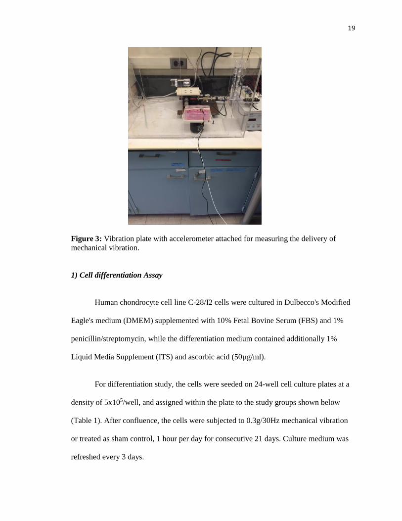
Figure 3: Vibration plate with accelerometer attached for measuring the delivery of
mechanical vibration.
1) Cell differentiation Assay
Human chondrocyte cell line C-28/I2 cells were cultured in Dulbecco's Modified
Eagle's medium (DMEM) supplemented with 10% Fetal Bovine Serum (FBS) and 1%
penicillin/streptomycin, while the differentiation medium contained additionally 1%
Liquid Media Supplement (ITS) and ascorbic acid (50µg/ml).
For differentiation study, the cells were seeded on 24-well cell culture plates at a
density of 5x10⁵/well, and assigned within the plate to the study groups shown below
(Table 1). After confluence, the cells were subjected to 0.3g/30Hz mechanical vibration
or treated as sham control, 1 hour per day for consecutive 21 days. Culture medium was
refreshed every 3 days.

20
Osteogenic medium Osteogenic medium + IL-1β
Normal medium Normal medium + IL-1β
Table 1: Representation of how 24-well plates were assigned for the study of cell
differentiation.
To examine the levels of differentiation of C-28/I2 cells, Alizarin Red staining
method was used: the cells were rinsed with PBS once. Then 1mL of 10% formalin was
added to each well and fixed for 15 minutes at room temperature. Each well was washed
with double distilled water (dH2O).
The staining protocol was as follows: add 500µL 40mM ARS (pH 4.1) per well,
incubate the plates at room temperature for 20 min with gentle shaking, wash wells 4
times, 5 minutes each time, with 1mL dH2O. The results of staining were observed under
a reverse microscope and scanned into .tiff files by using Photoshop software (version
5.0).
2) Gene Expression
Human chondrocyte cell line C-28/I2 cells were cultured and seeded onto 6-well
culture plates. The chondrocytes were routinely maintained in complete DMEM medium
(Sigma) with 10% FBS and 1% Penicillin/Streptomycin.
Prior to gene expression experiment, viability test was done using Trypan blue
exclusion assay to rule out the possibility of cell death before and after vibration, which
can bias the interpretation of gene expression results.
Four groups were evaluated in the study. The experiment was designed as follows:

21
(-)IL-1β, (-) Vibration (control) (-)IL-1β, (+) Vibration
(+)IL-1β, (-) Vibration (+)IL-1β, (+) Vibration
Table 2: Experimental design for evaluation gene expression.
The experimental groups were placed onto the vibration apparatus described above and
subjected to 0.3g/30Hz for 1 hour. IL-1β was added to the designated group right at the
start of vibration at a concentration of 10ng/mL. The control group was placed onto the
vibration apparatus but subjected to 0g/0Hz for 1 hour. The cells were lysed immediately
after 1 hour of vibration.
RNA extraction and Reverse transcription reaction.
Total RNA was extracted from the chondrocytes using TRIzol, following the
protocol from the manufacturer (Bio-RAD). Next, the RNA was treated with DNase (Life
Technologies Corporation) to remove any genomic DNA. A spectrophotometer was used
to determine the concentration of the total RNA using the formula:
Concentration of RNA= OD260 x 40 (constant) x dilution factor (ug/ml)
After DNase treatment, reverse transcription was performed using Oligo (dT) 20 primers
and SuperScript II as instructed by the manufacturer (Life Technologies Corporation).
End-point PCR
22
End-point PCR was performed to validate the primers used in the qPCR. The end-
point PCR reaction was run at 94°C for 2 minutes; followed by 94°C for 30 seconds,
58°C for 30 seconds, and 72°C for 45 seconds. 32 cycles were performed. Then the
reaction was run at 72°C for 10 minutes and finally 4°C. Taq polymerase was used for
the reaction.
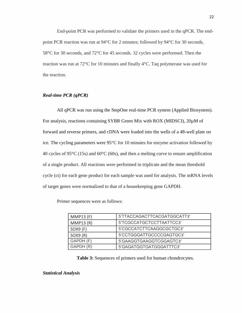
Real-time PCR (qPCR)
All qPCR was run using the StepOne real-time PCR system (Applied Biosystem).
For analysis, reactions containing SYBR Green Mix with ROX (MIDSCI), 20µM of
forward and reverse primers, and cDNA were loaded into the wells of a 48-well plate on
ice. The cycling parameters were 95°C for 10 minutes for enzyme activation followed by
40 cycles of 95°C (15s) and 60°C (60s), and then a melting curve to ensure amplification
of a single product. All reactions were performed in triplicate and the mean threshold
cycle (ct) for each gene product for each sample was used for analysis. The mRNA levels
of target genes were normalized to that of a housekeeping gene GAPDH.
Primer sequences were as follows:
MMP13 (F) 5’TTACCAGACTTCACGATGGCATT3’
MMP13 (R) 5’TCGCCATGCTCCTTAATTCC3’
SOX9 (F) 5’CGCCATCTTCAAGGCGCTGC3’
SOX9 (R) 5’CCTGGGATTGCCCCGAGTGC3’
GAPDH (F) 5’GAAGGTGAAGGTCGGAGTC3’ GAPDH (R) 5’GAGATGGTGATGGGATTTC3’
Table 3: Sequences of primers used for human chondrocytes.
Statistical Analysis

23
Statistical analysis was performed using one-way ANOVA. A p-value less than or
equal to 0.05 was considered statistically significant. Tukey post hoc was performed to
find the significant differences between groups. Descriptive statistics were also presented
for all experimental groups (SPSS version 23).

24
CHAPTER 4
RESULTS
Cell Differentiation
Cell differentiation is seen when a cell becomes more specialized. Cell
differentiation of each cell type is controlled by a specific program of gene expression.
This is the result of gene expression being either up-regulated or down-regulated. This in
turn will affect how the cell can function. In the case of TMJ chondrocytes, this
differentiation may allow for the cell to better repair itself from injury as well as maintain
its ability to function properly. In addition, the cells showed more differentiation in
osteogenic medium as expected (Fig. 4, 5). The results of our study indicate that
mechanical vibration (0.3g/30Hz) increases cell differentiation of human chondrocyte
cell line C-28/I2 cells in vitro when compared with controls (Fig. 4, 5). The presence of
IL-1β (1ng/ml) seemed to have minimal impact on differentiation of the cells. A large
difference in the amount of differentiation occurred when comparing the sham group (no
vibration) with the mechanical vibration group and also between the two types of growth
medium. Overall, our study shows that cultured human chondrocytes in osteogenic
medium are able to differentiate and enhanced by vibrational stimulation. There was no
significant effect of IL-1β on the differentiation of the C-28/I2 cells.

25
Sham Group (No vibration)
Figure 4: Differentiation of human chondrocytes in the sham (control) subjected to no
mechanical vibration.
30 Hz Vibration
Figure 5: Differentiation of human chondrocytes in the sham (control) subjected to
0.3g/30Hz mechanical vibration.
Osteogenic medium Osteogenic medium + IL-1β
Normal medium Normal medium + IL-1β
Table 4: Layout of the subgroups of 24-well plates for study of cell differentiation.
26
Gene Expression
Validation of PCR primers:
End-point PCR reactions were done for quality assurance that the primers
(GAPDH, SOX9, and MMP13) were working properly (Fig. 6, 7). The end-point PCR
reaction measures the amount of accumulated PCR product at the end of the PCR cycles.
Superscript II enzyme in the reaction system is for reverse transcripting mRNA to cDNA
which is further amplified through PCR. Without Superscript II enzyme the observed gel
bands represented contamination of genomic DNA, based on which DNAse was used to
purify the RNA samples before RT-PCR reaction. With Superscript II enzyme, the single
gel bands represented the signals of the targeting genes. Once the primers’ validity was
proved by the end-point PCR, qPCR reactions were used because of their higher
precision, sensitivity, and quantitative results. The data from the end-point PCR testing
are qualitative and presented below.

27
Figure 6: End-point PCR testing of housekeeping gene, GAPDH.
Figure 7: End-point PCR testing of primers MMP13 and SOX9.
28
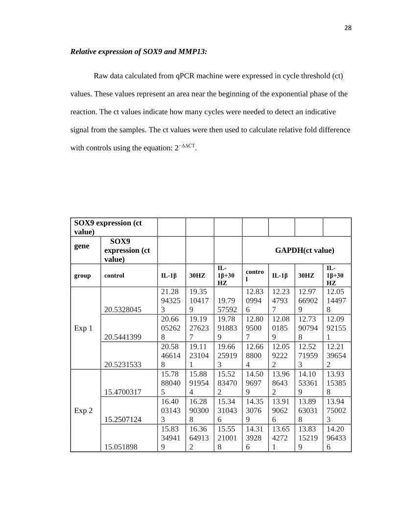
Relative expression of SOX9 and MMP13:
Raw data calculated from qPCR machine were expressed in cycle threshold (ct)
values. These values represent an area near the beginning of the exponential phase of the
reaction. The ct values indicate how many cycles were needed to detect an indicative
signal from the samples. The ct values were then used to calculate relative fold difference
with controls using the equation: 2−ΔΔCT.
SOX9 expression (ct
value)
gene
SOX9
expression (ct
value)
GAPDH(ct value)
group control IL-1β 30HZ
IL-
1β+30
HZ
contro
l IL-1β 30HZ
IL-
1β+30
HZ
Exp 1
20.5328045
21.28
94325
3
19.35
10417
9
19.79
57592
12.83
0994
6
12.23
4793
7
12.97
66902
9
12.05
14497
8
20.5441399
20.66
05262
8
19.19
27623
7
19.78
91883
9
12.80
9500
7
12.08
0185
9
12.73
90794
8
12.09
92155
1
20.5231533
20.58
46614
8
19.11
23104
1
19.66
25919
3
12.66
8800
4
12.05
9222
2
12.52
71959
3
12.21
39654
2
Exp 2
15.4700317
15.78
88040
5
15.88
91954
4
15.52
83470
2
14.50
9697
9
13.96
8643
2
14.10
53361
9
13.93
15385
8
15.2507124
16.40
03143
3
16.28
90300
8
15.34
31043
6
14.35
3076
9
13.91
9062
6
13.89
63031
8
13.94
75002
3
15.051898
15.83
34941
9
16.36
64913
2
15.55
21001
8
14.31
3928
6
13.65
4272
1
13.83
15219
9
14.20
96433
6
29
Exp 3
19.9958019
20.46
00563
20.01
89743
20.34
64202
9
13.34
1119
8
13.28
6344
5
12.17
72613
5
12.83
16240
3
20.0295467
19.90
028
20.03
09639
20.34
50050
4
12.67
2685
6
13.30
0914
8
12.55
28049
5
12.51
35860
4
20.0123425
19.84
20009
6
19.92
65937
8
20.22
98240
7
12.40
8614
2
13.14
1422
3
12.87
21838
12.71
69256
2
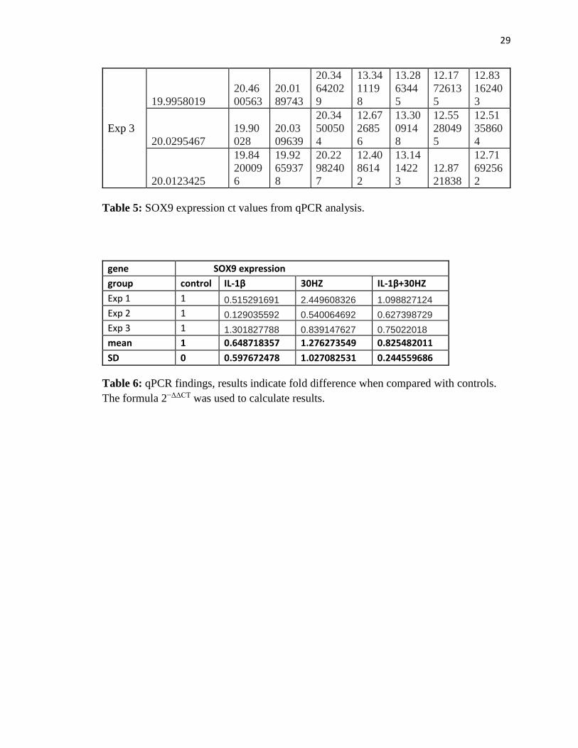
Table 5: SOX9 expression ct values from qPCR analysis.
gene SOX9 expression
group control IL-1β 30HZ IL-1β+30HZ
Exp 1 1 0.515291691 2.449608326 1.098827124
Exp 2 1 0.129035592 0.540064692 0.627398729
Exp 3 1 1.301827788 0.839147627 0.75022018
mean 1 0.648718357 1.276273549 0.825482011
SD 0 0.597672478 1.027082531 0.244559686
Table 6: qPCR findings, results indicate fold difference when compared with controls.
The formula 2−ΔΔCT was used to calculate results.

30
SOX9 Expression
Figure 8: Graph representing relative fold difference in SOX9 expression when
compared with controls.
Relative SOX9 expressions when compared with controls were as follows: 1
(control), 0.65 (IL-1β), 1.28 (30Hz vibration), 0.83 (30Hz vibration + IL-1β).
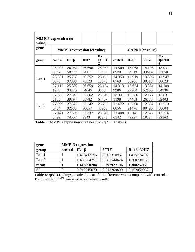
31
MMP13 expression (ct
value)
gene
MMP13 expression (ct value) GAPDH(ct value)
group control IL-1β 30HZ
IL-
1β+30H
Z
control IL-1β 30HZ
IL-
1β+30H
Z
Exp 1
26.907
6347
26.064
50272
26.696
04111
26.067
13486
14.509
6979
13.968
64319
14.105
33619
13.931
53858
26.981
6875
25.789
97803
26.752
73323
26.162
18376
14.353
0769
13.919
06261
13.896
30318
13.947
50023
27.117
1246
25.892
94243
26.659
04045
26.184
3338
14.313
9286
13.654
27208
13.831
52199
14.209
64336
Exp 2
27.687
2158
27.349
39194
27.362
85782
26.810
67467
13.341
1198
13.286
34453
12.177
26135
12.831
62403
27.399
0784
27.325
92583
27.242
90657
26.755
48935
12.672
6856
13.300
91476
12.552
80495
12.513
58604
27.141
6492
27.309
74007
27.337
8849
26.842
95845
12.408
6142
13.141
42227
12.872
1838
12.716
92562
Table 7: MMP13 expression ct values from qPCR analysis.
gene MMP13 expression
group control IL-1β 30HZ IL-1β+30HZ
Exp 1 1 1.455417156 0.902310967 1.415774107
Exp 2 1 1.430364251 0.883544624 1.200730133
mean 1 1.442890704 0.892927796 1.30825212
SD 0 0.017715079 0.013269809 0.152059052
Table 8: qPCR findings, results indicate fold difference when compared with controls.
The formula 2−ΔΔCT was used to calculate results.

32
MMP13 expression
Figure 9: Graph representing relative fold difference in MMP13 expression when
compared with controls.
Relative MMP13 expression when compared with controls was as follows: 1
(control), 1.44 (IL-1β), 0.89 (30Hz vibration + IL-1β), 1.30 (30Hz vibration + IL-1β).
A general trend was noticeable with both gene expressions of SOX9 and MMP13.
IL-1β decreased the gene expression of SOX9. Mechanical vibration increased the
expression of SOX9. When mechanical vibration was combined with IL-1β the effects of
IL-1β seemed to be countered to a small degree. With MMP13, IL-1β increased gene

33
expression. Mechanical vibration decreased expression of MMP13. When combined
(mechanical vibration + IL-1β) the effects of IL-1β were countered to some degree.
Statistical Analysis
Descriptives for SOX9
SOX9
N Mean
Std.
Deviation Std. Error
95% Confidence
Interval for Mean
Minimum Maximum
Lower
Bound
Upper
Bound
1.0000 3 1.000000 .0000000 .0000000 1.000000 1.000000 1.0000 1.0000
2.0000 3 .648718 .5976725 .3450664 -.835982 2.133419 .1290 1.3018
3.0000 3 1.276274 1.0270825 .5929864
-
1.275141 3.827688 .5401 2.4496
4.0000 3 .825482 .2445597 .1411966 .217962 1.433002 .6274 1.0988
Total 12 .937618 .5710987 .1648620 .574760 1.300477 .1290 2.4496
Table 9: Descriptive statistics for SOX9 gene expression.
ANOVA
SOX9
Sum of Squares df Mean Square F Sig.
Between Groups .644 3 .215 .583 .643
Within Groups 2.944 8 .368
Total 3.588 11
Table 10: One-way ANOVA for SOX9 analysis. Results are not statistically significant
(P≤0.05 considered statistically significant).

34
Multiple Comparisons
Dependent Variable: SOX9
Tukey HSD
(I)
group (J) group
Mean Difference (I-
J)
Std.
Error Sig.
95% Confidence Interval
Lower
Bound
Upper
Bound
1.0000 2.0000 .3512816 .4952980 .891 -1.234837 1.937400
3.0000 -.2762735 .4952980 .942 -1.862392 1.309845
4.0000 .1745180 .4952980 .984 -1.411600 1.760636
2.0000 1.0000 -.3512816 .4952980 .891 -1.937400 1.234837
3.0000 -.6275552 .4952980 .606 -2.213674 .958563
4.0000 -.1767637 .4952980 .983 -1.762882 1.409355
3.0000 1.0000 .2762735 .4952980 .942 -1.309845 1.862392
2.0000 .6275552 .4952980 .606 -.958563 2.213674
4.0000 .4507915 .4952980 .800 -1.135327 2.036910
4.0000 1.0000 -.1745180 .4952980 .984 -1.760636 1.411600
2.0000 .1767637 .4952980 .983 -1.409355 1.762882
3.0000 -.4507915 .4952980 .800 -2.036910 1.135327
Table 11: Tukey HSD analysis of SOX9 gene expression. No significance found.

35
Descriptives for MMP13
MMP-13
N Mean
Std.
Deviation
Std.
Error
95% Confidence Interval
for Mean
Minimum Maximum
Lower
Bound
Upper
Bound
1.0000 2 1.000000 .0000000 .0000000 1.000000 1.000000 1.0000 1.0000
2.0000 2 1.442891 .0177151 .0125265 1.283727 1.602054 1.4304 1.4554
3.0000 2 .892928 .0132698 .0093832 .773703 1.012152 .8835 .9023
4.0000 2 1.308252 .1520591 .1075220 -.057944 2.674449 1.2007 1.4158
Total 8 1.161018 .2453774 .0867540 .955877 1.366158 .8835 1.4554
Table 12: Descriptive statistics for MMP13 gene expression.
ANOVA
MMP13
Sum of
Squares df Mean Square F Sig.
Between Groups .398 3 .133 22.467 .006
Within Groups .024 4 .006
Total .421 7
Table 13: One-way ANOVA for MMP13 analysis (P≤0.05 considered statistically
significant). P=0.006 and is thus considered significant.
36
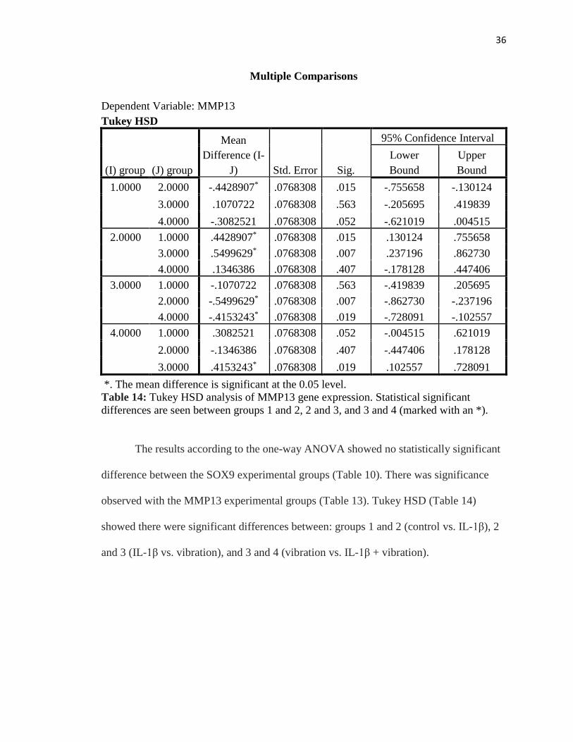
Multiple Comparisons
Dependent Variable: MMP13
Tukey HSD
(I) group (J) group
Mean
Difference (I-
J) Std. Error Sig.
95% Confidence Interval
Lower
Bound
Upper
Bound
1.0000 2.0000 -.4428907* .0768308 .015 -.755658 -.130124
3.0000 .1070722 .0768308 .563 -.205695 .419839
4.0000 -.3082521 .0768308 .052 -.621019 .004515
2.0000 1.0000 .4428907* .0768308 .015 .130124 .755658
3.0000 .5499629* .0768308 .007 .237196 .862730
4.0000 .1346386 .0768308 .407 -.178128 .447406
3.0000 1.0000 -.1070722 .0768308 .563 -.419839 .205695
2.0000 -.5499629* .0768308 .007 -.862730 -.237196
4.0000 -.4153243* .0768308 .019 -.728091 -.102557
4.0000 1.0000 .3082521 .0768308 .052 -.004515 .621019
2.0000 -.1346386 .0768308 .407 -.447406 .178128
3.0000 .4153243* .0768308 .019 .102557 .728091
*. The mean difference is significant at the 0.05 level.
Table 14: Tukey HSD analysis of MMP13 gene expression. Statistical significant
differences are seen between groups 1 and 2, 2 and 3, and 3 and 4 (marked with an *).
The results according to the one-way ANOVA showed no statistically significant
difference between the SOX9 experimental groups (Table 10). There was significance
observed with the MMP13 experimental groups (Table 13). Tukey HSD (Table 14)
showed there were significant differences between: groups 1 and 2 (control vs. IL-1β), 2
and 3 (IL-1β vs. vibration), and 3 and 4 (vibration vs. IL-1β + vibration).
37
CHAPTER 5
DISCUSSION
Cell differentiation
Our data show that mechanical vibration like that exerted by AcceleDent
(0.3g/30Hz) does not pose a negative influence on chondrocytes, rather enhance them to
be differentiated in vitro. This process of differentiation allows for cells to become
further specialized than they were previously. Although the results are qualitative it is
clear that differentiation increased significantly by mechanical vibration. IL-1β seemingly
did not cause a significant change in the amount of differentiation. It is interesting that
IL-1β – the potent inflammatory mediator was not shown to be a significant inhibitor of
differentiation. This can be due to the used concentration (1ng/mL) was not high enough
to affect the C-28/I2 chondrocytes. Aside from being of qualitative measure, this study
did have other limitations. The chondrocyte cell line C-28/I2 was used in place of
primary chondrocytes. These cells were utilized because primary chondrocytes are
difficult to obtain, cannot be controlled, and the number of cells is often inadequate. Due
to the complexity of the human body and cells within it there may be a difference in the
way the cells respond to mechanical vibration than that of our study. Further evaluation
should be done regarding the effects of mechanical vibration on cell differentiation.

38
Gene expression
SOX9
IL-1β was used to mimic an inflammatory environment. SOX9, an important
transcription factor, promotes cell survival (Lefebvre V, 2016). When cells were
stimulated with IL-1β, the expression of SOX9 decreased. When mechanical vibration of
0.3g/30Hz was applied to the group with IL-1β, the level of SOX9 expression was
increased (Fig. 8). Up-regulation of SOX9 indicates that the effect of mechanical
vibration are acting to counter the effect of IL-1β. Experiments using primary
chondrocytes from the TMJ region should be used to verify if the same relationship
exists. If mechanical vibration can decrease levels of inflammation, it may prove to be a
valuable therapy in treating patients who suffer from TMD that is of an inflammatory
nature. Although our data do not show a statistical significance, it is suggestive of a
pattern that mechanical vibration anabolically up-regulates SOX9 expression in both
normal and inflammatory conditions.
MMP13
MMP13 is a proteinase that breaks down collagen and other matrix components.
It is known for its catabolic activity (Liang Z, et. al, 2012). Our data showed an increase
in MMP13 expression with the addition of IL-1β (Fig. 9), which is in agreement with
previous work from Lee and Salter. They showed that IL-1β causes downstream events
leading to the up-regulation of MMPs. The effects of mechanical vibration were shown to
down-regulate expression of MMP13. Our data suggest that mechanical vibration
counters the effect of IL-1β. (Fig. 9). This model may be more representative of how
mechanical vibration can alter the cellular pathways present in an inflammatory state
39
because of the direct relationship between IL-1β and MMP13. Due to the complexity of
the human body and the multitude of biological pathways, it should not be assumed that
this exact relationship exists in an in vivo model. Further study should be done to better
understand the molecular pathways of inflammation and how cells respond to therapies
targeting on inflammation.
Overall, mechanical vibration does not appear to have a negative effect on human
chondrocytes in vitro. Our data suggest it may be beneficial to patients who suffer from
osteoarthritis of an inflammatory nature. We tested the hypothesis that mechanical
vibration would help reduce inflammation within the TMJ. Our data indicate that
inflammation can be reduced to some degree in an in vitro model (Fig. 8, 9). Studies
should be done using primary chondrocytes from the temporomandibular joints to
evaluate whether the same relationships exist. Due to time constraints the expression of
MMP13 was only run through two trials. Further testing will be done to further evaluate
this relationship. Therapies used to treat OA should aim to target the inflammatory
nature. Mechanical vibration may work well to help alleviate inflammation and slow the
progression of OA.
Although the preliminary data are not statistically significant, it indicates a
general trend that mechanical vibration counters the effects of IL-1β. Since increased IL-
1β levels seems to be an early indicator of progression of OA and also that of
inflammation (Alstergren et. al, 2003), it makes sense that decreasing this interleukin
would help halt this pathological progression. Again, further studies are needed using an
in vivo model to gain a better understanding of the relationship that exists between
mechanical vibration and pathologies involving inflammation.

40
CHAPTER 6
CONCLUSION
Mechanical vibration (0.3g/30Hz) is able to increase differentiation of human
chondrocytes C-28/I2 with and without treatment of IL-1β (1ng/mL), while IL-1β
(1ng/mL) itself shows minimal effect on the differentiation of human chondrocytes C-
28/I2. Mechanical vibration anabolically regulates gene expressions of SOX9
(upregulated, p > 0.05) and MMP13 (downregulated, p<0.05), and can partially counter
the catabolic changes of SOX9 and MMP13 induced by the treatment of IL-1β
(10ng/mL). Under the specific experimental conditions and limitations of this study, it
can be concluded that mechanical vibration (0.3g/30Hz) does not appear to harm human
chondrocytes C-28/I2 in vitro and may help enhance the normal function of chondrocytes
and potentially reduce inflammation. Further studies should be done to gain a better
understanding of the effects of mechanical vibration and the chondrocytes (cartilage) of
TMJ, in order to safely use vibration in orthodontics as well as potentially help manage
TMD.

41
BIBLIOGRAPHY
Acceledent. Accessed online at: http://acceledent.com/.
Alcaraz MJ, et. al. New Molecular Targets for the Treatment of Osteoarthritis.
Biochemical Pharmacology. 2010;80(1):13-21.
Alstergren P, Benavente C, Kopp S. Interleukin-1beta, interleukin-1 receptor antagonist,
and interleukin-1 soluble receptor II in temporomandibular joint synovial fluid from
patients with chronic polyarthritides. J Oral Maxillofac Surg. 2003;61(10):1171-8.
Bi W, et. al. Sox9 is required for cartilage formation. Nat Genet. 1999;22(1):85-9.
Bonnet CS, Walsh DA. Osteoarthritis, angiogenesis and inflammation. Rheumatology
(Oxford). 2005;44(1):7-16.
Borzi RM, et. al. Matrix metalloproteinase 13 loss associated with impaired extracellular
matrix remodeling disrupts chondrocyte differentiation by concerted effects on
multiple regulatory factors. Arthritis Rheum. 2010;62(8):2370-81.
Bowman SJ. The effect of vibration on the rate of leveling and alignment. J Clin Orthod.
2014;48(11):678-88.
Chen CS, Ingber DE. Tensegrity and mechanoregulation: from skeleton to cytoskeleton.
Osteoarthritis Cartilage. 1999;7(1):81-94.
Consensus Development Conference. Diagnosis, prophy-laxis, and treatment of
osteoporosis. Am J Med. 1993;94:646–650.
Eming, et. al. Inflammation in Wound Repair: Molecular and Cellular Mechanisms.
Journal of Investigative Dermatology. 2007;127(3):514-25.
Felson DT. Osteoarthritis: New Insights. Part 1: The Disease and Its Risk Factors. Annals
of Internal Medicine Ann Intern Med. 2000;133(8):635.
Finger F, et. al. Molecular Phenotyping of Human Chondrocyte Cell Lines T/C-28a2,
T/C-28a4, and C-28/I2. Arthritis & Rheumatism. 2003;48(12):3395-403.
Gidarakou IK, et. al. Comparison of skeletal and dental morphology in asymptomatic
volunteers and symptomatic patients with bilateral degenerative joint disease. Angle
Orthod. 2003;73:71-78.
Gómez-Cabello A, et. al. Effects of a Short-term Whole Body Vibration Intervention on
Bone Mass and Structure in Elderly People. Journal of Science and Medicine in
Sport. 2014;17(2):160-64.

42
Greco KV, et. al. High Density Micromass Cultures of a Human Chondrocyte Cell Line:
A Reliable Assay System to Reveal the Modulatory Functions of Pharmacological
Agents. Biochemical Pharmacology. 2011;82(12):1919-929.
Hamamura K, et. al. Knee Loading Reduces MMP13 Activity in the Mouse Cartilage.
BMC Musculoskeletal Disorders BMC Musculoskelet Disord. 2013;14(1):312.
Hunziker EB. Articular cartilage repair: basic science and clinical progress. A review of
the current status and prospects. Osteoarthritis Cartilage. 2002;10(6):432-63.
Kau CH, Nguyen JT, English JD. The clinical evaluation of a novel cyclical force
generating device in orthodontics. Orthodontic Practice US. 2010;1(1):10-15.
Kaupp JA, Waldman SD. Mechanical Vibrations Increase the Proliferation of Articular
Chondrocytes in High-density Culture. Proceedings of the Institution of Mechanical
Engineers, Part H: Journal of Engineering in Medicine. 2008;222(5):695-703.
Kim MR, et. al. Orthodontics and temporomandibular disorder: A meta-analysis. Am J
Orthod Dentofacial Orthop. 2002;121:438-46.
Knudson CB, Knudson W. Cartilage proteoglycans. Semin Cell Dev Biol. 2001;12(2):69-
78.
Kubota E, et. al. Interleukin 1β and Stromelysin (MMP3) Activity of Synovial Fluid as
Possible Markers of Osteoarthritis in the Temporomandibular Joint. Journal of Oral
and Maxillofacial Surgery. 1997;55(1):20-27.
Kulkarni RN, Voglewede PA, Liu D. Mechanical Vibration Inhibits Osteoclast
Formation by Reducing DC-STAMP Receptor Expression in Osteoclast Precursor
Cells. Bone. 2013;57(2):493-98.
LeBlanc A, et. al. Bone mineral and lean tissue loss after long duration space flight. J
Musculoskelet Neuronal Interact. 2000;1(2):157-60.
Lee HS, Salter DM. Biomechanics of Cartilage and Osteoarthritis. Osteoarthritis -
Progress in Basic Research and Treatment. 2015; Ch.3: 41-52.
Leethanakul C, et. al. Vibratory stimulation increases interleukin-1 beta secretion during
orthodontic tooth movement. Angle Orthodontist. 2016;86(1):74-80.
Lefebvre V, Dvir-Ginzberg M. SOX9 and the many facets of its regulation in the
chondrocyte lineage. Connect Tissue Res. 2016;29:1-13.
Liang Z, et. al. MiRNA-140 Is a Negative Feedback Regulator of MMP-13 in IL-1β-
stimulated Human Articular Chondrocyte C-28/I2 Cells. Inflamm. Res.
Inflammation Research. 2012;61(5):503-09.

43
Lin Z, et. al. The Chondrocyte: Biology and Clinical Application. Tissue Engineering.
2006;12(7):1971-984.
Liu D. Transmission of Mechanical Vibration from AcceleDent to Dentition and Skull.
Journal of Dental Research. 2013;92(Spec Iss A):Abstr. No. 1773.
Liu J. Biosynthetic Response of Cultured Articular Chondrocytes to Mechanical
Vibration. Res Exp Med. 2001;200:183-193.
Malemud CJ. Matrix metalloproteinases (MMPs) in health and disease: an overview.
Front Biosci. 2006;11:1696-1701.
Martin P. Wound Healing--Aiming for Perfect Skin Regeneration. Science.
1997;276(5309):75-81.
National Institute of Health. Management of temporomandibular disorders. NIH
Technology Assessment Conference. Bethesda, Md:1996.
Okeson JP. Management of Temporomandibular Disorders and Occlusion. St. Louis,
MO: Mosby Elsevier. 2008;Ch1: 2-24.
Osteoarthritis. Accessed online at: http://www.arthritis.org/about-
arthritis/types/osteoarthritis/.
Osteoarthritis. Accessed online at: http://www.rheumatology.org/I-Am-A/Patient-
Caregiver/Diseases-Conditions/Osteoarthritis.
Papadopoulou AK, et. al. Load Application Induces Changes in the Expression Levels of
SOX9, FGFR-3 and VEGF in Condylar Chondrocytes. FEBS Letters.
2007;581(10): 2041-046.
Pavlin D, et. al. Cyclic loading (vibration) accelerates tooth movement in orthodontic
patients: A double-blind, randomized controlled trial. Seminars in Orthodontics.
2015;21(3):187-94.
Rubin C, et. al. Prevention of Postmenopausal Bone Loss by a Low-Magnitude, High-
Frequency Mechanical Stimuli: A Clinical Trial Assessing Compliance, Efficacy,
and Safety. J Bone Miner Res Journal of Bone and Mineral Research.
2004;19(3):343-51.
Sabatini M, Pastoureau P, and De Ceuninck F. Cartilage and Osteoarthritis. Ch. 3-4.
Totowa, NJ: Humana, 2004.
Singer AJ, Clark RA. Cutaneous wound healing. N Engl J Med. 1999; 341(10):738-46.
44
Thie NM, Prasad NG, Major PW. Evaluation of glucosamine sulfate compared to
ibuprofen for the treatment of temporomandibular joint osteoarthritis: a randomized
double blind controlled 3 month clinical trial. J Rheumatology. 2001;28(6):1347-
1355.
Thompson WR, Sherwin SY, Rubin J. Vibration Therapy. Current Opinion in
Endocrinology & Diabetes and Obesity 2014;21(6):447-53.
Vane J, Botting R. Inflammation and the mechanism of action of anti-inflammatory
drugs. Faseb J. 1987;1:89-96.
Vane JR, Botting RM. Anti-inflammatory Drugs and Their Mechanism of Action.
Inflammation Research. 1998;47(0):78-87.
Walsh DA, et. al. Angiogenesis in the synovium and at the osteochondral junction in
osteoarthritis. Osteoarthritis Cartilage. 2007;15(7):743-51.
Ward K, et. al. Low Magnitude Mechanical Loading Is Osteogenic in Children With
Disabling Conditions. J Bone Miner Res Journal of Bone and Mineral Research.
2004;19(3):360-69.
Westesson P, Rohlin M. Internal Derangement Related to Osteoarthrosis in
Temporomandibular Joint Autopsy Specimens. Oral Surgery, Oral Medicine, Oral
Pathology. 1984;57(1):17-22.
Woodhouse NR, et. al. Supplemental Vibrational Force During Orthodontic Alignment:
A Randomized Trial. Journal of Dental Research. 2015;94(5):682-89.
Xiong H, Rabie AB, Hagg U. Mechanical strain leads to condylar growth in adult rats.
Front. Biosci. 2005;10:67–73.