the effect of water deprivation on the tonicity responsive ... · of highly concentrated urine is...
TRANSCRIPT
852
INTRODUCTIONDesert mammals can survive for long periods without free waterby obtaining preformed water from food and metabolic water fromthe oxidation of hydrogen, and by reducing the amount of waterlost during respiration and excretion (Degen, 1997). The excretionof highly concentrated urine is dependent on the regulation of twoprocesses: ultrafiltration of plasma in the glomerulus andreabsorption of water in the renal tubules. Accordingly, desertmammals have a reduced glomerular filtration rate and enhancedtubular water reabsorption (Degen, 1997). Water reabsorption relieson the medullary osmotic gradient created by the loop of Henle(Bankir and Rouffignac, 1985). Desert rodents, in particular, havelong loops of Henle, and routinely excrete urine in the range of4000–7000·mOsm·l–1 (Degen, 1997). A high osmolality of the innermedulla and papilla of the kidney exposes the cells in that regionto a hypertonic environment, which can severely compromise cellfunction and lead to DNA damage and apoptosis (Burg et al., 2007;Jeon et al., 2006). The most abundant osmolytes in the renal medullaand papilla of the kidney are Na+, Cl– and urea (Garcia-Perez andBurg, 1991). High NaCl causes hypertonicity and disturbances incell volume, but urea has no effect on tonicity as it readily permeatescell membranes (Yancey et al., 1982).
In response to hypertonicity, renal cells will rapidly accumulateinorganic ions, but in conjunction with elevated urea concentrations,this will compromise the function of cellular proteins andmacromolecules (Yancey et al., 1982). The intracellular ionic
concentration is then lowered by the replacement of the inorganicions with compatible osmolytes, which occurs over 1–3·days(Rauchman et al., 1997; Jeon et al., 2006; Burg et al., 2007). Sincecompatible osmolytes do not contribute to ionic strength, theintracellular ionic concentration remains within an optimalphysiological range, while the osmolality of the interstitium andintracellular fluid is the same (Beck et al., 1998; Garcia-Perez andBurg, 1991). The accumulation of compatible osmolytes is reversiblesince rehydration leading to diuresis, causes swelling of themedullary cells and the release of compatible osmolytes into theinterstitial fluid (Beck et al., 1998; Garcia-Perez and Burg, 1991).
Sorbitol (D-glucitol), myo-inositol, betaine and taurine arecompatible osmolytes that are found in abundance in the renalmedulla. In response to hypertonicity, sorbitol is produced fromglucose by the aldose reductase (AR) enzyme, whereas myo-inositol, betaine and taurine are transported into cells by Na+- orNa+/Cl–-dependent transporters (Burg et al., 1996). The transcriptionof AR and the myo-inositol (SMIT; also known as SLC5A3),betaine/GABA (BGT-1; also known as SLC6A12) and taurine(TauT; also known as SLC6A6) transporters is regulated by thetonicity-responsive enhancer binding protein (TonEBP; also knownas NFAT5) (Burg et al., 1996). The AR, SMIT, BGT-1 and TauTgenes have osmotic response elements (ORE) in their 5� flankingregions that contain the tonicity-responsive enhancer (TonE)consensus sequence (Takenaka et al., 1994). The binding of TonEBPto OREs increases the transcription of AR, SMIT, BGT-1 and TauT,
The Journal of Experimental Biology 211, 852-859Published by The Company of Biologists 2008doi:10.1242/jeb.006395
The effect of water deprivation on the tonicity responsive enhancer binding protein(TonEBP) and TonEBP-regulated genes in the kidney of the Spinifex hopping mouse,
Notomys alexis
Ray C. Bartolo* and John A. DonaldSchool of Life and Environmental Sciences, Deakin University, Geelong, Victoria 3217, Australia
*Author for correspondence at present address: Department of Physiology, University of Otago, PO Box 913, Dunedin, 9054, New Zealand(e-mail: [email protected])
Accepted 8 January 2008
SUMMARYIn desert rodents, the production of concentrated urine is essential for survival in xeric environments in order to conserve water.Reabsorption of water in the kidney is dependent on large osmotic gradients in the renal medulla. This causes the renal cells tobe bathed in a hypertonic extracellular fluid that can compromise cellular function. In response to hypertonicity, kidney cellsaccumulate compatible, non-ionic osmolytes that lower the ionic strength within the cells to isotonic levels by replacingintracellular ionic electrolytes. The tonicity-responsive enhancer binding protein (TonEBP) is a transcription factor that regulatesthe expression of genes that encode proteins that catalyse the accumulation of compatible osmolytes. We investigated theexpression of TonEBP mRNA and protein and compatible osmolyte genes in the Spinifex hopping mouse, Notomys alexis, anAustralian desert rodent that produces a highly concentrated urine. TonEBP mRNA expression was unchanged after 3·days ofwater deprivation but was significantly increased after 7·and 14·days of water deprivation. Immunohistochemistry showed thatduring water deprivation TonEBP had translocated from the cytoplasm into the nucleus of cells in the renal medulla and papilla.In addition, 3, 7 and 14·days of water deprivation caused a significant increase in aldose reductase (AR), myo-inositol (SMIT),betaine/GABA (BGT-1) and taurine (TauT) transporter mRNA expression, which is indicative of an increase in TonEBP activity. Indesert rodents, TonEBP regulation of gene transcription is probably an important mechanism to protect renal cells in the face ofthe large corticomedullary gradient that is required to concentrate urine and conserve water.
Key words: Notomys alexis, tonicity responsive enhancer binding protein (TonEBP), compatible osmolytes, kidney, water deprivation.
THE JOURNAL OF EXPERIMENTAL BIOLOGY
853Expression of TonEBP and associated genes in water-deprived hopping mice
which in turn leads to an increase in the intracellular accumulationof the respective compatible osmolyte (Woo and Kwon, 2002; Wooet al., 2002). TonEBP has a basal activity level under isotonicconditions that is decreased by hypotonicity and increased byhypertonicity. An increase in TonEBP activity is reflected by anincrease in the expression of AR, SMIT, BGT-1 and TauT mRNAs;mice lacking the TonEBP gene have a reduced expression of AR,SMIT, BGT-1 and TauT (Lopez-Rodriguez et al., 2004). Thebidirectionality of TonEBP activity has been demonstrated inMadin-Darby canine kidney (MDCK) cells (Woo et al., 2000a). InMDCK cells grown under isotonic conditions, TonEBP is distributedbetween the nucleus and cytoplasm, but under hypertonic stressTonEBP increases in abundance and translocates to the nucleus toact as a transcription factor of osmoprotective genes. By contrast,when the MDCK cells are transferred to a hypotonic medium,TonEBP translocates to the cytoplasm, its abundance decreases, andcompatible osmolytes move out of the cell (Woo et al., 2000a).
The Spinifex hopping mouse, Notomys alexis, is a small rodentthat is highly adapted to survive in arid environments where it canlive without drinking water (MacMillen and Lee, 1969; Weaver etal., 1994). N. alexis has been reported to produce the mostconcentrated urine of any mammal (9370·mOsm·l–1) (MacMillenand Lee, 1969). In the laboratory, N. alexis can tolerate waterdeprivation for 28·days without changes in plasma osmolality,vasopressin or renin, as seen in other species of desert rodent(Weaver et al., 1994; Heimeier et al., 2002). By contrast, waterdeprivation experiments using laboratory rats and mice are short-term because of the inability of the animals to survive withoutdrinking, and the animals show a marked increase in plasmaosmolality and vasopressin levels (see Degen, 1997). Thus, themechanisms underpinning survival during long-term waterdeprivation in desert mammals may be different from mesic species.Given the extraordinary urine-concentrating ability of N. alexis, wepredicted that the expression of TonEBP and the genes encodingproteins that regulate intracellular compatible osmolytes would playan important role and be upregulated in the renal medulla of N.alexis during periods of water stress. Thus, the aim of the currentstudy was to analyse the expression of TonEBP, AR, BGT-1 SMITand TauT mRNAs and determine the distribution of TonEBP proteinin the kidney of water-deprived N. alexis, in comparison to micewith ad libitum access to water.
MATERIALS AND METHODSAnimals
Spinifex hopping mice Notomys alexis Thomas 1922 were obtainedfrom a breeding colony at the Deakin University Animal House,Geelong, Australia. The animals were housed in rat boxescontaining straw and sawdust for bedding with wire mesh lids at21–24°C and a 13·h:11·h light:dark cycle (lights on at 08:00·h).The animals received fresh tapwater ad libitum and were fed FrenchWhite millet seed.
Water deprivation experimentsAll experiments involving N. alexis were performed with approvalfrom the Deakin University Animal Welfare Committee (Projectnumber A31/2003). The water deprivation experiments were carriedout as previously described (Donald and Bartolo, 2003). For eachwater deprivation time point, there was a water-replete (control)group in which N. alexis had ad libitum access to water, and a water-deprived (experimental) group in which N. alexis was subjected to3, 7 or 14·days without access to free water; this was termed waterdeprivation. All mice were ear-tagged to enable identification, and
were approximately 6·months old. There were eight animals in eachgroup and they were housed in groups of four in sand-filled glassaquaria (W·100·cm�H·40·cm�L·50·cm), which allowed forcommunal sleeping burrows. The mice were weighed and fed 20·gof millet seed per cage daily; a group of four mice eat a maximumof 4.5·g seed per day during water deprivation (R.C.B., unpublished).The mass of the animals in the 14-day water deprivation experimentwas used for the analysis shown in Fig.·1. At the end of the respectivewater deprivation periods, the mice were anaesthetised by halothaneinhalation followed by cervical dislocation. The kidneys weredissected free, and the left kidney was frozen in liquid nitrogen andstored at –80°C until RNA was isolated, while the right kidneywas fixed overnight at 4°C in 4% formaldehyde (pH·7.4;~1100·mOsm·kg–1), and then stored in 70% ethanol until processingfor immunohistochemistry.
Collection of urine for the measurement of osmolalityA separate water deprivation experiment using six N. alexis wasperformed to measure urine osmolality. The mice were water-deprived for 14·days and urine was collected on day·0, 3, 7 and 14.Urine was collected from individual mice placed for 1·h incylindrical containers (90·mm diameter and 150·mm high) that hada wire mesh floor. The containers were suspended above a collectiontray that contained light paraffin oil, thus preventing evaporationof the urine. Urine osmolality was then measured using a Vapro®
vapour pressure osmometer (Wescor Model 5520, Logan, UT,USA).
Amplification, cloning and sequencing of putative cDNAsKidney total RNA was isolated using TRIzol (Invitrogen, MountWaverley, Victoria, Australia), which utilises the single step acidguanidinium thiocyanate-phenol-chloroform extraction method(Chomczynski and Sacchi, 1987). The RNA concentration wasdetermined by spectrophotometry at 260·nm. First strand cDNA wassynthesised from kidney total RNA using Superscript II (Invitrogen)as per the manufacturer’s protocol. Primers were designed basedon Mus musculus sequence data obtained from GenBank (NationalCentre for Biotechnology, NCBI). The accession numbers for theM. musculus sequences are as follows: TonEBP, AF453571; AR,NM_009658; BGT-1, NM_133661; SMIT, AF220915; and TauT,AAB54039.
Primer sequences, annealing temperature, and the size of thepredicted PCR amplicons are shown in Table·1. PCR was performedin a total volume of 20·�l with a final concentration of: 1� PCR
50
60
70
80
90
100
110
14131211109876543210–1–2–3–4
Control Water deprived
Days of water deprivation
Bod
y m
ass
(%)
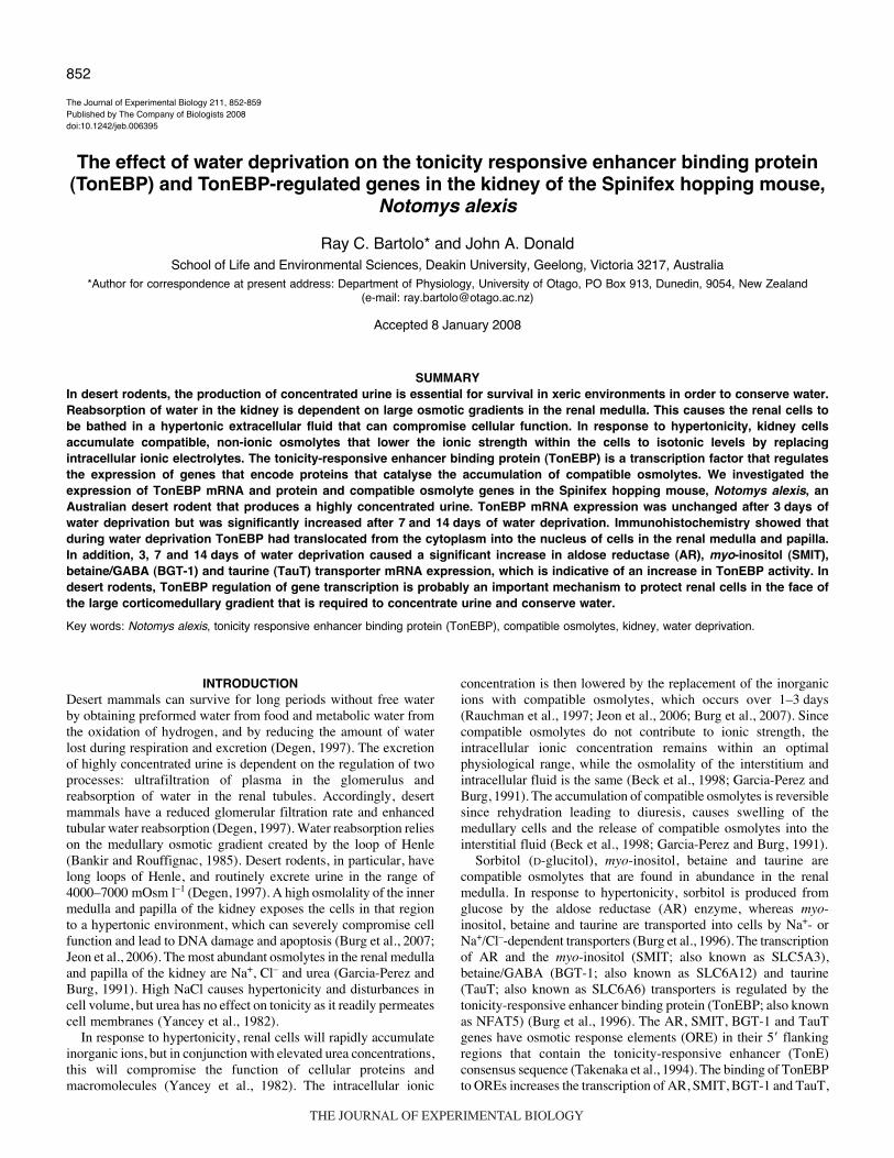
Fig.·1. The effect of 14·days of water deprivation on body mass in N.alexis. There was a significant decrease in mean body mass during theinitial period of water deprivation, but after day·7 it stabilised and began toincrease. Water-replete N. alexis showed no significant change in bodymass.
THE JOURNAL OF EXPERIMENTAL BIOLOGY
854
buffer, 0.2·mmol·l–1 dNTPs, 1·�mol·l–1 of each forward and reverseprimer, 1.0 i.u. of Taq DNA polymerase (Scientifix, Melbourne,Australia), 2.5·mmol·l–1 MgCl2 and 1·�l of the cDNA synthesisreaction. Amplification of the various cDNAs was performed asfollows: initial denaturation of 300·s at 94°C, 35 cycles of 45·s at94°C, 30·s at the annealing temperature (Table·1), 45·s at 72°C, anda final extension of 300·s at 72°C. The PCR products were purifiedand cloned into a pCR2.1 vector, which was then transformed intoOne Shot TOP10 chemically competent Escherichia coli cells usinga TA cloning® Kit (Invitrogen). The cloned cDNAs were sequencedon an Applied Biosystems automated sequencer (Australian GenomeResearch Facility, Brisbane, Australia). The BLAST (Basic LocalAlignment Search Tool) program on the NCBI database was usedto search GenBank for similar sequences (Altschul et al., 1997).Alignments of N. alexis and M. musculus nucleotide and amino acidsequences were carried out to determine homology between clonedN. alexis cDNAs and the sequences from which PCR primers weredesigned, using ClustalW (http://www.ebi.ac.uk/clustalw/).
mRNA expression analysisReverse transcription PCR was used to detect changes in theexpression of TonEBP, AR, BGT-1, SMIT and TauT mRNAs inthe kidney of water-replete and water-deprived N. alexis. Total RNAisolation and cDNA synthesis were performed as described above.For the analysis of mRNA expression, 2·�g of total RNA wasreverse transcribed, and then 1·�l of the cDNA reaction was usedin PCR. To quantify the level of mRNA expression between controland water-deprived N. alexis, the expression of glyceraldehyde-3-phosphate dehydrogenase (GAPDH) mRNA was used as an internalcontrol, as the expression of GAPDH does not change in responseto water deprivation (Heimeier et al., 2002; Sturzenbaum and Kille,2001). The GAPDH primers were as follows: forward 5�-GAA -GGTCGGTGTGAACGGATTTG-3�, and reverse 5�-TTACTCC -TTGGAGGCCATGTAGG-3�; these primers generated a 999 basepair amplicon.
In preliminary experiments, the linear amplification range of thegenes of interest and GAPDH were determined by running PCRreactions for a varying number of cycles between 17 and 35. Thelinear range was also determined in samples from control and water-deprived mice to ensure that there was no variation betweenindividual animals or sample groups. The PCRs for gene expressionanalysis were performed for varying numbers of cycles, dependingon the gene: GAPDH, 20; TonEBP, 22; AR, 24; BGT-1, 28; SMIT,24; TauT, 31. The parameters for the PCR cycles were the same aspreviously mentioned.
For the quantification of the PCR products, the reactions werespiked with 2.5·�Ci of [�-32P]dCTP. As the radio-labelled dCTPis randomly incorporated into the PCR products, the level ofradiation emitted by a PCR product is directly proportional to theamount of PCR product. Equal aliquots of the GAPDH PCR andgene of interest PCR were mixed (to avoid gel loading error) and
R. C. Bartolo and J. A. Donald
subjected to electrophoresis on a 1.5% agarose gel with a 1�Tris–borate–EDTA (TBE) running buffer at 100·V. The gel wasincubated in 0.5·�g·ml–1 of ethidium bromide, visualised on an UVlight box and the cDNA bands were excised, and placed inmicrocentrifuge tubes. The amount of isotope [�-32P] incorporatedinto the PCR products was measured by placing the microcentrifugetubes in vials and counting in a scintillation counter (Tri-Carb2000CA Liquid Scintillation Counter, United Technology Packard,Downers Grove, IL, USA). The expression of the various mRNAswas determined as a ratio of GAPDH mRNA expression (gene ofinterest/GAPDH), and the difference in the ratios between water-replete and water-deprived groups were analysed for statisticalsignificance. The mRNA expression data are expressed as apercentage of the control where the mean values from controlanimals represent 100% for illustrative purposes only.
TonEBP ImmunohistochemistryOne kidney from all mice (N=8 for each time point) was analysedfor TonEBP immunoreactivity (TonEBP-IR). Fixed tissues wereprocessed in a Leica TP 1010 automated tissue processor (Wetzlar,Germany), which dehydrated the tissue through a series of ethanoland xylene washes. The kidneys were then embedded in ParaplastTM
tissue embedding medium, and 5·�m sections were placed on slidescoated in 2% 3-aminopropyltiethoxysilane (Sigma) and allowed todry overnight. Sections were prepared for immunohistochemistryby dewaxing in xylene and rehydration through a graded series ofethanol to water. Endogenous peroxidase activity was quenched byincubating the sections in 3% hydrogen peroxide for 10·min. Thesections then underwent heat-induced epitope retrieval; sectionswere incubated in 1.0·mmol·l–1 EDTA buffer (pH·8.0) for 10·min,heated for 3�5·min in a 650·W microwave oven, cooled to roomtemperature and washed in phosphate-buffered saline (PBS; pH·7.4;2�5·min washes). Endogenous biotinylated proteins were blockedby the use of an Avidin–Biotin blocking kit (Vector Laboratories,Burlingame, CA, USA), which involved incubating sections inAvidin D solution for 15·min, a 1·min rinse with PBS, andincubation in Biotin solution for 15·min, followed by incubationwith an affinity-purified rabbit anti-mouse TonEBP antiserum for2·h at room temperature. The TonEBP antiserum was diluted(1:5000) with PBS, and was kindly donated by Prof. Seung KyoonWoo, University of Maryland, Baltimore, USA (Miyakawa et al.,1999). The sections were then washed in PBS for 2�10·min. AVectastain ABC kit (Vector Laboratories) was used for the detectionof the TonEBP antiserum. The sections were incubated withbiotinylated secondary antibody solution (1:200) for 30·min, washedin PBS for 2�10·min, and incubated with the Vectastain ABCreagent (Vector Laboratories) for 45·min. Sections were thenwashed in PBS for 10·min, rinsed in 0.1·mol·l–1 Tris (pH·7.4) andincubated in 0.02% diaminobenzidine tetrahydrochloride (DAB; in0.1·mol·l–1 Tris, pH 7.4) for 10·min. The slides were examined undera light microscope (Axioskop 20, Carl Zeiss, Göttingen, Germany)
Table·1. PCR amplification of TonEBP, AR, BGT-1, SMIT and TauT from N. alexis
Forward primer (5�–3�) Reverse primer (5�–3�) AT PS (bp)
TonEBP ATGCAATTTCAGAATCAGCC GCATTTGCTGAGAAAGAAG 60°C 514AR TTGACTGCGCCCAGGTGTAC TATATGCTGTCACCACGATGC 60°C 504BGT-1 ATGGACCAGAAAGGCAAGGAC CTCTCCCAGAATTCCATGACAG 60°C 509SMIT CACTGTGAGTGGATACTTCC TCTCTTAACTTCCTCAAACC 52°C 544TauT TCCACAAAGACATCCTGAAGC GGTGAAGTTGGCAGTGCTAAG 60°C 539
AR, aldose reductase; BGT-1, betaine/GABA transporter; SMIT, myo-inositol transporter; TauT, taurine transporter; TonEBP, tonicity-responsive enhancerbinding protein; AT, annealing temperature; PS, expected size of the PCR amplicons (bp).
THE JOURNAL OF EXPERIMENTAL BIOLOGY
855Expression of TonEBP and associated genes in water-deprived hopping mice
and sections were photographed with a digital colour system(Spot 35 Camera System, Diagnostic Instruments, SterlingHeights, MI, USA). The specificity of staining wasdetermined by running negative controls omitting primaryand/or secondary antibody.
Data analysisTo test the difference in mRNA expression between controland experimental groups, a Student’s t-test was performed.Changes in body mass during the 14-day water deprivationexperiment were analysed using a two-way ANOVA and aStudent’s t-test, and urine osmolalities were analysed usinga one-way ANOVA; each used a Tukey’s post-hoc test. Allstatistical probabilities were calculated using SPSS forWindows 14, and P�0.05 was considered significant (Quinnand Keogh, 2002).
Materials[�-32P]dCTP (3000·Ci·mmol·l–1) was purchased from GE LifeSciences (Rydalmere, NSW, Australia). The Vectastain ABC kitwas purchased from Abacus ALS, Brisbane, Australia. All otherchemicals were either reagent or molecular grade and werepurchased from Sigma-Aldrich (Castle Hill, NSW, Australia) orScientifix (Melbourne, Australia).
RESULTSCloning and sequencing of N. alexis cDNAs
TonEBP, AR, BGT-1, SMIT and TauT cDNAs were cloned andsequenced from N. alexis kidney cDNA. The nucleotide anddeduced amino acid sequences of the cDNAs are available formthe NCBI GenBank; accession numbers for the respective sequencesare shown in Table·2. Sequence analysis of the cloned N. alexiscDNAs showed that they have high homology to the respective M.musculus sequences from which the PCR primers were designed.Table·2 also summarises the sequence homology (as a percentage)of N. alexis nucleotide and deduced amino acid sequences, to theirrespective M. musculus sequences.
Effect of water deprivation on body mass and urine osmolalityof N. alexis
Control (water-replete) N. alexis showed no significant change inbody mass over the course of the experiment (Fig.·1). By contrast,N. alexis subjected to water deprivation lost mass over the first7·days, after which it stabilised (Fig.·1, Table·3). Compared to thebody mass at the beginning of water deprivation, there was asignificant decrease in mass after 3, 7 and 14·days of waterdeprivation; body mass at day·7 and day·14 was significantly lessthan that at day·0 and day·3, respectively. Mean urine osmolalitysignificantly increased in response to 3, 7 and 14·days of water
deprivation, when compared to water-replete N. alexis (Table·3).The urine osmolality at day·14 was significantly higher than thatat day 0, and 3 and 7·days of water deprivation (Table·3).
TonEBP mRNA expression and protein immunolocalisation inthe kidney
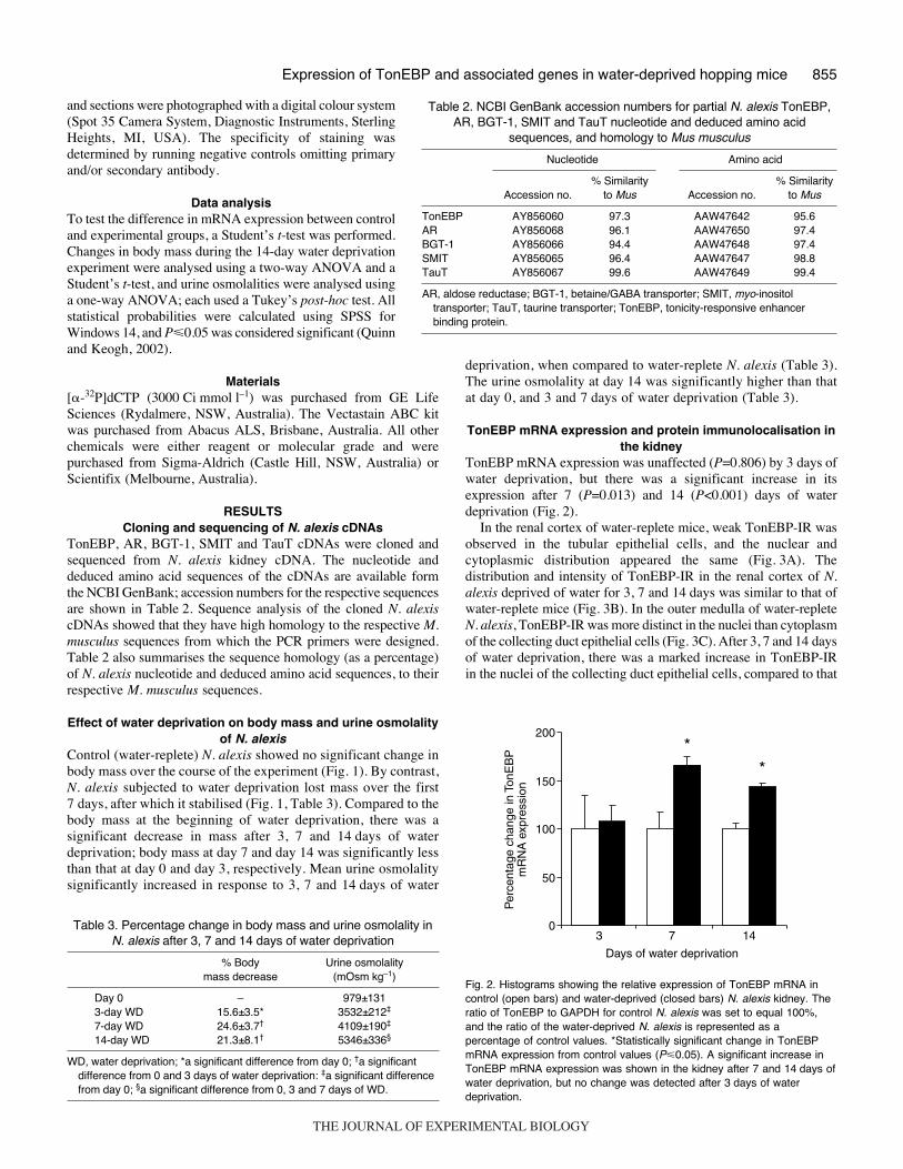
TonEBP mRNA expression was unaffected (P=0.806) by 3·days ofwater deprivation, but there was a significant increase in itsexpression after 7 (P=0.013) and 14 (P<0.001) days of waterdeprivation (Fig.·2).
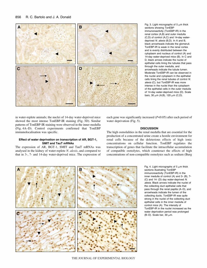
In the renal cortex of water-replete mice, weak TonEBP-IR wasobserved in the tubular epithelial cells, and the nuclear andcytoplasmic distribution appeared the same (Fig.·3A). Thedistribution and intensity of TonEBP-IR in the renal cortex of N.alexis deprived of water for 3, 7 and 14·days was similar to that ofwater-replete mice (Fig.·3B). In the outer medulla of water-repleteN. alexis, TonEBP-IR was more distinct in the nuclei than cytoplasmof the collecting duct epithelial cells (Fig.·3C). After 3, 7 and 14·daysof water deprivation, there was a marked increase in TonEBP-IRin the nuclei of the collecting duct epithelial cells, compared to that
Table 2. NCBI GenBank accession numbers for partial N. alexis TonEBP,AR, BGT-1, SMIT and TauT nucleotide and deduced amino acid
sequences, and homology to Mus musculus
Nucleotide Amino acid
% Similarity % Similarity Accession no. to Mus Accession no. to Mus
TonEBP AY856060 97.3 AAW47642 95.6AR AY856068 96.1 AAW47650 97.4BGT-1 AY856066 94.4 AAW47648 97.4SMIT AY856065 96.4 AAW47647 98.8TauT AY856067 99.6 AAW47649 99.4
AR, aldose reductase; BGT-1, betaine/GABA transporter; SMIT, myo-inositoltransporter; TauT, taurine transporter; TonEBP, tonicity-responsive enhancerbinding protein.
Table·3. Percentage change in body mass and urine osmolality inN. alexis after 3, 7 and 14 days of water deprivation
% Body Urine osmolality mass decrease (mOsm·kg–1)
Day 0 – 979±1313-day WD 15.6±3.5* 3532±212‡
7-day WD 24.6±3.7† 4109±190‡
14-day WD 21.3±8.1† 5346±336§
WD, water deprivation; *a significant difference from day 0; †a significantdifference from 0 and 3·days of water deprivation: ‡a significant differencefrom day 0; §a significant difference from 0, 3 and 7·days of WD.
Fig.·2. Histograms showing the relative expression of TonEBP mRNA incontrol (open bars) and water-deprived (closed bars) N. alexis kidney. Theratio of TonEBP to GAPDH for control N. alexis was set to equal 100%,and the ratio of the water-deprived N. alexis is represented as apercentage of control values. *Statistically significant change in TonEBPmRNA expression from control values (P�0.05). A significant increase inTonEBP mRNA expression was shown in the kidney after 7 and 14·days ofwater deprivation, but no change was detected after 3·days of waterdeprivation.
Per
cent
age
chan
ge in
Ton
EB
Pm
RN
A e
xpre
ssio
n
*
0
50
100
150
200
*
14 7
Days of water deprivation
3
THE JOURNAL OF EXPERIMENTAL BIOLOGY
856
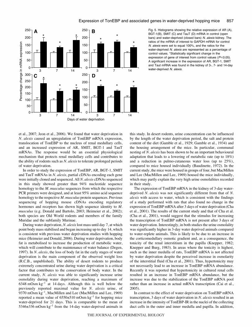
in water-replete animals; the nuclei of 14-day water-deprived miceshowed the most intense TonEBP-IR staining (Fig.·3D). Similarpatterns of TonEBP-IR staining were observed in the inner medulla(Fig.·4A–D). Control experiments confirmed that TonEBPimmunolocalisation was specific.
Effect of water deprivation on transcription of AR, BGT-1,SMIT and TauT mRNAs
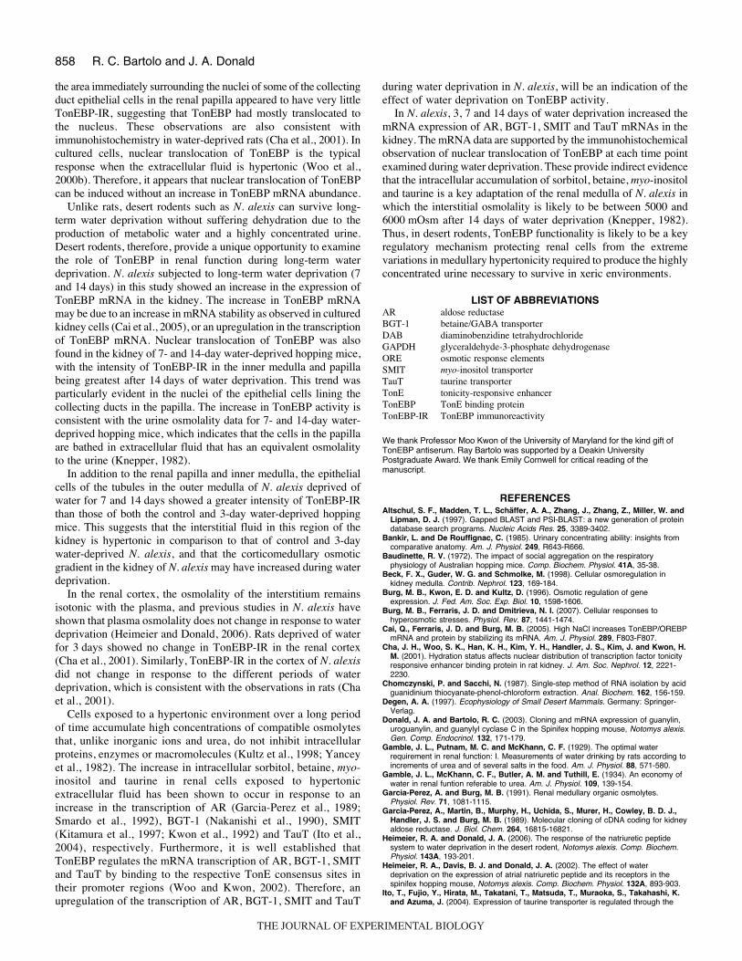
The expression of AR, BGT-1, SMIT and TauT mRNAs wasanalysed in the kidney of water-replete N. alexis, and compared tothat in 3-, 7- and 14-day water-deprived mice. The expression of
R. C. Bartolo and J. A. Donald
each gene was significantly increased (P<0.05) after each period ofwater deprivation (Fig.·5).
DISCUSSIONThe high osmolalities in the renal medulla that are essential for theproduction of a concentrated urine create a hostile environment forrenal cells because of the deleterious effects of high ionicconcentrations on cellular function. TonEBP regulates thetranscription of genes that facilitate the intracellular accumulationof compatible osmolytes, which counteract the effects of highconcentrations of non-compatible osmolytes such as sodium (Burg
Fig.·3. Light micrographs of 5·�m thicksections showing TonEBPimmunoreactivity (TonEBP-IR) in therenal cortex (A,B) and outer medulla(C,D) of control (A,C) and 14-day water-deprived N. alexis (B,D). In A and B,black arrowheads indicate the glomeruli.TonEBP-IR is weak in the renal cortexand is evenly distributed between thecytoplasm and nucleus of control (A) and14-day water-deprived mice (B). In C andD, black arrows indicate the nuclei ofepithelial cells lining the tubules that passthrough the outer medulla, andarrowheads indicate the tubule lumen.Moderate TonEBP-IR can be observed inthe nuclei and cytoplasm in the epithelialcells lining the renal tubules of control N.alexis (C), but TonEBP-IR was moreintense in the nuclei than the cytoplasmof the epithelial cells in the outer medullaof 14-day water-deprived mice (D). Scalebars, 30·�m (A,B), 120·�m (C,D).
Fig.·4. Light micrographs of 5·�m thicksections illustrating TonEBPimmunoreactivity (TonEBP-IR) in theinner medulla of control (A) and 3- (B), 7-(C) and 14- (D) day water-deprived N.alexis. Black arrows indicate the nuclei ofthe collecting duct epithelial cells thatpass through the renal papilla (A–D), andarrowheads indicate the lumen of thecollecting ducts. TonEBP-IR was quitestrong in the nuclei of the collecting ductepithelial cells in the inner medulla ofcontrol mice (A). The intensity ofTonEBP-IR in the nuclei increased as thewater deprivation period was prolonged(B–D). Scale bar, 30·�m.
THE JOURNAL OF EXPERIMENTAL BIOLOGY
857Expression of TonEBP and associated genes in water-deprived hopping mice
et al., 2007; Jeon et al., 2006). We found that water deprivation inN. alexis caused an upregulation of TonEBP mRNA expression,translocation of TonEBP to the nucleus of renal medullary cells,and an increased expression of AR, SMIT, BGT-1 and TauTmRNAs. The response would be an essential physiologicalmechanism that protects renal medullary cells and contributes tothe ability of rodents such as N. alexis to tolerate prolonged periodsof water deprivation.
In order to study the expression of TonEBP, AR, BGT-1, SMITand TauT mRNAs in N. alexis, partial cDNAs encoding each genewere initially cloned and sequenced. All N. alexis cDNAs sequencedin this study showed greater than 94% nucleotide sequencehomology to the M. musculus sequences from which the respectivePCR primers were designed, and at least 95% amino acid sequencehomology to the respective M. musculus protein sequences. Previoussequencing of hopping mouse cDNAs encoding regulatoryhormones and receptors has shown high sequence identity to M.musculus (e.g. Donald and Bartolo, 2003; Heimeier et al., 2002);both species are Old World rodents and members of the familyMuridae and the subfamily Murinae.
During water deprivation, N. alexis lost mass until day·7, at whichpoint body mass stabilised and began increasing up to day·14, whichis consistent with previous water deprivation studies with hoppingmice (Heimeier and Donald, 2006). During water deprivation, bodyfat is metabolised to increase the production of metabolic water,which will contribute to the maintenance of water balance (Degen,1997). In N. alexis, the loss of body fat in the early stages of waterdeprivation is the main component of the observed weight loss(R.C.B., unpublished). The ability of desert rodents to produceextremely concentrated urine during water deprivation is a criticalfactor that contributes to the conservation of body water. In thecurrent study, N. alexis was able to significantly increase urineosmolality during water deprivation, reaching a maximum of6348·mOsm·kg–1 at 14·days. Although this is well below thepreviously reported maximal value for N. alexis urine, of9370·mOsm·kg–1, MacMillen and Lee (MacMillen and Lee, 1969)reported a mean value of 6550±510·mOsm·kg–1 for hopping micewater-deprived for 21 days. This is comparable to the mean of5346±336·mOsm·kg–1 from the 14-day water-deprived animals in
this study. In desert rodents, urine concentration can be influencedby the length of the water deprivation period, the salt and proteincontent of the diet (Gamble et al., 1929; Gamble et al., 1934) andthe housing arrangement of the mice. In particular, communalnesting of N. alexis has been shown to be an important behaviouraladaptation that leads to a lowering of metabolic rate (up to 18%)and a reduction in pulmo-cutaneous water loss (up to 25%),compared to mice housed individually (Baudinette, 1972). In thecurrent study, the mice were housed in groups of four, but MacMillenand Lee (MacMillen and Lee, 1969) housed the mice individually,which may partly explain the very high urine osmolalities recordedin their study.
The expression of TonEBP mRNA in the kidney of 3-day water-deprived N. alexis was not significantly different from that of N.alexis with access to water, which is consistent with the findingsof a study performed with rats that also found no change in theexpression of TonEBP mRNA after 3·days of water deprivation (Chaet al., 2001). The results of the current study and that of Cha et al.(Cha et al., 2001), would suggest that the stimulus for increasingthe transcription of TonEBP mRNA is not present after 3·days ofwater deprivation. Interestingly, in both studies the urine osmolalitywas significantly higher in 3-day water deprived animals comparedto water-replete animals. This is likely to be due to an increase inthe corticomedullary osmotic gradient and, as a consequence, thetonicity of the renal interstitium in the papilla (Knepper, 1982;Knepper and Burg, 1983). In areas where the tonicity is highest,such as the inner medulla of rats, TonEBP mRNA is not affectedby water deprivation despite the perceived increase in osmolarityof the interstitial fluid (Cha et al., 2001). Thus, hypertonicity maynot necessarily lead to an increase in TonEBP mRNA abundance.Recently it was reported that hypertonicity in cultured renal cellsresulted in an increase in TonEBP mRNA abundance, but theincrease was due to the stabilization of the TonEBP mRNA poolrather than an increase in actual mRNA transcription (Cai et al.,2005).
In contrast to the effect of water deprivation on TonEBP mRNAtranscription, 3·days of water deprivation in N. alexis resulted in anincrease in the intensity of TonEBP-IR in the nuclei of the collectingduct cells in the outer and inner medulla and papilla. In addition,
Per
cent
age
chan
ge in
mR
NA
exp
ress
ion
0
50
100
150
200
250
0
50
100
150
200
250
0
50
100
150
200
250
0
50
100
150
200
250
300 D
* * *
A * *
*
B
*
*
*
C *
* *
14 7Days of water deprivation
3 14 73
14 73 14 73
Fig.·5. Histograms showing the relative expression of AR (A),BGT-1(B), SMIT (C) and TauT (D) mRNA in control (openbars) and water-deprived (closed bars) N. alexis kidney. Theratios of the mRNA of interest to GAPDH mRNA for controlN. alexis were set to equal 100%, and the ratios for thewater-deprived N. alexis are represented as a percentage ofcontrol values. *Statistically significant change in theexpression of gene of interest from control values (P�0.05).A significant increase in the expression of AR, BGT-1, SMITand Taut mRNA was found in the kidney of 3-, 7- and 14-daywater-deprived N. alexis.
THE JOURNAL OF EXPERIMENTAL BIOLOGY
858
the area immediately surrounding the nuclei of some of the collectingduct epithelial cells in the renal papilla appeared to have very littleTonEBP-IR, suggesting that TonEBP had mostly translocated tothe nucleus. These observations are also consistent withimmunohistochemistry in water-deprived rats (Cha et al., 2001). Incultured cells, nuclear translocation of TonEBP is the typicalresponse when the extracellular fluid is hypertonic (Woo et al.,2000b). Therefore, it appears that nuclear translocation of TonEBPcan be induced without an increase in TonEBP mRNA abundance.
Unlike rats, desert rodents such as N. alexis can survive long-term water deprivation without suffering dehydration due to theproduction of metabolic water and a highly concentrated urine.Desert rodents, therefore, provide a unique opportunity to examinethe role of TonEBP in renal function during long-term waterdeprivation. N. alexis subjected to long-term water deprivation (7and 14·days) in this study showed an increase in the expression ofTonEBP mRNA in the kidney. The increase in TonEBP mRNAmay be due to an increase in mRNA stability as observed in culturedkidney cells (Cai et al., 2005), or an upregulation in the transcriptionof TonEBP mRNA. Nuclear translocation of TonEBP was alsofound in the kidney of 7- and 14-day water-deprived hopping mice,with the intensity of TonEBP-IR in the inner medulla and papillabeing greatest after 14·days of water deprivation. This trend wasparticularly evident in the nuclei of the epithelial cells lining thecollecting ducts in the papilla. The increase in TonEBP activity isconsistent with the urine osmolality data for 7- and 14-day water-deprived hopping mice, which indicates that the cells in the papillaare bathed in extracellular fluid that has an equivalent osmolalityto the urine (Knepper, 1982).
In addition to the renal papilla and inner medulla, the epithelialcells of the tubules in the outer medulla of N. alexis deprived ofwater for 7 and 14·days showed a greater intensity of TonEBP-IRthan those of both the control and 3-day water-deprived hoppingmice. This suggests that the interstitial fluid in this region of thekidney is hypertonic in comparison to that of control and 3-daywater-deprived N. alexis, and that the corticomedullary osmoticgradient in the kidney of N. alexis may have increased during waterdeprivation.
In the renal cortex, the osmolality of the interstitium remainsisotonic with the plasma, and previous studies in N. alexis haveshown that plasma osmolality does not change in response to waterdeprivation (Heimeier and Donald, 2006). Rats deprived of waterfor 3·days showed no change in TonEBP-IR in the renal cortex(Cha et al., 2001). Similarly, TonEBP-IR in the cortex of N. alexisdid not change in response to the different periods of waterdeprivation, which is consistent with the observations in rats (Chaet al., 2001).
Cells exposed to a hypertonic environment over a long periodof time accumulate high concentrations of compatible osmolytesthat, unlike inorganic ions and urea, do not inhibit intracellularproteins, enzymes or macromolecules (Kultz et al., 1998; Yanceyet al., 1982). The increase in intracellular sorbitol, betaine, myo-inositol and taurine in renal cells exposed to hypertonicextracellular fluid has been shown to occur in response to anincrease in the transcription of AR (Garcia-Perez et al., 1989;Smardo et al., 1992), BGT-1 (Nakanishi et al., 1990), SMIT(Kitamura et al., 1997; Kwon et al., 1992) and TauT (Ito et al.,2004), respectively. Furthermore, it is well established thatTonEBP regulates the mRNA transcription of AR, BGT-1, SMITand TauT by binding to the respective TonE consensus sites intheir promoter regions (Woo and Kwon, 2002). Therefore, anupregulation of the transcription of AR, BGT-1, SMIT and TauT
R. C. Bartolo and J. A. Donald
during water deprivation in N. alexis, will be an indication of theeffect of water deprivation on TonEBP activity.
In N. alexis, 3, 7 and 14·days of water deprivation increased themRNA expression of AR, BGT-1, SMIT and TauT mRNAs in thekidney. The mRNA data are supported by the immunohistochemicalobservation of nuclear translocation of TonEBP at each time pointexamined during water deprivation. These provide indirect evidencethat the intracellular accumulation of sorbitol, betaine, myo-inositoland taurine is a key adaptation of the renal medulla of N. alexis inwhich the interstitial osmolality is likely to be between 5000 and6000·mOsm after 14 days of water deprivation (Knepper, 1982).Thus, in desert rodents, TonEBP functionality is likely to be a keyregulatory mechanism protecting renal cells from the extremevariations in medullary hypertonicity required to produce the highlyconcentrated urine necessary to survive in xeric environments.
LIST OF ABBREVIATIONS AR aldose reductaseBGT-1 betaine/GABA transporterDAB diaminobenzidine tetrahydrochlorideGAPDH glyceraldehyde-3-phosphate dehydrogenaseORE osmotic response elements SMIT myo-inositol transporterTauT taurine transporterTonE tonicity-responsive enhancerTonEBP TonE binding proteinTonEBP-IR TonEBP immunoreactivity
We thank Professor Moo Kwon of the University of Maryland for the kind gift ofTonEBP antiserum. Ray Bartolo was supported by a Deakin UniversityPostgraduate Award. We thank Emily Cornwell for critical reading of themanuscript.
REFERENCESAltschul, S. F., Madden, T. L., Schäffer, A. A., Zhang, J., Zhang, Z., Miller, W. and
Lipman, D. J. (1997). Gapped BLAST and PSI-BLAST: a new generation of proteindatabase search programs. Nucleic Acids Res. 25, 3389-3402.
Bankir, L. and De Rouffignac, C. (1985). Urinary concentrating ability: insights fromcomparative anatomy. Am. J. Physiol. 249, R643-R666.
Baudinette, R. V. (1972). The impact of social aggregation on the respiratoryphysiology of Australian hopping mice. Comp. Biochem. Physiol. 41A, 35-38.
Beck, F. X., Guder, W. G. and Schmolke, M. (1998). Cellular osmoregulation inkidney medulla. Contrib. Nephrol. 123, 169-184.
Burg, M. B., Kwon, E. D. and Kultz, D. (1996). Osmotic regulation of geneexpression. J. Fed. Am. Soc. Exp. Biol. 10, 1598-1606.
Burg, M. B., Ferraris, J. D. and Dmitrieva, N. I. (2007). Cellular responses tohyperosmotic stresses. Physiol. Rev. 87, 1441-1474.
Cai, Q., Ferraris, J. D. and Burg, M. B. (2005). High NaCl increases TonEBP/OREBPmRNA and protein by stabilizing its mRNA. Am. J. Physiol. 289, F803-F807.
Cha, J. H., Woo, S. K., Han, K. H., Kim, Y. H., Handler, J. S., Kim, J. and Kwon, H.M. (2001). Hydration status affects nuclear distribution of transcription factor tonicityresponsive enhancer binding protein in rat kidney. J. Am. Soc. Nephrol. 12, 2221-2230.
Chomczynski, P. and Sacchi, N. (1987). Single-step method of RNA isolation by acidguanidinium thiocyanate-phenol-chloroform extraction. Anal. Biochem. 162, 156-159.
Degen, A. A. (1997). Ecophysiology of Small Desert Mammals. Germany: Springer-Verlag.
Donald, J. A. and Bartolo, R. C. (2003). Cloning and mRNA expression of guanylin,uroguanylin, and guanylyl cyclase C in the Spinifex hopping mouse, Notomys alexis.Gen. Comp. Endocrinol. 132, 171-179.
Gamble, J. L., Putnam, M. C. and McKhann, C. F. (1929). The optimal waterrequirement in renal function: I. Measurements of water drinking by rats according toincrements of urea and of several salts in the food. Am. J. Physiol. 88, 571-580.
Gamble, J. L., McKhann, C. F., Butler, A. M. and Tuthill, E. (1934). An economy ofwater in renal funtion referable to urea. Am. J. Physiol. 109, 139-154.
Garcia-Perez, A. and Burg, M. B. (1991). Renal medullary organic osmolytes.Physiol. Rev. 71, 1081-1115.
Garcia-Perez, A., Martin, B., Murphy, H., Uchida, S., Murer, H., Cowley, B. D. J.,Handler, J. S. and Burg, M. B. (1989). Molecular cloning of cDNA coding for kidneyaldose reductase. J. Biol. Chem. 264, 16815-16821.
Heimeier, R. A. and Donald, J. A. (2006). The response of the natriuretic peptidesystem to water deprivation in the desert rodent, Notomys alexis. Comp. Biochem.Physiol. 143A, 193-201.
Heimeier, R. A., Davis, B. J. and Donald, J. A. (2002). The effect of waterdeprivation on the expression of atrial natriuretic peptide and its receptors in thespinifex hopping mouse, Notomys alexis. Comp. Biochem. Physiol. 132A, 893-903.
Ito, T., Fujio, Y., Hirata, M., Takatani, T., Matsuda, T., Muraoka, S., Takahashi, K.and Azuma, J. (2004). Expression of taurine transporter is regulated through the
THE JOURNAL OF EXPERIMENTAL BIOLOGY
859Expression of TonEBP and associated genes in water-deprived hopping mice
TonE (tonicity-responsive element)/TonEBP (TonE-binding protein) pathway andcontributes to cytoprotection in HepG2 cells. Biochem. J. 382, 177-182.
Jeon, U. S., Kim, J. A., Sheen, M. R. and Kwon, H. M. (2006). How tonicity regulatesgenes: story of TonEBP transcriptional activator. Acta Physiol. 187, 241-247.
Kitamura, H., Yamauchi, A., Nakanishi, T., Takamitsu, Y., Sugiura, T., Akagi, A.,Moriyama, T., Horio, M. and Imai, E. (1997). Effects of inhibition of myo-inositoltransport on MDCK cells under hypertonic environment. Am. J. Physiol. 272, F267-F272.
Knepper, M. A. (1982). Measurement of osmolality in kidney slices using vapourpressure osmometry. Kidney Int. 21, 653-655.
Knepper, M. A. and Burg, B. (1983). Organization of nephron function. Am. J.Physiol. 244, F579-F589.
Kultz, D., Madhany, S. and Burg, M. B. (1998). Hyperosmolality causes growth arrestof murine kidney cells-Induction of GADD45 and GADD153 by osmosensing viastress-activated protein kinase. J. Biol. Chem. 273, 13645-13651.
Kwon, H. M., Yamauchi, A., Uchida, S., Preston, A. S., Garcia-Perez, A., Burg, M.B. and Handler, J. S. (1992). Cloning of the cDNA for a Na+/myo-inositolcotransporter, a hypertonicity stress protein. J. Biol. Chem. 267, 6297-6301.
Lopez-Rodriguez, C., Antos, C. L., Shelton, J. M., Richardson, J. A., Lin, F.,Novobrantseva, T. I., Bronson, R. T., Igarashi, P., Rao, A. and Olson, E. N.(2004). Loss of NFAT5 results in renal atrophy and lack of tonicity-responsive geneexpression. Proc. Natl. Acad. Sci. USA 101, 2392-2397.
MacMillen, R. E. and Lee, A. K. (1969). Water metabolism of Australian hoppingmice. Comp. Biochem. Physiol. 28, 493-514.
Miyakawa, H., Woo, S. K., Dahl, S. C., Handler, J. S. and Kwon, H. M. (1999).Tonicity-responsive enhancer binding protein, a Rel-like protein that stimulatestranscription in response to hypertonicity. Proc. Natl. Acad. Sci. USA 96, 2538-2542.
Nakanishi, T., Turner, R. J. and Burg, M. B. (1990). Osmoregulation of betainetransport in mammalian renal medullary cells. Am. J. Physiol. 258, F1061-F1067.
Quinn, G. P. and Keogh, M. J. (2002). Experimental Design and Data Analysis forBiologists. Cambridge: Cambridge University Press.
Rauchman, M., Pullman, J. and Gullans, S. R. (1997). Induction of molecularchaperones by hyperosmotic stress in mouse inner medullary collecting duct cells.Am. J. Physiol. 42, F9-F17.
Smardo, F. L., Burg, M. B. and Garcia-Perez, A. (1992). Kidney aldose reductasegene transcription is osmotically regulated. Am. J. Physiol. 262, C776-C782.
Sturzenbaum, S. R. and Kille, P. (2001). Control genes in quantitative molecularbiological techniques: the variability of invariance. Comp. Biochem. Physiol. 130B,281-289.
Takenaka, M., Preston, A. S., Kwon, H. M. and Handler, J. S. (1994). The tonicity-sensitive element that mediates increased transcription of the betaine transportergene in response to hypertonic stress. J. Biol. Chem. 269, 29379-29381.
Weaver, D., Walker, L., Alcorn, D. and Skinner, S. (1994). The contributions of reninand vasopressin to the adaptation of the Australian spinifex hopping mouse(Notomys alexis) to free water deprivation. Comp. Biochem. Physiol. Comp. Physiol.108, 107-116.
Woo, S. K. and Kwon, H. M. (2002). Adaptation of kidney medulla to hypertonicity:role of the transcription factor TonEBP. Int. Rev. Cytol. 215, 189-202.
Woo, S. K., Dahl, S. C., Handler, J. S. and Kwon, H. M. (2000a). Bidirectionalregulation of tonicity-responsive enhancer binding protein in response to changes intonicity. Am. J. Physiol. 278, F1006-F1012.
Woo, S. K., Nahm, O. and Kwon, H. M. (2000b). How salt regulates genes: functionof a rel-like transcription factor TonEBP. Biochem. Biophys. Res. Commun. 278,269-271.
Woo, S. K., Lee, S. D. and Kwon, H. M. (2002). TonEBP transcriptional activator inthe cellular response to increased osmolality. Pflugers Arch. 444, 579-585.
Yancey, P. H., Clarke, M. E., Hand, S. C., Bowlus, D. R. and Somero, G. N. (1982).Living with water stress: evolution of osmolyte stress. Science 217, 1214-1222.
THE JOURNAL OF EXPERIMENTAL BIOLOGY