the differential distribution of bacteria between
TRANSCRIPT

RESEARCH Open Access
The differential distribution of bacteriabetween cancerous and noncancerousovarian tissues in situQi Wang1,2†, Lanbo Zhao3†, Lu Han1†, Guoxing Fu4, Xiaoqian Tuo1, Sijia Ma1, Qing Li1, Yiran Wang1,Dongxin Liang1, Miaomiao Tang1, Chao Sun1, Qing Wang1, Qing Song1,5* and Qiling Li1*
Abstract
Background: With the improvement of bacterial detection, the theory of the sterile female upper reproductivetract has been frequently challenged in recent years. However, thus far, no researchers have used ovaries as studytargets.
Methods: Six women who were diagnosed with ovarian cancer were included in the cancer group, and tenwomen who were diagnosed with a noncancerous ovarian condition (including three patients with uterine myomaand seven patients with uterine adenomyosis) were included in the control group. Immunohistochemistry stainingusing an antibacterial lipopolysaccharide (LPS) antibody was used to confirm the presence of bacteria in the ovariantissues. In addition, 16S rRNA sequencing was used to compare the differences in the bacteria between ovariancancer tissues and noncancerous ovarian tissues. BugBase and Phylogenetic Investigation of Communities byReconstruction of Unobserved States (PICRUSt) were used to predict the functional composition of the bacteria.
Results: Bacterial LPS was present in ovarian cancer tissue and noncancerous ovarian tissue, which implied thepresence of bacteria in ovarian tissue. When compared to the noncancerous ovarian bacteria at the phylum level,the cancerous ovarian bacteria were composed of increased Aquificae and Planctomycetes and decreasedCrenarchaeota. When predicting metagenomes, gene functions associated with the potentially pathogenic and theoxidative stress-tolerant phenotype were enriched in the ovaries of the cancer group. Forty-six significantly differentKEGG pathways existed in the ovarian bacteria of the cancer group compared to that of the control group.
Conclusions: Different bacteria compositions were present in cancerous and noncancerous ovarian tissues.
Trial registration: Chines Clinical Trail Registry, CHiCTR1800020018, Registered 11 September 2018, http://www.chictr.org.cn/
Keywords: Ovarian cancer, Bacteria, 16S rRNA sequencing, Lipopolysaccharide, KEGG
IntroductionAbdominal solid viscera, including the pancreas, kid-ney, spleen, liver and ovary, have always been believedto be absolutely sterile. However, this concept isbeing challenged. Leore et al. found that the bacteriain pancreatic tumors could mediate tumor resistanceto the chemotherapeutic drug gemcitabine [1]. S.
Manfredo Vieira et al. confirmed that Enterococcusgallinarum can translocate to the lymph nodes, liverand spleen and drive autoimmunity [2].The upper female reproductive tract, including the
uterus, fallopian tubes and ovaries, has been believed tobe absolutely sterile due to the obstacle of the cervix,which is also being challenged. The change in mucins inthe cervix during the menstrual cycle may lead to thepassage of bacteria [3, 4]. In addition, research has con-firmed that the uterus and fallopian tubes represent afunctionally united peristaltic pump under the endocrine
© The Author(s). 2020 Open Access This article is distributed under the terms of the Creative Commons Attribution 4.0International License (http://creativecommons.org/licenses/by/4.0/), which permits unrestricted use, distribution, andreproduction in any medium, provided you give appropriate credit to the original author(s) and the source, provide a link tothe Creative Commons license, and indicate if changes were made. The Creative Commons Public Domain Dedication waiver(http://creativecommons.org/publicdomain/zero/1.0/) applies to the data made available in this article, unless otherwise stated.
* Correspondence: [email protected]; [email protected]†Qi Wang, Lanbo Zhao and Lu Han contributed equally to this work.1Department of Obstetrics and Gynecology, First Affiliated Hospital, Xi’anJiaotong University, Xi’an, Shaanxi, ChinaFull list of author information is available at the end of the article
Wang et al. Journal of Ovarian Research (2020) 13:8 https://doi.org/10.1186/s13048-019-0603-4

control of the ovaries [5], which may aid the bacteria toenter the endometrium, fallopian tubes, and ovaries.With the improvement of bacterial detection, re-
searchers have been investigating the upper reproductivetract. Verstraelen et al. aimed to explore the presence of auterine bacteria using a barcoded Illumina paired-endsequencing method targeting the V1–2 hypervariable re-gion of the 16S RNA gene [6]. Fang et al. revealed diverseintrauterine bacteria in patients with endometrial polypsusing barcoded sequencing [7]. Miles and Chen also inves-tigated the bacteria of the reproductive tract in womenundergoing hysterectomy and salpingo-oophorectomyusing the 16S RNA gene [4, 8]. However, all the above-mentioned researchers used endometrial diseases as theirresearch targets, so the question of whether the ovariesare sterile is still unclear.In recent years, the bacteria of tumor tissues have be-
come a hot topic for researchers. Aleksandar et al. con-firmed that Fusobacterium was enriched in colorectaltumors [9]. In addition, Bullman et al. discovered thatthe colonization of human colorectal cancers with Fuso-bacterium is maintained in distal metastases and bacteriastability between paired primary and metastatic tumors[10]. Bacteria-driven or-associated carcinogenesis hasbeen demonstrated not only in CRC but also in the can-cers of stomach, lung, prostate, breast, cervix and endo-metrium [11–15]. However, whether the bacteria inovarian tissue are associated with ovarian cancer was stilla question. Therefore, in this study we compared com-positional and functional differences of bacteria in can-cerous ovarian tissue and normal ovaries.In this study, we used immunohistochemistry staining
and 16S rRNA sequencing to confirm the presence ofbacteria in the ovaries. First, we compared the differ-ences in the ovarian bacteria and its predicted functionbetween cancerous and noncancerous ovarian tissues.
Material and methodsPatient characteristicsSixteen patients were enrolled at the First AffiliatedHospital of Xi’an Jiaotong University. Patients with fol-lowing criteria were included in cancer group: patientwith a preliminary diagnosis of suspected ovariancancer and undergoing laparotomy, and the pathologywas serous ovarian cancer. Patients with following cri-teria were included in control group: patients with apreliminary diagnosis of uterine myoma or uterine ade-nomyosis and undergoing hysterectomy and salpingo-oophorectomy. The exclusion criteria were as follows:patients who were pregnant or nursing, patients whoused antibiotics within 2 months before surgery,patients who had a fever or elevated inflammatorymarkers, patients with any types of inflammation, andpatients with neoadjuvant chemotherapy.
Sample collectionOnce removed, the ovaries were cut into approximately1-cm thick ovarian tissue samples using a pair of sterilenew tweezers without touching anything else. Then, thecollected sample was placed into a sterile tube andplaced in liquid nitrogen. Specimens were then trans-ferred to the laboratory and stored at − 80 °C.
Immunohistochemistry for bacterial lipopolysaccharide(LPS) in ovariesImmunohistochemistry staining was performed on 5 μmserial sections from routine formalin-fixed, paraffin-embedded (FFPE) tissues. The samples were deparaffi-nized and rehydrated, and antigen retrieval was performedby microwave treatment for 10min in EDTA buffer (pH9.0). Endogenous peroxidase activity was stopped by incu-bating samples with 0.3% hydrogen peroxide in PBS for20min. A DAB substrate kit was used to detect HRP(Zytomed Systems, Berlin, Germany). A ZytoChem PlusHRP Polymer Anti-Rabbit secondary antibody was usedaccording to the manufacturer’s instructions (ZytomedSystems). To find the bacteria, the antibody to LPS core(Hycult Biotech, Uden, Netherlands; Clone WN1 222–5)was used at a concentration of 1:300 overnight at 4 °C.
16S rRNA sequencingDNA extractions were performed by using the Mag-Bind®Pathogen DNA 96 Kit (Omega Biotek, Norcross, USA).DNA was quantified using the QuantiFluor dsDNA Sys-tem (Promega, Madison, USA). The libraries were pre-pared using an Illumina 16S Metagenomic Sequencing kit(Illumina, Inc., San Diego, USA) according to the manu-facturer’s protocol. The V3-V4 region of the bacterial 16SrRNA gene sequences was amplified using the primer paircontaining the gene-specific sequences and Illuminaadapter overhang nucleotide sequences. The full-lengthprimer sequences were as follows: 16S Amplicon PCRForward primer: 5′ TCGTCGGCAGCGTCAGATGTGTATAAGA GACAG-[CCTACGGGNGGCWGCAG] and16S Amplicon PCR Reverse primer: 5′ GTCTCGTGGGCTCGGAGATGTGTATAAGAGACAG-[GACTACHVGGGTATCTAATCC].Amplicon polymerase chain reaction (PCR) was per-
formed to amplify the template from the DNA sampleinput. Briefly, each 25 μL PCR contained 12.5 ng of sam-ple DNA as an input, 12.5 μL of 2x KAPA HiFi HotStartReadyMix (Kapa Biosystems, Wilmington, USA) and5 μL of 1 μM of each primer. PCRs were carried outusing the following protocol: an initial denaturation stepwas performed at 95 °C for 3 min followed by 25 cyclesof denaturation (95 °C, 30 s), annealing (55 °C, 30 s) andextension (72 °C, 30 s), and a final elongation for 5 minat 72 °C. The reaction mix was removed from the PCR
Wang et al. Journal of Ovarian Research (2020) 13:8 Page 2 of 13

product with Mag-Bind RxnPure Plus magnetic beads(Omega Biotek).A second index PCR amplification, used to incorporate
the barcodes and sequencing adapters into the final PCRproduct, was performed in 25 μL reactions using thesame master mix conditions as described above. Thecycling conditions were as follows: 95 °C for 3 min,followed by 8 cycles of 95 °C for 30 min, 55 °C for 30 minand 72 °C for 30 min. A final 5-min elongation step wasperformed at 72 °C.The library was checked using an Agilent 2200 TapeS-
tation and quantified using a QuantiFluor dsDNA Sys-tem (Promega). Libraries were then normalized, pooledand sequenced (2 × 300 bp paired-end read setting) onthe MiSeq (Illumina, San Diego, USA) using a 600 cycleV3 standard flowcell producing approximately 100,000paired-end 2 × 300 base reads (Omega Bioservices, Nor-cross, USA).
16S rRNA sequencing analysisFor each sample, the raw reads were filtered based onsequencing quality using Trimmomatic [16]. The primerand adaptor sequences were removed. Sequence readswith both pair-end qualities lower than 25 were trun-cated. The software package QIIME was used to performthe 16S rRNA analyses. Sequences were clustered intooperational taxonomic units (OTUs) at a 97% similaritycutoff, and the relative abundance was calculated for theOTUs in each sample. All sequences were classifiedusing a native Bayesian classifier trained against the RDPtraining set (version 9; http://sourceforge.net/projects/rdp-classifier/), and OTUs were assigned a classificationbased on which taxonomy had the majority consensus ofthe sequences within a given OTU. The OTUs werethen aligned to the Silva database. Alpha diversity (in-cluding the Chao 1 index, the ACE index, the Shannonindex, the Simpson index and the Evenness index) andthe UniFrac-based principal coordinates analysis (PCoA)were performed based on the sample group information.
The prediction of bacteria functionThe relative representation of the bacteria characteristicswas predicted using BugBase on the basis of six phenotypecategories (Ward et al. unpublished) (https://bugbase.cs.umn.edu/): Gram staining, oxygen tolerance, ability to formbiofilms, mobile element content, pathogenicity, and oxida-tive stress tolerance. This software balances the KyotoEncyclopedia of Genes and Genomes (KEGG) database, theIntegrated Microbial Genomes (IMG4) platform and thePathosystems Resource Integration Center (PATRIC) sys-tem to confirm the contribution of specific OTUs to acommunity-level phenotype [17–19]. PICRUSt was used topredict the functional composition of a metagenome usingmarker gene data and a database of reference genomes.
Functional differences among the different groups werecompared using STAMP software [20, 21].
StatisticsAnalyses were performed in SPSS unless stated above. P <0.05 was considered an indication of statistical signifi-cance. The differences in age and parity of patients wereassessed with the use of Student’s t-test. The differencesin menopausal status, history of hypertension and diabeteswere assessed using the chi-square test. Differences in thenumber of ovarian bacteria taxa were assessed with theuse of the Mann-Whitney U test.
ResultsParticipant patientsSixteen patients who were undergoing oophorectomy orhysterectomy and salpingo-oophorectomy were includedin this study. In this study, ten women who were diag-nosed with benign endometrial conditions with noncan-cerous ovaries (including three patients with uterinemyoma and seven patients with uterine adenomyosis)were set as the control group, and six women who werediagnosed with ovarian cancer (including two patientswho were diagnosed in stage II and four patients whowere diagnosed in stage Ш) were set as the cancergroup. All diagnoses were based on final surgical path-ology after oophorectomy or hysterectomy and salpingo-oophorectomy. Compared with the control group, theage, menopausal status, parity, history of hypertensionand history of diabetes in patients diagnosed with ovar-ian cancer were not significantly different (Table 1).
The presence of bacteria in the ovariesTo confirm the presence of bacteria in ovaries using non-PCR-based methods, we performed immunohistochemistrystaining using an antibacterial LPS antibody. The resultsshowed that bacterial LPS were present in the cancerousovarian tissue and noncancerous ovarian tissue, which im-plied the presence of bacteria in ovarian tissue (Fig. 1).
Ovarian bacterial richness and diversity between cancerand control groupsTo detect the ovarian bacterial species richness and di-versity between the two groups, we analyzed the alphadiversity of the microbes. The observed number of spe-cies in the ovarian cancer tissues was lower than that inthe ovaries of the control group, but the difference wasnot significant. Moreover, we found that not only thebacterial species richness (represented by the Chao 1index and the ACE index) but also the diversity (repre-sented by the Shannon Index, the Simpson Index, andthe Evenness Index) in the ovarian cancer group werenot significantly different from those in the controlgroup (Fig. 2).
Wang et al. Journal of Ovarian Research (2020) 13:8 Page 3 of 13

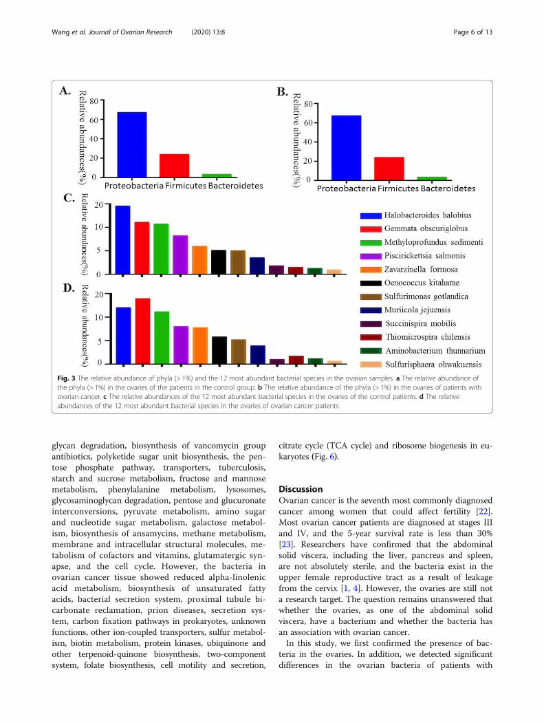
Ovarian bacteria characterization between cancer andcontrol groupsTo understand the ovarian bacteria in cancer and con-trol groups, we performed deep sequencing of the V3-V4 16S rRNA region of all sixteen collected samples. Inthe ovaries, our results showed that Proteobacteria was
the most abundant phylum (67.1% in the control groupand 67.20% in the cancer group). Firmicutes was the sec-ond most abundant phylum (23.77% in the controlgroup and 23.82% in the cancer group), and the thirdmost abundant phylum was Bacteroidetes (3.26% in thecontrol group and 3.41% in the cancer group) (Fig. 2a,
Table 1 Clinical characteristics of patients enrolled in the study
Control group (n = 10) Cancer group (n = 6) P value
Age 51.6(45–57) 57.3(46–75) 0.29
Menopausal status 0.12
Pre/Peri 8 2
Post 2 4
Parity 5.1(1–13) 3.1(2–5) 0.17
History of hypertension 0.52
Yes 1 2
NO 9 4
History of diabetes 0.70
Yes 1 1
NO 9 5
Stage (%)
II 2(33.3)
III 4(66.7)
Histotype (%)
Uterine myoma 3(30) –
Uterine adenomyosis 7(70) –
Ovarian serous carcinoma – 6(100)
The P-value of age and parity were assessed by Student’s t-test. The P-value of menopausal status, history of hypertension and diabetes were calculated by thechi-square test
Fig. 1 BugBase analysis of predicted metagenomes. The potentially pathogenic and immunohistochemistry of ovaries using an antibacterial LPSantibody. a control group (10x). Scale bars, 200 μm. b control group (40x). Scale bars, 50 μm. c cancer group (10x). Scale bars, 200 μm. d cancergroup (40x). Scale bars, 50 μm. Arrows point to LPS staining in the ovarian tissue
Wang et al. Journal of Ovarian Research (2020) 13:8 Page 4 of 13

b). At the species level, the ovarian bacterial communi-ties were dominated by Halobacteroides halobius(14.53%), followed by Gemmata obscuriglobus (11.07%)and Methyloprofundus sedimenti (10.69%) in the controlgroup. The ovarian bacterial communities in the cancergroup were dominated by Gemmata obscuriglobus(13.89%), followed by Halobacteroides halobius (11.99%)and Methyloprofundus sedimenti (11.12%) (Fig. 3).
Ovarian bacterial community composition differencesbetween cancer and control groupsWe carried out a comparison of differences in the over-all bacterial communities using PCoA, which showedthat the ovarian bacteria of the control group displayedsome differences compared to that of the cancer group(Fig. 4a and b).
Ovarian bacterial composition at different levels in cancerand control groupsTo detect the differences in ovarian bacteria between theseventeen samples, we analyzed the ovarian bacterialcomposition at different levels in cancer and controlgroups. In the Table 2, we showed the statistical differ-ence of ovarian bacteria in cancer and control groups atphylum, class, order, family, genus and species level. In
particular, the relative abundance of Anoxynatronumsibiricum may be associated with the stage of the tumor(Fig. 4c), and Methanosarcina vacuolata may be used todiagnose ovarian cancer (Fig. 4d).
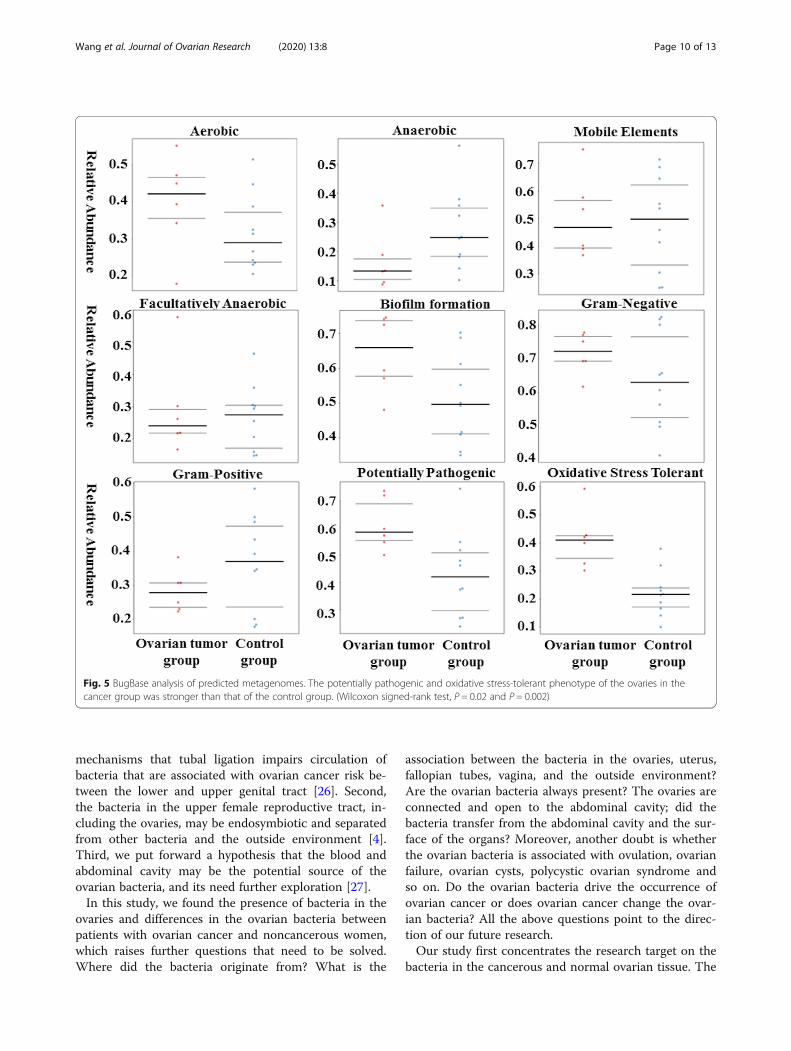
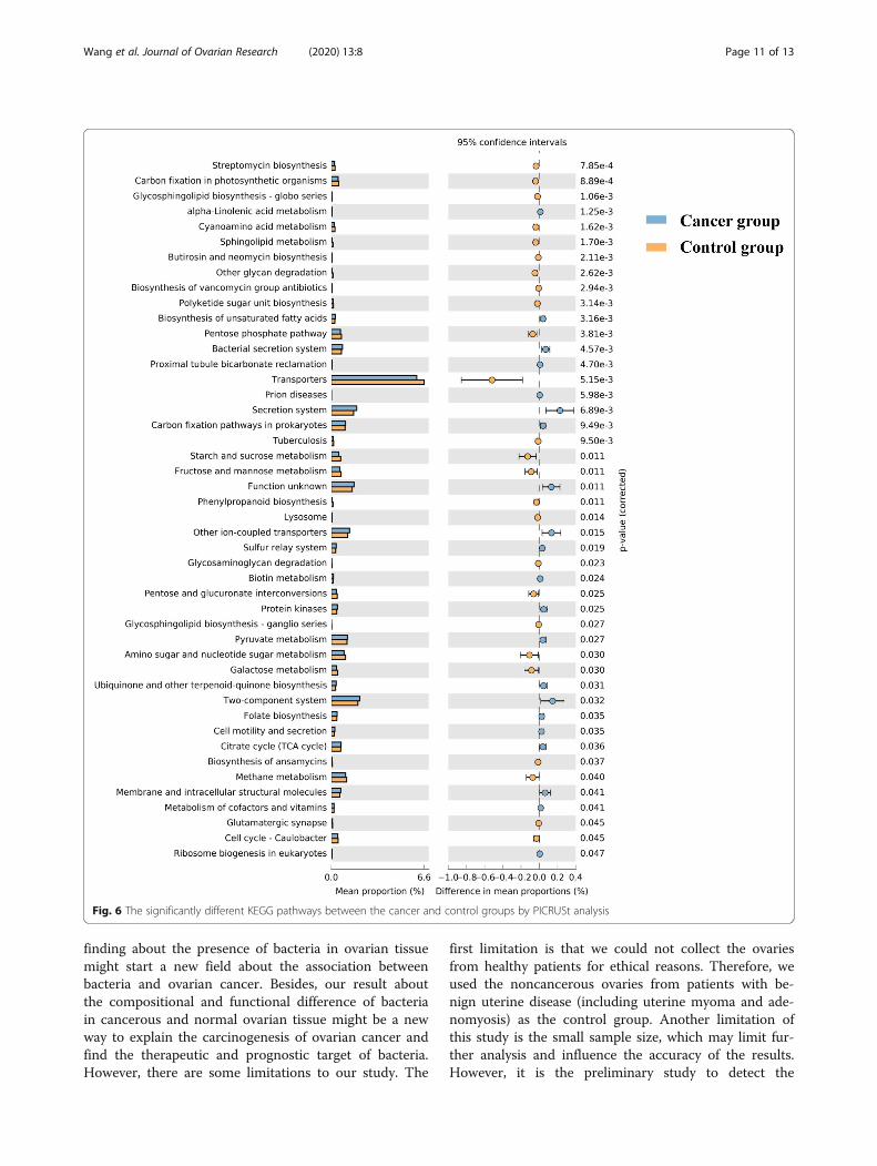
Predicted function of the ovarian bacteria showsphenotypic conservation between cancer and controlgroupsBugBase identified that gene functions associated withthe potentially pathogenic and the oxidative stress-tolerant phenotype were enriched in the ovaries of thecancer group (Wilcoxon signed-rank test, P = 0.02 andP = 0.002). The aerobic, anaerobic, facultatively anaer-obic, gram-positive, and gram-negative phenotypes; mo-bile elements; and biofilm formation of the ovarianbacteria showed no significant difference between ovar-ian cancer and control groups (Fig. 5). PICRUSt wasused to identify the KEGG pathways between the bac-teria of ovaries in cancer and control groups and found46 different KEGG pathways. The ovaries in the cancergroup showed increased pathways related to strepto-mycin biosynthesis, carbon fixation in photosyntheticorganisms, glycosphingolipid biosynthesis-globo series,cyanoamino acid metabolism, glycerophospholipid me-tabolism, butirosin and neomycin biosynthesis, other
Fig. 2 Bacterial richness and diversity in the cancer and control groups revealed by 16S rRNA sequencing a Observed species index (P = 0.06,Mann-Whitney U test); b Chao 1 index (P = 0.06, Mann-Whitney U test); c ACE index (P = 0.06, Mann-Whitney U test); d Shannon index (P = 0.32,Mann-Whitney U test); e Evenness index (P = 0.48, Mann-Whitney U test); f Simpson index (P = 0.46, Mann-Whitney U test)
Wang et al. Journal of Ovarian Research (2020) 13:8 Page 5 of 13
glycan degradation, biosynthesis of vancomycin groupantibiotics, polyketide sugar unit biosynthesis, the pen-tose phosphate pathway, transporters, tuberculosis,starch and sucrose metabolism, fructose and mannosemetabolism, phenylalanine metabolism, lysosomes,glycosaminoglycan degradation, pentose and glucuronateinterconversions, pyruvate metabolism, amino sugarand nucleotide sugar metabolism, galactose metabol-ism, biosynthesis of ansamycins, methane metabolism,membrane and intracellular structural molecules, me-tabolism of cofactors and vitamins, glutamatergic syn-apse, and the cell cycle. However, the bacteria inovarian cancer tissue showed reduced alpha-linolenicacid metabolism, biosynthesis of unsaturated fattyacids, bacterial secretion system, proximal tubule bi-carbonate reclamation, prion diseases, secretion sys-tem, carbon fixation pathways in prokaryotes, unknownfunctions, other ion-coupled transporters, sulfur metabol-ism, biotin metabolism, protein kinases, ubiquinone andother terpenoid-quinone biosynthesis, two-componentsystem, folate biosynthesis, cell motility and secretion,
citrate cycle (TCA cycle) and ribosome biogenesis in eu-karyotes (Fig. 6).
DiscussionOvarian cancer is the seventh most commonly diagnosedcancer among women that could affect fertility [22].Most ovarian cancer patients are diagnosed at stages IIIand IV, and the 5-year survival rate is less than 30%[23]. Researchers have confirmed that the abdominalsolid viscera, including the liver, pancreas and spleen,are not absolutely sterile, and the bacteria exist in theupper female reproductive tract as a result of leakagefrom the cervix [1, 4]. However, the ovaries are still nota research target. The question remains unanswered thatwhether the ovaries, as one of the abdominal solidviscera, have a bacterium and whether the bacteria hasan association with ovarian cancer.In this study, we first confirmed the presence of bac-
teria in the ovaries. In addition, we detected significantdifferences in the ovarian bacteria of patients with
Fig. 3 The relative abundance of phyla (> 1%) and the 12 most abundant bacterial species in the ovarian samples. a The relative abundance ofthe phyla (> 1%) in the ovaries of the patients in the control group. b The relative abundance of the phyla (> 1%) in the ovaries of patients withovarian cancer. c The relative abundances of the 12 most abundant bacterial species in the ovaries of the control patients. d The relativeabundances of the 12 most abundant bacterial species in the ovaries of ovarian cancer patients
Wang et al. Journal of Ovarian Research (2020) 13:8 Page 6 of 13

ovarian cancer when compared with samples from non-cancerous women.To avoid bacterial contamination, all instruments used
were sterilized, and the reagent we used was new. Whenoperating, the surgeon wore an autoclaved mask, capand suit and did not talk. The sample did not touch any-thing in the operating room except for the tweezers andwas immediately put into the sterilized tube. When thesample was transferred to the laboratory, as many of theprocedures as possible were performed on the asepsiswork table except the procedures that required largeequipment, such as centrifugal machines and se-quencers. More importantly, we used ovaries from pa-tients with benign uterine disease as the control groupto counteract possible contamination.
There are three possible reasons to explain the origin-ation of the ovarian bacteria. First, a new opinion is thatthe upper female reproductive tract is not sterile [4], anddifferent bacteria exist throughout the female reproduct-ive tract, forming a continuum from the vagina to theovaries [23]. The bacteria in the ovaries may originatefrom the fallopian tubes, uterine cavity, cervix canal orvagina, which is in contact with the outside environ-ment. Besides, many researches have confirmed tuballigation decreases the risk of EOC by unknown mecha-nisms [24, 25]. Walther et al. were able to amplify bac-terial DNA from 94% of the cervical/vaginal samples and87% of the uterine samples [23]. However, they wereonly able to amplify bacterial DNA from 50 and 61% offallopian tubes and ovaries, which imply the potential
Fig. 4 Communities clustered using PCoA and the relative abundance of Anoxynatronum sibiricum and Methanosarcina vacuolata. a Communitieswere clustered using PCoA. PC1 and PC2 are plotted on the x and y axes. The red block is equal to a sample in the ovarian cancer group. Theblue circle is equal to a sample in the control group. The samples from the ovarian cancer group can be separated from other samples in thecontrol group. b Communities clustered using Principal Component Analysis (PCoA). PC1 and PC2 are plotted on the x and y axes. The red blockis equal to a sample in the ovarian cancer group. The blue solid circle is equal to a sample from a patient with uterine myoma, and the bluehollow circle is equal to a sample of a patient with uterine adenomyosis. c The relative abundance of Anoxynatronum sibiricum (Control group:n = 10, cancer group: n = 6, P = 0.034, Mann-Whitney U test). d The relative abundance of Methanosarcina vacuolata (Control group: n = 10, cancergroup: n = 6, P = 0.001, Mann-Whitney U test)
Wang et al. Journal of Ovarian Research (2020) 13:8 Page 7 of 13

Table 2 Differential relative abundance of the taxa in ovarian communities between patients in cancer and control group
Control cohort (n = 10, %) Ovarian tumor cohort (n = 6, %) P value
Phylum Planctomycetes 0.5144 ± 0.1420 0.8655 ± 0.2638 0.023
Crenarchaeota 0.2840 ± 0.0787 0.1592 ± 0.0775 0.023
Aquificae 0.0352 ± 0.0137 0.0697 ± 0.0291 0.017
Class Spartobacteria 0.3149 ± 0.0923 0.4795 ± 0.1205 0.026
Sphingobacteriia 0.1280 ± 0.0695 0.0423 ± 0.0706 0.039
Order Planctomycetales 7.2700 ± 1.3880 9.1183 ± 0.8594 0.039
Pseudomonadales 0.1332 ± 0.0746 0.4283 ± 0.4019 0.023
Enterobacteriales 0.6038 ± 0.1237 2.0105 ± 2.5829 0.030
Methanobacteriales 0.1626 ± 0.0496 0.2602 ± 0.0859 0.030
Halobacteriales 0.0648 ± 0.0117 0.0439 ± 0.0287 0.039
Campylobacterales 0.0776 ± 0.0158 0.1133 ± 0.0232 0.009
Family Flavobacteriaceae 24.7500 ± 0.6712 21.7167 ± 3.0732 0.014
Methanobacteriaceae 0.1720 ± 0.0540 0.2667 ± 0.0867 0.039
Moraxellaceae 0.1328 ± 0.0658 0.4347 ± 0.4054 0.030
Petrotogaceae 0.0452 ± 0.0178 0.0638 ± 0.0112 0.039
Thermaceae 0.0078 ± 0.0089 0.0188 ± 0.0086 0.017
Archaeoglobaceae 0.0611 ± 0.0221 0.0381 ± 0.0123 0.045
Leptotrichiaceae 0.1018 ± 0.0524 0.0442 ± 0.0284 0.030
Microbacteriaceae 0.1493 ± 0.0618 0.2740 ± 0.1320 0.039
Staphylococcaceae 0.0281 ± 0.0545 0.0822 ± 0.0536 0.029
Thermogemmatisporaceae 0.7381 ± 0.1925 1.4583 ± 0.6982 0.013
Methanocorpusculaceae 0.0233 ± 0.0139 0.0091 ± 0.0063 0.023
Geodermatophilaceae 0.0552 ± 0.0335 0.0144 ± 0.0145 0.030
Genus Paenibacillus 0.7990 ± 0.4563 0.3207 ± 0.2151 0.039
Haloferula 0.1811 ± 0.0623 0.1156 ± 0.0263 0.023
Subdivision 0.0801 ± 0.0314 0.0465 ± 0.0188 0.039
Zavarzinella 0.0741 ± 0.0238 0.1234 ± 0.0305 0.009
Photorhabdus 0.0013 ± 0.0029 0.0068 ± 0.0050 0.023
Volucribacter 0.0081 ± 0.0062 0.0021 ± 0.0046 0.042
Blastococcus 0.0552 ± 0.0335 0.0144 ± 0.0145 0.030
Mesotoga 0.2509 ± 0.0703 0.3675 ± 0.1057 0.039
Defluviitoga 0.0550 ± 0.0252 0.0216 ± 0.0114 0.030
Dorea 0.0063 ± 0.0065 0.0000 ± 0.0000 0.025
Species Rhodopirellularubra 0.4011 ± 0.1433 0.7563 ± 0.2398 0.013
Haloferulasargassicola 0.1534 ± 0.0629 0.0999 ± 0.0227 0.030
Thermogemmatisporafoliorum 0.7813 ± 0.2152 1.4957 ± 0.6735 0.023
Mycoplasmaequigenitalium 0.5463 ± 0.0684 0.6820 ± 0.1108 0.039
Bifidobacteriumsubtile 0.0924 ± 0.0269 0.2584 ± 0.1958 0.026
Natroniellaacetigena 0.0075 ± 0.0078 0.0000 ± 0.0000 0.012
Flammeovirgakamogawensis 0.6966 ± 0.3523 0.2488 ± 0.1349 0.026
Eubacteriumyurii 0.0231 ± 0.0111 0.0091 ± 0.0074 0.030
Enterococcusdiestrammenae 0.2549 ± 0.0859 0.1458 ± 0.0809 0.030
Pelagicoccusalbus 0.0127 ± 0.0057 0.0047 ± 0.0024 0.017
Fodinibacterluteus 0.1588 ± 0.0461 0.0935 ± 0.0498 0.039
Wang et al. Journal of Ovarian Research (2020) 13:8 Page 8 of 13

Table 2 Differential relative abundance of the taxa in ovarian communities between patients in cancer and control group(Continued)
Control cohort (n = 10, %) Ovarian tumor cohort (n = 6, %) P value
Prosthecobacteralgae 0.0210 ± 0.0121 0.0080 ± 0.0050 0.030
Emticiciaoligotrophica 0.0743 ± 0.0297 0.0308 ± 0.0251 0.013
Leuconostoccitreum 0.0417 ± 0.0281 0.0108 ± 0.0125 0.039
Methanimicrococcusblatticola 0.2138 ± 0.0527 0.1572 ± 0.0383 0.039
Methanosarcinavacuolata 0.0156 ± 0.0061 0.0007 ± 0.0015 0.001
Lactobacillussucicola 0.0160 ± 0.0063 0.0081 ± 0.0053 0.030
Caldicoprobacteroshimai 0.0014 ± 0.0041 0.0044 ± 0.0042 0.048
Caldicellulosiruptorsaccharolyticus 0.3268 ± 0.1880 0.1082 ± 0.1296 0.039
Methylomicrobiumalbum 0.0013 ± 0.0021 0.0069 ± 0.0051 0.013
Novispirillum itersonii 0.0031 ± 0.0036 0.0000 ± 0.0000 0.048
Paenibacillusodorifer 0.6905 ± 0.4128 0.2356 ± 0.1583 0.039
Mycoplasmagenitalium 0.0023 ± 0.0038 0.0073 ± 0.0048 0.043
Sulfurospirillumhalorespirans 0.0630 ± 0.0163 0.0948 ± 0.0306 0.039
Streptococcuscastoreus 0.0514 ± 0.0415 0.0190 ± 0.0329 0.030
Spongiivirgacitrea 0.2355 ± 0.1391 0.0921 ± 0.0784 0.039
Staphylococcuscapitissubsp 0.0245 ± 0.0504 0.0752 ± 0.0506 0.021
Xanthomonasbromi 0.0094 ± 0.0117 0.0000 ± 0.0000 0.025
Vulcanisaeta thermophila 0.0457 ± 0.0106 0.0720 ± 0.0247 0.039
Volucribacter amazonae 0.0081 ± 0.0062 0.0021 ± 0.0046 0.042
Thalassotalea fusca 0.0316 ± 0.0202 0.0027 ± 0.0045 0.004
Thermus islandicus 0.0051 ± 0.0049 0.0000 ± 0.0000 0.025
Prevotella veroralis 0.0055 ± 0.0074 0.0000 ± 0.0000 0.048
Pseudobutyrivibrio xylanivorans 0.0072 ± 0.0063 0.0021 ± 0.0046 0.030
Peptoniphilus methioninivorax 0.0000 ± 0.0000 0.0031 ± 0.0033 0.017
Sphingobacterium arenae 0.2488 ± 0.1235 0.0861 ± 0.0529 0.030
Campylobacter rectus 0.0050 ± 0.0064 0.0000 ± 0.0000 0.048
Blautia glucerasea 0.0166 ± 0.0091 0.0056 ± 0.0067 0.033
Calditerricola yamamurae 0.0745 ± 0.0158 0.1084 ± 0.0306 0.023
Clostridium thermosuccinogenes 0.0036 ± 0.0051 0.0127 ± 0.0089 0.030
Alkalibacillus haloalkaliphilus 0.0058 ± 0.0066 0.0000 ± 0.0000 0.025
Acholeplasma oculi 0.0038 ± 0.0041 0.0000 ± 0.0000 0.025
Aureimonas phyllosphaerae 0.0013 ± 0.0029 0.0068 ± 0.0050 0.023
Azonexus hydrophilus 0.0773 ± 0.0316 0.0285 ± 0.0190 0.007
Anaerostipes rhamnosivorans 0.0005 ± 0.0015 0.0045 ± 0.0043 0.025
Anoxynatronum sibiricum 0.1172 ± 0.0708 0.0460 ± 0.0513 0.034
Legionella taurinensis 0.0029 ± 0.0031 0.0000 ± 0.0000 0.048
Mesonia phycicola 0.0119 ± 0.0087 0.0031 ± 0.0033 0.019
Luteolibacter cuticulihirudinis 0.2389 ± 0.1090 0.4292 ± 0.1517 0.030
Megasphaera indica 0.0052 ± 0.0055 0.0000 ± 0.0000 0.025
Dorea formicigenerans 0.0063 ± 0.0065 0.0000 ± 0.0000 0.025
Fuchsiella alkaliacetigena 0.0082 ± 0.0075 0.0014 ± 0.0031 0.043
Geobacillus thermodenitrificans 0.0063 ± 0.0051 0.0006 ± 0.0013 0.024
The P-value was calculated by the Mann-Whitney U test
Wang et al. Journal of Ovarian Research (2020) 13:8 Page 9 of 13
mechanisms that tubal ligation impairs circulation ofbacteria that are associated with ovarian cancer risk be-tween the lower and upper genital tract [26]. Second,the bacteria in the upper female reproductive tract, in-cluding the ovaries, may be endosymbiotic and separatedfrom other bacteria and the outside environment [4].Third, we put forward a hypothesis that the blood andabdominal cavity may be the potential source of theovarian bacteria, and its need further exploration [27].In this study, we found the presence of bacteria in the
ovaries and differences in the ovarian bacteria betweenpatients with ovarian cancer and noncancerous women,which raises further questions that need to be solved.Where did the bacteria originate from? What is the
association between the bacteria in the ovaries, uterus,fallopian tubes, vagina, and the outside environment?Are the ovarian bacteria always present? The ovaries areconnected and open to the abdominal cavity; did thebacteria transfer from the abdominal cavity and the sur-face of the organs? Moreover, another doubt is whetherthe ovarian bacteria is associated with ovulation, ovarianfailure, ovarian cysts, polycystic ovarian syndrome andso on. Do the ovarian bacteria drive the occurrence ofovarian cancer or does ovarian cancer change the ovar-ian bacteria? All the above questions point to the direc-tion of our future research.Our study first concentrates the research target on the
bacteria in the cancerous and normal ovarian tissue. The
Fig. 5 BugBase analysis of predicted metagenomes. The potentially pathogenic and oxidative stress-tolerant phenotype of the ovaries in thecancer group was stronger than that of the control group. (Wilcoxon signed-rank test, P = 0.02 and P = 0.002)
Wang et al. Journal of Ovarian Research (2020) 13:8 Page 10 of 13
finding about the presence of bacteria in ovarian tissuemight start a new field about the association betweenbacteria and ovarian cancer. Besides, our result aboutthe compositional and functional difference of bacteriain cancerous and normal ovarian tissue might be a newway to explain the carcinogenesis of ovarian cancer andfind the therapeutic and prognostic target of bacteria.However, there are some limitations to our study. The
first limitation is that we could not collect the ovariesfrom healthy patients for ethical reasons. Therefore, weused the noncancerous ovaries from patients with be-nign uterine disease (including uterine myoma and ade-nomyosis) as the control group. Another limitation ofthis study is the small sample size, which may limit fur-ther analysis and influence the accuracy of the results.However, it is the preliminary study to detect the
Fig. 6 The significantly different KEGG pathways between the cancer and control groups by PICRUSt analysis
Wang et al. Journal of Ovarian Research (2020) 13:8 Page 11 of 13

ovarian bacteria in patients with ovarian cancer, and wewill conduct further explorations with larger samplesizes.
ConclusionsThe ovaries contained several kinds of bacteria and werenot sterile in a noninflammatory environment. Besides,there were significant differences between the ovarianbacterial compositions of patients in the cancer and con-trol groups.
AbbreviationsFFPE: Formalin-fixed paraffin-embedded; IMG4: Integrated MicrobialGenomes; KEGG: Kyoto Encyclopedia of Genes and Genomes;LPS: Lipopolysaccharide; OTUs: Operational taxonomic units;PATRIC: Pathosystems Resource Integration Center; PCoA: Principalcoordinates analysis; PCR: Polymerase chain reaction; PICRUSt: PhylogeneticInvestigation of Communities by Reconstruction of Unobserved States
AcknowledgementsWe thank the colleagues in the Department of Gynecology of First AffiliatedHospital in Xi’an Jiatong University for their contributions to collectingsamples.
Authors’ contributionsWQ: Project development, Data collection, Data analysis, Manuscript writing.ZL, HL, TX, MS and LQ: Data collection. WY and LD: Data analysis and datacollection. TM and SC: Project development. FG and WQing: Projectdevelopment and data analysis. SQ and LQL: Experimental design andproject development. All authors read and approved the final manuscript.
FundingThis work was supported by grants from the Fundamental Research Fundsfor Xi’an Jiaotong University (xjj2015093), and Major Basic Research Project ofNatural Science of Shaanxi Provincial Science and Technology Department(2017ZDJC-11), the Key Research and Development Project of ShaanxiProvincial Science and Technology Department (2017ZDXM-SF-068), andShaanxi Provincial Collaborative Technology Innovation Project (2017XT-026,2018XT-002). The funders had no role in study design, data collection andanalysis, decision to publish, or preparation of the manuscript.
Availability of data and materialsPlease contact the corresponding author Qiling Li ([email protected]).
Ethics approval and consent to participateThis study was approved by the Medical Institutional Ethics Committee ofthe First Affiliated Hospital of Xi’an Jiaotong University. Informed consentwas obtained from all the enrolled patients.
Consent for publicationNot applicable.
Competing interestsThe authors declare that they have no competing interests.
Author details1Department of Obstetrics and Gynecology, First Affiliated Hospital, Xi’anJiaotong University, Xi’an, Shaanxi, China. 2Department of GynecologicalOncology, Shaanxi Provincial Cancer Hospital, Xi’an, Shaanxi, China. 3Guipei77, Health Science Center, Xi’an Jiaotong University, Xi’an, Shaanxi, China.4Omega Bioservices Inc, Norcross, GA, USA. 5Cardiovascular ResearchInstitute, Morehouse School of Medicine, Atlanta, Georgia, USA.
Received: 5 October 2019 Accepted: 17 December 2019
References1. Geller LT, Barzily-Rokni M, Danino T, Jonas OH, Shental N, Nejman D, et al.
Potential role of intratumor bacteria in mediating tumor resistance to thechemotherapeutic drug gemcitabine. Science. 2017;357(6356):1156–60.
2. Manfredo Vieira S, Hiltensperger M, Kumar V, Zegarra-Ruiz D, Dehner C,Khan N, et al. Translocation of a gut pathobiont drives autoimmunity inmice and humans. Science. 2018;359(6380):1156–61.
3. Brunelli R, Papi M, Arcovito G, Bompiani A, Castagnola M, Parasassi T, et al.Globular structure of human ovulatory cervical mucus. FASEB J. 2007;21(14):3872–6.
4. Chen C, Song X, Wei W, Zhong H, Dai J, Lan Z, et al. The microbiotacontinuum along the female reproductive tract and its relation to uterine-related diseases. Nat Commun. 2017;8(1):875.
5. Zervomanolakis I, Ott HW, Hadziomerovic D, Mattle V, Seeber BE, Virgolini I,et al. Physiology of upward transport in the human female genital tract.Ann N Y Acad Sci. 2007;1101:1–20.
6. Verstraelen H, Vilchez-Vargas R, Desimpel F, Jauregui R, Vankeirsbilck N,Weyers S, et al. Characterisation of the human uterine microbiome in non-pregnant women through deep sequencing of the V1-2 region of the 16SrRNA gene. PeerJ. 2016;4:e1602.
7. Fang RL, Chen LX, Shu WS, Yao SZ, Wang SW, Chen YQ. Barcodedsequencing reveals diverse intrauterine microbiomes in patients sufferingwith endometrial polyps. Am J Transl Res. 2016;8(3):1581–92.
8. Miles SM, Hardy BL, Merrell DS. Investigation of the microbiota of thereproductive tract in women undergoing a total hysterectomy and bilateralsalpingo-oopherectomy. Fertil Steril. 2017;107(3):813–20 e1.
9. Kostic AD, Gevers D, Pedamallu CS, Michaud M, Duke F, Earl AM, et al.Genomic analysis identifies association of Fusobacterium with colorectalcarcinoma. Genome Res. 2012;22(2):292–8.
10. Bullman S, Pedamallu CS, Sicinska E, Clancy TE, Zhang X, Cai D, et al.Analysis of Fusobacterium persistence and antibiotic response in colorectalcancer. Science. 2017;358(6369):1443.
11. Wang L, Zhou J, Xin Y, Geng C, Tian Z, Yu X, et al. Bacterial overgrowth anddiversification of microbiota in gastric cancer. Eur J Gastroenterol Hepatol.2016;28(3):261–6.
12. Hosgood HD 3rd, Sapkota AR, Rothman N, Rohan T, Hu W, Xu J, et al. Thepotential role of lung microbiota in lung cancer attributed to householdcoal burning exposures. Environ Mol Mutagen. 2014;55(8):643–51.
13. Kwon M, Seo SS, Kim MK, Lee DO, Lim MC. Compositional and FunctionalDifferences between Microbiota and Cervical Carcinogenesis as Identifiedby Shotgun Metagenomic Sequencing. Cancers. 2019;11(3):309.
14. Urbaniak C, Gloor GB, Brackstone M, Scott L, Tangney M, Reid G. Themicrobiota of breast tissue and its association with breast Cancer. ApplEnviron Microbiol. 2016;82(16):5039–48.
15. Feng Y, Ramnarine VR, Bell R, Volik S, Davicioni E, Hayes VM, et al.Metagenomic and metatranscriptomic analysis of human prostate microbiotafrom patients with prostate cancer. BMC Genomics. 2019;20(1):146.
16. Bolger AM, Lohse M, Usadel B. Trimmomatic: a flexible trimmer for Illuminasequence data. Bioinformatics. 2014;30(15):2114–20.
17. Kanehisa M, Goto S, Sato Y, Furumichi M, Tanabe M. KEGG for integrationand interpretation of large-scale molecular data sets; 2012.
18. Markowitz VM, Chen IM, Palaniappan K, Chu K, Szeto E, Grechkin Y, et al.IMG: the integrated microbial genomes database and comparative analysissystem. Nucleic Acids Res. 2012;40(Database issue):115–22.
19. Snyder EE, Kampanya N, Lu J, Nordberg EK, Karur HR, Shukla M, et al.PATRIC: the VBI PathoSystems resource integration center. Nucleic AcidsRes. 2007;35(Database issue):D401–D6.
20. Langille MGI, Zaneveld J, Caporaso JG, Mcdonald D, Dan K, Reyes JA, et al.Predictive functional profiling of microbial communities using 16S rRNAmarker gene sequences. Nat Biotechnol. 2013;31(9):814.
21. Parks DH, Tyson GW, Hugenholtz P, Beiko RG. STAMP: statistical analysis oftaxonomic and functional profiles. Bioinformatics. 2014;30(21):3123.
22. Leranth C, Hamori J. "Dark" Purkinje cells of the cerebellar cortex. Acta BiolAcad Sci Hung. 1970;21(4):405–19.
23. Walther-Antonio MR, Chen J, Multinu F, Hokenstad A, Distad TJ, Cheek EH,et al. Potential contribution of the uterine microbiome in the developmentof endometrial cancer. Genome Med. 2016;8(1):122.
Wang et al. Journal of Ovarian Research (2020) 13:8 Page 12 of 13

24. Sieh W, Salvador S, McGuire V, Weber RP, Terry KL, Rossing MA, et al. Tuballigation and risk of ovarian cancer subtypes: a pooled analysis of case-control studies. Int J Epidemiol. 2013;42(2):579–89.
25. Rice MS, Hankinson SE, Tworoger SS. Tubal ligation, hysterectomy, unilateraloophorectomy, and risk of ovarian cancer in the Nurses' health studies.Fertil Steril. 2014;102(1):192–8 e3.
26. Mert I, Walther-Antonio M, Mariani A. Case for a role of the microbiome ingynecologic cancers: Clinician's perspective. J Obstet Gynaecol Res. 2018;44(9):1693–704.
27. Paisse S, Valle C, Servant F, Courtney M, Burcelin R, Amar J, et al.Comprehensive description of blood microbiome from healthy donorsassessed by 16S targeted metagenomic sequencing. Transfusion. 2016;56(5):1138–47.
Publisher’s NoteSpringer Nature remains neutral with regard to jurisdictional claims inpublished maps and institutional affiliations.
Wang et al. Journal of Ovarian Research (2020) 13:8 Page 13 of 13